High Expression of p21 as a Potential Therapeutic Target in Ovarian Clear-cell Carcinoma
←
→
Page content transcription
If your browser does not render page correctly, please read the page content below
ANTICANCER RESEARCH 40: 5631-5639 (2020)
doi:10.21873/anticanres.14576

                High Expression of p21 as a Potential Therapeutic
                    Target in Ovarian Clear-cell Carcinoma
                  YUKI MINAGAWA1,2, KOUSUKE ISHINO1, RYUICHI WADA1,3, MITSUHIRO KUDO1,
                         ZENYA NAITO1,3, TOSHIYUKI TAKESHITA2 and RYUJI OHASHI1,3

                 1Department of Integrated Diagnostic Pathology, Nippon Medical School, Tokyo, Japan;
                     2Departmentof Obstetrics and Gynecology, Nippon Medical School, Tokyo, Japan;
                   3Department of Diagnostic Pathology, Nippon Medical School Hospital, Tokyo, Japan

Abstract. Background/Aim: DNA damage response (DDR),             organize cell-cycle arrest and repair of damaged DNA. Tumour
wherein p21 is a cell fate determinant, is a potential cancer    protein p53 (TP53) and 21-kDa protein (p21) suppress the cell
therapeutic target. Molecular expression during DDR was          cycle, and damaged DNA is repaired. If the DNA repair is
explored in ovarian clear-cell carcinoma (CCC). Materials        incomplete, the cell undergoes apoptosis. The susceptibility of
and Methods: CHK1, CHK2, TP53 and p21 expression in              tumours to chemotherapeutic agents, such as cisplatin, cis-
DDR was examined using immunostaining in surgical sections       diamminedichloridoplatinum(II), is regulated by molecules
of CCC (n=22). Molecular alterations in two types of CCC         expressed during DDR (1, 2), which can be a target of
cell lines, JHOC-5 and JHOC-9, were investigated using           molecular targeting therapy (1, 3). p21, one of the molecules
western blot analysis. Results: Expression of DDR-associated     involved in DDR, is an inhibitor of cyclin-dependent kinase
molecules was noted in most patients. While high p21             (CDK) and a key regulator of the cell cycle in DDR (4-6). It
expression was found in half of the patients, the remaining      is also suggested that p21 regulates apoptotic cell death (4-7).
patients exhibited low p21 expression. Treatment with               Epithelial ovarian cancer (EOC) is the most lethal
UC2288, a p21 inhibitor, attenuated proliferation of both cell   malignancy, and a leading cause of gynaecological cancer-
lines, more prominently in JHOC-9, resulting in reduced          related deaths worldwide. Clear-cell carcinoma (CCC), a
viability and subsequent apoptosis. Conclusion: p21 Inhibitor    subtype of EOC, accounts for more than 20% of EOC in
induced cell death in cells with high p21 expression,            Japan (8). Compared to other subtypes, the prognosis of
suggesting that p21 suppression can be a therapeutic strategy    patients with CCC is poor due to a high recurrence rate and
to treat patients with CCC.                                      chemotherapy resistance (9, 10). Owing to its aggressive
                                                                 behaviour, patients with CCC predominantly follow an
The DNA damage response (DDR) plays an important role in         unfavourable clinical course.
the maintenance of genetic integrity in normal as well as in        The molecules highly expressed during DDR in CCC have
carcinoma cells (1). When DNA is damaged, sensors such as        been described in literature (3). For example, the expression
poly (ADP ribose) polymerase (PARP) and 80 kDa Ku protein        levels of CHK1, CDK2, and p27 correlated with the degree
(KU80) activate ataxia-telangiectasia mutated (ATM) and          of chemoresistance in patients with CCC (11, 12). Although
ATM- and RAD3-related (ATR). Subsequently, transducers,          understanding the functional state of DDR is beneficial to
checkpoint kinase 1 (CHK1) and checkpoint kinase 2 (CHK2),       predicting sensitivity to treatment, molecular expression in
                                                                 DDR has not been fully elucidated in CCC due to the limited
                                                                 number of studies.
                                                                    In the present study, we explored the clinicopathological
This article is freely accessible online.                        significance of DDR in CCC. The expression of CHK1, CHK2,
                                                                 TP53 and p21 were examined by immunohistochemical (IHC)
Correspondence to: Kousuke Ishino, Department of Integrated      staining of surgical sections of CCC. As a DNA-damaging
Diagnostic Pathology, Nippon Medical School, 1-1-5 Sendagi,
                                                                 agent, we chose cisplatin in order to observe its effect on
Bunkyo-ku 113-8602, Tokyo, Japan. Tel: +81 338222131, e-mail:
kishino@nms.ac.jp
                                                                 molecular expression in DDR using two types of CCC cell
                                                                 lines. The effect of UC2288, an attenuator of p21 (13-15), was
Key Words: Ovarian clear-cell carcinoma, DNA damage response,    also examined to investigate how functional modulation of p21
cisplatin, p21.                                                  may alter CCC cell viability.

                                                                                                                            5631
ANTICANCER RESEARCH 40: 5631-5639 (2020)

Materials and Methods                                                   Merck KGaA, St. Louis, MO, USA) were added to culture medium,
                                                                        and the cells were cultured at 37˚C. Viable cells were determined
Cases of CCC. A total of 22 CCC cases were retrieved from               using Cell Counting Kit-8 (DOJINDO Molecular Technologies, Inc.,
pathological records in the archives at the Department of Diagnostic    Kumamoto, Japan) at 0, 24, 48, and 72 h. Experiments were
Pathology, Nippon Medical School Hospital, and used for                 performed in triplicate. The viable cells were indicated as
histological and IHC studies. These cases underwent ovarian             absorbance at 450 nm.
resection from 2013 to 2019. Histology was reviewed by two
investigators (YM and RW). The clinicopathological features of the      Preparation of protein samples. Cultured cell of JHOC-5 and
cases are listed in Table I. Pathological T-factor and lymph node       JHOC-9 were plated in 100 mm dish at density of 5×10⁵ cells/dish
metastasis were reviewed. The study was approved by the Ethical         and 1×10⁶ cells/dish, respectively. The cells were cultured at 37˚C
Committee of Nippon Medical School Hospital (Approval no. 30-           for 72 h. Then, 10 μg/ml cisplatin and 10 μM UC2288 were added
06-944). Written informed consent was obtained from all patients.       to the culture medium, and cells cultured at 37˚C for 24 h.
                                                                        Thereafter, cells were washed three times with PBS and
IHC staining and semi-quantitative evaluation. IHC staining of          subsequently lysed in 50 mM Tris-HCl (pH 7.6)/0.5% sodium
molecules involved in DDR was performed using the polymer-based         dodecylsulfate and sonicated in iced water for 60 min. Protein
two-step method. Briefly, paraffin sections were deparaffinized and     samples were used for western blot analysis.
hydrated in phosphate-buffered saline (PBS). Following blocking of
endogenous peroxidase, sections were pre-treated with appropriate       Western blot analysis. Protein samples were mixed with 2 ×
buffer, if necessary. Thereafter, sections were incubated with          Laemmli Sample Buffer (Bio-Rad Laboratories, Inc., Hercules, CA,
antibodies against CHK1, CHK2, TP53, and p21 at 4˚C overnight           USA) and boiled at 95˚C for 10 min. Protein samples were
(Table II). Following washing three times with PBS, sections were       electrophoresed in 5-20% polyacrylamide gel (e-PAGEL, ATTO
subsequently incubated with MAX-PO (cat. no. 424134 Mouse,              Corporation, Tokyo, Japan) and transferred onto a polyvinylidene
424144 Rabbit; Nichirei Biosciences Inc., Tokyo, Japan). Peroxidase     difluoride membrane. Following blocking with 5% skim milk in
activity was visualized with diaminobenzidine (DAB), using the          Tris-buffered saline/0.05% Tween 20 at room temperature for 30
DAB Substrate Kit (Nichirei Biosciences Inc.).                          min, the membrane was incubated with the antibodies listed in Table
   The expression of molecules was evaluated by a semi-                 II at 4˚C overnight. Following washing with 25 mM Tris-HCl
quantitative method. For CHK1, CHK2, and TP53, nuclear staining         (pH8.0)/150 mM NaCl/0.01% Triton X, membranes were incubated
was considered as a positive reaction. Case were considered positive    with horseradish peroxidase-conjugated anti-mouse immunoglobulin
when ≥20% of tumour cells showed nuclear staining, whereas cases        (dilution, 1:10,000) or anti-rabbit immunoglobulin (dilution,
were considered negative when positive cells were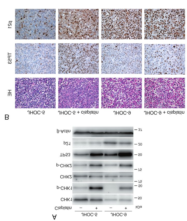
Minagawa et al: p21 Expression in Ovarian Clear-cell Carcinoma

Table I. Clinicopathological characteristics and immunohistochemical results of the cases of clear-cell carcinoma of the ovary.

Case     Age (years)         TNM           Stage*      CHK1      CHK2     TP53         p21      p21 score        Time of              Outcome
no.      at diagnosis                                                                                           recurrence

1            55           pT3aN0M0          IIIA2         +        +        +          High       200           2 Months          DOD, 4 months
2            48           pT1cN0M0          IC(1)         +        +        −          High       210               −
3            56           pT1cN0M0          IC(1)         +        +        +          Low        100               −
4            49           pT1cN0M0          IC(1)         +        +        +          High       210               −
5            40           pT1cN0M0          IC(3)         +        +        +          High       200               −
6            43           pT1aN0M0            IA          +        +        +          High       240               −
7            67           pT1cN0M0          IC(1)         +        +        −          Low         30               −
8            46           pT1aN0M0            IA          +        +        +          Low         20               −
9            79           pT1aN0M0            IA          −        +        −          Low        110               −
10           53          pT1c3N0M0           IC3          +        +        +          High       270               −
11           62          pT1c1N0M0           IC1          +        +        +          Low        170               −
12           69           pT1cNXM0          IC(1)         +        +        +          Low         30               −
13           78           pT1cN0M0          IC(3)         +        +        +          High       230               −             DOOD, 3 months
14           58           pT1cN0M0          IC(1)         +        +        +          Low        150               −
15           65           pT1aN0M0            IA          +        +        +          High       240               −
16           60          pT1c3N0M0           IC3          +        +        +          High       230               −
17           53          pT3cNXMX            IIIC         −        +        +          High       230           36 Months
18           62          pT3bNXM0            IIIB         +        +        +          High       260               −
19           48           pT1cN0M0          IC(2)         +        +        +          Low        180               −
20           58           pT1aN0M0            IA          +        +        +          Low         20               −
21           39          pT1c3N0M0           IC3          +        +        +          Low        140            9 Months         DOD, 15 months
22           59          pT1c1NXM0           IC1          +        +        +          Low         10           70 Months

DOD: Died of disease; DOOD: died of other disease; High: score ≥200; Low: score
ANTICANCER RESEARCH 40: 5631-5639 (2020)
Figure 1. Representative images of immunostaining for checkpoint kinase 1 (CHK1), checkpoint kinase 2 (CHK2), tumour protein p53 (TP53) and p21
in clear-cell carcinoma cases. Significant expression of CHK1, CHK2, and TP53 was noted for the majority of these cases. In contrast, p21 expression
levels by immunohistochemistry were categorized as low (score=20, case 8) and high (score=270, case 10). Original magnification ×400.
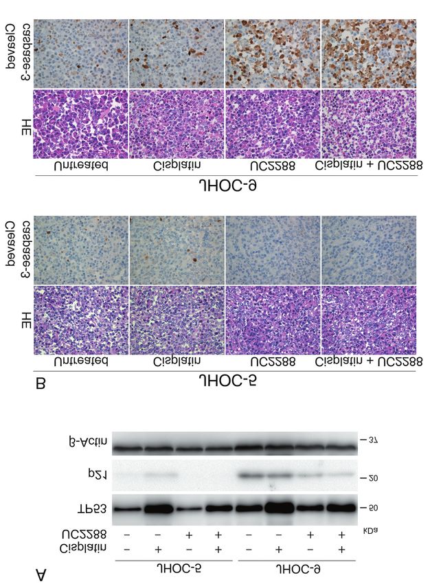
level of TP53 was noted after cisplatin treatment of both cell alone, or a combination of cisplatin and UC2288. The
lines. Prior to treatment, p21 expression was higher in concentrations of cisplatin and UC2288 were 10 μg/ml and
JHOC-9 cells, while its expression was minimal in JHOC-5 10 μM, respectively. In both cultured cell lines, proliferation
cells. Cisplatin treatment had a mild suppressive effect on was inhibited by treatment with cisplatin alone, and was
JHOC-9 cells, preserving significant p21 expression. suppressed to a greater extent by UC2288 alone, and by
cisplatin plus UC2288 (Figure 3). Notably, JHOC-9
IHC expression and location of TP53 and p21 before and underwent cell death after treatment with UC2288 alone, or
after cisplatin treatment. TP53 and p21 expression was combined with cisplatin (Figure 3). No significant difference
examined in cell blocks (Figure 2B). TP53 was sporadically was observed in the suppressive effect between UC2288
positive in untreated JHOC-5 and JHOC-9 cells, and the when used alone and in combination with cisplatin,
number of positive cells increased after cisplatin treatment in suggesting that combining these two agents did not result in
both groups. Regarding p21, a clonal positive reaction was a synergistic effect.
evident in untreated JHOC-9 cells compared to JHOC-5 cells.
The nuclear expression of p21 in JHOC-9 cells remained high Cell fate-determinant proteins in cultured cells and cell block
even after cisplatin treatment, whereas the number of p21- with cisplatin and UC2288 treatment. TP53 and p21
positive cells increased in the JHOC-5 cell line. expression was investigated in cultured cells treated with
Collectively, the above results using cell culture and cell either cisplatin or UC2288 alone, or their combination. p21
block indicated that the sensors and transducers we examined expression was eliminated by UC2288 in JHOC-5 cells. In
in the DDR pathway were operational in two types of JHOC-9 cells, the expression level of p21 was reduced by
cultured CCC cells. Notably, the expression pattern of p21 UC2288 with and without cisplatin (Figure 4A). Caspase-3
was different between JHOC-5 and JHOC-9 cells; p21 was activation was examined in cell blocks (Figure 4B). No
highly expressed in JHOC-9 cells regardless of cisplatin significant positivity for cleaved caspase-3, the active form
treatment. Hence, we subsequently performed the study of caspase-3, was noted in JHOC-5 cells treated with agents
using UC2288, an attenuator of p21. alone nor in combination. In contrast, the number of cleaved
caspase-3-positive JHOC-9 cells significantly increased after
Susceptibility of cultured cells to cisplatin and UC2288. UC2288 treatment with and without cisplatin. Cisplatin alone
Cultured cells were treated with either cisplatin or UC2288 induced caspase-3 activation in a few JHOC-9 cells. These
5634Minagawa et al: p21 Expression in Ovarian Clear-cell Carcinoma
Figure 2. Expression and phosphorylation of molecules in DNA-damage response of cultured ovarian clear-cell cancer cell lines JHOC-5 and
JHOC-9 treated with 10 μg/ml cis-diamminedichloridoplatinum(II) (cisplatin) for 24 h. A: Western blot analysis. B: Immunohistochemical staining
of cell block sections.
results indicate that p21 inhibition is more likely to reduce phosphorylation or activation of CHK1, CHK2, and TP53
cell viability, causing cell death in CCC cells with high p21 was also identified. There was a different pattern of p21
expression compared to those with low p21 expression. expression between the cell types. Prior to stimulation, p21
was constitutively expressed in JHOC-9 cells, whereas its
Discussion expression was minimal in JHOC-5 cells. p21 Inhibition by
UC2288 reduced cell viability in both JHOC-5 and JHOC-9
The present study investigated molecular expression during cells; however, its suppressive effect was more substantial in
DDR occurring in CCC using surgical sections and two types JHOC-9 cells causing cell death, which was confirmed by
of cultured cells. We first evaluated IHC expression of CHK1, cleaved caspase-3 activation.
CHK2, and TP53 in DDR (Figure 1 and 5). Half of our cases We examined the DDR pathway in cultured CCC cells and
demonstrated high p21 expression, whereas the remainder found that CHK1 and CHK2 were phosphorylated after
exhibited low p21 expression. In treated cultured CCC cells, incubation with cisplatin, indicative of preserved functions
5635ANTICANCER RESEARCH 40: 5631-5639 (2020) Figure 3. Cultured cells were treated with 10 μg/ml cis-diamminedichloridoplatinum(II) (cisplatin) or 10 μM UC2288, a p21-specific inhibitor, or their combination and proliferation was analysed. A: The microscopic appearance of JHOC-5 and JHOC-9 cells treated for 24 h. B: Proliferation of JHOC-5 and JHOC-9 cells. Significantly different at *p
Minagawa et al: p21 Expression in Ovarian Clear-cell Carcinoma

Figure 4. Cultured cells were treated with 10 μg/ml cis-diamminedichloridoplatinum(II) (cisplatin) or 10 μM UC2288, a p21-specific inhibitor, or
their combination for 24 h and then analysed for proteins involved in determining cell fate. A: Western blot analysis of JHOC-5 and JHOC-9 cells.
B: Immunohistochemical staining of cleaved caspase-3 in JHOC-5 and JHOC-9 cell blocks.

                                                                                                                                           5637
ANTICANCER RESEARCH 40: 5631-5639 (2020)
and ovarian cancer (24, 25). Others demonstrated that
enhanced nuclear expression of p21 was associated with a
favourable clinical course in localized RCC (21) and ovarian
cancer (25), whereas the opposite was reported in metastatic
RCC (21) and prostate cancer (22). In the present study, we
classified surgical sections from patients with CCC into groups
with high and low p21 expression. However, we were unable
to demonstrate an association between the p21 expression
pattern and patients’ clinicopathological characteristics. This is
because most patients had disease at an early pathological stage
and were followed-up for a short period of time. Based on our
findings, in which UC2288 induced death in cells with high
p21 expression, it is anticipated that UC2288 may serve as a
therapeutic agent to treat patients with CCC highly expressing
p21. However, future studies using a larger cohort of patients
with CCC with a longer follow-up period are required to
validate our hypothesis.
Conflicts of Interest
The Authors declare no conflicts of interest and that no funding was
received for this study.
Authors’ Contributions
YM, KI, and RW were involved in the conception and design of the
Figure 5. The signalling pathway of the DNA-damage response. PARP:
study. YM, RW, and RO performed the histological examination and
Poly (ADP ribose) polymerase; KU80: 80-kDa Ku protein; ATR: ATM-
analysis. YM, KI, and TT acquired the clinical data. YM and KI
and RAD3-related; ATM: ataxia-telangiectasia mutated; CHK1:
checkpoint kinase 1; CHK2: checkpoint kinase 2; TP53: tumour protein performed the biological and cell culture experiments. YM, KI, RW,
p53; p21; 21-kDa protein; PCNA: proliferating cell nuclear antigen; and RO analysed and interpreted the data. RW and MK performed
CDK2: cyclin-dependent kinase 2. the statistical analysis. YM, KI, and RW drafted the manuscript.
MK, ZN, TT and RO provided critical revisions to the article. All
Authors read and approved the final article.
Acknowledgements
growth of CCC cell lines was inhibited by a UC2288. To our
knowledge, this is the first report to demonstrate the anticancer The Authors would like to acknowledge the excellent assistance of
property of a p21 inhibitor in CCC cell lines. Although p21 Ms Kiyoko Kawahara for cell culture, Mr Takenori Fujii and Ms
Yoko Kawamoto for help with histology and immunohistochemical
acts as an inhibitor of CDK in the nucleus, it also acts as a
staining, and Mr Kiyoshi Teduka for help with western blot analysis
regulator of cell death in the cytoplasm. p21 binds to caspase- (Department of Integrated Diagnostic Pathology, Nippon Medical
3 and inhibits cleavage and transformation to the activated School, Tokyo, Japan).
form of caspase-3 (20). Additionally, it was shown that p21
forms a complex with apoptosis signal-regulating kinase 1 and References
inhibits the apoptosis signal (4). Therefore, it is conceivable
that UC2288 attenuates the inhibitory effect of the p21- 1 Blanpain C, Mohrin M, Sotiropoulou PA and Passegue E: DNA-
dependent mechanism on pro-apoptotic molecules in damage response in tissue-specific and cancer stem cells. Cell
cytoplasm. Our findings, in which caspase-3 cleavage was Stem Cell 8: 16-29, 2011. PMID: 21211780. DOI: 10.1016/
enhanced in JHOC-9 cells supports this hypothesis. However, j.stem.2010.12.012
the exact aetiology underlying the different expression levels 2 Dasari S and Tchounwou PB: Cisplatin in cancer therapy:
Molecular mechanisms of action. Eur J Pharmacol 740: 364-378,
of p21 between JHOC-5 and JHOC-9 cells leading to a
2014. PMID: 25058905. DOI: 10.1016/j.ejphar.2014.07.025
subsequent cellular response is unclear, and additional studies
3 Shigetomi H, Higashiura Y, Kajihara H and Kobayashi H:
may reveal the mechanism in future. Targeted molecular therapies for ovarian cancer: An update and
Nuclear expression of p21 was documented in renal cell future perspectives. Oncol Rep 28: 395-408, 2012. PMID:
carcinoma (RCC) (21), prostate cancer (22), breast cancer (23), 22641286. DOI: 10.3892/or.2012.1833
5638Minagawa et al: p21 Expression in Ovarian Clear-cell Carcinoma
4 Dutto I, Tillhon M, Cazzalini O, Stivala LA and Prosperi E: in situ hybridization study. Lung Cancer 68(3): 375-382, 2010.
Biology of the cell cycle inhibitor p21(CDKN1A): Molecular PMID: 19712993. DOI: 10.1016/j.lungcan.2009.07.014
mechanisms and relevance in chemical toxicology. Arch Toxicol 17 Kashiyama T, Oda K, Ikeda Y, Shiose Y, Hirota Y, Inaba K,
89: 155-178, 2015. PMID: 25514883. DOI: 10.1007/s00204- Makii C, Kurikawa R, Miyasaka A, Koso T, Fukuda T, Tanikawa
014-1430-4 M, Shoji K, Sone K, Arimoto T, Wada-Hiraike O, Kawana K,
5 Kreis NN, Louwen F and Yuan J: The multifaceted p21 Nakagawa S, Matsuda K, McCormick F, Aburatani H, Yano T,
(CIP1/WAF1/CDKN1A) in cell differentiation, migration and Osuga Y and Fujii T: Antitumor activity and induction of TP53-
cancer therapy. Cancers 11: 1220, 2019. PMID: 31438587. DOI: dependent apoptosis toward ovarian clear cell adenocarcinoma
10.3390/cancers11091220 by the dual PI3K/mTOR inhibitor DS-7423. 9(2): e87220, 2014.
6 Cazzalini O, Scovassi AI, Savio M, Stivala LA and Prosperi E: PMID: 24504419. DOI: 10.1371/journal.pone.0087220
Multiple roles of the cell cycle inhibitor p21(CDKN1A) in the 18 Abbas T and Dutta A: p21 In cancer: Intricate networks and
DNA damage response. Mutation Res 704: 12-20, 2010. PMID: multiple activities. Nat Rev Cancer 9: 400-414, 2009. PMID:
20096807. DOI: 10.1016/j.mrrev.2010.01.009 19440234. DOI: 10.1038/nrc2657
7 Gartel AL and Tyner AL: The role of the cyclin-dependent 19 Al Bitar S and Gali-Muhtasib H: The role of the cyclin-
kinase inhibitor p21 in apoptosis. Mol Cancer Ther 1: 639-649, dependent kinase inhibitor p21CIP1/WAF1 in targeting cancer:
2002. PMID: 12479224. Molecular mechanisms and novel therapeutics. Cancers 11:
8 Machida H, Matsuo K, Yamagami W, Ebina Y, Kobayashi Y, Tabata 1475, 2019. PMID: 31575057. DOI: 10.3390/cancers11101475
T, Kanauchi M, Nagase S, Enomoto T and Mikami M: Trends and 20 Suzuki A, Tsutomi Y, Akahane K, Araki T and Miura M:
characteristics of epithelial ovarian cancer in Japan between 2002 Resistance to Fas-mediated apoptosis: Activation of caspase 3 is
and 2015: A JSGO-JSOG joint study. Gynecol Oncol 153: 589-596, regulated by cell cycle regulator p21WAF1 and IAP gene family
2019. PMID: 30905436. DOI: 10.1016/j.ygyno.2019.03.243 ILP. Oncogene 17: 931-939, 1998 PMID: 9747872. DOI:
9 Takahashi K, Takenaka M, Kawabata A, Yanaihara N and 10.1038/sj.onc.1202021
Okamoto A: Rethinking of treatment strategies and clinical 21 Robert HW, Borowsky AD, Seligson D, Lin PY, Dillard-Telm L,
management in ovarian clear-cell carcinoma. Int J Clin Oncol Belldegrun AS, Figlin RA and Pantuck AD: p21 Is a prognostic
25: 425-431, 2020. PMID: 31989349. DOI: 10.1007/s10147- marker for renal cell carcinoma: implications for novel
020-01625-w therapeutic approaches. J Urol 177: 63-69, 2007. PMID:
10 Chan JK, Teoh D, Hu JM, Shin JY, Osann K and Kapp DS: Do 17162001. DOI: 10.1016/j.juro.2006.08.073
clear-cell ovarian carcinomas have poorer prognosis compared 22 Baretton GB, Klenk U, Diebold J, Schmeller N and Löhrs U:
to other epithelial cell types? A study of 1411 clear-cell ovarian Proliferation- and apoptosis-associated factors in advanced
cancers. Gynecol Oncol 109: 370-376, 2008. PMID: 18395777. prostatic carcinomas before and after androgen deprivation
DOI: 10.1016/j.ygyno.2008.02.006 therapy: Prognostic significance of p21/WAF1 in advanced
11 Itamochi H, Nishimura M, Nishimura M, Oumi N, Kato M, prostate cancer. Br J Cancer 80(3-4): 546-555, 1999. PMID:
Oishi T, Shimada M, Sato S, naniwa J, Sato S, Kudoh A, Kigawa 10408865. DOI: 10.1038/sj.bjc.6690390
J and Harada T: Checkpoint kinase inhibitor AZD7762 23 Zohny SF, Al-Malki AL, Zamzami MA and Choudhry H:
overcomes cisplatin resistance in clear cell carcinoma of the p21WAF1/CIP1: Its paradoxical effect in the regulation of breast
ovary. Int J Gynecol Cancer 24(1): 61-69, 2014. PMID: cancer. Breast Cancer 26(2): 131-137, 2019. PMID: 30255294.
24362713. DOI: 10.1097/IGC.0000000000000014 DOI: 10.1007/s12282-018-0913-1
12 Itamochi H, Yoshida T, Walker CL, Bartholomeusz C, Aoki D, 24 Shimizu M, Nikaido T, Toki T, Shiozawa T and Fujii S: Clear-cell
Ishihara H, Suzuki N, Kigawa J, Terakawa N and Ueno NT: carcinoma has an expression pattern of cell cycle regulatory
Novel mechanism of reduced proliferation in ovarian clear cell molecules that is unique among ovarian adenocarcinomas. Cancer
carcinoma cells: Cytoplasmic sequestration of CDK2 by p27. 85(3): 669-677, 1999. PMID: 10091740. DOI: 10.1002/(sici)1097-
Gynecol Oncol 122: 641-647, 2011. PMID: 21652059. DOI: 0142(19990201)85:33.0.co;2-f
10.1016/j.ygyno.2011.05.003 25 Anttila MA, Kosma VM, Puolakka J, Juhola M, Saarikoski S
13 Liu R, Wettersten HI, Park SH and Weiss RH: Small-molecule and Syrjänen: p21/WAF1 Expression as related to p53, cell
inhibitors of p21 as novel therapeutics for chemotherapy- proliferation and prognosis in epithelial ovarian cancer. Br J
resistant kidney cancer. Future Med Chem 5(9): 991-994, 2013. Cancer 79(11/12): 1870-1878, 1999. PMID: 10206307. DOI:
PMID: 23734682. DOI: 10.4155/fmc.13.56 10.1038/sj.bc.6690298
14 Wettersten HI, Hwang SH, Li C, Shiu EY, Wecksler AT, 26 Prat J and FIGO Committee on Gynecologic Oncology: Staging
hammock BD and Weiss RH: A novel p21 attenuator which is classification for cancer of the ovary, fallopian tube, and
structurally related to sorafenib. Cancer Biol Ther 14: 278-285, peritoneum. Int J Gynaecol Obstet 124(1): 1-5, 2014. PMID:
2013. PMID: 23298903. DOI: 10.4161/cbt.23374 24219974. DOI: 10.1016/j.ijgo.2013.10.001
15 Tay VSY, Devaraj S, Koh T, Ke G, Crasta KC and Ali Y:
Increased double-strand breaks in diabetic β-cells with a p21
response that limits apoptosis. Sci Rep 9(1): 19341, 2019.
PMID: 31852915. DOI: 10.1038/s41598-019-54554-8
16 Lee HJ, Xu X, Choe G, Chung DH, Seo JW, Lee JH, Lee CT,
Jheon S, Sung SW and Chung JH: Protein overexpression and
gene amplification of epidermal growth factor receptor in non- Received July 31, 2020
small cell lung carcinomas: Comparison of four commercially Revised August 14, 2020
available antibodies by immunohistochemistry and fluorescence Accepted August 23, 2020
5639You can also read