Negative regulation between the expression levels of receptor for hyaluronic acid mediated motility and hyaluronan leads to cell migration in ...
←
→
Page content transcription
If your browser does not render page correctly, please read the page content below
ONCOLOGY LETTERS 20: 199, 2020

                Negative regulation between the expression levels of
                 receptor for hyaluronic acid‑mediated motility and
               hyaluronan leads to cell migration in pancreatic cancer
         XIAO‑BO CHENG1*, SHUWEN WANG1*, HUA YANG1, HANXING TONG2, GUODONG SHI1, LILI WU3,
          JIAN ZHOU1, LEI SHI1, HONGWEI LI1, SONGTAO REN1, YUHONG ZHOU4 and NORIHIRO SATO5

           1
            Shanghai Public Health Clinical Center, Fudan University, Shanghai 201508; Departments of 2General Surgery,
            3
              Radiotherapy and 4Medical Oncology, Zhongshan Hospital, Fudan University, Shanghai 200032, P.R. China;
                           5
                             Department of Surgery 1, School of Medicine, University of Occupational and
                                   Environmental Health, Kitakyushu, Fukuoka 807‑8555, Japan

                                        Received February 15, 2019; Accepted July 22, 2020

                                                      DOI: 10.3892/ol.2020.12060

Abstract. Receptor for hyaluronic acid (HA)‑mediated motility          mRNA expression and HA concentration in PDAC cells and
(RHAMM) expression is upregulated in pancreatic ductal                 tissues. The negative correlation between RHAMM mRNA
adenocarcinoma (PDAC). In the present study, small inter‑              expression and HA concentration was demonstrated in other
fering RNA knockdown was used to investigate the regulatory            models, including SUIT2 cells treated with an HA inhibitor or
mechanism and function of RHAMM in PDAC cells. Reverse                 stimulator and a system involving co‑culture of SUIT2 cells
transcription‑quantitative PCR was used to measure the                 and stromal fibroblasts. The present findings demonstrated a
mRNA expression levels of RHAMM, hyaluronan synthases                  negative correlation between RHAMM mRNA expression and
(HAS1, HAS2 and HAS3) and hyaluronidases (HYAL1,                       HA production in a subset of PDAC cell lines. The efficacy of
HYAL2 and HYAL3) in eight PDAC cell lines. Cell migration              a therapeutic strategy targeting RHAMM should be carefully
was assessed using a Transwell assay, while HA concentra‑              evaluated in future studies.
tion was measured using an ELISA. The results revealed that
RHAMM‑knockdown significantly increased migration in                   Introduction
two PDAC cell lines, significantly decreased migration in one
cell line and did not affect migration in the other cell lines,        Pancreatic ductal adenocarcinoma (PDAC) is an aggressive
and was positively associated with changes in HA production.           and lethal neoplasm, with a 5‑year survival rate
2 CHENG et al: NEGATIVE REGULATION BETWEEN RHAMM AND HA IN PDAC
MAPK, leading to ERK phosphorylation and activation of TPA (100 ng/ml) on cell viability were analyzed using TBDE
the transcription effectors activator protein‑1 and NF κ B, assays as measurements of cytotoxicity. PDAC cells (~2x10⁵) were
resulting in cell migration (6). cultured at 37˚C in the medium. The untreated and RHAMM
High CD44 expression is associated with a poor prog‑ siRNA‑treated cells were harvested after 24 h of transfection,
nosis in patients with PDAC, and its knockdown inhibits the while the untreated, 4‑MU‑ and TPA‑treated cells were harvested
migration of PDAC cells (7). In our previous study, it was after 48 h of treatment. Subsequently, 4% trypan blue was added
demonstrated that RHAMM is upregulated in PDAC cells and at room temperature for 3 min, and the cells were counted using
tissues, and is associated with a poor prognosis in patients with a LUNA™ automated cell counter (Logos Biosystems, Inc.),
PDAC following surgical resection (8). Previously, RHAMM according to the manufacturer's protocol. Cytotoxicity was
has been used as a promising target for antibody therapy to determined from the number of viable cells (no color) in treated
inhibit the extracellular function of RHAMM on the surface samples as a percentage of that in the untreated control.
of cancer cells (9). Therefore, the present study hypothesized
that the migration of PDAC cells may decrease following inhi‑ si R NA t a rget i ng R H A M M. si R NA for R H A M M
bition of RHAMM. To test this hypothesis, small interfering (ON‑TARGETplus Human HMMR siRNA‑SMARTpool
(si)RNA knockdown was used to determine the effects of L‑010409‑00‑0005) and negative control siRNA (siGENOME
RHAMM on the migratory ability of PDAC cells. Non‑Targeting siRNA Pool D‑001206‑13‑05) were purchased
from Cytiva. All PDAC cell lines were transfected with 100 nM
Materials and methods siRNA using DhermaFECT 1 Transfection Reagent (Cytiva)
for 24 h at 37˚C, according to the manufacturer's protocol.
Cell lines and reagents. A total of eight PDAC cell lines The target sequences were: GUUA ACAGCCAGUGAGAUA
were used in the present study. BxPC‑3, Panc‑1, Capan‑2, [molecular weight (MW), 13,414.8 g/mol]; GGACUAAUG
CFPAC1 and AsPC‑1 were purchased from American Type AACUACUAAA (MW, 13,384.8 g/mol); GAUACUACCUUG
Culture Collection, NOR‑P1 from the RIKEN BioResource CCUGCUU (MW, 13,429.9 g/mol); CAAGUGGCGUCUCCU
Center, and SUIT‑2 and KP‑2 from the Japanese Collection of CUAU (MW, 13,444.8 g/mol).
Research Bioresources Cell Bank. The characteristics of the
eight PDAC cell lines are shown in Table I. These cell lines had Reverse transcription‑quantitative (RT‑q)PCR. Total RNA
recently been obtained when the experiments were performed was isolated from all cells (untreated and treated) and
and their identities were routinely monitored by short tandem tissues (matched pairs of primary pancreatic tumor and
repeat profiling. Primary fibroblasts (sk‑f2) derived from adjacent non‑tumor tissues) using the RNeasy ® Mini kit
PDAC tissues were donated by Kyushu University (Fukuoka, (Qiagen, Inc.). Single‑stranded cDNA was synthesized from
Japan). PDAC cell lines and sk‑f2 cells were maintained in 1 µg total RNA using the SuperScript® VILO™ MasterMix
RPMI‑1640 medium supplemented with 10% FBS and 1% (Invitrogen; Thermo Fisher Scientific, Inc.) for 10 min at 25˚C,
streptomycin and penicillin (all Thermo Fisher Scientific, 1 h at 42˚C and 5 min at 85˚C. mRNA expression analyses
Inc.) in a 5% CO2 incubator at 37˚C. 4‑methylumbelliferone of RHAMM (Hs00234864_m1), HAS1 (Hs00758053_m1),
(4‑MU) and 12‑O‑tetradecanoyl‑phorbol‑13‑acetate (TPA) HAS2 (Hs00193435_m1), HAS3 (Hs00193436_m1), HYAL1
were purchased from Sigma‑Aldrich (Merck KGaA). (Hs00201046_m1), HYAL2 (Hs01117343_g1), HYAL3
(Hs00185910_m1) and the housekeeping gene GAPDH
Tissue samples. Patients aged ≥18 years old with histologically (Hs02758991_g1), used as a control, were performed using
confirmed PDAC diagnosed and a Karnofsky Performance TaqMan® Fast Universal PCR Master Mix, TaqMan Gene
Score ≥80 were enrolled. The main exclusion criteria were Expression Assays and Step One Plus real‑time PCR instru‑
as follows: i) Central nervous system metastasis; and ii) other ment (all Applied Biosystems; Thermo Fisher Scientific, Inc)
malignant tumors. Surgical tissue specimens (matched pairs of at room temperature for 45 min. The thermocycling conditions
primary pancreatic tumor and adjacent non‑tumor tissues, 1 cm used for qPCR consisted of an initial denaturation step for
away from the tumor tissue) collected from 20 patients with 20 sec at 95˚C, followed by 40 cycles of denaturation for 1 sec
PDAC (11 men and 9 women; age range, 34‑82 years; median at 95˚C, annealing for 20 sec at 60˚C and extension for 30 sec
age, 64 years) between August 2012 and December 2014 at at 72˚C. The relative quantification was performed using the
the School of Medicine of the University of Occupational and comparative 2‑ΔΔCq method (10).
Environmental Health (Kitakyushu, Japan) were evaluated by
an experienced pathologist. All samples were collected and Migration assay. After 24 h of treatment with siRNAs, the
handled in an identical manner. Tissue aliquots were placed cells were harvested and counted, and ~2x10 4 cells were
in RNAlater® Stabilization Solution (Ambion; Thermo Fisher seeded in 100 µl medium with 1% FBS in the top chamber with
Scientific, Inc.) at ‑80˚C until further use. The present study an uncoated membrane (24‑well Transwell insert; 8‑µm pore
was approved by the Ethics Committee of the University of size; BD Biosciences). A total of 900 µl medium containing
Occupational and Environmental Health (Kitakyushu, Japan), 10% FBS was used as a chemoattractant in the lower chamber,
and written informed consent was obtained from all patients and the cells were allowed to migrate at 37˚C with 5% CO2
who approved the use of their tissues for unspecified research for 48 h. Non‑migrating cells on the upper surface of the
purposes. membrane were removed with a cotton swab. Migrating cells
that penetrated onto the lower surface of the membrane were
Trypan blue dye exclusion (TBDE) assay of cell viability. The fixed with 70% methanol for 10 min, stained with hematoxylin
effects of RHAMM siRNA (100 nM), 4‑MU (1,000 µM) and for 10 min and eosin for 5 min, all at room temperature, andONCOLOGY LETTERS 20: 199, 2020 3 Table I. Characteristics of eight pancreatic ductal adenocarcinoma cell lines. Cell line Derivation Metastasis Proliferation, h Differentiation BxPC‑3 Primary tumor No 48‑60 Moderate to poor PANC‑1 Primary tumor Yes 52 Poor NOR‑P1 Liver metastasis Yes 16 Poor SUIT‑2 Liver metastasis Yes 38.2 Moderate KP‑2 Primary tumor No 48 Moderate Capan‑2 Primary tumor No 96 Well CFPAC‑1 Liver metastasis Yes 31 Well AsPC‑1 Ascites Yes 38‑40 Poor Figure 1. Changes in RHAMM mRNA expression in eight PDAC cell lines transfected with siRNA targeting RHAMM. Transfection of PDAC cell lines with 100 nM siRNA targeting RHAMM resulted in 18‑86% knockdown of RHAMM mRNA expression. Data are presented as the mean ± SD of three replicates. * P
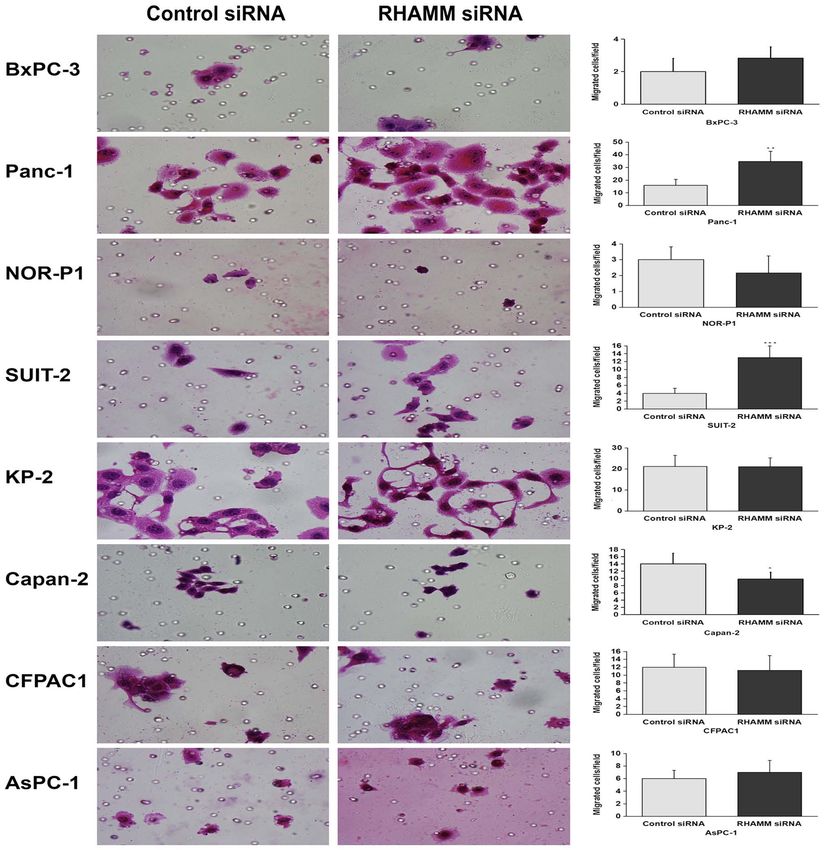
4                                CHENG et al: NEGATIVE REGULATION BETWEEN RHAMM AND HA IN PDAC

Figure 2. Changes in migration of eight pancreatic ductal adenocarcinoma cell lines following knockdown of RHAMM analyzed using Transwell migration
assays (magnification, x400). The numbers of migrating cells following knockdown of RHAMM were significantly increased in two cell lines (Panc‑1 and
SUIT‑2), significantly decreased in Capan‑2 cells and remained unchanged in the other cell lines. Data are presented as the mean ± SD of six replicates.
*
 P
ONCOLOGY LETTERS 20: 199, 2020                                                                  5

Figure 3. Changes in HA production in eight pancreatic ductal adenocarcinoma cell lines following knockdown of RHAMM‑ analyzed via ELISA. HA
production was significantly increased in two cell lines (Panc‑1 and SUIT‑2), significantly decreased in three cell lines (NOR‑P1, CFPAC1 and Capan‑2) and
remained unchanged in the other cell lines following RHAMM‑knockdown. Data are presented as the mean ± SD of three replicates. *P
6                                 CHENG et al: NEGATIVE REGULATION BETWEEN RHAMM AND HA IN PDAC

Figure 4. Changes in HAS1 mRNA expression in eight pancreatic ductal adenocarcinoma cell lines following treatment with siRNA targeting RHAMM. Data
are presented as the mean ± SD of three replicates. The white boxes at the bottom are a comprehensive representation of HAS1 mRNA expression in all cell
lines, with the data presented as the median and interquartile range, revealing that HAS1 mRNA expression was not significantly changed following treatment.
HAS1, hyaluronan synthase 1; RHAMM, receptor for hyaluronic acid‑mediated motility; siRNA, small interfering RNA; RQ, relative quantity.

cells, while no significant difference was observed in the                      AsPC‑1; Fig. 7), HYAL2 mRNA expression in three cell lines
remaining cell lines (NOR‑P1, BxPC‑3, AsPC‑1, CFPAC1 and                        (NOR‑P1, Capan‑2 and AsPC‑1; Fig. 8) and HYAL3 mRNA
KP‑2; Fig. 2) compared with control siRNA‑treated groups.                       expression in two cell lines (Panc‑1 and AsPC‑1; Fig. 9),
Furthermore, HA production was significantly increased in                       compared with the control siRNA‑treated groups. As afore‑
two cell lines (Panc‑1 and SUIT‑2) and significantly decreased                  mentioned, RHAMM‑knockdown led to increased production
in three cell lines (NOR‑P1, CFPAC1 and Capan‑2), while                         of HA in two cell lines (Panc‑1 and SUIT‑2), which may have
no significant differences were observed in KP‑2, BxPC‑3                        been induced by a significant increase in combined HAS2
and AsPC‑1 cell lines compared with control‑siRNA treated                       mRNA expression compared with the control siRNA‑treated
groups (Fig. 3).                                                                groups (Fig. 5), and decreased production of HA in three cell
    To elucidate the mechanism underlying HA modulation                         lines (NOR‑P1, CFPAC1 and Capan‑2), which may have been
via RHAMM‑knockdown, the mRNA expression levels                                 induced by significantly increased combined HYAL1 mRNA
of HA‑synthesizing enzymes, HAS1‑3 (Figs. 4‑6), and                             expression compared with the control siRNA‑treated groups
HA‑degrading enzymes, HYAL1‑3 (Figs. 7‑9), in PDAC                              (Fig. 7).
cells were analyzed using RT‑qPCR. RHAMM‑knockdown
led to a significantly increased production of HAS2 mRNA                        Correlation between RHAMM mRNA expression and HA
expression in six cell lines (BxPC‑3, Panc‑1, SUIT‑2, KP‑2,                     concentration. The present study hypothesized that there may
CFPAC1 and AsPC‑1; Fig. 5), HYAL1 mRNA expression                               be a correlation between RHAMM mRNA expression and HA
in five cell lines (Panc‑1, NOR‑P1, Capan‑2, CFPAC1 and                         concentration. To test this, RHAMM mRNA expression and
ONCOLOGY LETTERS 20: 199, 2020 7 Figure 5. Changes in HAS2 mRNA expression in eight pancreatic ductal adenocarcinoma cell lines following treatment with siRNA targeting RHAMM. Data are presented as the mean ± SD of three replicates. The white boxes at the bottom are a comprehensive representation of HAS2 mRNA expression in all cell lines, with the data presented as the median and interquartile range, revealing that HAS2 mRNA expression was significantly increased following treatment. *P
8                                 CHENG et al: NEGATIVE REGULATION BETWEEN RHAMM AND HA IN PDAC

Figure 6. Changes in HAS3 mRNA expression in eight pancreatic ductal adenocarcinoma cell lines following treatment with siRNA targeting RHAMM. Data
are presented as the mean ± SD of three replicates. The white boxes at the bottom are a comprehensive representation of HAS3 mRNA expression in all cell
lines, with the data presented as the median and interquartile range, revealing that HAS3 mRNA expression was not significantly changed following treatment.
HAS3, hyaluronan synthase 3; RHAMM, receptor for hyaluronic acid‑mediated motility; siRNA, small interfering RNA; RQ, relative quantity.

Figure 7. Changes in HYAL1 mRNA expression in eight pancreatic ductal adenocarcinoma cell lines following treatment with siRNA targeting RHAMM.
Data are presented as the mean ± SD of three replicates. The white boxes at the bottom are a comprehensive representation of HYAL1 mRNA expression in
all cell lines, with the data presented as the median and interquartile range, revealing that HYAL1 expression was significantly increased following treatment.
*
 P
ONCOLOGY LETTERS 20: 199, 2020                                                                  9

Figure 8. Changes in HYAL2 mRNA expression in eight pancreatic ductal adenocarcinoma cell lines following treatment with siRNA targeting RHAMM.
Data are presented as the mean ± SD of three replicates. The white boxes at the bottom are a comprehensive representation of HYAL2 mRNA expression in
all cell lines, with the data presented as the median and interquartile range, revealing that HYAL2 mRNA expression was not significantly changed following
treatment. *P
10                               CHENG et al: NEGATIVE REGULATION BETWEEN RHAMM AND HA IN PDAC

Figure 9. Changes in HYAL3 mRNA expression in eight pancreatic ductal adenocarcinoma cell lines after treatment with siRNA targeting for RHAMM.
Data are presented as the mean ± SD of three replicates. The white boxes at the bottom are a comprehensive representation of HYAL3 mRNA expression in
all cell lines, with the data presented as the median and interquartile range, revealing that HYAL3 mRNA expression was not significantly changed following
treatment. *P
ONCOLOGY LETTERS 20: 199, 2020                                                              11

Figure 11. SUIT‑2‑based models proved that RHAMM mRNA expression is significantly and negatively associated with HA concentration. (A) HA concentra‑
tion was significantly decreased and RHAMM mRNA expression levels were significantly increased when SUIT‑2 cells were treated with an HA inhibitor
(4‑MU; 1,000 µM). (B) HA concentration was significantly increased and RHAMM mRNA expression levels were significantly decreased when SUIT‑2
cells were treated with an HA stimulator (TPA; 100 ng/ml). (C) HA concentration was significantly increased and RHAMM mRNA expression levels were
significantly decreased when SUIT‑2 cells were co‑cultured with primary fibroblasts (sk‑f2). An independent‑samples t‑test was used to analyze the data.
Data are presented as the mean ± SD of three replicates. 4‑MU, 4‑methylumbelliferone; TPA, 12‑O‑tetradecanoyl‑phorbol‑13‑acetate; HA, hyaluronic acid;
RHAMM, receptor for HA‑mediated motility; RQ, relative quantity.

    The present findings suggest a complex mechanism                          Funding from Shanghai Public Health Clinical Center (grant
regulating RHAMM mRNA expression and HA synthesis                             no. KY‑GW‑2018‑03), the Shanghai Municipal Committee
in PDAC cells at the genetic level. There may be a negative                   for Health and Family Planning (grant no. 201740194), the
correlation between RHAMM mRNA expression and HA                              Clinical Science and Technology Innovation Foundation of
production in a subset of PDAC cell lines. Future studies                     Shanghai Shenkang Hospital Development Center (grant
should confirm whether there is a negative correlation between                no. SHDC12018112) and the National 13th Five‑Year
RHAMM expression and HA production in PDAC cell lines                         Grand Program on Key Infectious Disease Control (grant
and tissues at the protein level, and explore its mechanism in                no. 2018ZX10302103‑003).
depth in order to provide a stronger scientific basis for the
evaluation of the risk and benefit of a therapeutic strategy                  Availability of data and materials
targeting RHAMM.
                                                                              All data generated or analyzed during this study are included
Acknowledgements                                                              in this published article.

Not applicable.                                                               Authors' contributions

Funding                                                                       NS, YZ and XC conceived the experimental design. XC,
                                                                              SW, HY and HT performed the experiments. XC, SW, GS,
The present study was supported by the Medical and Health                     LW, JZ, LS, HL and SR analyzed the data. XC, SW, NS and
Science and Technology Innovation Foundation of Jinshan                       YZ wrote the paper. All authors read and approved the final
District of Shanghai (grant no. 2018‑3‑23), the Intramural                    manuscript.
12 CHENG et al: NEGATIVE REGULATION BETWEEN RHAMM AND HA IN PDAC
Ethics approval and consent to participate 9. Gust KM, Hofer MD, Perner SR, Kim R, Chinnaiyan AM,
Varambally S, Moller P, Rinnab L, Rubin MA, Greiner J, et al:
RHAMM (CD168) is overexpressed at the protein level and may
The present study was approved by the Ethics Committee of constitute an immunogenic antigen in advanced prostate cancer
the University of Occupational and Environmental Health disease. Neoplasia 11: 956‑963, 2009.
10. Livak KJ and Schmittgen TD: Analysis of relative gene expression
(Kitakyushu, Japan), and written informed consent was data using real‑time quantitative PCR and the 2(‑Delta Delta
obtained from all patients who approved the use of their tissues C(T)) method. Methods 25: 402‑408, 2001.
for unspecified research purposes. 11. Cheng XB, Sato N, Kohi S and Yamaguchi K: Prognostic impact
of hyaluronan and its regulators in pancreatic ductal adenocar‑
cinoma. PLoS One 8: e80765, 2013.
Patient consent for publication 12. Cheng XB, Kohi S, Koga A, Hirata K and Sato N: Hyaluronan
stimulates pancreatic cancer cell motility. Oncotarget 7:
4829‑4840, 2016.
Not applicable. 13. Mahlbacher V, Sewing A, Elsasser HP and Kern HF: Hyaluronan
is a secretory product of human pancreatic adenocarcinoma
cells. Eur J Cell Biol 58: 28‑34, 1992.
Competing interests 14. Fries H, Elsasser HP, Mahlbacher V, Neumann K and Kern HF:
Localisation of hyaluronate (HA) in primary tumors and nude
The authors declare that they have no competing interests. mouse xenografts of human pancreatic carcinomas using a bioti‑
nylated HA‑binding protein. Virchows Arch 424: 7‑12, 1994.
15. McBride WH and Bard JB: Hyaluronidase‑sensitive halos around
References adherent cells. Their role in blocking lymphocyte‑mediated
cytolysis. J Exp Med 149: 507‑515, 1979.
16. Suzuki Y, Nishida Y, Naruse T, Gemba T and Ishiguro N:
1. Siegel R, Naishadham D and Jemal A: Cancer statistics, 2013. Pericellular matrix formation alters the efficiency of intracellular
CA Cancer J Clin 63: 11‑30, 2013. uptake of oligonucleotides in osteosarcoma cells. J Surg Res 152:
2. Hidalgo M: Pancreatic cancer. N Engl J Med 362: 1605‑1617, 148‑156, 2009.
2010. 17. Toole BP: Hyaluronan‑CD44 interactions in cancer: Paradoxes
3. Bardeesy N and DePinho RA: Pancreatic cancer biology and and possibilities. Clin Cancer Res 15: 7462‑7468, 2009.
genetics. Nat Rev Cancer 2: 897‑909, 2002. 18. Sohr S and Engeland K: RHAMM is differentially expressed in
4. Mahadevan D and Von Hoff DD: Tumor‑stroma interactions the cell cycle and downregulated by the tumor suppressor p53.
in pancreatic ductal adenocarcinoma. Mol Cancer Ther 6: Cell cycle 7: 3448‑3460, 2008.
1186‑1197, 2007. 19. Godar S and Weinberg RA: Filling the mosaic of p53 actions:
5. Erkan M, Reiser‑Erkan C, Michalski CW and Kleeff J: Tumor P53 represses RHAMM expression. Cell cycle 7: 3479, 2008.
microenvironment and progression of pancreatic cancer. Exp 20. Lin SL, Chang D, Chiang A and Ying SY: Androgen receptor
Oncol 32: 128‑131, 2010. regulates CD168 expression and signaling in prostate cancer.
6. Schwertfeger KL, Cowman MK, Telmer PG, Turley EA and Carcinogenesis 29: 282‑290, 2008.
McCarthy JB: Hyaluronan, inflammation, and breast cancer
progression. Front Immunol 6: 236, 2015. This work is licensed under a Creative Commons
7. Li XP, Zhang XW, Zheng LZ and Guo WJ: Expression of CD44 Attribution-NonCommercial-NoDerivatives 4.0
in pancreatic cancer and its significance. Int J Clin Exp Pathol 8: International (CC BY-NC-ND 4.0) License.
6724‑6731, 2015.
8. Cheng XB, Sato N, Kohi S, Koga A and Hirata K: Receptor
for hyaluronic acid‑mediated motility is associated with poor
survival in pancreatic ductal adenocarcinoma. J Cancer 6:
1093‑1098, 2015.You can also read