Hormone Circuits Lecture notes Uri Alon (Spring 2021) Lecture 9 The thyroid and its discontents
←
→
Page content transcription
If your browser does not render page correctly, please read the page content below
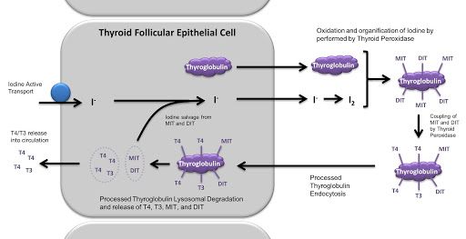
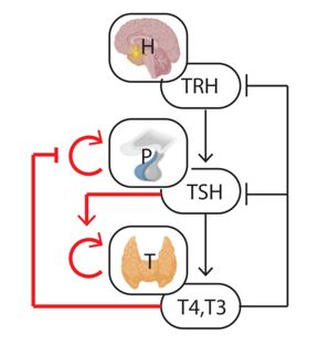
Hormone Circuits Lecture notes
 Uri Alon (Spring 2021)
 Lecture 9

 The thyroid and its discontents
Goal: Understand the biology and diseases of thyroid, to set up the next lecture on the
origin of autoimmune diseases

The HP axes control pillars of life- growth, reproduction, stress, and metabolism. So far, we
studied two, the HPA axis for stress, and the HP-Ovary axis for ovulation. We now study a third,
the HP-thyroid axis, or HPT axis for short, that controls metabolic rate. It is biologically fascinating,
and the origin of many common diseases. Especially diseases in which the body attacks itself-
autoimmune diseases. So, this lecture is the first of a two-lecture mini-series, where we
understand the origin of autoimmune diseases: what is the logic of the body attacking itself. Today
we will use the thyroid to understand the biology, dynamics, and treatment of such diseases. To
do so, we will revisit some of the principles that we studied, to reap the fruit of our work in this
course by understanding otherwise mysterious features of these diseases.

The HPT axis is designed to keep a steady level of thyroid hormone T4

The thyroid is a 10-gram gland at the front of our throat shaped like a butterfly
(Fig. 1). It secretes thyroid hormone, T4, that goes to all cells and has far-
reaching effects on the heart and on metabolism. It is the effector organ of the
HPT axis (Fig. 2). The hypothalamus H secretes hormone x1 (thyroid releasing
hormone TRH), that makes the pituitary P secrete hormone x2 (thyroid
stimulating hormone) from specialized thyrotroph cells. TSH makes the thyroid
secrete T4, our x3 hormone. T4 is converted in the tissues to a more active
form T3. As in all HP axes, T4 inhibits the production of the two upstream Figure 1
hormones x2 and x1, TSH and TRH.

As in the other HP axes, the hormones also control the size of the glands. TSH
makes the thyroid cells proliferate and grow. In fact, growth of the thyroid is
infamous, it is called goiter. It happens when iodine, essential for making T4, is
low in the diet, as in areas far from the sea which in old times lacked access to
the iodine in seafood. Low T4 releases the inhibitors from TSH, and makes the
thyroid grow sometimes by a factor of 10, like a small melon at the throat, to try Figure 2
to make enough thyroid hormone.
You can already see that when T4 is low, TSH is high (Fig.
3). TSH is a very sensitive indicator of problems with the
thyroid. It varies by a factor of a thousand while T4 varies by
a factor of less than 10, with a normal range of 10-20 pmol/L.
The relation between TSH and T4 is steep, almost
exponential. That is why TSH is such a common blood test.
It can even tell if there will be future problems, even though
now T4 is fine- stay tuned.

The pituitary cells that secrete TSH also have size control-
 Figure 3
in this axis, it is T4 that inhibits P cell growth. This is a
variation on the other axes (where x1 enhances P growth, not x3 inhibits it). Why this different
design? Perhaps this axis is designed to keep constant T4 levels, whereas other axes like the
HPA are designed to have a wide range of x3 (cortisol) according to the stress inputs. Indeed, the
equation for P in the HPT axis reads dP/dt=P(b_P/x3-a_P). This locks x3 to a constant steady-
state 3 = ! / ! equal to the ratio of P cell production and removal parameters. In contrast, in
the HPA axis, / = ( ! 2 − ! ) which locks x2 to a constant 2 = ! / ! , and keeps x3 free
to vary with input u.

Indeed, free T4 levels in the circulation are rather constant. We say free T4 because most of T4
is bound in the blood to carrier proteins and is not active. The normal free T4 range in the
population is 10-20 pmol/L as mentioned above, and a given healthy individual varies around their
own personal set point over about 50% of this range.

In some situations, the setpoint can change, however. The hypothalamus sends signals to change
the setpoint. One such case is starvation or serious illness, in which both TSH and T4 are held
low to reduce metabolic activity. This is called sick euthyroid syndrome. Another signal that can
change T4 is temperature, where cold climate increases T4 because it has a role in
thermogenesis (making heat).

The crazy production pipeline of T4

For our understanding of diseases, we need some details of how t4 is made
in the thyroid. T4 is a molecule made of two carbon hexagonal rings from
tyrosine (Fig. 4). Each ring has two iodines attached. In the cells of the
body, T4 is converted to a more active form T3, so called because one of
four iodines is removed by iodinase enzymes, leaving three iodines. Figure 4
T4 is made in modular ‘chemical factories’ in the thyroid called the
thyroid follicles (Fig. 5). Each follicle is a layer of epithelial cells
called thyrocytes surrounding a spherical pore filled with colloid.
Total size is about 400 microns. The thyrocytes import iodine from
the blood vessels surrounding the follicle, then export the iodine
into the colloid (Fig. 6). They make huge amounts of a protein Tg
that has many tyrosines, and dump this also into the colloid. There,
 Figure 5
iodine is added to the tyrosines by an enzyme on the surface of
the thrycytes called TPO. TPO also links two
tyrosines together to make T4. Then, the cells
import the iodinated Tg back inside, break it
up into small pieces, and extract the T4, and
export it out of the cell into the circulation.
Crazy, no?

Remember Tg and TPO for future use. TSH
increases all of the above steps, and the
production of Tg, TPO and the other pumps
and enzymes. Figure 6

Hyperthyroidism and hypothyroidism

To understand the importance and function of T4, let's see what happens when there is too much
or too little of it. Diseases with too much T4 result in hyperthyroidism. The heart beats fast and
irregularly, which can lead to dangerous arrhythmias. You eat more but lose weight. This harks
back to our first lecture, where we saw the appetite and diet lines. Hyperthyroidism increases
metabolic rate and shifts the diet line towards the opposite effect of higher food intake and reduced
fat. Emotions and thoughts race, sometimes resembling mania. There is a feeling of heat and
sweat, due to thermogenesis. Muscles hurt. Hands might shake due to effects on the nervous
system.

When thyroid hormone is too low, hypothyroidism, the effects are opposite. The heart beats
slowly and weakly, and you can be out of breath when climbing the stairs. You gain weight despite
not eating more, with reduced appetite and constipation. Emotions tend towards depression.
There is a clammy feeling of cold. In infants, hypothyroidism can lead to cognitive problems due
to impaired development of the brain- that is why a TSH test at birth is so critical.

Hashimotos disease, a case of self-attack

The most common cause of hypothyroidism is Hashimoto's disease (Fig. 7). Devastatingly
common, it affects 4% of the population, primarily women. In Hashimoto's disease, white blood
cells called T-cells attack and kill thyrocytes. Antibodies against thyrocytes participate in the
damage.
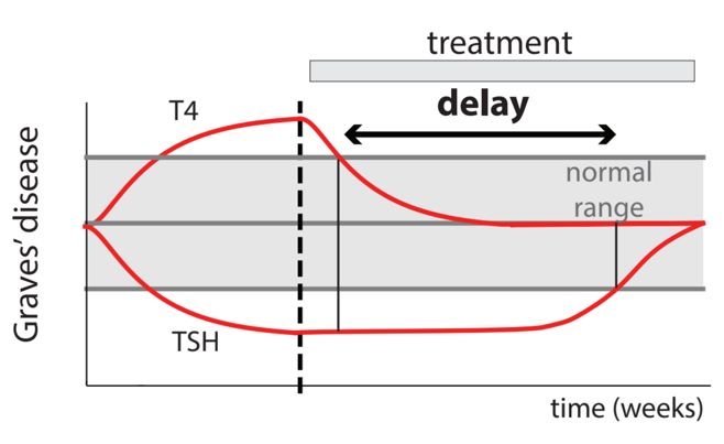
Figure 7 Figure 8

The T-cells normally attack virus infected cells, not healthy cells of the body (Fig. 8). Cells in the
body display small pieces of the protein they make on “identity cards” on their surface called
MHCs. When a virus infects a cell, it displays pieces of the virus proteins in some of its MHCs.
The MHCs are scanned by T cells. Each T-cell has a special receptor called the TCR, which can
sense specific pieces of protein, called antigens, in an MHC. If a T-cell recognizes the viral
antigen, it kills the cell by injecting poison and setting off suicide pathways. T-cells that sense
normal body proteins are eliminated in an “education“ process, and so the only remaining T cells
respond to foreign proteins and can eliminate virus infected cells. The T-cells also activate B-cells
to make antibodies against the same targets, and the antibodies mark the virus for destruction.

Unfortunately, in Hashimotos, T-cells attack healthy thyrocytes and damage the thyroid. More in
the next lecture.

The antigens recognized by T -cells in Hashimotos are pieces of the Tg and TPO enzymes
discussed above.

The T-cells also activate B-cells to make antibodies against Tg and TPO. The antibodies
participate in damaging the thyroid. Hashimotos is clinically identified by anti-Tg and Anti-TPO
antibodies in the blood.

Sometimes, early stages of Hashimoto's cause T4 to spill from destroyed cells, causing
hyperthyroidism. This is soon followed by hypothyroidism as the thyroid is destroyed.

Hashimotos is treated by supplying T4 in pills. The pills are taken for life since the autoimmune
attack never stops (though it can come in waves). Most people live fine with these pills. A small
percentage has problems adjusting the dose. Thus, this causes problems for millions of people.
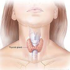
TSH shows a delay after Hashimoto’s is treated

When Hashiotos is treated by T4 pills, T4 recovers
to its normal range comparatively rapidly. TSH
however remains high for 6 weeks after T4
normalizes (Fig. 9). This delay is usually not
explained in endocrinology textbooks. It cannot be
explained by the protein lifetimes which are much
faster.

In fact, the disease can start quietly with no
symptoms, long before T4 levels drop below Figure 9
normal (Fig. 10). In this subclinical stage,
thyroid cells are killed by the immune system.
TSH is high (above 5IU), but T4 is normal. The
origin of the subclinical stage is the dynamical
compensation we discussed before. The
pituitary cell total mass grows and so more TSH
is secreted, to compensate for the killing of
thyrocytes. Figure 10

To understand the subclinical stage in terms of equations, we can build on our experience with
the HPA axis. The thyroid mass growth is stimulated by TSH, denoted x2, so that / =
 ( " 2 − " ). The steady state of TS, 2 = " / " , is proportional to the removal rate of thyroid
cells, " . The autoimmune killing increases thyrocyte removal rate " , so that Hashimoto raises
TSH, as observed. The pituitary mass grows to supply this extra TSH. However, the level of T3,
denoted x3, is kept constant by the previously mentioned equation for the pituitary mass

 / = ( ! / 3 − ! ), so that 4 = 3 = ! / ! . Clinically, doctors recommend a followup to
see if T4 levels fall below their normal range.

Eventually however, the pituitary cannot grow beyond its carrying capacity. We need a more
accurate equation with carrying capacity K: / = ( ! (1 − / ) / 3 − ! ). Now x3 drops and
we have overt hypothyroidism, the clinical stage of the disease. These dynamical findings are by
Yael Korem, who just finished her PhD in our group.
Hyperthyroidism due to toxic nodule: a case of a hypersensing mutant
We now turn to hyperthyroidism, too much T4. One
major cause is a batch of growing cells in the thyroid
that form a nodule that secretes too much T4 (Fig. 11).
These can be imaged by ultrasound, felt by touch, and
seen to be active in secreting T4 by a radioactive
iodine scan. Since they show up on the scae, they are
called ‘hot nodules’ or toxic nodules. They occur in
about 1% of the population.

There are also so called ‘cold nodules’ that do not
secrete T4. Cold nodules are 10 times more likely than
hot nodules to become cancerous (to have the Figure 11
potential for cancer in other body parts called
metastases). Hot nodules are very rarely cancerous.

Toxic nodules are an example of a principle we have seen before. The thyroid cells are controlled
by a feedback circuit in which a signal, TSH, makes the cells both secrete more hormone and to
proliferate. We have seen this in the beat cell circuit where glucose is the signal. This circuit has
a fragility. Mis-sensing mutants that “think” there is too much signal can divide and form a nodule
that secretes too much hormone.

This is exactly what happens in a toxic thyroid nodule. The nodule cells are all copies 9a clone)
of a mutant cell with a mutation, usually in the TSH receptor, that makes it more sensitive to TSH.
There are at least 50 such known mutations in the receptor gene. The mutant proliferates and
over many years can grow to a toxic nodule that causes hyperthyroidism.

Treatment of toxic nodules is their removal.

It is interesting to consider why the
thyroid does not have a biphasic mutant
resistance mechanism like beta cells,
where cells that sense too much glucose
kill themselves (Fig. 12). This effect,
called glucotoxicity, gives strong
 Figure 12
missensing mutants a selective
disadvantage. We discussed this in lecture 3-4.

Why does the thyroid lack a biphasic effect, something we might call TSH-toxicity? It would be
nice if a cell that senses high TSH would kill itself to avoid toxic nodules. However, unlike beta
cells, the signal in the thyroid case, TSH, varies over a 1000-fold range to compensate for
physiological variation, such as iodine levels in the nutrition which can vary over at least a 100-
fold range. Blood glucose has a much smaller range of variation. Perhaps the thyroid is thus
prevented from using a TSH-toxicity due to its need for extensive compensation. In the next
lecture we will see how the need for mutant resistance can explain the physiological role of
autoimmune attack of the thyroid. But we are getting ahead of ourselves.

Graves disease, a common autoimmune disease shrouded in mystery

An even more common cause of hyperthyroidism, too much thyroid hormone, is Graves disease.
It occurs often at ages 20-50, in as much as 2-3% of the population, more in females than males.

In Graves disease, the body produces antibodies that bind and activate the TSH receptor. They
thus mimic TSH. As a result, the thyroid grows and secretes more T4. As in Hashimotos, there is
a subclinical stage with low TSH and normal T4. The pituitary shrinks, or more accurately the total
mass of TSH-secreting cells in the pituitary shrinks. TSH drops to undetectable levels. When the
pituitary shrinks to very small mass, T4 begins to rise, and the clinical stage sets in.

Graves is treated differently in the US and in Europe/Israel. In the US the thyroid is typically
destroyed by radioactive iodine or removed, and the person takes T4 pills for life. In Europe, often
the thyroid is first given a chance. The patient takes antithyroid drugs that inhibit T4 production
by inhibiting TPO. T4 levels normalize. Remarkably, in about 50% of the patients, Graves goes
away: antithyroid drugs are reduced (tapered) over months until after a year or so T4 and TSH
return to normal and one can stop taking drugs, the antibodies go away. In the rest of the 50% of
the patients, Graves doesn't go away, and the
thyroid is removed. Most patients also take beta-
blocker drugs to alleviate the heart palpitations and
muscle tremors that characterize Graves' disease.

Note that here again there is a delay, and it is even
longer than in Hshimotos (Fig. 13). After T4
normalizes it takes months for TSH to go back up to
normal. Again, this delay is due to pituitary mass.
During Graves the pituitary thyrotrophs atrophy and Figure 13
take months to recover.

Why are there autoimmune diseases of hormone glands?

Why is the thyroid attacked by the body? Why risk 5% of the population with a potentially deadly
disease, before medicine turned it into a curable disease? The same question for beta cells, where
type-1 diabetes is an autoimmune attack on beta cells that was a death sentence to about 1% of
children before the advent of insulin treatment.

Also, why these particular cell types and not others? For example, right next to the beta cells are
alpha cells that produce glucagon. Why is there no autoimmune disease that attacks alpha cells?
Similarly, there is almost no autoimmune disease of the parathyroid gland, which sits right on top
of the thyroid. The parathyroid cells, which make parathyroid hormone for calcium control, are
almost never attacked. They are only attacked after immune checkpoint drugs are taken for
cancer treatment, or in very rare congenital mutations in immune tolerance.
And why the weird antibodies that activate TSH receptors in Graves disease? Where are the
corresponding antibody diseases for the thousands of other types of receptors in the body?

If you want to consider some answers, and you have a curious mind, tune in to the next lecture
in our 2-lecture miniseries.

 I am just a T cell and my stories seldom told

 Educated in the thymus

 I learned how to defend my country

 Laying low

 seeking out the lonely lymph nodes where the other T cells go

 Seeking out the places only they would know
 Nay nay nay…
References

Good reviews on HPT physiology and responses to environmental conditions:

 Chatzitomaris, A., Hoermann, R., Midgley, J.E., Hering, S., Urban, A., Dietrich, B.,
 Abood, A., Klein, H.H., and Dietrich, J.W. (2017). Thyroid Allostasis–Adaptive
 Responses of Thyrotropic Feedback Control to Conditions of Strain, Stress, and
 Developmental Programming. Front. Endocrinol. 8.

 Mariotti, S., and Beck-Peccoz, P. (2000). Physiology of the Hypothalamic-Pituitary-
 Thyroid Axis. In Endotext, K.R. Feingold, B. Anawalt, A. Boyce, G. Chrousos, W.W. de
 Herder, K. Dhatariya, K. Dungan, A. Grossman, J.M. Hershman, J. Hofland, et al., eds.
 (South Dartmouth (MA): MDText.com, Inc.), p.

Mathematical modeling of HPT:

 Dietrich, J.W., Landgrafe, G., and Fotiadou, E.H. (2012). TSH and Thyrotropic Agonists:
 Key Actors in Thyroid Homeostasis.

 Goede, S.L., Leow, M.K.-S., Smit, J.W.A., and Dietrich, J.W. (2014). A novel minimal
 mathematical model of the hypothalamus–pituitary–thyroid axis validated for
 individualized clinical applications. Math. Biosci. 249, 1–7.

Papers about the robustness of T4 and TSH set points:

 Andersen, S., Pedersen, K.M., Bruun, N.H., and Laurberg, P. (2002). Narrow Individual
 Variations in Serum T4 and T3 in Normal Subjects: A Clue to the Understanding of
 Subclinical Thyroid Disease. J. Clin. Endocrinol. Metab. 87, 1068–1072.

 Wartofsky, L., and Dickey, R.A. (2005). The Evidence for a Narrower Thyrotropin
 Reference Range Is Compelling. J. Clin. Endocrinol. Metab. 90, 5483–5488.

Review on hysteresis in the HPT:

 Leow, M.K.-S. (2016). A Review of the Phenomenon of Hysteresis in the Hypothalamus–
 Pituitary–Thyroid Axis. Front. Endocrinol. 7.

Papers that experimentally show the shrinkage/enlargement of the pituitary gland in
hyper/hypothyroidism:

 Scheithauer, B.W., Kovacs, K., Randall, R.V., and Ryan, N. (1985). Pituitary gland in
 hypothyroidism. Histologic and immunocytologic study. Arch. Pathol. Lab. Med. 109,
 499–504.

 Scheithauer, B.W., Kovacs, K.T., Young, W.F., and Randall, R.V. (1992). The Pituitary
 Gland in Hyperthyroidism. Mayo Clin. Proc. 67, 22–26.

Review about autoimmune disorders of the thyroid:

 Rapoport, B., and McLachlan, S.M. (2001). Thyroid autoimmunity. J. Clin. Invest. 108,
 1253–1259.
You can also read