A Histological and Histochemical Study on the Gallbladder of the Alburnus tarichi (Güldenstädt, 1814) (Cyprinidae) - SciELO
←
→
Page content transcription
If your browser does not render page correctly, please read the page content below
Int. J. Morphol.,
38(4):869-875, 2020.

      A Histological and Histochemical Study on the Gallbladder
        of the Alburnus tarichi (Güldenstädt, 1814) (Cyprinidae)

                                   Estudio Histológico e Histoquímico de la Vesícula Biliar
                                      de Alburnus tarichi (Güldenstädt, 1814) (Cyprinidae)

                                               Burak Kaptaner1; Handan Aykut2 & Emine Dogan2

KAPTANER, B.; AYKUT, H. & DOGAN, E. A histological and histochemical study on the gallbladder of the Alburnus tarichi
(Güldenstädt, 1814) (Cyprinidae). Int. J. Morphol., 38(4):869-875, 2020.

         SUMMARY: In the current study, the histological structure of the gallbladder of Alburnus tarichi (Güldenstädt, 1814) was
investigated. Hematoxylin and eosin were used to stain the histological sections for routine examinations, in addition to using periodic
acid Schiff (PAS) for the neutral mucins, aldehyde fuchsin (AF) for the sulphated mucins, and Alcian blue (AB; pH: 2.5) for the acidic
mucins. In addition, proliferating cell nuclear antigen (PCNA) immune-staining was performed for the detection of dividing cells among
the epithelium. The gallbladder of A. tarichi was composed of mucosa, muscularis, and serosa or adventitia layers. The mucosa covering
the wavy pleomorphic folds was made up of tall columnar epithelium and a lamina propria. The apical surface of the epithelial cells was
lined by continuous short microvilli. On the epithelium, the luminal surface was remarkably stained with PAS, AF, and AB. Slight to
moderate staining was observed on the epithelial cells in the apical zone with PAS. The cytoplasm of epithelial cells were stained in a
slight manner with AF. No goblet cells were observed among the epithelium. According to the PCNA immune-staining, some epithelial
cells were observed to proliferate. The lamina propria consisted of loose connective tissue that contained fibrocytes, collagen and elastic
fibers, capillaries, and small blood vessels. The muscularis layer displayed muscle fibers that were circular, smooth, and surrounded by
collagen fibers. The subserosal and serosal or adventitial layers had typical morphology to those of other fish and vertebrates.

           KEY WORDS: Gallbladder; Alburnus tarichi; Fish; Histology; Histochemistry; PCNA.

INTRODUCTION

        Gallbladder carries out 2 main functions, in the case                  (Togari & Okada, 1960; Viehberger, 1982; Madrid et al.,
of an animal, that first is storing bile and concentrating bile-               1989; Gillotaux et al., 2011; Gillotaux et al., 2013), they are
absorbing water and electrolytes (sodium chloride, HCO3)                       quantitatively inadequate, because fish are a diversified
by its mucosa. The second is secreting concentrated bile into                  group of animals that can be categorized into 3 different
intestine via smooth muscle contraction and assisting of                       classes: Agnatha or Cyclostomata, Chondrichthyes, and
absorption of lipid soluble compounds (Banfield 1975).                         Osteichthyes. Moreover, the diet of fish varies depending
Among its other functions, the gallbladder epithelium is                       on their feeding habitat and niche adaptation. Those factors
responsible for the secretion and excretion of substances like                 also affect digestive system morphology and its accessory
hormones and xenobiotics. Additionally, it produces and                        organ gallbladder between classes (Oldham-Ott &
secrets mucus as a protective film layer at the surface of                     Gilloteaux). Therefore, more histological and histochemical
gallbladder and some parts of bile tract (Oldham-Ott &                         investigations are necessary to elucidate the gallbladder
Gilloteaux, 1997). Accordingly, a variety of studies on                        among different fish species.
gallbladder mucins have been performed using
histochemistry in mammalian species and showed their                                  Alburnus tarichi (Güldenstädt, 1814) is an
possible pathophysiological roles on cholelithogenesis (Ma-                    anadromous Cyprinid species endemic to the Lake Van basin
drid et al., 1997; Gilloteaux, 1997).                                          of Turkey. Lake Van is the Earth’s largest soda lake and it
                                                                               has highly alkaline (total alkalinity of 153 meq/L; pH 9.8)
       Despite the presence of several histological and                        and brackish (total salinity of 0.22 %) water. The lake exhibits
histochemical studies on gallbladder in various fish species                   an extreme living environment. The littoral zone displays

1
    Department of Biology, Faculty of Science, University of Van Yuzuncu Yil, Tusba, Van, Turkey.
2
    Department of Biology, Institute of Natural and Applied Sciences, University of Van Yuzuncu Yil, Tusba, Van, Turkey.

                                                                                                                                         869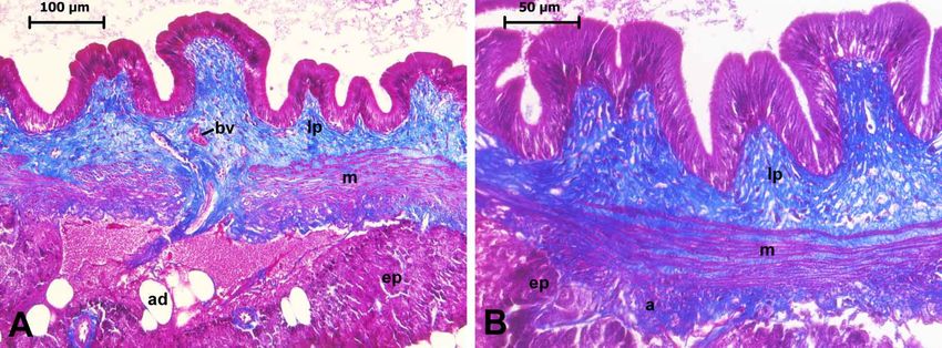
KAPTANER, B.; AYKUT, H. & DOGAN, E. A histological and histochemical study on the gallbladder of the Alburnus tarichi (Güldenstädt, 1814) (Cyprinidae). Int. J. Morphol., 38(4):869-875, 2020.

an absence of higher plants and the living conditions are                                        In some of the sections, counterstaining was performed for
generally lethal for other fish and invertebrates. However,                                      the PAS staining with Mayer’s hematoxylin and for the AF
A. tarichi has physiological abilities allowing it to adapt to                                   or AB staining, neutral red was used. Figures for both the
the lake’s harsh conditions (Danulat & Selcuk, 1992).                                            counterstained and uncounterstained sections stained with
However, to date, no study regarding histological                                                PAS, AF, and AB were presented in the Results section for
organization and histochemical features of gallbladder in A.                                     comparison. Canada balsam was used to mount all of the
tarichi has been performed. In the present study, it was aimed                                   preparations, and a Leica DMI 6000B microscope (Leica
to understand the microscopic anatomy of the gallbladder                                         Microsystems, Wetzlar, Germany) was used to examine and
in this endemic species and learn how they differed from                                         photograph them. The heights and widths of the epithelial
other fish species. Those results were reported herein for                                       cells of the gallbladder were measured using ImageJ 1.46
the first time, and will provide reference knowledge for future                                  software (https://imagej.nih.gov/ij/download.html).
studies of A. tarichi.
                                                                                                 PCNA immunohistochemistry. For detection of
                                                                                                 proliferating cells in the epithelial layer
MATERIAL AND METHOD                                                                              immunohistochemical proliferating cell nuclear antigen
                                                                                                 (PCNA) staining was conducted. The sections of the neutral
                                                                                                 buffered formalin-fixed tissues were then deparaffinized,
Fish. Herein, 8 adult Lake Van fish (Alburnus tarichi                                            rehydrated, and incubated for 10 min with 3 % hydrogen
Güldenstädt, 1814), with a fork length of 18 to 20 cm and                                        peroxide to prevent endogenous peroxidase activity. Tris-
total weight of between 80 and 100 g, were examined. The                                         buffered saline (TBS) (20 mM Tris and 140 mM NaCl) at a
fish used as the study material were caught by professional                                      pH of 7.6 was used for washing the sections, followed by
fishermen, during the fishing season in November, 2019, in                                       the application of a protein block for 10 min to inhibit
Lake Van (43°13′E, 38°26′N). The deaths of the fish were                                         nonspecific binding. The sections were then incubated with
the result of natural causes. Therefore, in accordance with                                      primary antibody (a mouse monoclonal PC10 to PCNA, cat.
the Turkish Ministry of Forestry and Water Affairs                                               number: ab29, Abcam plc., Cambridge, UK) at a dilution of
regulations, the use of animal tissue post mortem did not                                        1:200 overnight. After the sections had been washed with
require Animal Experiments Local Ethics Committee                                                TBS, the remaining procedures were performed using the
approval (The Official Gazette of Republic of Turkey on                                          EXPOSE mouse and rabbit specific IHC/DAB detection IHC
February 15, 2014, No: 28914); however, a decision protocol                                      kit (cat. no: ab80436; Abcam, UK), following the
was issued by the above mentioned Ethics Committee of                                            manufacturer’s instructions, as described by Ceylan &
Van Yuzuncu Yil University (VAN YUHADYEK) under                                                  Kaptaner (2019). To perform the counterstaining, Mayer’s
decision number: YUHADYEK-2019/10, protocol number:                                              hematoxylin was used. Next, the slides were rinsed in graded
27552122-604.01.02-E.86004.                                                                      ethanol concentrations (95 % and 100 %) and xylene, and
                                                                                                 Canada balsam was used to seal them (Merck, KGaA,
Histological procedures. Immediately after the death of                                          Darmstadt, Germany). To test the antibodies’ specificities,
the animals, careful dissection of the gallbladder and its                                       negative control slides were used, to which TBS was added
annex tissues was performed. Next, Bouin’s solution, in                                          in place of the primary antibody. A Leica DMI 6000B
addition to a concentration of neutral buffered formalin (10                                     microscope was used to perform the examinations and take
%), separately, was used to fix the tissues at 4 °C for 24 h.                                    photographs of the slides.
After which, a graduated ethanol series was used to dehydrate
the tissues, and these were embedded in paraffin. As a next
step, 5-µm-thick cross-sections of the tissues were taken and                                    RESULTS
then placed on slides coated with adhesive (Marienfeld
GmbH, Lauda-Königshofen, Germany). Following
deparaffinization and rehydration, hematoxylin and eosin                                                 The gallbladder is located under the anterior portion
(H&E) was used to stain the tissue sections for the routine                                      of right liver lobe. It is spheroid to ellipsoid in shape and
histological examinations, while Mallory’s triple (Crossmon,                                     yellow to greenish in color. Approximately, two-thirds of
1937) was used for the collagen fibers; periodic acid-Schiff                                     the gallbladder is surrounded by adipose tissue containing
(PAS) (McManus, 1948) was used for the neutral mucins,                                           pancreatic tissues (Fig. 1A). The gallbladder is composed
sialomucins, and glycogen; aldehyde fuchsin (AF), as                                             of 3 layers, including the mucosa, muscularis, and serosa or
described by Lamar Jones (2002), was used for the sulphated                                      adventitia. The mucosa covering the wavy pleomorphic folds
mucins and elastic fibers; and Alcian blue (AB) at pH 2.5,                                       is made up an epithelium lining the inner surface and a la-
according to Totty (2002), was used for the acidic mucins.                                       mina propria, which lies beneath the epithelial layer (Figs.
870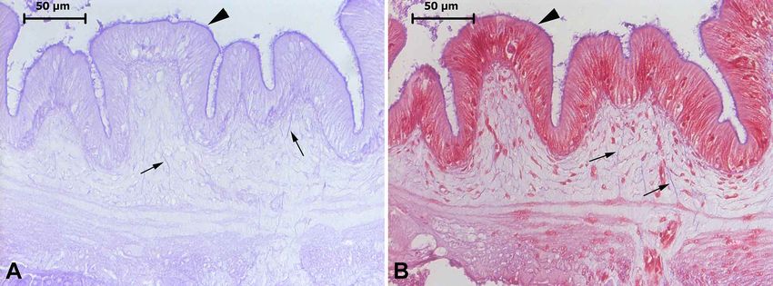
KAPTANER, B.; AYKUT, H. & DOGAN, E. A histological and histochemical study on the gallbladder of the Alburnus tarichi (Güldenstädt, 1814) (Cyprinidae). Int. J. Morphol., 38(4):869-875, 2020.
1B and 2). The prominent oval nuclei of the epithelial cells
are located basally or close to the basal region. The apical
surface of those cells was covered by a margin of short
microvilli. Goblet cells were not determined among the
epithelial layer (Figs. 2A-B). The epithelium of the
gallbladder was arranged as tall columnar cells ranging from
16.76 to 73.80 µm and averagely measuring 44.03 µm in
height and from 1.83–6.29 to 3.22 µm in width, which were
and located on basal membrane. The positively immune-
stained proliferating cells in their nuclei with PCNA were
observed among the epithelium (Fig. 3). Loose connective
tissue was observed in the lamina propria, which comprised
fibrocytes, collagen, and AF-stained elastic fibers, and
Fig. 1. A) Photograph of the dissected gallbladder of A. tarichi
surrounded by adipose tissue (at) containing pancreatic tissues.
Asterisks on the gallbladder display free surface. B) Cross-section
of the gallbladder stained with Mallory’s triple displaying the typical Fig. 3. Proliferating cells (arrowheads) containing nuclei that were
layers and adjacent tissue (a: adventitia; e: epithelium; ep: exocrine stained brown and detected via PCNA immunohistochemistry in
pancreas; L: lumen; lp: lamina propria; m: muscularis; M: muco- the epithelial layer of the gallbladder of A. tarichi.
sa; s: serosa).
capillaries, usually including erythrocytes
and in a few extent small blood vessels
(Figs. 2A, 4 and 6). The muscularis was
comprised of circular muscle fibers
surrounded by collagen fibers and was
invaded by a few capillaries (Figs. 2A and
4). The outermost serosa layer of the free
surface of the gallbladder was covered by
a mesothelium and made up of a thin
subserosal loose connective tissue
containing collagen fibers and
occasionally determined small vessels
(Fig. 2B). The surface of the gallbladder
adjacent to the adipose tissue or pancreas
included an adventitial layer comprised of
loose connective tissue, including collagen
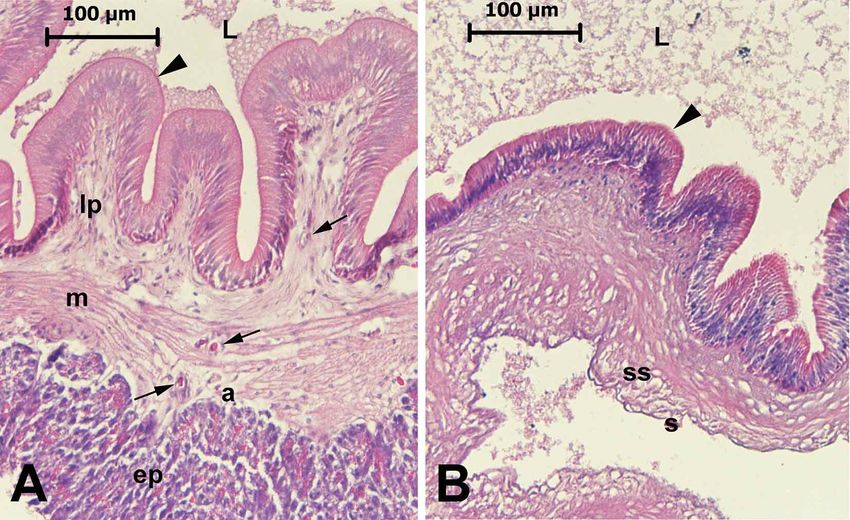
Fig. 2. Cross-sections from the attached (A) and free (B) surfaces of the gallbladder wall fibers, capillaries, and blood vessels (Figs.
stained with H&E. The apical surface of the epithelial cells was lined by short microvilli 2A and 4).
(arrowheads). An area of the exocrine pancreas (ep) in the attached surface and serosa
(s) and subserosa (ss) in the free surface can be seen. Some capillaries are indicated with The luminal surface of the
arrows among the lamina propRia (lp), muscularis (m), and adventitia (a). epithelial cells showed a remarkable
871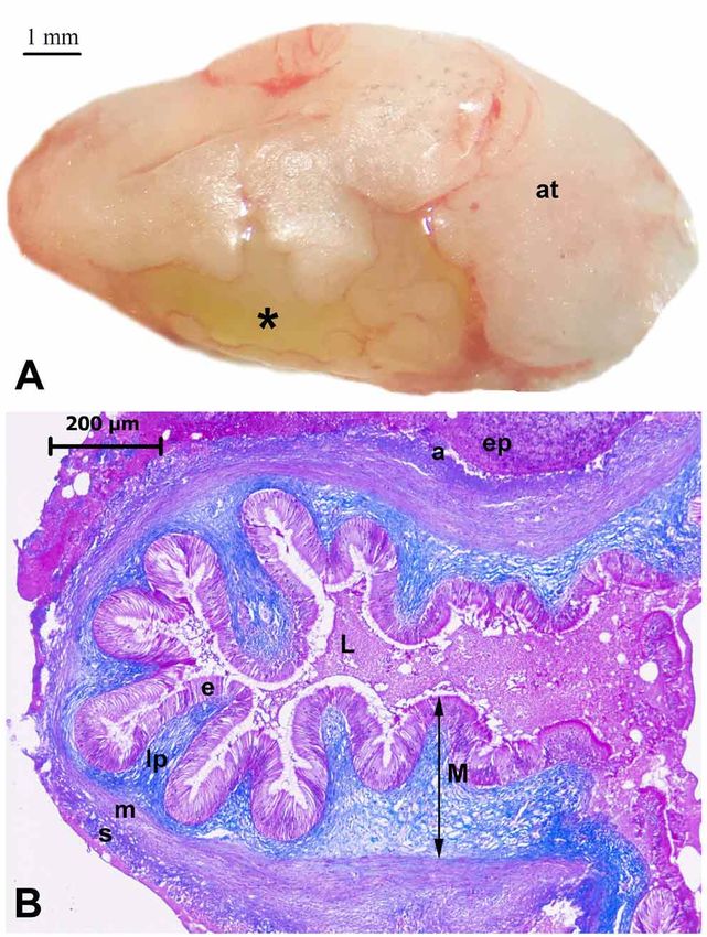
KAPTANER, B.; AYKUT, H. & DOGAN, E. A histological and histochemical study on the gallbladder of the Alburnus tarichi (Güldenstädt, 1814) (Cyprinidae). Int. J. Morphol., 38(4):869-875, 2020.

        Fig. 4. Mallory’s triple staining in the gallbladder wall of A. tarichi at different magnifications; 20× (A) and 40× (B). Collagen
        fibers stained in blue throughout the loose connective tissue of the lamina propria (lp), and among the muscle fibers of the
        muscularis (m) and in the adventitia (ad) can be seen. Blood vessels (bv) were found in lamina propria and adipocytes (ad),
        and exocrine pancreas (ep) tissue was observed in the outer surface of the gallbladder.

        Fig. 5. PAS staining without counterstaining (A) and with counterstaining using Mayer’s hematoxylin (B) in the gallbladder
        sections. A remarkable staining with PAS was observed on the luminal surface of the epithelial layer (arrowheads). PAS
        reaction was also found in the apical zone of the epithelial cells.

        Fig. 6. AF staining without counterstaining (A) and with counterstaining using neutral red (B) in the gallbladder sections. A
        remarkable reaction with AF was observed at the luminal surface of the epithelial layer (arrowheads). Weak reaction was also
        observed in the cytoplasmic zone of the epithelial cells. AF-stained elastic fibers (arrows) were also identified in the lamina
        propria.

872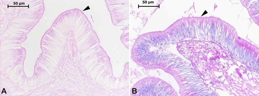
KAPTANER, B.; AYKUT, H. & DOGAN, E. A histological and histochemical study on the gallbladder of the Alburnus tarichi (Güldenstädt, 1814) (Cyprinidae). Int. J. Morphol., 38(4):869-875, 2020.

positive histochemical reactivity by PAS staining, indicating                                    luminal surface of the epithelial cells. In addition, the
the presence of neutral mucins, sialomucins, and glycogen.                                       cytoplasm showed a slight staining with AF (Figs. 6A-B).
Slight to moderate luminal surface PAS reactivity was also                                       AB histochemical staining for acidic mucopolysaccharides
detected at the apical zone of the epithelial cells (Figs. 5 A-                                  was restricted to the luminal surface of the epithelial layer
B). AF staining for sulphated groups was detected in the                                         (Figs. 7A-B).

Fig. 7. AB staining without counterstaining (A) and with counterstaining using neutral red (B) in the gallbladder sections. A remarkable
reaction with AB was observed at the luminal surface of the epithelial layer (arrowheads). No cytoplasmic staining was found in the
epithelial cells.

DISCUSSION

        The histological layers of the gallbladder wall were                                     The heights of the epithelial cells were averagely measured
composed of mucosa including surface epithelium and la-                                          as 60 µm in Cyprinus carpio, 50 µm in Carassius auratus,
mina propria, circular smooth muscle, and serosa. Those                                          45 µm Leuciscus hakonensis and Leuciscus hakonensis, 40
layers are also present in other fish species (Western, 1969;                                    µm in Salmo irideus and Parasilurus asotus, 35 µm in Chana
Gupta, 1971; Gilloteaux et al., 2011, 2013; Nazlic et al.,                                       argus, 32 µm in Zacco platypus, 28 µm in Misgurnus
2014) and in agreement with other vertebrates and mammals                                        anguillicaudatus, 25 µm in Zacco temminckii, and 16 µm in
(Oldham-Ott & Gilloteaux; Mobini & Poursafar, 2019). The                                         Plecoglessus altivelis (Togari & Okada). In the gallbladder,
surface epithelium lining the mucosal folds thrown into the                                      the mucosa epithelial cells of the stargazer of Uranoscopus
lumen of the gallbladder is comprised of tall columnar cells.                                    scaber ranged from 50 to 85 µm high and were ≤5 µm wide
Togari & Okada also reported single-layered high columnar                                        (Gilloteaux et al., 2011), and ranged from 25 to 45 µm high
cells in the gallbladder of the different fish species, such as                                  and from 4 to 5 wide for Torpedo marmorata (Gilloteaux et
sweetfish (Plecoglessus altivelis), rainbow trout (Salmo                                         al., 2013). Thus, it can be said that the heights of the mucosal
irideus), snake head fish (Chana argus), catfish (Parasilurus                                    epithelium cells in A. tarichi displayed similarities to other
asotus), loach (Misgurnus anguillicaudatus), chub                                                fish species, but mainly to Cyprinid species. The apical
(Ganathopogon elongates), and Zacco platypus, whereas                                            surface of the gallbladder epithelial cells was observed as
they observed pseudostratified epithelium scattered as islets                                    lined by a margin of continuous microvilli in A .tarichi. In
in a single epithelial layer of some other fish species, such                                    accordance with this study, similar findings have been
as carp (Cyprinus carpio), gibel (Carassius auratus), dace                                       reported in different studies conducted on different fish
(Leuciscus hakonensis), Zacco temminckii, and                                                    species (Western; Viehberger; Gilloteaux et al., 2011, 2013).
Acheilongnathus moriokae. Cholecystocytes were also                                              No goblet cells were observed among the gallbladder
arranged as simple columnar epithelium in the gallbladders                                       epithelium in the present study. The presence of goblet cells
of stargazer (Uranoscopus scaber) (Gilloteaux et al., 2011)                                      among the gallbladder epithelium may display differences
and Torpedo marmorata (Gilloteaux et al., 2013). The                                             between fish species. Although goblet cells have been
heights and widths of the mucosal epithelial cells of the                                        reported in some fish, they have not been reported in others
gallbladder can markedly vary depending on the fish species.                                     (Togari & Okada), as in the current study.
                                                                                                                                                                                      873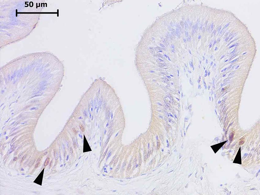
KAPTANER, B.; AYKUT, H. & DOGAN, E. A histological and histochemical study on the gallbladder of the Alburnus tarichi (Güldenstädt, 1814) (Cyprinidae). Int. J. Morphol., 38(4):869-875, 2020.

       PCNA, or cyclin, is a protein with multifunctional                                        CONCLUSION
properties that, in the dividing cells, is highly expressed
in phases G1 and S (Kelman, 1997; Kaptaner & Kankaya,
2013; Ceylan & Kaptaner). Immunohistochemical                                                            In conclusion, the histological and histochemical
analysis of the PCNA supplied important information for                                          features of the gallbladder were generally consistent with
evaluating proliferating cells among the gallbladder                                             reports to those of other fish species. Differences in the
epithelial cells of A. tarichi and some cells displayed                                          gallbladder structure between fish species are related to diet
positive immune staining in their nuclei. Consistently,                                          (Oldham-Ott & Gilloteaux). Thus, the differences, such as
Togari & Okada also observed mitotic divisions in the                                            the lack of goblet cells, may have arisen from its diet. To
gallbladder epithelium of different fish species, especially                                     date, there have been no studies on the histological structure
in Cyrinus carpio. The gallbladder epithelium exhibited                                          and histochemical properties of the gallbladder of A. tarichi.
repairing or reactive cell growth in the some damaged                                            Thus, the findings herein will provide basic knowledge for
regions in Torpedo marmorata (Gilloteaux et al., 2011).                                          studies in the future.
Thus, it can be concluded that epithelial cell renewal is a
common observation in the gallbladder epithelium of fish
species.                                                                                         KAPTANER, B.; AYKUT, H. & DOGAN, E. Estudio histológico e
                                                                                                 histoquímico sobre la vesícula biliar de Alburnus tarichi (Güldenstädt,
                                                                                                 1814) (Cyprinidae). Int. J. Morphol., 38(4):869-875, 2020.
        The luminal surface of the gallbladder
epithelium exhibited remarkable PAS staining, which                                                       RESUMEN: En este estudio, se investigó la estructura
indicates neutral mucins, whereas apical zones of the                                            histológica de la vesícula biliar de Alburnus tarichi (Güldenstädt,
cells were only slightly stained. On the other hand, only                                        1814). Las secciones histológicas se tiñeron con Hematoxilina-Eosina
the luminal surface of the epithelium was stained with                                           para los exámenes de rutina, además de usar el ácido periódico de
AF and AB, indicating acidic and sulphated groups,                                               Schiff (PAS) para las mucinas neutras, aldehído fucsina (FA) para
respectively. In accordance of our findings, glycogen                                            las mucinas sulfatadas y azul alcián (AB; pH: 2,5) para las mucinas
staining occurred in small amounts in the epithelium                                             ácidas. Además, se realizó una tinción inmune de antígeno nuclear
                                                                                                 de células proliferativas (PCNA) para la detección de células en di-
of the gallbladder of fish species and was restricted to
                                                                                                 visión entre el epitelio. La vesícula biliar de A. tarichi estaba com-
the basal, apical, or both sides of the nucleus in the                                           puesta de capas, mucosa, muscular y serosa o adventicia. La muco-
cells, depending on the fish species (Togari & Okada).                                           sa que cubría los pliegues pleomórficos ondulados estaba formada
A similar staining pattern with a small differences was                                          por un epitelio columnar alto y una lámina propia. Se observó una
also observed in the gilthead sea bream (Sparus aurata),                                         superficie apical de las células epiteliales revestida por
where only the luminal surfaces of the epithelial cells                                          microvellosidades cortas y continuas. En el epitelio se observó una
of the gallbladder reacted with the above mentioned                                              tinción importante de la superficie luminal teñida con PAS, FA y
                                                                                                 AB. Se observó una tinción leve a moderada en las células epiteliales
stains (Madrid et al., 1989). Luminal surface staining
                                                                                                 en la zona apical con PAS. El citoplasma de las células epiteliales se
in the epithelial cells of the gallbladder with PAS and                                          tiñó ligeramente con FA. No se observaron células caliciformes en-
AF was also observed in upper vertebrates, including                                             tre el epitelio. Según la tinción de PCNA, se observó que prolifera-
amphibian, avian, and mammalian species (Madrid et                                               ban algunas células epiteliales. La lámina propia consistía en tejido
al., 1989; Mobini, 2012; Mobini & Poursafar). This                                               conectivo laxo que contenía fibrocitos, colágeno y fibras elásticas,
suggests that mucin secretion on the epithelial surface                                          capilares y pequeños vasos sanguíneos. La capa muscular mostraba
might be attributed to a protective barrier role against                                         fibras musculares circulares, lisas y rodeadas de fibras de colágeno.
the possible damaging impact of highly concentrated                                              Las capas subserosas y serosas o adventicias tenían una morfología
                                                                                                 típica a la de otros peces y vertebrados.
bile salts, such as amphipathic and strong detergents
(Dray-Charier et al., 1997; Gilloteaux et al., 2013).                                                   PALABRAS CLAVE: Vesícula biliar; Alburnus tarichi;
                                                                                                 Pescado; Histología; histoquímica; PCNA.
       The lamina propria of the mucosa comprised loose
connective tissue that contained elastic fibers, collagen
fibers, and capillaries, and fibrocytes. This area was                                           REFERENCES
surrounded by smooth muscle bundles surrounded by
collagen fibers and was invaded by small vessels. The
histological features of all of the layers of the gallbladder                                    Banfield, W. J. Physiology of the gallbladder. Gastroenterology, 69(3):770-
wall, including outer subserosa and serosa or adventitia,                                           7, 1975.
were in agreement with findings obtained from other                                              Ceylan, S. & Kaptaner, B. Apoptosis and cell proliferation in the epithelia
                                                                                                    of the esophagus and intestine of Alburnus tarichi Güldenstädt, 1814
teleost fish species (Western; Gupta; Gilloteaux et al.,                                            (Cyprinidae) during migration from highly alkaline and brackish water
2011; Nazlic et al.).                                                                               to fresh water. Eur. Zool. J., 86(1):103-12, 2019.

874
KAPTANER, B.; AYKUT, H. & DOGAN, E. A histological and histochemical study on the gallbladder of the Alburnus tarichi (Güldenstädt, 1814) (Cyprinidae). Int. J. Morphol., 38(4):869-875, 2020.
Crossmon, G. A modification of Mallory's connective tissue stain with a Corresponding author:
discussion of the principles involved. Anat. Rec., 69(1):33-8, 1937. Burak Kaptaner
Danulat, E. & Selcuk, B. Life history and environmental conditions of the University of Van Yuzuncu Yil
anadromous ChalcAlburnus tarichi in the highly alkaline Lake Van,
Faculty of Science
eastern Anatolia, Turkey. Arch. Hydrobiol., 126(1):105-25, 1992.
Dray-Charier, N.; Paul, A.; Combettes, L.; Bouin, M.; Mergey, M.; Balladur,
Department of Biology
P.; Capeau, J. & Housset, C. Regulation of mucin secretion in human 65080 Tusba
gallbladder epithelial cells: predominant role of calcium and protein Van
kinase C. Gastroenterology, 112(3):978-90, 1997. TURKEY
Gilloteaux, J. Introduction to the biliary tract, the gallbladder, and gallstones.
Microsc. Res. Tech., 38(6):547-51, 1997. ORCID: B. Kaptaner (http://orcid.org/0000-0003-2366-6756)
Gilloteaux, J.; Ott, D. W. & Oldham-Ott, C. K. The gallbladder of
Uranoscopus scaber L. (teleost perciform fish) is lined by specialized
cholecystocytes. Anat. Rec. (Hoboken), 294(11):1890-903, 2011.
Gilloteaux, J.; Ott, D. W. & Oldham-Ott, C. K. The gallbladder of the electric Email: bkaptaner@yahoo.com, bkaptaner@yyu.edu.tr
ray Torpedo marmorata Risso displays excrescent cholecystocytes with
merocrine and apocrine-like secretions. Anat. Rec. (Hoboken),
296(1):79-95, 2013. Received: 07-01-2020
Gupta, O. P. Studies on the morphology, histology and the swallowing Accepted: 17-02-2020
mechanism of the digestive tract of a carnivorous fish, Xenentodon
cancila (Ham.). Okajimas Folia Anat Jpn., 48(1):29-51, 1971.
Kaptaner, B. & Kankaya, E. Analysis of germ cell proliferation, apoptosis,
and androgenesis in the Lake Van fish (Chalcalburnus tarichi) during
testicular development. Fish Physiol. Biochem., 39(6):1165-79, 2013.
Kelman, Z. PCNA: structure, functions and interactions. Oncogene,
14(6):629-40, 1997.
Lamar Jones, M. Connective Tissues and Stains. In: Bancroft, J. D. (Ed.).
The Theory and Practice of Histological Techniques. 5th ed. Edinburgh,
Churchill Livingstone, 2002. pp.139-62.
Madrid, J. F.; Ballesta, J.; Galera, T.; Castells, M. T. & Pérez-Tomás, R.
Histochemistry of glycoconjugates in the gallbladder epithelium of ten
animal species. Histochemistry, 91(5):437-43, 1989.
Madrid, J. F.; Hernández, F. & Ballesta, J. Characterization of glycoproteins
in the epithelial cells of human and other mammalian gallbladder. A
review. Microsc. Res. Tech., 38(6):616-30, 1997.
McManus, J. F. Histological and histochemical uses of periodic acid. Stain
Technol., 23(3):99-108, 1948.
Mobini, B. & Poursafar, M. Histological and histochemical studies of the
gall bladder of the graylag goose (Anser anser). Biotech. Histochem.,
94(6):404-9, 2019.
Mobini, B. Microscopic study of the gall bladder of the chukar partridge
(Alectoris chukar). Bulg. J. Vet. Med., 15(2):73-8, 2012.
Nazlic, M.; Paladin, A. & Bocina, I. Histology of the digestive system of
the black scorpionfish Scorpaena porcus L. Acta Adriat., 55(1):65-74,
2014.
Oldham-Ott, C. K. & Gilloteaux, J. Comparative morphology of the
gallbladder and biliary tract in vertebrates: variation in structure,
homology in function and gallstones. Microsc. Res. Tech., 38(6):571-
97, 1997.
Togari, C. & Okada, T. Cytological studies of the gallbladder epithelium of
the fish. Okajimas Folia Anat. Jpn., 35(1-3):11-25, 1960.
Totty, B. A. Mucins. In: Bancroft, J. D. (Ed.), The Theory and Practice of
Histological Techniques. 5th ed. Edinburgh, Churchill Livingstone, 2002.
pp.163-200.
Viehberger, G. Apical surface of the epithelial cells in the gallbladder of
the rainbow trout and the tench. Cell Tissue Res., 224(2):449-54, 1982.
Western, J. R. Studies on the diet, feeding mechanism and alimentary tract
in two closely related teleosts, the freshwater Cottus gobio L. and the
marine Parenophrys bubalis euphrasen. Acta Zool., 50(3):185-205,
1969.
875You can also read