Original Research Article
←
→
Page content transcription
If your browser does not render page correctly, please read the page content below
Original Research Article

Molecular docking and dynamics simulation of a screening library from Life Chemicals
database for potential protein-protein interactions (PPIs) inhibitors against coronavirus 2
receptor binding domain.

Abstract:
The ongoing pandemic of coronavirus 2 represents a major challenge for global public health
authorities. Coronavirus disease 2019 can be fatal especially in elderly people and those with
comorbidities. Currently, several vaccines against coronavirus 2 are under application in
multiple countries with emergency use authorization. In the same time, many vaccine
candidates are under development and assessment. It is worth noting that the design of some
of these vaccines depends on the expression of receptor binding domain for viral spike
protein to induce host immunity. As such, blocking the spike protein interface with
antibodies, peptides or small molecular compounds can impede the ability of coronavirus 2 to
invade host cells by intervention with interactions between viral spike protein and cellular
angiotensin converting enzyme 2. In this virtual screening study, we have used predictive
webservers, molecular docking and dynamics simulation to evaluate the ability of 3000
compounds to interact with interface residues of spike protein receptor binding domain. This
library of chemicals was focused by Life Chemicals as potential protein-protein interactions
inhibitor. Here, we report that hit compound 7, with IUPAC name of
3‐ cyclohexyl‐ N‐ (4‐ {[(1R,9R)         ‐ 6‐ oxo‐ 7,11‐          diazatricyclo      [7.3.1.02,7]
trideca‐ 2,4‐ dien‐ 11‐ yl] sulfonyl} phenyl) propenamide, may have the capacity to interact
with interface of receptor binding domain for viral spike protein and thereby reduce cellular
entry of the virus. However, in vitro and in vivo assessments may be required to validate
these virtual findings.
Keywords: SARS-CoV-2, COVID-19, protein-protein interactions inhibitor, rule of four,
docking, molecular dynamics simulation.
Introduction:
The severe acute respiratory syndrome coronavirus 2 (SARS-CoV-2) is the causative
pathogen responsible for the ongoing pandemic of coronavirus disease 2019 (COVID-19) (1).
SARS-CoV-2 is a novel RNA virus and has about 79% of genomic sequence identity with
SARS-CoV, a previously identified beta-coronavirus (2). SARS-CoV-2 can be transmitted
mainly through exposure to respiratory droplets generated by infected persons, airborne
transmission by aerosols is also possible during aerosol generating medical procedures like
intubation (3). Upon exposure to SARS-CoV-2, the mean incubation period can range
between 5.6 and 6.7 days after which symptoms of COVID-19 may develop (4). Common
clinical features of COVID-19 include fever, dry cough, shortness of breath, fatigue, myalgia,
nausea, vomiting and diarrhea. The COVID-19 infection fatality rate (IFR) is estimated to be
0.68% with 95% confidence interval between 0.53% and 0.82%. COVID-19 fatality is

                                               1
usually the result of complications like pneumonia, acute respiratory distress syndrome
(ARDS), cardiac injury, liver injury, kidney injury and coagulopathy (5,6). Therapeutic
options currently available to control COVID-19 complications involve oxygen supply,
management of sepsis, use of corticosteroids, heparin and antiviral agents like Remdesivir
(5,7–9). According to World health organization (WHO), as of 18 February 2021, seven anti-
COVID-19 vaccines are under emergency use authorization in multiple countries and more
than 60 vaccine candidates are under clinical development (10). Different platforms have
been utilized to produce these COVID-19 vaccines like inactivated virus, protein subunit,
recombinant viral-vector and nucleic acid (DNA and mRNA). Some of these vaccines have
focused on the expression of full-length SARS-CoV-2 spike protein inside human body to
elicit immune response, other vaccines used only the receptor binding domain (RBD) of spike
protein to induce immunity (11). SARS-CoV-2 can infect susceptible host through interaction
with angiotensin converting enzyme 2 (ACE2) on the surface of target cells. The receptor
binding domain (RBD) of SARS-CoV-2 is located within S1 subunit of the viral spike
protein (12). Virtual alanine scanning approach have been used to determine the interacting
residues at the interface of SARS-CoV-2 receptor binding domain and angiotensin converting
enzyme 2. Identification of these interface residues is considered important to construct a
protein-protein interactions (PPIs) inhibitor capable of blocking cellular entry of SARS-CoV-
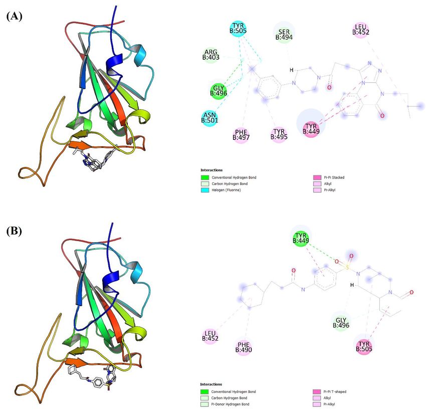
2 (13). A three-dimensional illustration for the complex between RBD of SARS-CoV-2 spike
protein and ACE2 can be seen in Figure 1 (A). A close view for interaction interface of
crystal complex can be seen in Figure 1 (B). The sequence of amino acids for RBD of
SARS-CoV-2 spike glycoprotein is presented in Figure 1 (C), RBD residues involved in
interaction with ACE2 are surrounded by orange line. In this virtual screening study, we have
used a protein-protein interactions (PPIs) focused library of compounds from Life Chemicals
database. Our aim was to identify potential inhibitor of SARS-CoV-2 spike protein binding to
ACE2 thereby blocking viral entry to target cells. The chemical compounds in this library
were curated based on rule of four (RO4) principle. According to rule of four, a compound
may have the potential to inhibit protein-protein interactions (PPIs) if it possesses the
following four chemical criteria: a molecular weight (M.W.) ≥ 400 g/ mol, a calculated
logarithm of partition coefficient (cLog P) ≥ 4, the number of rings ≥ 4 and the number of
hydrogen bond acceptor (HBA) ≥ 4 (14,15).
2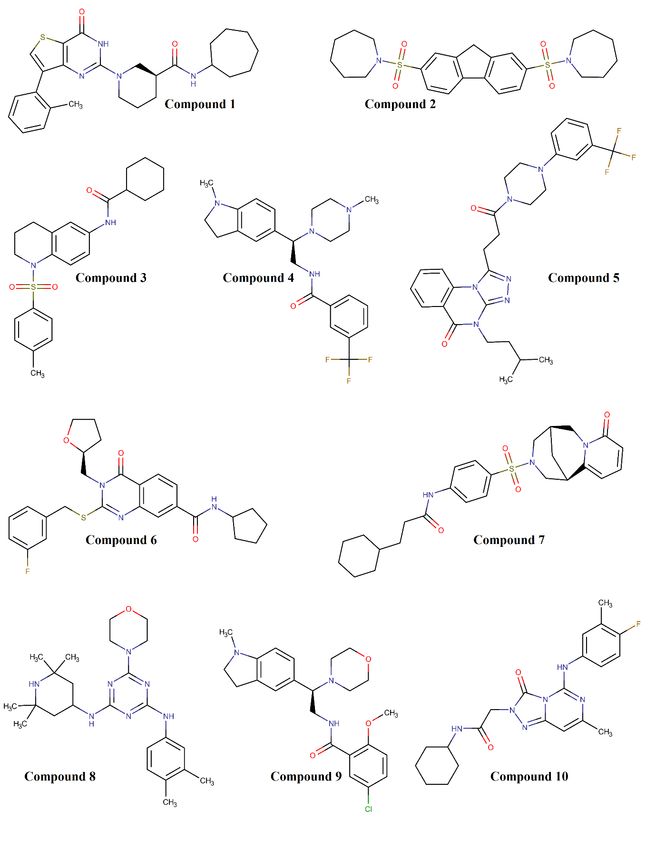
Figure 1: (A) A three-dimensional view for 6LZG crystal complex between receptor binding
domain RBD of SARS-CoV-2 spike protein and its receptor angiotensin converting enzyme 2
ACE2. (B) A close view for interaction interface between SARS-CoV-2 spike protein RBD
and ACE2, the amino acid residues of spike protein RBD are shown as sticks and labelled in
one letter format. (C) The amino acids sequence of SARS-CoV-2 spike protein RBD is
presented in one letter format, RBD residues that are involved in interaction with ACE2 are
encircled by orange line.
Materials and methods:
Preparation of receptor binding domain crystal and chemicals database:
The crystal complex of receptor binding domain RBD of SARS-CoV-2 spike protein and
angiotensin converting enzyme 2 ACE2 was downloaded from protein data bank website, the
identification code for the downloaded crystal PDB is 6LZG (16,17). We have used UCSF
chimera version 1.15 to prepare the crystal file for docking study and dynamics simulation
(18). By using UCSF chimera software, we have removed ACE2 peptidase domain (chain A)
from 6LZG crystal, we also removed any bound ligands, water molecules and ions. The
residues sequence of chain B (receptor binding domain) was visualized as one letter format
by using UCSF chimera. We have used 3000 compounds downloaded from Life Chemicals
website as a screening library, this library of chemicals was focused by Life Chemicals as
potential inhibitors for protein-protein interactions (PPIs) by using rule of four (RO4)
filtration criteria. Based on RO4 principle, any compound may have the capacity to interfere
with interactions between two proteins if it has the following criteria: a molecular weight ≥
400 g/ mol, a logarithm of partition coefficient ≥ 4, the number of hydrogen bond acceptor ≥
4 and the number of rings ≥ 4. Additionally, the compounds in this focused library have been
enriched for sp3 hybridization of carbon atoms to ensure high level of three-dimensional
diversity and hence sufficient drug-likeness score (14,15).
Structure-based virtual screening:
We have used MCULE online drug discovery website to screen the downloaded chemicals
library against the prepared crystal of SARS-CoV-2 spike protein RBD (chain B of 6LZG
crystal) (19). Here, we used a virtual screening workflow steps similar to those applied in our
previously published researches (20,21). In summary, we have used the default screening
steps with the addition of REOS (rapid elimination of swill) filter to reduce possibility of
frequent and non-selective hits. The MCULE website employs AutoDock Vina tool to
perform structure based virtual screening (22). Also, this online drug discovery platform uses
AutoDock tools to add polar hydrogen atoms and Gasteiger charges to the uploaded crystal
(23). We have used a binding site area of (X: 30, Y:30, Z:30) Angstrom while the coordinates
were (X: -33, Y: 26, Z: 7.5). The final hits were ranked according to their least binding
energy to the receptor binding domain (RBD) of SARS-CoV-2 spike protein crystal (chain
B). According to the minimum binding energy rank, we have selected the top ten hits for
further virtual characterization and visualization. For each compound of these top ten hits, we
have saved the complex of ligand and protein with least binding energy pose as PDB file. The
generated PDB file was then visualized as two and three-dimensional illustrations by using
PyMOL version 2.4.1 and discovery studio visualizer version 21.1.0.20298 respectively
(24,25). The two-dimensional chemical structures of these top ten hits were drawn by using
MarvinSketch version 20.1 (26).
Prediction of chemical, pharmacokinetics and mutagenic characteristics of hit
compounds:

                                              3
MCULE online platform provides the opportunity to predict various chemical features of the
generated hit compounds. Drug-likeness score for the top ten hits were predicted by using
Molsoft webserver (27). Various pharmacokinetics and mutagenic characteristics for the top
hits were predicted and calculated by using SwissADME and pkCSM webservers (28,29).
These webservers use predictive regression and molecular similarity to analyze molecules
under investigation (30,31).
Molecular Dynamics (MD) study:
We have employed YASARA Dynamics version 20.12.24 to perform molecular dynamics
(MD) simulation of the ligand-protein complex with the least binding energy pose (32). The
steps for molecular dynamics simulation study are similar to what we had followed in our
previously published researches (21,33). In summary, the MD protocol involves hydrogen
bonds network optimization and a pKa prediction to fine-tune the protonation of residues at
pH value equal to 7.4 (34). NaCl was added with a concentration of 0.9%, an excess of either
sodium or chloride ions were used to neutralize ligand-protein complex. To eliminate any
possible clashes, steepest descent and simulated annealing minimizations were applied. The
simulation period employed was 10 nanoseconds by using AMBER14 force field for solute,
TIP3P for water, AM1BCC and GAFF2 for ligand (35–37). The cutoff value for van der
Waals (vdW) forces was 8 Angstrom, the default parameters were employed by AMBER
(38). By applying Particle Mesh Ewald algorithm, no cutoff value was used for electrostatic
forces (39). The motions equations were used as a multiple timestep of 1.25 and 2.5
femtoseconds for bonded and non-bonded interactions respectively at a pressure of 1 atm and
a temperature of 298K (40). After assessment of root-mean-square deviation (RMSD) for the
solute as a function of simulation period, the first 10 nanoseconds were regarded as the
equilibration time and precluded from further analysis.
Results and discussion:
A prediction of different chemical characteristics for the top ten hit compounds that were
computationally screened against RBD crystal of SARS-CoV-2 spike protein can be seen in
Table 1. In this table, the hit compounds were ranked according to their minimum binding
energy to RBD crystal. According to Table 1, all these protein-protein interactions (PPIs)
inhibitor candidates have a molecular weight greater than 400 g/ mol and a number of
hydrogen bond accepters greater than 4. Regarding the prediction of partition coefficient
logarithm (Log P), all the reported hits have Log P value greater than 4 with the exception of
compounds 4, 9 and 10. The chemical structures for these top ten hits can be seen in Figure 2
where all these compounds have more than 4 rings within their structures. Based on chemical
data presented in Table 1 and Figure 2, we can assess the adherence of these hit compounds
to rule of four criteria.
Table 1: Chemical characteristics of the top ten protein-protein interactions (PPIs) inhibitors
that were virtually screened against receptor binding domain (RBD) crystal of SARS-CoV-2.
These top hits were ranked according to their minimum binding energy to SARS-CoV-2
RBD crystal.
Compound TPSA H-bond H-bond
Molecular Formula M.W. (g/ mol) Log P
No (Å2) acceptors donors
1 C26H32N4O2S 464.625 5.471 106.33 6 2
2 C25H32N2O4S2 488.667 6.425 91.52 6 0
3 C23H28N2O3S 412.547 5.874 74.86 5 1
4Compound TPSA H-bond H-bond
Molecular Formula M.W. (g/ mol) Log P
No (Å2) acceptors donors
4 C24H29F3N4O 446.508 3.748 38.82 5 1
5 C28H31F3N6O2 540.579 4.393 75.74 8 0
6 C26H28FN3O3S 481.584 5.070 98.52 6 1
7 C26H33N3O4S 483.624 5.047 96.86 7 1
8 C24H37N7O 439.597 4.330 87.23 8 3
9 C23H28ClN3O3 429.937 3.538 54.04 6 1
10 C21H25FN6O2 412.460 3.303 93.32 8 2
M.W.: molecular weight; Log P: logarithm of partition coefficient; TPSA: topological polar
surface area.
5Figure 2: Chemical structures of the top ten protein-protein interactions (PPIs) inhibitor
candidates as screened virtually against receptor binding domain (RBD) of SARS-CoV-2
spike protein crystal. These candidates are ranked according to their minimum binding
energy to RBD crystal.
6In Table 2, various pharmacokinetics properties together with mutagenic potential are
predicted and listed for the top ten hit compounds. These hits were ordered in Table 2
according to their predicted least binding energy as presented in this table. It is obvious that
compounds 4, 9 and 10 have one violation for rule of four (RO4) criteria as seen in Table 2.
The logarithm of partition coefficient (Log P) for these compounds is less than 4 as reported
in Table 1. All the reported hits have a high predicted drug-likeness score with the exception
of compounds 2, 3 and 8. Both compounds 1 and 6 were precluded from further analysis due
to their anticipated poor water solubility. Additionally, compounds 4 and 10 may have a
mutagenic capacity as predicted by AMES toxicity and therefore are excluded from any
further examination. According to Table 2, only compounds 5 and 7 pass all the criteria for
rule of four (RO4), have high drug-likeness score, possess moderate water solubility with no
mutagenic capacity as indicated by predictive webservers. As such, only compounds 5 and 7
were considered for further evaluation of docking images and dynamics simulation.
Careful examination of two and three-dimensional illustrations for compound 5 and 7
docking against RBD crystal with least binding energy pose, as presented in Figure 3, reveal
that both compounds may be involved in different kinds of chemical interactions with
interface residues of RBD crystal. Of interest is the possible ability of compound 5 to form
conventional hydrogen bond with Glycine 496 as seen in Figure 3 (A). On the other hand,
compound 7 may have the capacity to form conventional hydrogen bond with Tyrosine 449
as noticed in Figure 3 (B). Both Tyrosine 449 and Glycine 496 are considered interface
residues of RBD crystal as can be seen in Figure 1.
7Table 2: Predicted least binding energy, pharmacokinetic characteristics and mutagenic
capacity of the top inhibitors for protein-protein interactions (PPIs) as screened against RBD
crystal of SARS-CoV-2. These chemical compounds were arranged according to their
predicted minimum binding energy to RBD crystal.
Binding Drug- Water
RO4 Intestinal VDss AMES
No energy likeness solubility
violations absorption (%) (L/Kg) toxicity
(Kcal/ mol) score (mg/ ml)
7.43e-05
1 -8.7 0 1.16 92.95 3.43 No
(Poor)
6.39e-04
2 -8.5 0 -0.88 96.66 3.29 No
(Moderate)
4.76e-04
3 -8.5 0 0.52 93.22 3.11 No
(Moderate)
3.36e-02
4 -8.1 1 1.88 88.86 62.52 Yes
(Moderate)
1.03e-03
5 -8.0 0 1.23 93.94 1.03 No
(Moderate)
3.37e-04
6 -7.8 0 1.00 96.27 2.55 No
(Poor)
2.00e-03
7 -7.8 0 1.16 96.28 1.50 No
(Moderate)
5.71e-04
8 -7.7 0 -0.49 94.19 32.21 No
(Moderate)
4.77e-02
9 -7.6 1 1.76 92.26 8.41 No
(Soluble)
5.66e-03
10 -7.6 1 0.62 91.91 0.83 Yes
(Moderate)
RO4: role of four; VDss: steady state volume of distribution.
8Figure 3: Docking results of compound 5 and compound 7 against receptor binding domain
of SARS-CoV-2 spike protein crystal are illustrated in three-dimensional and two-
dimensional views as seen in (A) and (B) respectively. For each compound, only docking
pose with least binding energy was considered here. For two-dimensional illustrations,
colored discs represent amino acid residues in RBD interaction interface while dashed lines
refer to interaction bonds.
Finally, the results for compounds 5 and 7 molecular dynamics (MD) simulation were
reported in both Figure 4 and Figure 5. By superposing the receptor on its reference
structure, ligand proximity to RBD interface residues can be recorded as a function of
simulation period as seen in Figure 4. It is well-evident that compound 5 has moved away
from initial binding site by the end of simulation period as noticed in Figure 4 (A), this may
refer to weak interactions between compound 5 and RBD interface residues. On the other
hand, Figure 4 (B) shows that compound 7 can maintain a more constant distance from RBD
interface as reported by root-mean-square deviation (RMSD) of ligand movement throughout
10 nanoseconds. This may indicate that compound 7 has stronger interactions with RBD
9crystal interface than does compound 5. Conformational changes RMSD of compounds 5 and
7 can be seen in Figure 5 (A) and (B) respectively. By superposing the ligand on its reference
structure throughout simulation period, we can observe that the conformational changes
RMSD for compounds 5 and 7 in Figure 5 are consistent with ligand movement RMSD for
these two compounds in Figure 4. Based on these results, it is obvious that compound 7 may
have more capacity than do compound 5 to bind interface residues of receptor binding
domain for SARS-CoV-2 spike protein.
Figure 4: Root-mean-square deviation (RMSD) of ligand movement as a function of
simulation interval. Plot (A) is for compound 5 movement and plot (B) is for compound 7
movement.
10Figure 5: Root-mean-square deviation (RMSD) of ligand conformational changes as a
function of simulation interval. Plot (A) is for compound 5 conformational changes while plot
(B) is for compound 7 conformational changes.
Conclusion:
We report that compound 7, with IUPAC name of 3‐ cyclohexyl‐ N‐ (4‐ {[(1R,9R)
‐ 6‐ oxo‐ 7,11‐ diazatricyclo [7.3.1.02,7] trideca‐ 2,4‐ dien‐ 11‐ yl] sulfonyl} phenyl)
propenamide, may be able to interfere with interactions between RBD of SARS-CoV-2 spike
protein and AC2 peptidase domain. However, the outputs of this virtual screening study may
need further in vitro and in vivo evaluation.
11References:
1. Chauhan S. Comprehensive review of coronavirus disease 2019 (COVID-19). Biomed
J. 2020 Aug 1;43(4):334–40.
2. Lu R, Zhao X, Li J, Niu P, Yang B, Wu H, et al. Genomic characterisation and
epidemiology of 2019 novel coronavirus: implications for virus origins and receptor
binding. Lancet. 2020 Feb 22;395(10224):565–74.
3. Transmission of SARS-CoV-2: implications for infection prevention precautions
[Internet]. [cited 2021 Feb 27]. Available from: https://www.who.int/news-
room/commentaries/detail/transmission-of-sars-cov-2-implications-for-infection-
prevention-precautions
4. Quesada JA, López-Pineda A, Gil-Guillén VF, Arriero-Marín JM, Gutiérrez F,
Carratala-Munuera C. Incubation period of COVID-19: A systematic review and meta-
analysis. Rev Clínica Española (English Ed. 2021 Feb;221(2):109–17.
5. Wiersinga WJ, Rhodes A, Cheng AC, Peacock SJ, Prescott HC. Pathophysiology,
Transmission, Diagnosis, and Treatment of Coronavirus Disease 2019 (COVID-19): A
Review. JAMA - J Am Med Assoc. 2020 Aug 25;324(8):782–93.
6. Meyerowitz-Katz G, Merone L. A systematic review and meta-analysis of published
research data on COVID-19 infection fatality rates. Int J Infect Dis. 2020 Dec
1;101:138–48.
7. The RECOVERY Collaborative Group. Dexamethasone in Hospitalized Patients with
Covid-19. N Engl J Med. 2021 Feb 25;384(8):693–704.
8. Gozzo L, Viale P, Longo L, Vitale DC, Drago F. The Potential Role of Heparin in
Patients With COVID-19: Beyond the Anticoagulant Effect. A Review. Front
Pharmacol. 2020 Aug 21;11.
9. Beigel JH, Tomashek KM, Dodd LE, Mehta AK, Zingman BS, Kalil AC, et al.
Remdesivir for the Treatment of Covid-19 — Final Report. N Engl J Med. 2020 Nov
5;383(19):1813–26.
10. COVID-19 vaccines [Internet]. [cited 2021 Feb 27]. Available from:
https://www.who.int/emergencies/diseases/novel-coronavirus-2019/covid-19-vaccines
11. Belete TM. Review on Up-to-Date Status of Candidate Vaccines for COVID-19
Disease. Infect Drug Resist. 2021 Jan 19;Volume 14:151–61.
12. Han Q, Lin Q, Jin S, You L. Coronavirus 2019-nCoV: A brief perspective from the
front line. J Infect. 2020 Apr 1;80(4):373–7.
13. Baig MS, Alagumuthu M, Rajpoot S, Saqib U. Identification of a Potential Peptide
Inhibitor of SARS-CoV-2 Targeting its Entry into the Host Cells. Drugs R D. 2020
Sep 26;20(3):161–9.
14. Morelli X, Bourgeas R, Roche P. Chemical and structural lessons from recent
successes in protein-protein interaction inhibition (2P2I). Curr Opin Chem Biol. 2011
Aug;15(4):475–81.
15. PPI Focused Libraries by Ligand-based Approach | Protein-Protein Interactions (PPI)
12Screening Libraries | Targeted and Focused Screening Libraries | Life Chemicals
[Internet]. [cited 2021 Feb 28]. Available from: https://lifechemicals.com/screening-
libraries/targeted-and-focused-screening-libraries/ppi-libraries/ppi-focused-library
16. RCSB PDB - 6LZG: Structure of novel coronavirus spike receptor-binding domain
complexed with its receptor ACE2 [Internet]. [cited 2021 Mar 5]. Available from:
https://www.rcsb.org/structure/6LZG
17. Wang Q, Zhang Y, Wu L, Niu S, Song C, Zhang Z, et al. Structural and Functional
Basis of SARS-CoV-2 Entry by Using Human ACE2. Cell. 2020 May 14;181(4):894-
904.e9.
18. Pettersen EF, Goddard TD, Huang CC, Couch GS, Greenblatt DM, Meng EC, et al.
UCSF Chimera - A visualization system for exploratory research and analysis. J
Comput Chem. 2004 Oct;25(13):1605–12.
19. mcule [Internet]. [cited 2021 Mar 5]. Available from: https://mcule.com/
20. Odhar HA, Rayshan AM, Ahjel SW, Hashim AA, Albeer AAMA. Molecular docking
enabled updated screening of the matrix protein VP40 from Ebola virus with millions
of compounds in the MCULE database for potential inhibitors. Bioinformation. 2019
Sep 30;15(9):627–32.
21. Odhar HA, Ahjel SW, Albeer AAMA, Hashim AF, Rayshan AM, Humadi SS.
Molecular docking and dynamics simulation of FDA approved drugs with the main
protease from 2019 novel coronavirus. Bioinformation. 2020 Mar 31;16(3):236–44.
22. Trott O, Olson AJ. AutoDock Vina: Improving the speed and accuracy of docking with
a new scoring function, efficient optimization, and multithreading. J Comput Chem.
2009;31(2):NA-NA.
23. Morris GM, Huey R, Lindstrom W, Sanner MF, Belew RK, Goodsell DS, et al.
AutoDock4 and AutoDockTools4: Automated docking with selective receptor
flexibility. J Comput Chem. 2009 Dec;30(16):2785–91.
24. PyMOL | pymol.org [Internet]. [cited 2021 Mar 5]. Available from:
https://pymol.org/2/
25. Free Download: BIOVIA Discovery Studio Visualizer - Dassault Systèmes [Internet].
[cited 2021 Mar 5]. Available from: https://discover.3ds.com/discovery-studio-
visualizer-download
26. Marvin | ChemAxon [Internet]. [cited 2021 Mar 5]. Available from:
https://chemaxon.com/products/marvin
27. Molsoft L.L.C.: Drug-Likeness and molecular property prediction. [Internet]. [cited
2021 Mar 5]. Available from: https://molsoft.com/mprop/
28. SwissADME [Internet]. [cited 2021 Mar 5]. Available from:
http://www.swissadme.ch/
29. pkCSM [Internet]. [cited 2021 Mar 5]. Available from:
http://biosig.unimelb.edu.au/pkcsm/prediction
30. Daina A, Michielin O, Zoete V. SwissADME: A free web tool to evaluate
pharmacokinetics, drug-likeness and medicinal chemistry friendliness of small
13molecules. Sci Rep. 2017 Mar 3;7.
31. Pires DEV, Blundell TL, Ascher DB. pkCSM: Predicting small-molecule
pharmacokinetic and toxicity properties using graph-based signatures. J Med Chem.
2015 May 14;58(9):4066–72.
32. Krieger E, Vriend G. YASARA View - molecular graphics for all devices - from
smartphones to workstations. Bioinformatics. 2014 Oct 15;30(20):2981–2.
33. Odhar HA, Ahjel SW, Humadi SS. Towards the design of multiepitope-based peptide
vaccine candidate against SARS-CoV-2. bioRxiv. 2020 Jul 8;2020.07.07.186122.
34. Krieger E, Dunbrack RL, Hooft RWW, Krieger B. Assignment of protonation states in
proteins and ligands: Combining pK a prediction with hydrogen bonding network
optimization. Methods Mol Biol. 2012;819:405–21.
35. Maier JA, Martinez C, Kasavajhala K, Wickstrom L, Hauser KE, Simmerling C.
ff14SB: Improving the Accuracy of Protein Side Chain and Backbone Parameters from
ff99SB. J Chem Theory Comput. 2015 Jul 7;11(8):3696–713.
36. Wang J, Wolf RM, Caldwell JW, Kollman PA, Case DA. Development and testing of
a general Amber force field. J Comput Chem. 2004 Jul 15;25(9):1157–74.
37. Jakalian A, Jack DB, Bayly CI. Fast, efficient generation of high-quality atomic
charges. AM1-BCC model: II. Parameterization and validation. J Comput Chem. 2002
Dec;23(16):1623–41.
38. Hornak V, Abel R, Okur A, Strockbine B, Roitberg A, Simmerling C. Comparison of
multiple amber force fields and development of improved protein backbone
parameters. Proteins Struct Funct Genet. 2006 Nov 15;65(3):712–25.
39. Essmann U, Perera L, Berkowitz ML, Darden T, Lee H, Pedersen LG. A smooth
particle mesh Ewald method. J Chem Phys. 1995 Nov 15;103(19):8577–93.
40. Krieger E, Vriend G. New ways to boost molecular dynamics simulations. J Comput
Chem. 2015 May 15;36(13):996–1007.
14You can also read