Influence of patellofemoral pain syndrome on plantar pressure in the foot rollover process during gait
←
→
Page content transcription
If your browser does not render page correctly, please read the page content below
CLINICS 2011;66(3):367-372                                                                                          DOI:10.1590/S1807-59322011000300001

CLINICAL SCIENCE
Influence of patellofemoral pain syndrome on
plantar pressure in the foot rollover process
during gait
Sandra Aliberti,I Mariana de S.X. Costa,I Anice de Campos Passaro,I Antônio Carlos Arnone,II
Rogério Hirata,III Isabel C. N. SaccoI
I
  Laboratory of Biomechanics of the Human Movement and Posture; Physical Therapy, Speech and Occupational Therapy Department, School of Medicine,
University of São Paulo, São Paulo, Brazil. II Orthopedics Clinic, University Hospital, University of São Paulo, São Paulo, Brazil. III Research Assistant at the
Center for Sensory-Motor Interaction (SMI), Department of Health Science and Technology, Aalborg University, Aalborg, Denmark.

  BACKGROUND: Patellofemoral Pain Syndrome is one of the most common knee disorders among physically active
  young women. Despite its high incidence, the multifactorial etiology of this disorder is not fully understood.

  OBJECTIVES: To investigate the influence of Patellofemoral Pain Syndrome on plantar pressure distribution during
  the foot rollover process (i.e., the initial heel contact, midstance and propulsion phases) of the gait.

  MATERIALS AND METHODS: Fifty-seven young adults, including 22 subjects with Patellofemoral Pain Syndrome (30
  ¡ 7 years, 165 ¡ 9 cm, 63 ¡ 12 kg) and 35 control subjects (29 ¡ 7 years, 164 ¡ 8 cm, 60 ¡ 11 kg), volunteered for
  the study. The contact area and peak pressure were evaluated using the Pedar-X system (Novel, Germany)
  synchronized with ankle sagittal kinematics.

  RESULTS: Subjects with Patellofemoral Pain Syndrome showed a larger contact area over the medial (p = 0.004) and
  central (p = 0.002) rearfoot at the initial contact phase and a lower peak pressure over the medial forefoot
  (p = 0.033) during propulsion when compared with control subjects.

  CONCLUSIONS: Patellofemoral Pain Syndrome is related to a foot rollover pattern that is medially directed at the
  rearfoot during initial heel contact and laterally directed at the forefoot during propulsion. These detected
  alterations in the foot rollover process during gait may be used to develop clinical interventions using insoles,
  taping and therapeutic exercise to rehabilitate this dysfunction.

  KEYWORDS: Patellofemoral pain syndrome; Biomechanics; Gait; Plantar Pressure; Lower extremity.
  Aliberti S, Costa MSX, Passaro AC, Arnone AC, Hirata R, Sacco ICN. Influence of patellofemoral pain syndrome on plantar pressure in the foot rollover
  process during gait. Clinics. 2011;66(3):367-372.
  Received for publication on August 26, 2010; First review completed on October 1, 2010; Accepted for publication on November 9, 2010
  E-mail: icnsacco@usp.br
  Tel.: 55 11 3091-8426

INTRODUCTION                                                                          frequently cited intrinsic factors include femoral trochlear
                                                                                      anatomic alterations, weakness and/or imbalance of the
  Patellofemoral Pain Syndrome (PFPS) is one of the most                              quadriceps, peripatelar soft-tissue tightness and lower
common knee dysfunctions among physically active young                                extremity dynamic misalignments.1,4 The most commonly
women. Despite its high incidence, rehabilitation is challen-                         cited lower extremity dynamic misalignments are excessive
ging, as the multifactorial etiology of this syndrome is not                          hip adduction and internal rotation as well as excessive
fully understood.1-3                                                                  and/or prolonged rearfoot pronation during locomotion.6,7
  PFPS is believed to originate from a combination of                                    PFPS is believed to be related to a reduction in the contact
extrinsic and intrinsic risk factors.1,4,5 The most commonly                          area in the patellofemoral joint; this reduction occurs due to
described extrinsic risk factors related to PFPS include the                          alterations in the dynamic alignment of the tibiofemoral
surface used for running or physical activity practice, sport                         joint.8 One theory states that excessive and/or prolonged
shoes and the volume and intensity of training.5 The most                             pronation of the rearfoot leads to excessive medial rotation
                                                                                      of the tibia in a closed kinetic chain.7 This medial rotation of
                                                                                      the tibia would induce a compensatory medial rotation of
Copyright ß 2011 CLINICS – This is an Open Access article distributed under           the femur to maintain the relative lateral rotation of the
the terms of the Creative Commons Attribution Non-Commercial License (http://         tibial plateau in relation to the femoral condyles, which are
creativecommons.org/licenses/by-nc/3.0/) which permits unrestricted non-
commercial use, distribution, and reproduction in any medium, provided the            associated with knee extension during the midstance phase
original work is properly cited.                                                      of gait. When the femur medially rotates, the compression

                                                                                367
Patellofemoral pain syndrome CLINICS 2011;66(3):367-372
Aliberti S et al.
between the lateral surface of the patella and the lateral The PFPS subjects experienced pain in the patellofemoral
femoral condyle rises. As a result, patellofemoral joint stress joint region for at least two months (4¡3 years). Their pain
increases.6,9 occurred during at least one of the following situations:
Kinematic studies that have investigated the relationship resisted contraction of the femoral quadriceps, squatting,
between excessive /prolonged foot pronation and rearfoot prolonged sitting and descending or ascending stairs.
eversion with PFPS have produced controversial findings. Subjects were excluded from the study if they had under-
Some authors observed greater pronation in subjects with gone any previous knee surgery, had a history of patellar
PFPS during locomotor activities,13-15 while others could not dislocation or had any other limitations that would
confirm the coexistence of excessive rearfoot pronation and influence gait. The control subjects had no history or
PFPS.5,16 One of the potential limitations of these studies diagnosis of knee pathology or trauma, no knee pain with
was that rearfoot motion could not be differentiated from any of the activities mentioned and no limitations that
forefoot motion. Total foot pronation is composed of two would influence gait.
events, rearfoot eversion during weight acceptance and Overall exclusion criteria for both groups were a
midfoot/forefoot loading during early midstance. Models discrepancy of 1 cm21 or greater in lower leg length and
that show the foot as a rigid segment may miss information major foot deformities. The arch index was evaluated to
related to its flexibility during the foot rollover process in exclude major arch alterations (planus, equinus planus and
locomotor tasks.16 extra cavus feet) that could interfere with gait mechanics.22
Alternatively, an indirect way of evaluating the kinetic Knee pain intensity in individuals with PFPS was
chain results of the foot rollover process during gait is to measured with a Visual Analogue Pain Scale (VAS), in
assess plantar pressure distribution during the gait sub- which subjects rated their current pain on a 10-cm
phases. Higher pressures on the medial areas of the plantar horizontal scale that ranged from ‘‘no pain’’ on the left to
surface as well as excessive pronation during running were ‘‘worst pain imaginable’’ on the right. The VAS was shown
associated with the development of lower limb injuries.17 to be a reliable, valid and responsible means of assessing
Thijs et al.18 evaluated plantar pressure in soldiers during pain in studies of PFPS.23-25 The intensity of knee pain in the
barefoot gait and observed a relationship between PFPS and PFPS group was 1.7¡2.3 cm, and the control subjects
lateralized support of the feet, suggesting that individuals presented a knee pain intensity equal to 0.0 cm. To better
who developed PFPS exhibited a heel strike in a less- characterize knee function in PFPS and CG subjects, each
pronated position and a foot rollover that was more directed individual was evaluated with the Lysholm Functional
Knee Scale; the average (median) Lysholm score was 70 for
toward the lateral side of the foot. This study evaluated a
the PFPS group and 98 for the CG.24,25
specific military population that was going through an
intense training period, and the authors commented that As PFPS is common in physically active individuals, the
subjects in both groups answered a questionnaire about
caution should be used when generalizing these findings.
how many time they have been physically active, frequency
The plantar pressure distribution findings and their
and duration of physical activity; they were considered
relationship to lower limb injuries and pain suggest that
physically active when their physical activity frequency was
there is not a consensus in the literature and more
at least three times a week.26 Both groups had similar scores
investigation is needed.
for physically activity (GC = 51.3%, PFPS = 40.0%; p = 0.422),
To our knowledge, no studies have investigated plantar
practice time (GC = 5¡5 years, PFPS = 5¡2 years; p = 0.330),
pressure distribution during the subphases of the foot
frequency (GC = 3¡1 days/week, PFPS = 2¡1 days/week;
rollover process in a PFPS population. This investigation p = 0.246) and duration (GC = 85¡30 min, PFPS = 68¡
may identify the phases of the foot rollover process during 22 min; p = 0.534).
which plantar loading and foot contact alterations occur and
may contribute to elucidating foot mechanics in PFPS
individuals.
Gait Measurement
The contact area and peak pressure were evaluated
This study aimed to investigate the influence of
during barefoot gait using the Pedar-X System (Novel,
Patellofemoral Pain Syndrome on plantar pressure distribu-
Munich, Germany) synchronized to the ankle sagittal
tion during the initial heel contact, midstance and propul-
angular variation. This variation was evaluated by an
sion phases of gait.
electrogoniometer that was instrumented with a strain gage
(model SG110/A, Biometrics, Gwent, England) and fixed to
MATERIALS AND METHODS the ankle joint according to the manufacturer’s instructions
(Goniometer and torsiometer operating manual. Gwent:
Subjects Biometrics Ltd; 2002).
Fifty-seven adults of both sexes volunteered for the study The insoles of the Pedar-X System were 2.5 mm thick and
and were divided into two groups: the control group (CG) contained a matrix of 99 capacitive pressure sensors with a
(n = 35; 29¡7 yrs; 164¡ 8 cm; 60¡11 kg; 32 women) and the spatial resolution of 1.6 to 2.2 cm;2 the insoles were placed
PFPS group (PFPS) (n = 22; 30¡7 yrs; 165¡9 cm; 63¡12 kg; inside an anti-skid sock.27 Prior to the tests, the insoles were
20 women). The sample power calculation was based on the calibrated according to the manufacturer’s instructions, and
primary outcome (the pressure variables) with an expected the zero setting procedure was performed as recommended
proportion of PFPS development of 30%, a power of 80% by Novel prior to data acquisition.28
and an alpha error of 5%.19,20 The groups did not differ in The electrogoniometer was calibrated with the ankle in its
mean age (p = 0.698), height (p = 0.935), or body mass mechanically neutral position while the subject stood in a
(p = 0.734). All participants gave their written informed relaxed posture with his or her body weight equally
consent, and the Local Ethics Committee approved the distributed in both feet and in stationary equilibrium; the
study (protocol n.1237/05). output value was defined as the zero angle of the
368CLINICS 2011;66(3):367-372 Patellofemoral pain syndrome
Aliberti S et al.
goniometer. Forward motion of the lower segment was
regarded as flexion (positive values) and backward motion
as extension (negative values).
Before data acquisition, the subjects were instructed to
walk freely in a self-selected cadence on a 10-meter
walkway to reproduce their daily gait and adapt to the lab
environment and to the equipment attached to their feet.
The self-selected cadence was converted into an audible
digital signal using a metronome. The participants kept a
similar cadence between trials based on the audio feedback
provided by the metronome; this procedure helped avoid
great differences in gait cadence between trials. The
cadences performed during the gait trials did not differ
between the groups (CG = 108¡3 steps/min; PFPS = 107¡
3 steps/min; p = 0.871). A total of 15 steps were recorded,
and the mean was used for statistical purposes.
Pedar and electrogoniometer data were acquired and
synchronized using a 12-bit analog-to-digital converter
(AMTI, DT3002) at a sampling rate of 100 Hz. Both signals
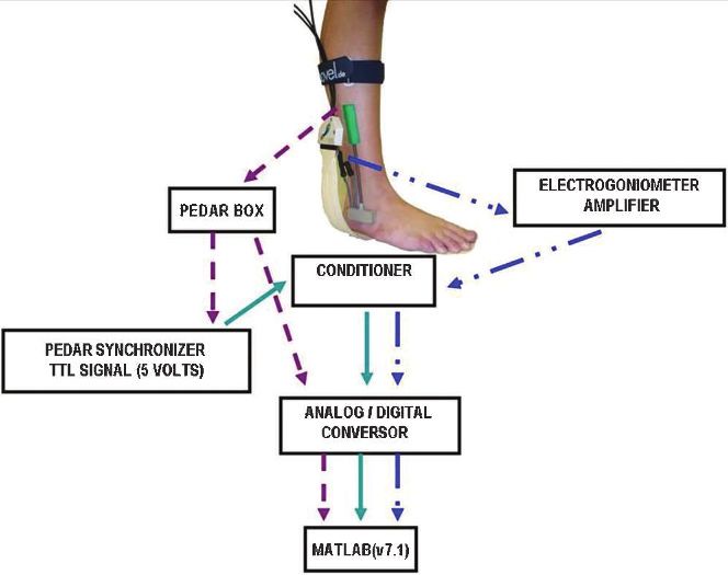
were synchronized by a TTL (transistor-transistor logic) Figure 1 - Synchronization between the Pedar-X System and the
signal that was emitted by the Pedar Sync Box. This box ankle electrogoniometer through the synchronizer box of the
Pedar-X System.
emitted 0 volts (V) when at least 20 kPa was detected by the
Pedar System while the foot was in contact with the surface
and 5 V when the pressure was lower than 20kPa while the to extension of the ankle and the toe-off instant (as seen in
foot was in the swing phase. the pressure data).
Although the Pedar-X system is most appropriate for Statistical inferential analysis was performed with
evaluating shod gait, we used the insoles to evaluate Statistica v.7 software (Statsoft Inc.). For statistical purposes,
barefoot gait as reported by Nurse and Nigg.29 Barefoot pressure data from only one foot per subject were analyzed
gait analysis was performed because our intention was to and compared. In the control group, a foot was randomly
investigate the complex behavior of the foot-floor interac- chosen for analysis, while in the PFPS group, the chosen foot
tion30,31 without any other interference such as the subject’s corresponded to the painful knee in subjects with unilateral
shoes. Additionally, the Pedar-X system could acquire pain and the most painful knee in subjects with bilateral
multiple steps without requiring the subject to alter their pain.
gait to make contact with any platform.32 Plantar pressure variables followed a normal distribution
The contact area (cm2) and peak pressure (kPa) were (Shapiro-Wilk’s Test), and variances were homogeneous
evaluated in six plantar areas that were adjusted propor- (Levene’s Test). Groups and areas were compared using
tionally to the length and width of each subject’s foot with 2 three-way ANOVAs (2X3X6) that considered the gait
the Novel Multiprojects software (Novel, Munich,
Germany). The plantar surface was first divided into three
larger areas, the rearfoot (30% of the foot length), midfoot
(30% of the foot length) and forefoot (40% of the foot length),
according to the scheme established by Cavanagh and
Ulbrecht.33 The rearfoot was subdivided into the medial
(30% of the rearfoot width), central (40% of the rearfoot
width) and lateral rearfoot (30% of the rearfoot width) areas,
and the forefoot was subdivided into the medial (55% of the
forefoot width) and lateral forefoot (45% of the forefoot
width).34
DATA ANALYSIS
The stance phase of the gait was divided into three
subphases using a customized mathematical function
developed using Matlab software (v.7.1). The initial heel
contact phase was defined as the time interval between the
first foot contact with the ground (as seen in the pressure
data) and the deflection instant in the transition from
extension to flexion of the ankle angular variation curve.
The midstance phase was defined as the time interval Figure 2 - Subphases of the gait stance obtained from ankle
sagittal angular variation: the initial heel contact occurred
between the prior ankle deflection instant (extension to
between A and B, the midstance phase occurred between B
flexion) and the deflection instant in the transition from the and C, and the propulsion phase occurred between C and D.
flexion to extension of the ankle. The propulsion phase was Positive values denote ankle flexion, and negative values denote
the interval between the last deflection instant from flexion extension.
369Patellofemoral pain syndrome CLINICS 2011;66(3):367-372
Aliberti S et al.
Figure 3 - Mean values of the contact area (cm2) in six plantar areas (MR = medial rearfoot, CR = central rearfoot, LR = lateral rearfoot,
M = midfoot, MF = medial forefoot and LF = lateral forefoot) during initial contact, midstance and propulsion. (CG = control group,
PFPS = patellofemoral pain syndrome, * p,0.05, # p , 0.1).
Figure 4 - Mean values of the peak pressure (kPa) in six plantar areas (MR = medial rearfoot, CR = central rearfoot, LR = lateral rearfoot,
M = midfoot, MF = medial forefoot and LF = lateral forefoot) during initial contact, midstance and propulsion. (CG = control group,
PFPS = patellofemoral pain syndrome, * p,0.05).
370CLINICS 2011;66(3):367-372 Patellofemoral pain syndrome
Aliberti S et al.
subphases (3) and the plantar areas (6) as repeated measure- revealing changes in this pattern that would be difficult to
ments; these analyses were followed by a Newman-Keuls perceive through a clinical visual observation of gait. The
post-hoc test. The level of significance was set at a = 5% and decision to divide the foot rollover mechanism into three
marginal significance was set at 1% , a , 5%. phases allowed us to investigate in more detail what was
happening at initial contact, midstance and propulsion in this
RESULTS population. The rollover pattern observed in the PFPS
individuals was different from that observed in the healthy
The PFPS group showed a greater contact area at the individuals and could induce alterations in the load attenua-
medial (p = 0.004) and central (p = 0.002) rearfoot during tion within the kinetic chain of the lower limbs. Plantar
initial heel contact, a greater contact area at the medial pressure that is medially distributed at the initial contact and
(p = 0.072) and lateral (p = 0.005) forefoot in the midstance laterally distributed during propulsion would probably
and a greater contact area at the lateral forefoot in the result in torque alterations in the lower kinetic chain.
propulsion phase (p = 0.079). A plantar contact that is medially oriented in the rearfoot
The PFPS group also presented a smaller peak pressure at is probably related to an everted rearfoot and could lead to
the medial forefoot during propulsion than the CG (p = an excessive medial rotation of the tibia. This rotation could
0.003). induce a compensatory medial rotation of the femur and a
lateralization of the patella in relation to the femur,
DISCUSSION increasing the patellofemoral joint stress.7 This medially
directed contact in the rearfoot has already been detected in
Our main findings showed that subjects with PFPS
individuals with PFPS during stair descent.34 Plantar
presented larger contact areas at the medial and central
pressure that is laterally distributed during propulsion is
rearfoot during initial heel contact, at the medial and lateral
probably related to a greater re-inversion that is performed
forefoot in the midstance, and at the lateral forefoot during
to provide a rigid lever for push-off, as the more everted
propulsion. PFPS individuals also presented a smaller peak
initial contact leads to a less stable foot.17 This event can
pressure at the medial forefoot that was followed by a larger
possibly cause lower limb torque modification during gait,
contact area at the lateral forefoot during propulsion.
inducing alterations in the load attenuation within the
These results suggest that PFPS individuals exhibit a foot
kinetic chain.
rollover process characterized by an initial heel contact that
This detailed characterization of the rocker mechanism is
is performed more medially at the rearfoot and a propulsion
clinically relevant because these findings can be used to
phase that is performed more laterally at the forefoot. In the
develop clinical interventions such as insoles, taping and
midstance, the larger contact area at both the medial and
therapeutic exercises useful for the rehabilitation of this
lateral forefoot suggests that PFPS individuals have a
dysfunction.
greater excursion of the foot during this phase both
Furthermore, this study evaluated a population that was
medially and laterally.
predominantly female who participated in this study and
According to Willems et al.,17 this laterally directed
exhibited similar levels of physical activity compared with
support during propulsion, which causes the terminal
the control group, which decreased the possibility that
push-off to occur laterally rather than predominantly across
gender and variable levels of physical activity interfered
the hallux as expected, occurs because an increase in
with our results.
eversion during initial heel contact leads to a less stable
foot. Consequently, a greater re-inversion is performed to
provide a rigid lever for optimal push-off. Willems et al.17 CONCLUSION
performed a prospective study of physically active subjects Individuals with PFPS exhibit a foot rollover pattern that
who developed exercise-related injuries. Plantar pressure is medially directed at the rearfoot during initial heel
and rearfoot 3D kinematics were evaluated, and the contact and laterally directed at the forefoot during
researchers observed a foot rollover pattern during running propulsion. These alterations during the foot rollover
that was similar to what we observed during gait. Subjects process can be used to develop clinical interventions that
who developed injuries showed a more central initial use insoles, taping and therapeutic exercise to rehabilitate
contact that was associated with a more everted rearfoot this dysfunction.
as well as a more laterally directed propulsion. In our study,
PFPS individuals presented a more medially directed
initial contact and more laterally directed support during REFERENCES
propulsion. This result may also be attributed to a greater 1. LaBella C. Patellofemoral pain syndrome: evaluation and treatment.
evertion of the rearfoot at heel strike followed by an increase Prim Care. 2004;31:977-1003.
2. DeHaven KE, Lintner DM. Athletic injuries: comparison by age, sport,
in supination during the propulsion phase. and gender. Am J Sports Med. 1986;14:218-24.
The laterally directed propulsion observed in this study, 3. Davis I S, Powers CM. Patellofemoral Pain Syndrome: proximal,distal
inferred from the larger lateral forefoot contact area and the and local factors:an international research retreat. J Orthop Sports Phys
Ther. 2010;40:A1-A48.
smaller peak pressure at the medial forefoot during 4. Powers CM. The influence of altered lower-extremity kinematics on
propulsion, is compatible with the pattern reported by patellofemoral joint dysfunction: a theoretical perspective. J Orthop
Thijs et al.18 These authors prospectively evaluated plantar Sports Phys Ther. 2003;33:639-46.
5. Messier SP, Davis SE, Curl WW, Lowery RB, Pack RJ. Etiologic factors
pressure during the gait of military subjects who developed associated with patellofemoral pain in runners. Med Sci Sports Exerc.
PFPS. They observed a more lateralized foot rollover pattern 1991;23:1008-15.
in subjects who developed PFPS than in subjects who did 6. Powers CM. The influence of abnormal hip mechanics on knee injury: a
not develop the disorder. biomechanical perspective. J Orthop Sports Phys Ther. 2010; 40:42-51.
7. Tibério D. The effect of excessive subtalar joint pronation on patellofe-
The present study contributes to discussions about the moral mechanics:a theoretical model. J Orthop Sports Phys Ther.
influence of PFPS in the foot rollover mechanism during gait, 1987;9:160-5.
371Patellofemoral pain syndrome CLINICS 2011;66(3):367-372
Aliberti S et al.
8. Salsich GB, Perman WH. Patellofemoral joint contact area is influenced 22. Cavanagh PR, Rodgers MM. The Arch Index: a useful measure from foot-
by tibiofemoral rotation alignment in individuals who have patellofe- prints. J. Biomechanics. 1987;20:547-51, doi: 10.1016/0021-9290(87)90255-7.
moral pain. J Orthop Sports Phys Ther. 2007;37:521-8. 23. Piva SR, Fitzgerald GK, Irrgang JJ, Fritz JM, Wisniewski S, McGinty GT
9. Gross MT, Foxworth JL. The role of Foot Orthoses as an Intervention for et al. Associates of physical function and pain in patients with
Patellofemoral Pain. J Orthop Sports Phys Ther. 2003; 33:661-70. patellofemoral pain syndrome. Arch Phys Med Rehabil. 2009 ;90:285-
10. Moya GB, Siqueira CM, Caffaro RR, Fu C, Tanaka C. Can quiet standing 95, doi: 10.1016/j.apmr.2008.08.214.
posture predict compensatory postural adjustment? Clinics. 2009;64:791- 24. Sacco ICN, Konno GK, Rojas GB, Arnone AC, Passaro AC, et al.
6, doi: 10.1590/S1807-59322009000800014. Functional and EMG responses to a physical therapy treatment in
11. Bacarin TA, Sacco IC, Hennig EM. Plantar pressure distribution patterns patellofemoral syndrome patients. J Electromyogr Kinesiol. 2006;16: 167-
during gait in diabetic neuropathy patients with a history of foot ulcers. 74, doi: 10.1016/j.jelekin.2004.06.010.
Clinics. 2009;64:113-20, doi: 10.1590/S1807-59322009000200008. 25. Natri A, Kannus P, and Jarvinen M. Which factors predict the long-term
12. Greve JM, Grecco MV, Santos-Silva PR. Comparison of radial shock- outcome in chronic patellofemoral pain syndrome? A 7-yr prospective
waves and conventional physiotherapy for treating plantar fasciitis. follow-up study. Med Sci Sports Exerc. 1998;30:1572-7, doi: 10.1097/
Clinics. 2009;64:97-103, doi: 10.1590/S1807-59322009000200006. 00005768-199811000-00003.
13. Earl JE, Hertel J, Denegar CR. Patterns of Dynamic Malalignment, 26. Shephard RJ. Limits to the measurement of habitual physical activity by
Muscle Ativation, Joint Motion and Patellofemoral-Pain Syndrome. J questionnaires. Br J Sports Med. 2003;37:197-206; discussion 206.
Sport Rehabil, 2005;14:215-33. 27. Burnfield JM, Few CD, Mohamed OS, Perry J. The influence of walking
14. Levinger P, Gilleard W. The heel strike transient during walking in speed and footwear on plantar pressures in older adults. Clin Biomech.
subjects with patellofemoral pain syndrome. Phys. Ther. in Sport.
2004;19:78-84, doi: 10.1016/j.clinbiomech.2003.09.007.
2005;6:83-8, doi: 10.1016/j.ptsp.2005.02.005.
28. Hsiao H, Guan J, Weatherly M. Accuracy and precision of two in-shoe
15. Levinger P, Gilleard W. Tibia and rearfoot motion and ground reaction
pressure measurement systems. Ergonomics. 2002;45:537-55, doi: 10.
forces in subjects with patellofemoral pain syndrome during walking.
1080/00140130210136963.
Gait Posture. 2007;25:2-8, doi: 10.1016/j.gaitpost.2005.12.015.
16. Powers CM, Chen PY, Reischl SF, Perry J. Comparison of foot pronation 29. Nurse MA, Nigg BM. The effect of changes in foot sensation on plantar
and lower extremity rotation in persons with and without patellofemoral pressure and muscle activity. Clin Biomech. 2001;16:719-27, doi: 10.1016/
pain. Foot Ankle Int. 2002;23:634-40. S0268-0033(01)00090-0.
17. Willems TM, De Clercq D, Delbaere K, Vanderstraeten G, De Cock A,. 30. Macellari V, Giacomozzi C, Saggini R. Spatial-temporal parameters of
Witvrouw E. A prospective study of gait related risk factors for exercise- gait: reference data and a statistical method for normality assessment.
related lower leg pain. Gait Posture. 2006;23:91-8, doi: 10.1016/j.gaitpost. Gait Posture. 1999;10:171-81, doi: 10.1016/S0966-6362(99)00021-1.
2004.12.004. 31. Macellari V, Giacomozzi C. Multistep pressure platform as a stand-alone
18. Thijs Y, Van Tiggelen D, Roosen P, De Clercq D, Witvrouw E. A system for gait assessment. Med Biol Eng Comput. 1996;34:299-304, doi:
prospective study on gait-related intrinsic risk factors for patellofemoral 10.1007/BF02511242.
pain. Clin J Sport Med. 2007;17:437-45. 32. Shaw JE, Van Shie CHM, Carrington AL, Abbott CA, Boulton AJM.
19. Breslow NE, Day NE. Statistical Methods in Cancer Research .The Analysis Ananalysis of dynamic forces transmitted through the foot in diabetic
of Case-Control Studies: Lyon. IARC Scientific Publications. 1987;2:1-406. neuropathy. Diabetes Care. 1998;21:1955-9, doi: 10.2337/diacare.21.11.1955.
20. Taunton JE, Ryan MB, Clement DB, McKenzie DC, Lloyd-Smith DR, 33. Cavanagh P, Ulbrecht J. Clinical plantar pressure measurement in
Zumbo BD. A retrospective case-control analysis of 2002 running diabetes: rationale and methodology. The Foot. 1994;4:123-35, doi: 10.
injuries. British Journal of Sports Medicine. 2002;36:95, doi: 10.1136/ 1016/0958-2592(94)90017-5.
bjsm.36.2.95. 34. Aliberti S, Costa MS, Passaro AC, Arnone AC, Sacco ICN. Medial contact
21. Eng JE, Pierrynowski MR. The effect of soft foot orthotics on three- and smaller plantar loads characterize individuals with Patellofemoral
dimensional lower-limb kinematics during walking and running. Pain Syndrome during stair descent. Phys Ther in Sport. 2010;11:30-4,
Physical Therapy. 1994;74:45-9. doi: 10.1016/j.ptsp.2009.11.001.
372You can also read