Nail Manifestations in COVID-19: Insight into a Systemic Viral Disease
←
→
Page content transcription
If your browser does not render page correctly, please read the page content below
Review Article

                                                           Skin Appendage Disord                                                           Received: May 13, 2021
                                                                                                                                           Accepted: June 21, 2021
                                                           DOI: 10.1159/000518087                                                          Published online: August 17, 2021

Nail Manifestations in COVID-19:
Insight into a Systemic Viral Disease
Ana Preda-Naumescu a Kayla Penney b Ross L. Pearlman a Robert T. Brodell a
Carlton Ralph Daniel c Vinayak K. Nahar a, d
aUniversity
           of Alabama at Birmingham School of Medicine, Birmingham, AL, USA; bLSU Health Shreveport School of
Medicine, Shreveport, LA, USA; cDepartment of Dermatology, School of Medicine, University of Mississippi
Medical Center, Jackson, MS, USA; dDepartment of Preventive Medicine, School of Medicine/John D. Bower School
of Population Health, University of Mississippi Medical Center, Jackson, MS, USA

Keywords                                                                                      with the onset of symptom onset or as late as 16 weeks fol-
SARS-CoV-2 · Nails · Red half-moon sign · Transverse                                          lowing the disease. Some of these findings are shared with
orange nail lesions · Mees’ lines · Beau’s lines · Periungual                                 other conditions associated with a proinflammatory state.
pernio-like changes                                                                           Nail changes offer unique insight into the pathophysiologic
                                                                                              basis for SARS-CoV-2 and they may serve as diagnostic clues.
                                                                                                                                            © 2021 The Author(s)
Abstract                                                                                                                                    Published by S. Karger AG, Basel

Nail manifestations are 1 of the several extrapulmonary find-
ings associated with COVID-19 caused by the severe acute                                          Introduction
respiratory syndrome coronavirus 2 (SARS-CoV-2). Nail
changes, however, have been largely ignored and not yet                                          COVID-19 is the contagious respiratory tract disease
summarized. This article is intended to increase awareness of                                 caused by severe acute respiratory syndrome coronavirus
nail manifestations of SARS-CoV-2, which occur weeks to                                       2 (SARS-CoV-2). It became a household name in spring
months after acute infection and the periungual pernio-like                                   of 2020 when the World Health Organization declared a
changes may occur concomitantly with infection. An elec-                                      world-wide pandemic [1]. Since the first outbreak was re-
tronic search was carried out in PubMed (Medline), Science                                    ported in Wuhan, China, in December of 2019, >132 mil-
Direct, and Scopus databases. The following keywords and all                                  lion global cases have been confirmed with over 2.8 mil-
of their possible combinations were used to identify studies:                                 lion global deaths as of April 7, 2021 [2]. Cutaneous find-
“SARS-CoV-2,” “COVID-19,” “Coronavirus,” “2019-ncov,”                                         ings vary with patient demographics and the timing of
“nail,” and “nails.” Six case reports were included in this study.                            infection. They are classified as erythema-edema with
Manifestations identified included red half-moon sign, trans-                                 vesicles or pustules (pseudo-chilblain), other vesicular
verse orange nail lesions, Mees’ lines, and Beau’s lines.                                     eruptions, urticarial lesions, maculopapular eruptions,
Though largely nonspecific, these findings can be recognized                                  and livedo or necrosis [3].

karger@karger.com      © 2021 The Author(s)                                                   Correspondence to:
www.karger.com/sad     Published by S. Karger AG, Basel                                       Vinayak K. Nahar, naharvinayak @ gmail.com
                       This article is licensed under the Creative Commons Attribution 4.0
                       International License (CC BY) (http://www.karger.com/Services/
                       OpenAccessLicense). Usage, derivative works and distribution are
                       permitted provided that proper credit is given to the author and the
                       original publisher.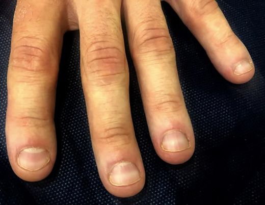
Table 1. A summary of the reviewed case reports

Author                 Condition           Description                              Relation to COVID-19                Other

Neri et al. [5]        The red half-moon   Red convex lesion at the distal        Two weeks after symptoms with         Reported widening of bands
                       sign                margin of the lunula on the fingernail confirmed diagnosis                   1 month after appearance
                                           beds
Méndez-Flores et al.   The red half-moon   Red convex lesion at the distal        Two days after symptoms with          Resolved within 1 week
[6]                    sign                margin of the lunula on the fingernail confirmed diagnosis
                                           beds
Tammaro et al. [8]     Transverse orange   Orange discolorations at the distal      Sixteen weeks after symptoms,       Remained unchanged at
                       nails               tips of the fingernail beds              appearing simultaneously with a     1 month
                                                                                    positive IgG test for SARS-CoV-2
Fernandez-Nieto        Transverse          White, transverse lines across the       Appeared concurrently with       Still present after 45 days,
et al. [11]            leukonychia         fingernail beds                          symptoms and confirmed diagnosis but diminishing as nail
                       (Mees’ lines)                                                                                 growth ensued
Alobaida et al. [12]   Beau’s lines        Transverse grooves along the             Three and a half months after
                                           proximal finger and toenail beds         symptoms and confirmed diagnosis
Ide et al. [13]        Beau’s lines        Transverse grooves along the             One month after symptoms and        Associated with leukonychia
                                           proximal fingernail beds                 confirmed diagnosis                 and periungual desquamation

    SARS-CoV-2, severe acute respiratory syndrome coronavirus 2.

   Because some nail manifestations of SARS-CoV-2                             ing keywords were utilized: “SARS-CoV-2,” “COVID-19,” “Coro-
may occur months after the acute infection, they have                         navirus,” “2019-ncov,” “nail,” and “nails.” Additional searches in
                                                                              the Google Scholar were performed to ensure that no studies were
been largely overlooked. Nonspecific ungual changes as-
                                                                              overlooked.
sociated with a variety of infectious diseases have been
described and may offer insight into the more serious sys-                        Search Outcome
temic manifestations of these conditions [4]. This article                        Two independent investigators performed the search and re-
highlights these findings and the unique nail lesions that                    viewed the titles, abstracts, full texts to determine if they met the
                                                                              inclusion criteria. A total of 1,094 studies were identified using the
have been described following infection with the novel
                                                                              electronic search processes. After removing duplicates, the titles
coronavirus.                                                                  and abstracts of 56 articles were reviewed, and then 14 articles were
                                                                              left for full-text review. During full-text review, 8 articles were
                                                                              omitted based on the inclusion criterion, and 6 articles met the
    Methods                                                                   predetermined eligibility requirements. Table 1 provides a brief
                                                                              overview of the COVID-19 cases with nail manifestations focusing
    Eligibility Criteria                                                      on clinical presentation, location, and temporal relationship to vi-
    Case reports were considered for inclusion based on the follow-           ral infection. The remainder of the review provides more detailed
ing criteria if: (1) nail changes in COVID-19 were assessed; (2)              insight to these unique nail presentations in hopes of helping
publication occurred in peer-reviewed journals; and (3) writing               health care providers recognize signs of this systemic disease.
was in English. Articles were excluded based on the following cri-
teria: (1) duplicates; (2) information appeared as conference ab-
stracts, opinion, editorials, perspective, viewpoints, news, letters to          Results
the editor, commentaries, reviews, feature articles, white papers,
and guidelines; (3) studies were incomplete or ongoing; and (4)
unpublished articles.                                                            The Red Half-Moon Sign
                                                                                 The red half-moon sign is a novel manifestation of
   Literature Search Strategy                                                 coronavirus infection. It has been described as appearing
   A literature review analyzed currently available published cases           following symptom onset. Presentation includes appear-
of documented SARS-CoV-2 with nail manifestations. An elec-
tronic search was carried out in PubMed (Medline), Science Di-                ance of a distally convex half-moon shaped red band sur-
rect, and Scopus databases from December 1, 2019, to April 2,                 rounding the distal margin of the lunula, which may ap-
2021. To identify the articles, possible combinations of the follow-          pear on all fingernails [5]. The affected patient denied as-

2                        Skin Appendage Disord                                                        Preda-Naumescu/Penney/Pearlman/
                         DOI: 10.1159/000518087                                                       Brodell/Daniel/Nahar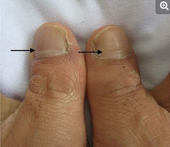
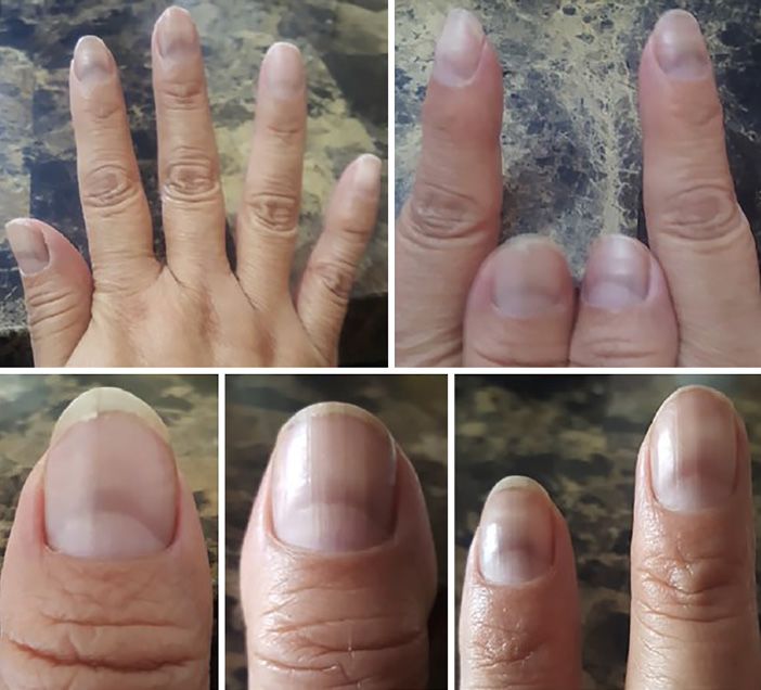
Fig. 1. The red half-moon sign. Red, hori-
zontally oriented, convex bands span the
distal margin of the lunula 2 days after the
onset of SARS-CoV-2 infection. Permis-
sion to use this image was granted by the
International Journal of Dermatology on
April 23, 2021. Fig. 1, page 1414 [6]. SARS-
CoV-2, severe acute respiratory syndrome
coronavirus 2.

sociated symptoms and had no other cutaneous
manifestations of COVID-19 [5]. This novel finding was
corroborated in a second patient 2 days following onset
of COVID-19 symptoms (Fig. 1) [6]. These clinical find-
ings may represent microvascular injury of the capillary
network of the distal subungual arcade secondary inflam-
matory immune response and a procoagulant mileu that
has been previously associated with SARS-CoV-2 infec-
tion [5]. Biopsies were not performed in either case. Sim-
ilarly, transverse nail-bed lines have been previously re-
ported in association with inflammatory conditions such
as Kawasaki disease [5]. Also appearing similar to the
above findings, erythematous periungual findings known
as “pernio-like” have been found in inflammatory states       Fig. 2. Transverse orange nails. Distal orange discoloration ap-
that are frequently seen in various systemic diseases [7].    peared 16 weeks after SARS-CoV-2 infection with a straight border
                                                              separating this finding from the proximal healthy-appearing nail
                                                              bed. Permission to use this image was granted by Dermatologic
   Transverse Orange Nail Lesions                             Therapy on April 23, 2021. Fig. 1, page 1 [7]. SARS-CoV-2, severe
   Orange discolorations at the end of the fingernail beds    acute respiratory syndrome coronavirus 2.
were noted in an elderly, female patient 16 weeks after the
onset of COVID-19 symptoms [8]. Physical exam re-
vealed orange discoloration of the distal nail plate with a   border of the discoloration followed the shape of the nail
demarcation separating them from the healthy-appear-          lunula which is consistent with a systemic cause of this
ing nail bed areas (Fig. 2) [8]. The shape of the proximal    finding [8]. A positive PCR diagnosis and IgG against

Nail Manifestations in COVID-19                               Skin Appendage Disord                                          3
                                                              DOI: 10.1159/000518087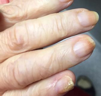
Fig. 4. Beau’s lines. Horizontal nail grooves were noted on all ten
                                                                     fingernails growing distally with nail growth. This photograph was
                                                                     taken 3 and a half months following a diagnosis of SARS-CoV-2
                                                                     [10]. Permission to use this image was granted by Canadian Med-
                                                                     ical Association Journal on April 23, 2021. Fig. 1, page E1040 [10].
Fig. 3. Transverse leukonychia (Mees’ lines). Transverse, non-       SARS-CoV-2, severe acute respiratory syndrome coronavirus 2.
blanchable white lines are present on all fingernails that slowly
grew out. This photograph was taken 45 days after the lines were
first noted during a hospitalization for SARS-CoV-2 infection.
Permission to use this image was granted by Dermatologic Thera-      Transverse leukonychia was recently described in all ten
py on April 23, 2021. Fig. 1, page 5 [9]. SARS-CoV-2, severe acute   fingernails in a previously healthy 57-year-old Spanish
respiratory syndrome coronavirus 2.                                  male with SARS-CoV-2 bilateral pneumonia confirmed
                                                                     by PCR [11]. The patient was treated with lopinavir/rito-
                                                                     navir 100 mg/400 mg bid for 10 days with adequate re-
SARS-CoV-2 confirmed the diagnosis. The nail findings                sponse [5]. The transverse, nonblanchable white lines
were unchanged 1 month later [8].The patient also had                progressively migrated with the growth of the nail and
ferropenic anemia [8]. The authors noted similar findings            were present 45 days later (Fig. 3) [11].
in the nails of Kawasaki patients, once more alluding to                Mees’ lines are also associated with a number of other
the possibility of complement-medicated microvascular                systemic stressors including acute renal failure, conges-
injury as the potential explanation behind this phenom-              tive heart failure, ulcerative colitis, cancer, infections, sys-
enon [7]. The significance of the low serum iron levels is           temic lupus erythematous, and the use of chemothera-
unknown, but this finding has been associated with more              peutic agents [11]. The lines appear to be the product of
severe SARS-CoV-2 disease [8].                                       altered keratinization of the nail plate that occurs as sys-
   A significant onychopathological indicator of chronic             temic conditions induce temporary dysfunction of nail
renal failure, Lindsay’s nail, or the half-and-half nail can         growth [11]. Importantly, they are nonblanchable which
resemble this finding. The key to distinguishing between             helps distinguish them from Muehrcke lines, which are
these phenomena may be the degree of orange discolor-                associated with hypoalbuminemia and altered nail bed
ation that is found in COVID affected nails [9]. Further-            vascularization [11].
more, this finding may also be confused for pseudo half-
and-half nails, a similar finding seen in patients with pso-            Beau’s Lines
riasis [9].                                                             Beau’s lines are transverse grooves in the nail plate
                                                                     caused by temporary diminution or suspension of nail
   Transverse Leukonychia (Mees’ Lines)                              growth approximately 2–3 weeks following acute stress to
   Transverse leukonychia or Mees’ lines are transverse              the nail matrix [4, 11]. These lines appear as the involved
white lines in the finger or toe nails. These exam findings          nail emerges from the proximal nail fold [6]. Beau’s lines
have been previously described in association with nu-               have been described following local trauma to the nail as
merous systemic disorders including arsenic poisoning                well as systemic illnesses, severe malnutrition, autoim-
due to deposition of arsenic in keratin rich tissues [4, 10].        mune conditions, including pemphigus and Raynaud dis-
Spanning the width of the nail plate, Mees’ lines often af-          ease, Kawasaki disease, and chemotherapy [12]. A
fect all fingernails and take 3–6 weeks to develop [4, 10].          45-year-old man recently presented with horizontal nail

4                      Skin Appendage Disord                                                Preda-Naumescu/Penney/Pearlman/
                       DOI: 10.1159/000518087                                               Brodell/Daniel/Nahar
grooves across the proximal nail folds of both finger and Conflict of Interest Statement
toe nails approximately 3 and a half months following a
Robert T. Brodell has participated in multi-center clinical trials
diagnosis of SARS-CoV-2 (Fig. 4) [12]. In another report, with Corevitas (Formerly Corrona) Psoriasis Registry and Novar-
sunken, white horizontal lines developed in the nails of a tis. He is also an associate editor of the Journal of the American
68-year-old Japanese man 1-month following hospital Academy of Dermatology, Faculty advisor for the American Med-
discharge for SARS-CoV-2 infection [13]. The nail find- ical Student Research Journal, and editor-in-chief of Practice Up-
ings were clinically defined as leukonychia and Beau’s date: Dermatology, and serves as Staff Dermatologist at the GV
(Sonny) MONTGOMERY VA HOSPITAL in Jackson, MS. Daniel
lines with periungual desquamation, a finding well-de- III C. Ralph is among the board of directors of Council for Nail
scribed in pediatric inflammatory multisystem syndrome Disorders, European Nail Society, and St. Dominic Health Ser-
[13]. No specific intervention is required for Beau’s lines vices Foundation. He is also Clinical Professor of Dermatology at
to resolve with continued nail growth. the University of Alabama at Birmingham. He is also American
Dermatological Association Chairman of Endowment Commit-
tee. He serves on the editorial board of the Skin Appendages Dis-
orders and on the advisory board of Ortho Pharmaceutical. He is
Discussion also the co-editor of a book Scher and Daniel’s Nails, Fourth edi-
tion, Springer, Philadelphia, 2018. He is also a stakeholder of Med-
Manifestations of COVID-19 infection in the nail unit imetriks. Ana Preda-Naumescu, Kayla Penney, Ross L. Pearlman,
are mostly non-specific possibly caused by the sensitive and Vinayak K. Nahar have no conflicts of interest.
nature of the nail matrix when impacted by trauma, in-
flammation, and hypercoagulability. This is certainly the Funding Sources
case with Beau’s lines which are well described in associa-
tion with coxsackievirus in addition to COVID-19 [14]. None declared.
Development of Beau’s lines after use of a tourniquet on
the upper extremity for hand surgery supports the hy-
Author Contributions
pothesis that nail matrix arrest may be secondary to isch-
emia [15]. Mees’ lines may similarly result from the dys- A.P.-N., K.P., and V.K.N. contributed to conception and de-
regulation of cell turnover at the matrix due to digital sign; A.P.-N., K.P., and V.K.N. contributed to the identification of
ischemia resulting from sepsis and hypotension, micro- studies. A.P-N., K.P., and V.K.N. contributed to the extraction of
vascular damage, or a hypercoagulable state resulting in data; all authors contributed to the analysis of data; all authors con-
tributed to the interpretation of data; all authors drafted the article
abnormal keratinization [16]. or revised it critically for important intellectual content; all authors
The red-half-moon nail sign is exceptional in that it gave final approval of the version of the article to be published; all
may represent a pathognomonic nail finding in CO authors agree to be accountable for all aspects of the work in ensur-
VID-19 infection [5]. These isolated case reports, how- ing that questions related to the accuracy or integrity of any part
ever, are not yet sufficient to establish this finding as a of the work are appropriately investigated and resolved; and all
authors have read and approved the manuscript.
unique characteristic of COVID-19 infection. COVID
“pernio” accounts for 2/3 of skin findings in patients with
COVID-19 and is associated with a lymphocytic vasculitis
of small dermal vessels with fibrin thrombi [17]. Similarly,
complement-mediated vascular injury results in throm- References 1 World Health Organization. Naming the
bus formation in small vessels in lung tissue, and comple- coronavirus disease (COVID19) and the virus
that causes it. 2020. Available from: https: //
ment binding to COVID-19 spike glycoproteins has been www.who.int/emergencies/diseases/novel-
identified in purpuric skin lesions of COVID-19 patients coronavirus-2019/technical-guidance/nam-
[18]. Neri et al. [5] have suggested the same mechanism ing-the-coronavirus-disease-(covid-2019)-
and-the-virus-thatcauses-it.
for erythronychia: inflammation and a procoagulant mi- 2 Johns Hopkins Corona Resource Center.
lieu produce microvascular injury of the nail bed. Available from: https: //coronavirus.jhu.edu.
Nail changes along with cutaneous manifestations Accessed 2021 April 7.
3 Galván Casas C, Català A, Carretero Hernán-
may provide valuable insight into underlying systemic dez G, Rodríguez-Jiménez P, Fernández-Nieto
manifestations of SARS-CoV-2. Clinicians should con- D, Rodríguez-Villa Lario A, et al. Classification
tinue to document these changes since they may become of the cutaneous manifestations of COVID-19:
a rapid prospective nationwide consensus
important diagnostic clues and help clinicians better un- study in Spain with 375 cases. Br J Dermatol.
derstand the pathophysiologic basis for this viral illness. 2020;183:71–7.
Nail Manifestations in COVID-19 Skin Appendage Disord 5
DOI: 10.1159/0005180874 Callen J, Jorizzo J, Zone J, Piette W, Rosen- SARS-CoV-2 infection. Dermatol Ther. 2021; foot-mouth disease: onychomadesis and
bach M, Vleugels RA. Nail signs of systemic 34(1):e14688. beau’s lines. Ann Dermatol. 2014 Apr; 26(2):
disease. Dermatological signs of systemic dis- 9 Lipner SP, Kroumpouzos G, Scher RK, Daniel 280–3.
ease. Philadelphia, PA: Elsevier; 2016. Vol. 5; CR. Nails in systemic disease. Sher and Dan- 15 Rubin AI, Jellinek NJ, Daniel CR III, Scher
p. 387–96. iel’s nails. 4th ed. Spinger; 2018. p. 343–82. RK. Scher and Daniel’s nails: diagnosis, sur-
5 Neri I, Guglielmo A, Virdi A, Gaspari V, Sta- 10 Sharma S, Gupta A, Deshmukh A, Puri V. Ar- gery, therapy. Philadelphia, PA: Springer In-
race M, Piraccini BM. The red half-moon nail senic poisoning and Mees’ lines. QJM. 2016; ternational Publishing; 2018. Vol. 4.
sign: a novel manifestation of coronavirus in- 109(8):565–6. 16 Fawcett RS, Linford S, Stulberg DL. Nail ab-
fection. J Eur Acad Dermatol Venereol. 2020; 11 Fernandez-Nieto D, Jimenez-Cauhe J, Orte- normalities: clues to systemic disease. Am
34(11):we663–5. ga-Quijano D, Diaz-Guimaraens B, Domin- Fam Physician. 2004 Mar 15;69(6):1417–24.
6 Méndez-Flores S, Zaladonis A, Valdes-Rodri- guez-Santas M, Martinez-Rubio J. Transverse 17 Cappel MA, Cappel JA, Wetter DA. Pernio
guez R. COVID-19 and nail manifestation: be leukonychia (Mees’ lines) nail alterations in a (chilblains), SARS-CoV-2, and COVID toes
on the lookout for the red half-moon nail sign. COVID-19 patient. Dermatol Ther. 2020; unified through cutaneous and systemic
Int J Dermatol. 2020;59(11):1414. 33(6):e13863. mechanisms. Mayo Clin Proc. 2021 Apr;
7 Daniel CR III, Bower JD, Danial CR. The half 12 Alobaida S, Lam JM. Beau lines associated 96(4):989–1005.
and half fingernail: the most significant ony- with COVID-19. CMAJ. 2020;192(36):E1040. 18 Magro C, Mulvey JJ, Berlin D, Nuovo G, Sal-
chopathological indicator of chronic renal 13 Ide S, Morioka S, Inada M, Ohmagari N. vatore S, Harp J, et al. Complement associated
failure. J Miss State Med Assoc. 1975;16:376. Beau’s lines and leukonychia in a COVID-19 microvascular injury and thrombosis in the
8 Tammaro A, Adebanjo GAR, Erasmus HP, patient. Intern Med. 2020;59(24):3259. pathogenesis of severe COVID-19 infection:
Chello C, Pezzuto A, Ramirez-Estrada S, et al. 14 Shin JY, Cho BK, Park HJ. A clinical study of a report of five cases. Transl Res. 2020 Jun;
Transverse orange nail lesions following nail changes occurring secondary to hand- 220:1–13.
6 Skin Appendage Disord Preda-Naumescu/Penney/Pearlman/
DOI: 10.1159/000518087 Brodell/Daniel/NaharYou can also read