A Mononuclear Nickel (II) Complex: Synthesis, X-ray Structure, Spectroscopic Characterization and Anti-tumor Activity
←
→
Page content transcription
If your browser does not render page correctly, please read the page content below
Advanced Chemicobiology Research
http://ojs.wiserpub.com/index.php/ACBR/

Research Article

A Mononuclear Nickel (II) Complex: Synthesis, X-ray Structure,
Spectroscopic Characterization and Anti-tumor Activity

Sangharaj Diyali1, Ayan Kumar Mohanty2, Subhajit Sarkar1, Md. Firoj Hossain1, Biplab Bhowmik3,
Bhaskar Biswas1*
1
 Department of Chemistry, University of North Bengal, Darjeeling 734013, India
2
 Department of Biotechnology, University of North Bengal, Darjeeling 734013, India
3
 Department of Zoology, Diamond Harbour Women’s University, West Bengal 743368, India
 E-mail: bhaskarbiswas@nbu.ac.in, icbbiswas@gmail.com

Received: 18 December 2020; Revised: 4 January 2021; Accepted: 14 January 2021

Abstract: In this work, an effort has been made to examine the counteranionic behavior of thiocyanide ion towards
the stabilization of a dicationic tris(2,2’-bipyridine)nickel (II) complex in the crystalline phase. The complex,
[Ni(bipy)3]·2NCS (1) [bipy = 2,2’-bipyridine] was designed, synthesized and structurally characterized. The crystal
structure analysis reveals that 1 crystallises in a hexagonal system with P6/mcc space group and adopts an octahedral
geometry. The nickel (II) complex exhibits important cytotoxic behaviour towards lung cancer cells (A549). IC50 value
is calculated through MTT assay and determined as 131 µG/mol. The relative percentage of morphological changes is
further determined and ~63% of the cells are destroyed through apoptosis mode within 24 h incubation.

Keywords: nickel (II), 2,2’-bipyridine, crystal structure, anti-tumor activity

1. Introduction
      Metallo-intercalators act as a pivotal role in developing the chemistry of nucleic acids, especially for their
importance in versatile applications like footprinting of DNA, design of structural probes, the development of
therapeutic agents, etc [1-7]. Thus, enormous numbers of metallo-intercalators consisting of planar N-donor
heterocyclics are widely utilized as vital chemical or photochemical reagents in the progress of nucleic acids chemistry.
One of the renowned scientist groups, Barton and co-workers [1, 8-12] contributed significantly to the chemical science
of metallo-intercalators and charge transport in DNA with the photo-physical and biological studies of polypyridyl
based metal complexes containing d-block metal ions. Meanwhile, Sigman and co-workers contributed immensely
to chemical nuclease activities by designing metal-phenanthroline chelates [6, 13-17]. During the last few decades,
2,2’-bipyridine, 1,10-phenanthroline, terpyridine and others are widely employed as effective chelators in advanced
chemical sciences [18]. Scientific studies illustrate that nickel-based coordination compounds play a pivotal role in
DNA damage particularly in aging and cancerous activity [19-21]. It is also documented that nickel complexes show an
important anti-proliferative effect against leukemic cells (HL-60), prostatic cells (PC-3) and human hepatocarcinoma
cells (Hep-G2) [22]. Therefore, the design and exploration of anti-proliferative activity of the coordination compounds
of nickel have been considered as an emerging area of biological and medicinal research [23-25].

Copyright ©2021 Bhaskar Biswas, et al.
DOI:
This is an open-access article distributed under a CC BY license
(Creative Commons Attribution 4.0 International License)
https://creativecommons.org/licenses/by/4.0/

Advanced Chemicobiology Research                                                                  22 | Bhaskar Biswas, et al.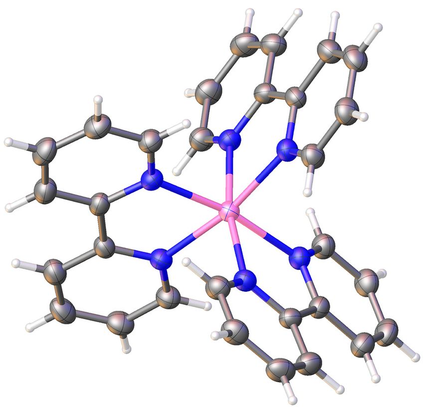
Among the transition metal ions, nickel (II) ion is very attractive towards the scientific community for its properties
to serve as bio-enzymes like hydrolage, generator of H2 gas, etc. Among the available bio-relevant metalloenzymes,
alkaline phosphatase enzyme contributes significantly to different biological functions ranging form DNA fragmentation
to RNA replication to bone metabolism as well as bio-remediation of organophosphate pesticides [26]. Previously,
several scientific groups made the effort to establish the crystal structures of tris(2,2’-bipyridine)nickel (II) in association
with different counteranions like perchlorate, hexamolybdate, chloride, tetrachlorozincate and even 2-thiobarbiturate
[27-31]. Herein, a tris(2,2’-bipyridine)nickel (II) thiocyanido complex has been synthesized and structurally
characterized. Studies of its antitumor activities suggest that ~63% of the cells are found as apoptosis in 24 h incubation
which was assessed from the relative percentage of morphological changes for lung cancer cells (A549).

2. Materials and methods
2.1 Starting materials
    Highly pure 2,2’-bipyridine (SRL, India), nickel (II) chloride hexahydrate (SRL, India), ammonium thiocyanide
(SRL, India) and all the reagents, chemicals and solvents were procured from the reputed outlets and used as received.

2.2 Physical measurements
    A FTIR-8400S spectrophotometer (SHIMADZU, Japan) with KBr palette was used to record FT-IR and a V-730
UV-Visible spectrophotometer (JASCO, Japan) was employed to measure the electronic spectrum in the solution phase.
A Q-Tof-micro quadruple mass spectrometer (Waters Corporation, USA) was employed to record ESI-Ms spectrum.
The pH value of the solutions was measured in PH meter (Systronics, India) at room temperature. A 2400 CHN
microanalyzer (Perkin Elmer, USA) was used to carry out elemental analyses.

2.3 Synthesis of [Ni(bipy)3 ]·2NCS (1)
     A methanolic solution of bipyridine (0.312 g, 2 mM) was mixed drop by drop to an aqueous solution of NiCl2 (0.240
g, 1 mM) followed by addition of aqueous solution of 2 mM of NH4NCS (0.152 g). The pink coloured solution was kept
in open atmosphere to obtain single crystals. Yield: 0.183 g (~76% based on metal salt). Anal. Calc. for C32H24N8S2Ni (1):
C, 59.74; H, 3.76; N, 17.42. Found: C, 59.79; H, 3.71; N, 17.47%. IR (KBr, cm-1): 2071 (vNCS), 1638,1601 (vC = N); UV-
Vis (λmax, Abs, nm, 10-4 M, H2O): 275, 305, 509, 705.

2.4 X-ray diffraction analysis
     An XtaLABmini diffractometer (Rigaku Corporation, Japan) equipped with a Mercury375R (2 × 2 bin mode) CCD
detector was employed to collect X-ray diffraction data. The data were obtained on a graphite monochromated Mo-Kα
radiation (λ = 0.71073 Å) at 293(2) K using w scans. The diffraction data were reduced with Crystal Clear suite. OLEX2
was used to determine the space group of the complex. Direct method was used to solve the crystal structure. The
refinement of diffraction data was done by full-matrix least-squares procedures with the SHELXL-97 software package
employing OLEX2 suite [32-33].

2.5 Anticancer activity of the nickel (II) complex
2.5.1 Cell culture

      The cell line (A549 human lung cancer) was collected from the National Center for Cell Science, Pune, India.
Dulbecco's Modified Eagle Medium (DMEM) high glucose medium (Sigma-Aldrich, USA) in supplement with 10%
fetal bovine serum (Gibco) was employed to culture the cells in 96-well culture plates. The culture plate was kept in
a humidified atmosphere of 5% CO2 at 37°C (Thermo Scientific, USA). The details of the experiment may be found
elsewhere.

Volume 1 Issue 1|2021| 23                                                                       Advanced Chemicobiology Research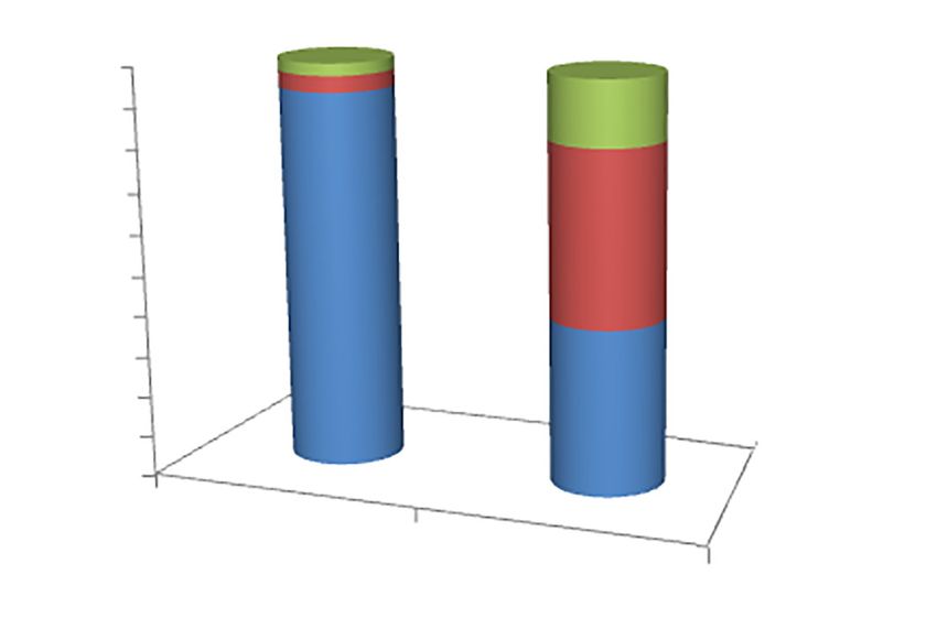
2.5.2 Cytotoxicity assay (MTT assay)
The viability of A549 cells was tested by cell viability assay using MTT reagent [34], a colorimetric assay. The
various concentrations of Ni (II) complex were prepared from stock solution of Ni (II) complex in dimethylsulfoxide
(DMSO) diluted using DMEM media. Two hundred microliters of the sample was added to wells having 5 × 10³ A549
cells per well. DMSO was considered as the solvent control. Twenty µL of MTT solution (5mg/mL in poly butylene
succinate (PBS)) was added to the well and the plate was wrapped in aluminum foil. Then, it was incubated at 37°C
for 4 h. The formazan product was dissolved by the addition of 100 μL of DMSO to the well. The absorbance was
monitored at 570 nm (measurement) and 630 nm (reference) using a 96-well plate reader (Bio-Rad, iMark, USA).
Data were collected for three replicates each and used to calculate the respective mean. The percentage inhibition was
determined following the formula:
Mean OD of untreated cells ( control ) - Mean OD of treated cells ( treated ) × 100
=
Mean absorbance of untreated cells ( control )
The IC50 value which was calculated using viability against concentration plot (IC50 is the concentration at which
the complex killed 50% of the cells) for 24 h was determined.
2.5.3 Acridine orange (AO) and ethidium bromide (EB) staining
The apoptosis or cell death was evaluated by the staining method described by Spector et al. [34]. The cells were
treated with an IC50 concentration of the nickel complex for 24 h. After incubation, the cells were harvested and cold
PBS was used to wash the cells. Cell pellets were re-suspended and diluted with PBS to a concentration of 5 × 10⁵ cells/
mL and mixed with 25 µL of AO/EB solution (3.8 µM of AO and 2.5 µM of EB in PBS) on clean microscope slide and
immediately examined under an Axio scope 2 plus fluorescent microscope (Zeiss, Japan) with UV filter (450-490 nm).
Among the 300 cells for each sample, viability was checked through apoptosis or necrosis following the staining of
membrane integrity and nucleus structure.
3. Results and discussion
3.1 Synthesis and formulation
The octahedral nickel (II) complex was produced by adding 2,2’-bipyridine to nickel (II) chloride followed by
ammonium thiocyanide in an aqueous-methanolic solution at room temperature. Previously, Czakis-Sulikowska et al.
prepared similar type of nickel (II)-thiocyanate complex in reaction with 4,4’-bipyridine in aq. ethanolic medium [35].
The primary zone of coordination for 1 was defined by mainly X-ray diffraction study and other spectroscopic and
analytical methods. The pink coloured single crystals were isolated after a couple of days. The structural formulations
were confirmed by different analytical techniques, and X-ray diffraction analysis.
Figure 1. Preparative route for the nickel (II) complex (1)
Advanced Chemicobiology Research 24 | Bhaskar Biswas, et al.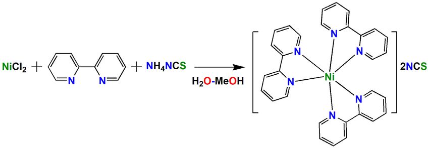
3.2 Crystal structure of [Ni(bipy)3 ]·2NCS (1)
     The X-ray structure of mononuclear Ni (II) complex indicates that mononuclear Ni (II) complex exists in a
hexagonal crystal system with P6/mcc space group and adopts a distorted octahedral geometry. An Oak Ridge Thermal
Ellipsoid Plot (ORTEP) view of 1 with 30% ellipsoidal probability is shown in Figure 2. Both the axial and equatorial
planes around the Ni (II) centre are formed by nitrogen atoms of the three bipyridine ligands. The dicationic charge
of nickel (II)-bipyridine chelate is counterbalanced by two units of thiocyanate ions. The crystallographic structural
parameters of 1 are listed in Table 1. The metal centric selected bond lengths and angles are presented in Table 2.
     Scientific documents show that Hong et al., Zhang et al., Rui Perez et al. reported the crystal structures of tris(2,2’-
bipyridine)nickel (II) chelate in combination with percholarate, molybdate and chloride anions respectively [27-28,
30]. The reported crystal structures showed different crystal systems and space groups and a comparison of structural
refinement parameters between the reported structures and our structure is given in Table 3. In our crystal structure, the
thiocyanate ions get distorted although we made several attempts to refine the structure. However, we have confirmed
the existence of thiocyanate through IR and elemental analysis.

                                    Table 1. Crystallographic refinement parameters of [Ni(bipy)3]·2NCS (1)

                                    Crystal parameters                                                 1
                                    Empirical formula                                             C31H24N7SNi
                                     Formula weight                                                  584.7
                                       Temperature                                                   293 K
                                       Wavelength                                                  0.71073 Å
                                      Crystal system                                               Hexagonal
                                       Space group                                                  P6/mcc
                                   Unit cell dimensions                                        a = 13.3747 (19) Å
                                                                                               b = 13.3747(19) Å
                                                                                                c = 21.440(4) Å
                                         Volume                                                   3321.4(9) Å3
                                             Z                                                         8
                                   Density (calculated)                                           1.110 Mg/m3
                                  Absorption coefficient                                          1.335 mm-1
                                          F (000)                                                     1152
                                   Reflections collected                                             6964
                                  Independent reflections                                             1122
                                          R (int)                                                    0.035
                                   Goodness-of-fit on F2                                              1.08
                                    R indices (all data)                                   R1 = 0.0504, wR2 = 0.170
                                Largest diff. peak and hole                                   0.50 and -0.40 e. Å-3

                Table 2. Metal centric bond lengths (Å) and bond angles (°) for [Ni(bipy)3]·2NCS obtained from the XRD structure

                                                 Bond lengths (Å)                             Bond angles
                       Ni1-N1                       2.091 (2)               N1-Ni1-N1*a                       94.35 (10)
                                                                            N1-Ni1-N1*c                        78.64 (9)
                                                                            N1-Ni1-N1*d                       169.86 (12)
                                                                            N1-Ni1-N1*e                       93.50 (10)

Volume 1 Issue 1|2021| 25                                                                                     Advanced Chemicobiology Research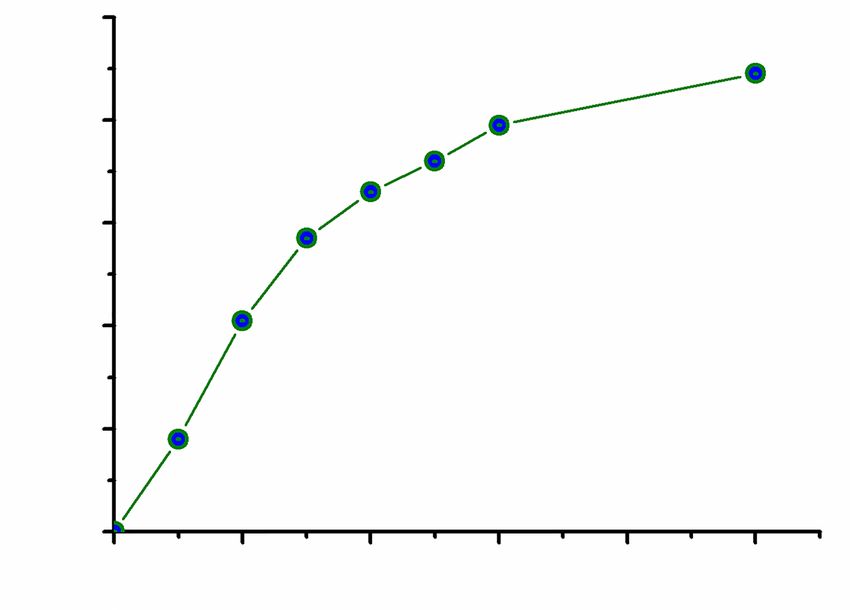
C2
                                                            C1
                                         C3

                                      C4
                                                            N1
                                                                              Ni1
                                                  C5

                          Figure 2. An ORTEP diagram of the dicationic [Ni(bipy)3]2+ with 30% ellipsoidal probability

           Table 3. A comparison of structural refinement parameters of [Ni(bipy)3]·2NCS (1) with the previously reported structures

           Parameter             [Ni(bipy)3]Cl2 ([30])    [Ni(bipy)3]·2ClO4 ([27])    [Ni(bipy)3][ZnCl4] ([31])            This work*

         Crystal system              Monoclinic                  Monoclinic                    Trigonal                    Hexagonal

          Space group                    C2/c                       C2/c                         R3c                         P6/mcc

               Z                           4                          4                           12                            8

      Unit cell dimensions

             a (Å)                    13.410 (2)                 17.502 (2)                  13.34.3 (2)                  13.3747 (19)

             b (Å)                    22.509 (4)                 10.777 (1)                                               13.3747 (19)

             c (Å)                    23.781 (4)                 16.092 (2)                  58.932 (12)                   21.440 (4)
                   3
            V (Å )                     6921 (2)                  3034.8 (6)                    9087 (3)                    3321.4 (9)

3.3 Solution phase behavior of the nickel (II) complex
     The absorption spectrum of the tris(bipy)nickel (II) complex was measured in aqueous phase at room temperature.
The electronic spectrum displayed that the two well resolved ligand field transitions at 705 and 509 nm in the visible
range corresponding to the spin-allowed 3A2g → 3T2g, and 3A2g → 3T1g(F) electronic transitions respectively. This
spectrum resembles very well with the electronic spectrum of the tris(2,2’-bipy)nickel (II) tetrafluoroborate complex
reported by Abramov. The electronic transition, 3A2g → 3T1g(F) is responsible for the development of pink color. Other
electronic transitions at 275 and 305 nm are assignable to π→π or n→π transitions of bipyridine ligands.

3.4 Antiproliferative activity of the nickel (II) complex
3.4.1 MTT Assay

     The human A549 lung cancer cell line was considered to examine the cytotoxic activities of the Ni (II) complex
following MTT assay [36]. The cytotoxic effect was studied based on dose-dependent manner which helps to determine

Advanced Chemicobiology Research                                                                                        26 | Bhaskar Biswas, et al.
the IC50 in comparison with untreated cells. The IC50 value for 24 h was calculated as 131 ± 0.5 µG/mL and the results
of MTT assay indicated that the nickel (II) complex is moderately toxic towards A549 cells (Figure 3).
100
80
60
% Inhibition
40
20
0
0 100 200 300 400 500
Concentration, μM
Figure 3. Cytotoxic effect of the [Ni(bipy)3]·2NCS complex on A549 cells after exposure for 24 h
3.4.2 AO/EB staining
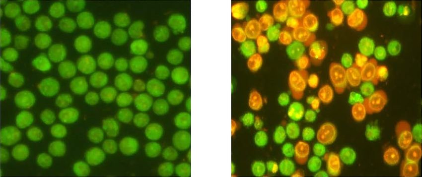
The morphological changes in the apoptosis induced by the nickel (II) complex were determined by AO/EB
staining. The early and late apoptosis of A549 cancer cell line in treatment with synthetic tris(chelate)nickel (II) complex
for 24 h was observed with AO/EB double-stained method and shown in Figure 4. It is commonly observed that the
viable cells display a green fluorescence which indicates the normal intact cell membrane featuring uniform chromatin.
However, a bright green region with yellowish-green colour presents the early apoptosis cells which recommend the
nuclear fragmentation with apoptotic bodies outside the cell membrane. The orange-yellow colour in AO/EB staining
suggests the late apoptosis cells with fragmented chromatin. The AO/EB results suggest that the nickel (II) complex
induced the majority of cell death through apoptosis mode and very fewer in necrosis for 24 h treatment (Figure 5). Cell
shrinkage, chromatin condensation, and fragmentation were also observed in Ni (II) complex treated cells. This finding
is correlated with the MTT assay (Figure 3).
Control Ni(II) Complex
Figure 4. Representative morphological changes produced in A549 human lung cancer cells by [Ni(bipy)3]·2NCS (1) with AO/EB staining after 24 h
incubation
Volume 1 Issue 1|2021| 27 Advanced Chemicobiology ResearchAO/EB Staining
100
90
80
Percentage of cells
70 Necrosis
60 Apoptosis
50
40 Normal
30
20
10
0
Control
Ni(II) Complex
Control and treated cells at 24 h
Figure 5. Relative percentage of morphological changes by the treatment of [Ni(bipy)3]·2NCS (1) with A549 human lung cancer cell following
AO/EB staining
4. Conclusions
To summarize, a mononuclear octahedral nickel (II)-phenanthroline complex, [Ni(bipy)3]·2NCS (1) was prepared
and structurally characterized using different analytical techniques and computational modeling. The crystal structural
analysis indicates that nickel (II) complex was crystallized in a hexagonal system and adopted an octahedral geometry.
The nickel (II) complex exhibited important cytotoxic behaviour towards lung cancer cells (A549) and IC50 value was
calculated through MTT assay and determined the value as 131 µG/mol. The changes in apoptosis and necrosis were
observed through changes in relative percentage of morphological behavior which ensures that ~63% of the cells are
destroyed compared to the control cells after 24 h incubation.
Supplementary data
Supplementary crystallographic data are available free of charge from The Director, CCDC, 12 Union Road,
Cambridge, CB2 1EZ, UK (fax: +44-1223-336033; E-mail: deposit@ccdc.cam.ac.uk or www: http://www.ccdc.cam.
ac.uk) upon request, quoting deposition number CCDC 2050513 for the nickel (II) complex 1.
References
[1] Erkkila K, Odom D, Barton J. Recognition and reaction of metallointercalators with DNA. Chemical Reviews.
1999; 99: 2777-2796.
[2] Armitage B. Photocleavage of nucleic acids. Chemical Reviews. 1998; 98: 1171-1200.
[3] Pogozelski W, Tullius T. Oxidative strand scission of nucleic acids: Routes initiated by hydrogen abstraction from
the sugar moiety. Chemical Reviews. 1998; 98: 1089-1108.
[4] Burrows C, Muller J. Oxidative nucleobase modifications leading to strand scission. Chemical Reviews. 1998; 98:
1109-1152.
[5] McMillin D, McNett K. Photoprocesses of copper complexes that bind to DNA. Chemical Reviews. 1998; 98:
1201-1220.
[6] Sigman D, Bruice T, Mazumder A and Sutton C. Targeted chemical nucleases. Accounts of Chemical Research.
Advanced Chemicobiology Research 28 | Bhaskar Biswas, et al.1993; 26: 98-104.
[7] Pratviel G, Bernadou J, Meunier B. DNA and RNA cleavage by metal complexes. Advanced Inorganic Chemistry.
1998; 45: 251-312
[8] Pyle AM. Barton JK. Probing nucleic acids with transition metal complexes. Progress in Inorganic Chemistry.
1990; 38: 413-475.
[9] Barton J. Metals and DNA: Molecular left-handed complements. Science. 1986; 233: 727-734.
[10] Barton JK, Raphael AL. Site-specific cleavage of left-handed DNA in pBR322 by lambda-tris (diphenyl
phenanthroline) cobalt (III). Proceedings of the National Academy of Sciences of the United States of America.
1985; 82: 6460-6464.
[11] Delaney S, Pascaly M, Bhattacharya P, Han K and Barton J. Oxidative damage by ruthenium complexes containing
the dipyridophenazine ligand or its derivatives: A focus on intercalation. Inorganic Chemistry. 2002; 41: 1966-
1974.
[12] Sontz P, Muren N, Barton J. DNA charge transport for sensing and signaling. Accounts of Chemical Research.
2012; 45: 1792-1800.
[13] Sigman D, Mazumder A, Perrin D. Chemical nucleases. Chemical Reviews. 1993; 93: 2295-2316.
[14] Sigman D. Nuclease activity of 1,10-phenanthroline-copper ion. Accounts of Chemical Research. 1986; 19: 180-
186.
[15] Sigman D, Graham D, D’Aurora V and Stern A. Oxygen-dependent cleavage of DNA by the 1,10-phenanthroline.
cuprous complex. Inhibition of escherichia coli DNA polymerase I. Journal of Biological Chemistry. 1979; 254:
12269-12272.
[16] Zelenko O, Gallagher J, Sigman D. Scission of DNA with Bis (1,10-phenanthroline) copper without intramolecular
hydrogen migration. Angewandte Chemie International Edition in English. 1997; 36: 2776-2778.
[17] Zelenko O, Gallagher J, Xu Y, Sigman D. Chemical nuclease activity of 1,10-phenanthroline-copper. Isotopic
probes of mechanism. Inorganic Chemistry. 1998; 37: 2198-2204.
[18] Reedijk J, Wilkinson G, Gillard RD and McCleverty JA. Comprehensive Coordination Chemistry, Pergamon Press,
Oxford. 1987; 2: 73.
[19] Saleem K, Wani W, Haque A, Lone M, Hsieh M, Jairajpuri M, et al. Synthesis, DNA binding, hemolysis assays
and anticancer studies of copper (II), nickel (II) and iron (III) complexes of a pyrazoline-based ligand. Future
Medicinal Chemistry. 2013; 5: 135-146.
[20] Muralisankar M, Haribabu J, Bhuvanesh N, Karvembu R and Sreekanth A. Synthesis, X-ray crystal structure,
DNA/protein binding, DNA cleavage and cytotoxicity studies of N (4) substituted thiosemicarbazone based copper
(II)/nickel (II) complexes. Inorganica Chimica Acta. 2016; 449: 82-95.
[21] Haleel A, Arthi P, Dastagiri RN, Veena V, Sakthivel N, Arun Y, et al. DNA binding, molecular docking and
apoptotic inducing activity of nickel (II), copper (ii) and zinc (ii) complexes of pyridine-based tetrazolo [1,5-a]
pyrimidine ligands. RSC Advances. 2014; 4: 60816-60830.
[22] Zhu T, Wang Y, Ding W, Xu J, Chen R, Xie J, et al. Anticancer activity and DNA-binding investigations of the Cu (II)
and Ni (II) complexes with coumarin derivative. Chemical Biology & Drug Design. 2014; 85: 385-393.
[23] Alomar K, Landreau A, Allain M, Bouet G, Larcher G. Synthesis, structure and antifungal activity of thiophene-
2,3-dicarboxaldehyde bis(thiosemicarbazone) and nickel (II), copper(II) and cadmium(II) complexes:
Unsymmetrical coordination mode of nickel complex. Journal of Inorganic Biochemistry. 2013; 126: 76-83.
[24] Ramírez-Macías I, Maldonado C, Marín C, Olmo F, Gutiérrez-Sánchez R, Rosales M, et al. In vitro anti-leishmania
evaluation of nickel complexes with a triazolopyrimidine derivative against leishmania infantum and leishmania
braziliensis. Journal of Inorganic Biochemistry. 2012; 112: 1-9.
[25] Betanzos-Lara S, Gómez-Ruiz C, Barrón-Sosa L, Gracia-Mora I, Flores-Álamo M, Barba-Behrens N. Cytotoxic
copper (II), cobalt (II), zinc (II), and nickel (II) coordination compounds of clotrimazole. Journal of Inorganic
Biochemistry. 2012; 114: 82-93.
[26] Kojima T, Morimoto T, Sakamoto T, Miyazaki S and Fukuzumi S. Ruthenium (II) pyridylamine complexes with
diimine ligands showing reversible photochemical and thermal structural change. Chemistry-A European Journal.
2008; 14: 8904-8915.
[27] Zhou Y, Li X, Xu Y, Cao R and Hong M. Tris(2,2’-bipyridine)nickel (II) diperchlorate. Acta Crystallographica
Section E Structure Reports Online. 2003; 59: m300-m302.
[28] Fan L, Wei P, Pang S and Zhang X. Tris(2,2’-bipyridine)nickel (II) hexamolybdate. Acta Crystallographica Section
E Structure Reports Online. 2010; 66: m1119-m1119.
[29] Golovnev N, Molokeev M, Sterkhova I, Lesnikov M and Samoilo A. Structure of bis (2-Thiobarbiturate) Tris(2,2-
Volume 1 Issue 1|2021| 29 Advanced Chemicobiology ResearchBipyridyl) Nickel (II) Hexahydrate. Journal of Structural Chemistry. 2019; 60: 111-116.
[30] Ruiz-Pérez C, Lorenzo Luis P, Lloret F and Julve M. Dimensionally controlled hydrogen-bonded nanostructures:
synthesis, structure, thermal and magnetic behaviour of the tris-(chelated) nickel (II) complex [Ni(bipy)3]
Cl2·5.5H2O (bipy = 2,2’-bipyridyl). Inorganica Chimica Acta. 2002; 336: 131-136.
[31] Lin SH, Wang ZK, Zhang BH, Hu HM and Huang, JS. Crystal structure of tris(2,2’-bipyridine)nickel (II)
tetrachlorozincate. Chinese Journal of Chemistry. 2000; 19: 95-98.
[32] CrystalClear 2.0. Japan, Tokyo: Rigaku Corporation.
[33] (a) Sheldrick G. A short history of SHELX. Acta Crystallographica Section A Foundations of Crystallography.
2007; 64: 112-122. (b) Dolomanov O, Bourhis L, Gildea R, Howard J and Puschmann H. OLEX2: A complete
structure solution, refinement and analysis program. Journal of Applied Crystallography. 2009; 42: 339-341.
[34] Spector DL, Goldman RD, Leinwand LA. Cell: A laboratory manual. Culture and Biochemical Analysis of Cells.
New York: Cold Spring Harbor Laboratory Press; 1998.
[35] Czakis-Sulikowska D, Katuzna J. Synthesis and thermal behaviour of 4,4’-bipyridyl and 2,4’bipyridyl complexes
of Co (II), Ni (II) and Cu (II) thiocyanates. Journal of Thermal Analysis. 1996; 47: 1763-1776.
[36] Mosmann T. Rapid colorimetric assay for cellular growth and survival: Application to proliferation and cytotoxicity
assays. Journal of Immunological Methods. 1983; 65: 55-63.
Advanced Chemicobiology Research 30 | Bhaskar Biswas, et al.You can also read