TCL1 and CLA Expression in Agranular CD4/CD56 Hematodermic Neoplasms (Blastic NK-Cell Lymphomas) and Leukemia Cutis
←
→
Page content transcription
If your browser does not render page correctly, please read the page content below
Hematopathology / TCL1 AND CLA IN CD4/CD56 HEMATODERMIC NEOPLASMS AND LEUKEMIA CUTIS

TCL1 and CLA Expression in Agranular CD4/CD56
Hematodermic Neoplasms (Blastic NK-Cell Lymphomas)
and Leukemia Cutis
Tony Petrella, MD,1,2 Chris J.L.M. Meijer, MD,6 Sophie Dalac, MD,3 Rein Willemze, MD,7
Marc Maynadié, MD,5 Laurent Machet, MD,8 Olivier Casasnovas, MD,4 Béatrice Vergier, MD,9
and Michael A. Teitell, MD, PhD10

Key Words: TCL1; CLA; HECA-452; CD4; CD56; Blastic NK cells; Plasmacytoid dendritic cells; Leukemia cutis

DOI: 10.1309/0QPPAVTUPCV9UCLV

Abstract                                                                  Agranular CD4/CD56 hematodermic neoplasm
    Agranular CD4/CD56 hematodermic neoplasm                         (CD4/CD56 HN) is a rare, recently described clinical entity
(CD4/CD56 HN), also termed blastic natural killer cell               with genetic, morphologic, and etiologic diagnostic criteria
lymphoma, is characterized by a peculiar                             that initially was proposed by the French Study Group on
immunophenotype and high skin tropism. The lineage of                Cutaneous Lymphomas in 1999.1 Since the first CD4/CD56
origin is not known, and a plasmacytoid dendritic cell               HN case published in 1994 by Adachi and colleagues,2
derivation has been proposed. CD4/CD56 HN                            several individual cases or small series of cases have been
generally is diagnosed by using tumor skin biopsy, with              reported as distinct diagnostic entities under different
the most important differential diagnosis being                      names.3,4 It has been suggested that CD4/CD56 HN origi-
myelomonocytic leukemia cutis. We evaluated the                      nates from the natural killer (NK)-cell lineage mainly
expression of 2 plasmacytoid dendritic cell antigens,                because the tumor cells express the CD56 antigen. In the
T-cell leukemia 1 (TCL1) and cutaneous lymphocyte-                   current World Health Organization classification of lymphoid
associated antigen (CLA), in 29 cases of CD4/CD56                    malignant neoplasms, the diagnostic entity termed blastic
HN and 18 cases of myelomonocytic leukemia cutis.                    NK-cell lymphoma has been proposed for tumors satisfying
TCL1 and CLA were expressed in 26 (90%) of 29                        the diagnostic criteria for CD4/CD56 HN.5 However, there
CD4/CD56 HN cases vs TCL1 expression in 3 (17%)                      remains little evidence of NK-cell lineage origins, and the
and CLA expression in 14 (78%) of 18 leukemia cutis                  precise lineage of blastic NK-cell lymphoma was not asserted
cases. Furthermore, CLA antiserum displays a peculiar                in the World Health Organization classification scheme.
small-dot staining pattern in CD4/CD56 HN. These                          As earlier suggested, CD4/CD56 HN might originate
results suggest that TCL1 and CLA are good markers                   from a myelomonocytic precursor, because both CD4/CD56
for CD4/CD56 HN tumor cells and add support for a                    HN and monocytic lineage cells commonly express CD4,
plasmacytoid dendritic cell origin. The high skin                    CD56, and CD68 surface antigens, and tumors from both cell
tropism of CD4/CD56 HN might be related to the skin-                 types might have a deletion of 5q.1 In a more recent study,
homing property of CLA.                                              CD4/CD56 HN expressed the CD123 antigen and showed a
                                                                     highly similar immunohistochemical profile to that of plas-
                                                                     macytoid dendritic cells (PDCs).6 We, therefore, revised our
                                                                     proposal and considered that CD4/CD56 HN might originate
                                                                     from a subpopulation of PDCs. Unfortunately PDCs remain a
                                                                     relatively poorly characterized cell type. Low numbers of
                                                                     PDCs reside in fetal cord and adult blood, bone marrow,
                                                                     lymph node, and tonsil, making analysis of their biochemical
                                                                     and immunophenotypic properties challenging.

© American Society for Clinical Pathology                                                           Am J Clin Pathol 2004;122:307-313   307
                                                                                              307    DOI: 10.1309/0QPPAVTUPCV9UCLV      307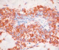
Petrella et al / TCL1 AND CLA IN CD4/CD56 HEMATODERMIC NEOPLASMS AND LEUKEMIA CUTIS

     The official designation blastic NK-cell lymphoma for         cases have been reported.1,6 All 29 cases had typical clinical
CD4/CD56 HN might be an unfortunate one, because an NK-            manifestations and conformed to established phenotypic
cell origin has not been demonstrated, and, in our experience,     criteria.6 Clinical data are given in ❚Table 1❚. All cases were
the main histopathologic differential diagnosis in skin tumor      CD4+ and CD123+. One case was CD56–.27 All cases were
biopsy specimens is myeloid leukemia cutis rather than NK-         lineage negative, particularly for the myelomonocytic differ-
cell or T-cell cutaneous lymphoma. True NK-cell or T-cell          entiation markers CD13, CD14, CD15, CD33, and CD117.
lymphomas usually are excluded with appropriate immuno-                 Eighteen cases of leukemia cutis (acute myeloid
histochemical staining. NK-cell lymphomas typically express        leukemia [AML], 17; chronic myelomonocytic leukemia
cytotoxic proteins, including granzyme B, TIA1, or perforin.7      [CMML], 1) also were evaluated. The 18 cases were assem-
T-cell lymphomas typically reflect their T-lineage derivation      bled from the archives of two of us (T.P., 14 cases;
and express CD3 and T-cell receptor (TCR)αβ or TCRγδ               C.J.L.M.M, 4 cases). All 18 cases were diagnosed between
protein, with monoclonal TCR gene rearrangements that are          1991 and 2001. In addition to identification in skin biopsy
determined by polymerase chain reaction or Southern blot           specimens, AML blastic cells also were identified by hema-
analysis.7 In contrast with these 2 entities, myeloid leukemias,   tocytologists during leukemic phases that occurred previ-
particularly those with monocytic differentiation, are pheno-      ously, simultaneously, or secondarily (AML-M5) to skin
typically close to CD4/CD56 HN. The CD4 antigen                    localization. According to the French-American-British
commonly is expressed by monocytes 8 and monocytic                 subclassification28 of AML, there was 1 case of AML-M1,
leukemias. Furthermore, a great number of myeloid                  11 of AML-M2, and 5 of AML-M5. Of the 17 AML cases, 3
leukemias express the CD56 antigen.9-13 To exclude myeloid         were secondary to refractory anemia with excess blasts, and
leukemias from the differential diagnosis, immunohistochem-        1 was secondary to CMML. Of the 18 cases, 4 were CD4+
ical stains must evaluate the myelomonocytic differentiation       only, and 4 expressed CD4 and CD56 ❚Table 2❚. None of the
antigens, including CD13, CD14, CD33, and CD117, all of            cases expressed only CD56. Myeloperoxidase staining was
which generally are negative in CD4/CD56 HN.6,14 These             positive in 12 of the 18 cases (Table 2).
antibodies require frozen tissue samples for robust detection           Four nonneoplastic lymph nodes with or without follic-
of their corresponding surface antigens.                           ular hyperplasia were used as positive control tissue samples.
     The histologic distinction between CD4/CD56 HN and            All case and control samples were available as formalin-fixed,
leukemia cutis could be very difficult on paraffin-embedded        paraffin-embedded blocks from which serial 5-µm-thick tissue
sections when the results of myeloperoxidase staining are          sections were prepared. Immunohistochemical staining was
negative. To provide an improved method for diagnosing             performed using steam retrieval (pH 6.0). TCL1 detection was
CD4/CD56 HN on formalin-fixed, paraffin-embedded tissue            with a polyclonal rabbit antiserum for which use has been
sections, we evaluated 2 additional immunohistochemical            described previously,24,29 and CLA detection was with HECA-
antigens that are expressed by PDCs: HECA-45215 and T-             452, a monoclonal antibody, described by Duijvestijn and
cell leukemia 1 (TCL1).16 HECA-452 antibody recognizes             colleagues30 and provided by one of us (C.J.L.M.M.). TCL1
the cutaneous lymphocyte-associated antigen (CLA). CLA is          antiserum was used at 1:50 dilution, and the HECA-452 anti-
a cell surface glycoprotein that specifically binds to E-          body was used without dilution. Immunoperoxidase detection
selectin.17 It might have a role in lymphocyte homing to the       was performed with the SuperSensitive immunohistochemical
skin. TCL1 is a small, 14-kd cytoplasmic protein that binds        detection system, Stavigen Multilink kit (BioGenex, San
to members of the serine/threonine Akt kinase family.18-21         Ramon, CA). Cases were scored as positive when more than
TCL1 has an unknown mechanistic role in promoting                  10% of the tumor cells expressed the surveyed antigen.
distinct subtypes of mature T- and B-cell leukemia/
lymphoma.22-26 We evaluated the expression of these 2 PDC
differentiation antigens in CD4/CD56 HN to determine
                                                                   Results
whether they could help distinguish between CD4/CD56 HN
and leukemia cutis in formalin-fixed, paraffin-embedded
tissue sections.                                                   HECA-452 Reactivity in CD4/CD56 HN
                                                                        Of 29 cases, 26 (90%) expressed CLA as detected by
                                                                   HECA-452 staining (Table 2). Immunostaining was similar
                                                                   in all positive cases, showing a peculiar paranuclear dot
Materials and Methods
                                                                   pattern within the region of the Golgi apparatus ❚Image 1A❚.
    We obtained 29 cases of CD4/CD56 HN: 23 from the               Sometimes the dots were very small and seen only at high
French Study Group on Cutaneous Lymphomas and 6 from               magnification. Interestingly, a similar paranuclear dot pattern
the Dutch Cutaneous Lymphoma Group. Fourteen of these              was not observed on lymph node control slides in which

308   Am J Clin Pathol 2004;122:307-313                                                            © American Society for Clinical Pathology
308   DOI: 10.1309/0QPPAVTUPCV9UCLV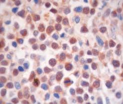
Hematopathology / ORIGINAL ARTICLE
❚Table 1❚
Clinical Summary of CD4/CD56 Hematodermic Neoplasm Cases
Case No./Sex/Age (y) Skin Lesions at Diagnosis Extension at Diagnosis Initial Treatment Outcome
1/M/56 Several nodules LN, BM, blood PC Dead, 13 mo
2/M/82 1 nodule (forehead) None RT and PC Dead, 24 mo
3/M/81 10 bruise-like papules (trunk) None PC Dead, 11 mo
4/F/96 1 nodule (cheek) NA None Dead, 1 mo
5/M/77 Plaques and nodules (trunk) None PC Dead, 7 mo
6/F/54 1 plaque (leg) None PC and AG Alive, 38 mo
7/F/33 1 nodule (leg) LN PC Dead, 27 mo
8/M/69 Papules and nodules (back) NA None Dead, 2 mo
9/M/8 1 bruise-like tumefaction (knee) LN PC Dead, 33 mo
10/M/37 1 papule (leg) None PC Dead, 40 mo
11/F/67 Several nodules and plaques (trunk) None PC Dead, 17 mo
12/M/84 1 bruise-like tumefaction (forehead) LN, BM PC Dead, 5 mo
13/M/62 Several nodules and plaques (abdomen) None RT and PC Dead, 13 mo
14/M/75 Plaques and nodules (trunk) LN PC Dead, 26 mo
15/M/64 Several plaques and papules (arms and trunk) BM PC Dead, 12 mo
16/M/69 Multiple disseminated nodules None PC Dead, 21 mo
17/F/70 1 nodule (arm) BM PC Alive, 9 mo
18/M/70 1 nodule (cheek) None PC Alive, 14 mo
19/F/88 Multiple disseminated nodules None PC Dead, 8 mo
20/M/72 1 nodule (shoulder) LN, BM, blood PC Dead, 3 mo
21/F/65 1 nodule (thigh) LN PC Dead, 17 mo
22/F/86 2 nodules (thigh and leg) LN PC Dead, 5 mo
23/F/49 1 nodule (cervical) LN PC Dead, 9 mo
24/M/60 Plaques (scalp) None RT Dead, 22 mo
25/M/56 Plaques and nodules (trunk) BM PC Alive, 14 mo
26/M/74 Generalized plaques and nodules None PC Dead, 12 mo
27/M/77 Generalized nodules None PUVA Dead, 7 mo
28/F/43 Generalized plaques None PC and RT Alive, 5 mo
29/M/64 1 nodule (arm) None RT Alive, 22 mo
AG, allograft; BM, bone marrow; LN, lymph node; NA, data not available; PC, polychemotherapy; PUVA, psoralen plus ultraviolet A; RT, radiotherapy.
cells from distinct developmental lineages stained positive pattern ❚Image 1B❚. Epidermal Langerhans cells and dermal
with the HECA-452 antibody (monocytes, PDCs, and high dendritic cells also reacted with the HECA-452 antibody.
endothelial venules). HECA-452 immunostaining on tumor The case of CMML (case 17, Table 2) was reactive but
sections was generally diffuse, involving more than 80% of without a dot-like staining pattern.
the tumor cells in each positive case. In addition, as previ-
ously described,31 HECA-452 was expressed on epidermal TCL1 Reactivity in Leukemia Cutis
Langerhans cells and dermal dendritic cells. Only 3 (17%) of 18 cases of leukemia cutis were posi-
tive for TCL1-specific antiserum staining (Table 2). These
TCL1 Reactivity in CD4/CD56 HN cases showed only cytoplasmic staining. Two of the 15
Of 29 cases, 26 (90%) were positive for staining with TCL1-negative cases showed fewer than 10% of the cells in
TCL1-specific antiserum (Table 2). Of the 26 positive cases, the cutaneous infiltrate expressing TCL1, probably repre-
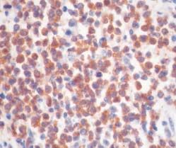
19 showed cytoplasmic ❚Image 2A❚ and nuclear staining senting tumor-infiltrating lymphocytes rather than true tumor
❚Image 2B❚. The 7 remaining positive cases showed only cells. The CMML case was TCL1-negative.
cytoplasmic staining. Similar to HECA-452–positive cases,
TCL1 immunostaining on tumor sections generally was
diffuse and strongly positive in more than 50% of the tumor
Discussion
cells in each positive case.
In our series of cases, we showed that 26 (90%) of 29
HECA-452 Reactivity in Leukemia Cutis cases of CD4/CD56 HN were reactive for the CLA antibody
Of 18 cases of leukemia cutis, 14 (78%) expressed HECA-452. Of 18 cases of leukemia cutis, 14 (78%) also
CLA as detected by HECA-452 staining (Table 2). As for reacted with the HECA-452 antibody. Therefore, the HECA-
all cases of CD4/CD56 HN, 5 positive cutaneous AML 452 antibody alone cannot discriminate between CD4/CD56
cases showed a paranuclear small-dot immunostaining HN and cutaneous AML, and the difference in immuno-
pattern, while 9 positive cases showed a diffuse cytoplasmic staining for CLA between these entities is not significant.
© American Society for Clinical Pathology Am J Clin Pathol 2004;122:307-313 309
309 DOI: 10.1309/0QPPAVTUPCV9UCLV 309Petrella et al / TCL1 AND CLA IN CD4/CD56 HEMATODERMIC NEOPLASMS AND LEUKEMIA CUTIS
❚Table 2❚
Immunohistochemical Staining Profile for CD4/CD56 Hematodermic Neoplasms and Leukemia Cutis
Case No. HECA-452 TCL1 CD4 CD56 MPO
CD4/CD56 hematodermic neoplasms
1 + ++ + + –
2 + ++ + – –
3 + + + + –
4 + ++ + + –
5 + ++ + + –
6 + + + + –
7 – ++ + + –
8 + + + + –
9 + ++ + + –
10 + ++ + + –
11 + ++ + + –
12 + ++ + + –
13 + ++ + + –
14 + ++ + + –
15 + ++ + + –
16 + ++ + + –
17 + ++ + + –
18 + + + + –
19 + + + + –
20 – ++ + + –
21 + ++ + + –
22 + ++ + + –
23 + – + + –
24 – – + + –
25 + ++ + + –
26 + + + + –
27 + + + + –
28 + * + + –
29 + – + + –
Leukemia cutis†
1 (AML-M2) – – – – +w
2 (AML-M2) – – – – –
3 (AML-M2; RAEB) +* – – – +
4 (AML-M2) +* – – – –
5 (AML-M2) + – – – +
6 (AML-M2; RAEB) +* – – – +
7 (AML-M5) +* – – – +w
8 (AML-M2; RAEB) + – – – +
9 (AML-M1) – – – – –
10 (AML-M5; CMML) + – – – +
11 (AML-M2) + + + – +
12 (AML-M2) + – + – +w
13 (AML-M2) + + + – +
14 (AML-M5) + – + + –
15 (AML-M2) – + + + –
16 (AML-M5) + – + +w +
17 (CMML) + – +w – +w
18 (AML-M5) +* – + + –
AML, acute myeloid leukemia; CMML, chronic myelomonocytic leukemia; MPO, myeloperoxidase; RAEB, refractory anemia with excess blasts; TCL1, T-cell leukemia 1;
–, negative; +, cytoplasmic positivity; ++, cytoplasmic and nuclear positivity; +w, weak positivity.
* Additional nuclear staining.
† Numbers with AML denote French-American-British classification.
The HECA-452 epitope is part of an inducible carbohy- in reactive lymph nodes strongly expressed CLA. Furthermore,
drate on the P-selectin glycoprotein ligand-1.32 It specifically PDCs have been detected within the dermis, first by Facchetti
binds to E-selectin expressed by endothelial cells and particu- and colleagues36 in Jessner-Kanoff syndrome and more
larly on lymph node high endothelial venules.33 Furthermore, it recently by Wollenberg and colleagues37 in a variety of inflam-
is expressed on most T cells at cutaneous sites, on cutaneous T- matory skin diseases. These findings suggest that any potential
cell lymphomas,34 and on lymphoid cells derived from human homing role for CLA to skin is not restricted to lymphocytes
skin.35 Therefore, CLA might have an important role in but also might include other cell types, such as PDCs. These
lymphocyte homing to the skin. Facchetti and colleagues15 observations also suggest that the dermis is a target organ for
demonstrated that plasmacytoid T cells (former name of PDCs) PDCs. This may not be surprising because mature dendritic
310 Am J Clin Pathol 2004;122:307-313 © American Society for Clinical Pathology
310 DOI: 10.1309/0QPPAVTUPCV9UCLVHematopathology / ORIGINAL ARTICLE
A B
❚Image 1❚ A, CD4/CD56 hematodermic neoplasm. A small-dot paranuclear pattern of positive immunostaining (HECA-452,
×1,000). B, Leukemia cutis. Cytoplasmic and membranous immunostaining activity (HECA-452, ×630).
A B
❚Image 2❚ CD4/CD56 hematodermic neoplasm. A, Cytoplasmic immunostaining (T-cell leukemia 1 [TCL1], ×630). B, Nuclear
immunostaining (TCL1, ×1,000).
cells reside in the skin, where PDCs might have a major role in We further observed that 26 (90%) of 29 CD4/CD56
antigen presentation. Interestingly, mature dendritic cells also HN cases expressed the TCL1 cytoplasmic differentiation
express a HECA-452 epitope, which is likely from CLA, that is antigen. These results are consistent with the previous
weak in the dermis and strong in the epidermis.38 results of Herling and colleagues,16 which showed that 86%
It is tempting to speculate that intense HECA-452 of the CD4/CD56 HN cases tested expressed TCL1 protein.
staining supports an important role for CLA in the skin Of 3 TCL1-negative cases, 2 were distinct from the 3
tropism of CD4/CD56 HN and most cases of leukemia cutis. HECA-452–negative CD4/CD56 HN cases, indicating that
It remains unclear whether CD4/CD56 HN cases are primary these antigens are not invariantly coordinately regulated in
or secondary neoplasms of the skin. If skin is a target organ this disease. We conclude from these 2 studies that TCL1
for PDC localization, as suggested by CLA expression and indeed is a frequently positive antigenic marker for
dermal tropism, it is logical to consider CD4/CD56 HN as a CD4/CD56 HN. In contrast with the frequent expression of
primary skin tumor, in agreement with clinical data. TCL1 in CD4/CD56 HN, only 3 (17%) of 18 leukemia cutis
© American Society for Clinical Pathology Am J Clin Pathol 2004;122:307-313 311
311 DOI: 10.1309/0QPPAVTUPCV9UCLV 311Petrella et al / TCL1 AND CLA IN CD4/CD56 HEMATODERMIC NEOPLASMS AND LEUKEMIA CUTIS
cases expressed TCL1. This difference seems highly signifi- Pathology, Hôpital du Haut Lévêque, Pessac, France; and
10Departments of Pathology and Pediatrics, Center for Cell
cant (P < .001; χ2 test). However, the use of TCL1 alone to
distinguish between CD4/CD56 HN and cutaneous AML Mimetic Studies, Jonsson Comprehensive Cancer Center, David
Geffen School of Medicine at UCLA, Los Angeles, CA.
should be avoided because the small percentage of TCL1-
positive AML cases could result in misdiagnosis. Supported in part by grants R01CA90571 and
TCL1 is a small, β-barrel–shaped cytoplasmic protein R01CA107300 (Dr Teitell) from the National Cancer Institute,
National Institutes of Health, Bethesda, MD, and grant NCC2-
that augments the activation of the cell survival kinase Akt by
1364 (Dr Teitell) from the Center for Cell Mimetic Studies.
physical association and multimer formation.19,39,40 Its Address reprint requests to Dr Petrella: Centre de
pattern of expression during B- and T-cell development Pathologie, 33 rue Nicolas Bornier, 21000, Dijon, France.
suggests critical stage-specific developmental functions, Dr Teitell is a scholar of the Leukemia and Lymphoma
rather than a broad role throughout the life span of a lympho- Society, White Plains, NY.
Acknowledgments: We are grateful to Janine Wechsler, MD,
cyte.19 Reflecting its B-lineage pattern of expression, TCL1 is
Martine Bagot, MD, Pascal Joly, MD, Philippe Courville, MD,
expressed in a large number of B-cell malignant neoplasms, Jacques Bosq, MD, Marie-Françoise Avril, MD, Alain Devidas,
from pre–B-cell lymphoblastic leukemia/lymphoma through MD, Thierry Molina, MD, Stéphane Leprêtre, MD, Thérèse
germinal-center derived follicular, Burkitt, and diffuse large Rousset, MD, Marie Paule Cabrol, MD, Philippe Bernard, MD,
B-cell lymphomas.24,29,41,42 TCL1 also marks mature T cell Anne Durlach, MD, Anne De Muret, MD, Gérard Couillault, MD,
leukemias, such as T-cell chronic lymphocytic leukemia and Marie Beylot-Barry, MD, Michelle Delaunay, MD, Pierre
Déchelotte, MD, Pierre Souteyrand, MD, Michel D’incan, MD,
T-cell prolymphocytic leukemia, owing to overexpression by
and Anne Dompmartin, MD, and all members of the French Study
14q32.1 chromosomal translocations.43,44 As such, TCL1 has Group on Cutaneous Lymphomas. We also thank Sylvie Espin and
diagnostic usefulness in mature B- and T-lineage tumors. Its Marie-Antoinette Lignier for their technical assistance.
role in normal PDC biology or in malignant initiation and
progression of CD4/CD56 HN is less clear, although its
robust, high-frequency expression suggests an origin of References
CD4/CD56 HN from PDC rather than from the NK-cell
1. Petrella T, Dalac S, Maynadié M, et al. CD4+ CD56+
lineage, which is negative for TCL1 expression.16,25 cutaneous neoplasms: a distinct hematological entity? Groupe
CLA and TCL1 frequently are coexpressed markers for Francais d’Etude des Lymphomes Cutanes (GFELC). Am J
CD4/CD56 HN that are demonstrated easily in formalin- Surg Pathol. 1999;23:137-146.
fixed, paraffin-embedded tissue sections. Used in combina- 2. Adachi M, Maeda K, Takekawa M, et al. High expression of
CD56 (N-CAM) in a patient with cutaneous CD4-positive
tion with additional immunohistochemical stains, HECA-452 lymphoma. Am J Hematol. 1994;47:278-282.
antibody and TCL1 antiserum are useful for diagnosing this 3. Penven K, Macro M, Salaun V, et al. Skin manifestations in
clinical entity. In addition, we observed for cutaneous AML CD4+, CD56+ malignancies. Eur J Dermatol. 2003;13:161-165.
and particularly for CD4/CD56 HN a distinct paranuclear 4. Reimer P, Rudiger T, Kraemer D, et al. What is CD4+CD56+
HECA-452 immunostaining pattern in small dots within the malignancy and how should it be treated? Bone Marrow
Transplant. 2003;32:637-646.
area of the Golgi apparatus. The significance of this unique
5. Chan JKC, Jaffe ES, Ralfkiaer E. Blastic NK-cell lymphoma.
staining pattern, which was not observed in normal lymphoid In: Jaffe ES, Harris NL, Stein H, et al, eds. Pathology and
tissues, deserves further study. The clear-cut difference of Genetics of Tumours of Haematopoietic and Lymphoid Tissues.
TCL1 expression between CD4/CD56HN and AML strongly Lyon, France: IARC Press; 2001:214-215. World Health
Organization Classification of Tumours.
suggests that the 2 diseases are distinct. However, common
6. Petrella T, Comeau MR, Maynadié M, et al. “Agranular
patterns of HECA-452 staining also might underlie a possible CD4+ CD56+ hematodermic neoplasm” (blastic NK-cell
relationship between CD4/CD56 HN and AML or perhaps an lymphoma) originates from a population of CD56+ precursor
overlap between CD4/CD56 HN and some undifferentiated cells related to plasmacytoid monocytes. Am J Surg Pathol.
2002;26:852-862.
cases of AML. This area clearly requires further study.
7. Jaffe ES, Ralfkiaer E. Mature T-cell and NK-cell neoplasms.
Finally, the high skin tropism of CD4/CD56 HN might be In: Jaffe ES, Harris NL, Stein H, et al, eds. Pathology and
related to the skin-homing property of CLA. Genetics of Tumours of Haematopoietic and Lymphoid Tissues.
Lyon, France: IARC Press; 2001:191-194. World Health
From the 1Centre de Pathologie, the Departments of 2Pathology, Organization Classification of Tumours.
3Dermatology, 4Clinical Hematology, and the 5Laboratory of 8. Knapp W, Dorken E, Rieber H, et al. Leucocyte Typing, IV:
White Cell Differentiation Antigens. Oxford, England: Oxford
Hematology, Centre Hospitalo-Universitaire du Bocage, Dijon,
University Press; 1989:1093.
France; 6Department of Pathology, Vrije Universiteit Medical
9. Scott AA, Head DR, Kopecky KJ, et al. HLA-DR–, CD33+,
Center, Amsterdam, the Netherlands; 7Department of
CD56+, CD16– myeloid/natural killer cell acute leukemia: a
Dermatology, Leiden University Medical Center, Leiden, the previously unrecognized form of acute leukemia potentially
Netherlands; 8Department of Dermatology, Centre Hospitalo- misdiagnosed as French-American-British acute myeloid
Universitaire Trousseau, Tours, France; 9Department of leukemia-M3. Blood. 1994;84:244-255.
312 Am J Clin Pathol 2004;122:307-313 © American Society for Clinical Pathology
312 DOI: 10.1309/0QPPAVTUPCV9UCLVHematopathology / ORIGINAL ARTICLE
10. Thomas X, Vila L, Campos L, et al. Expression of N-CAM 28. Bennett JM, Catovsky D, Daniel MT, et al. Proposals for the
(CD56) on acute leukemia cells: relationship with disease classification of the acute leukaemias. French-American-
characteristics and outcome. Leuk Lymphoma. 1995;19:295- British (FAB) Co-operative Group. Br J Haematol.
300. 1976;33:451-458.
11. Lee JJ, Chung IJ, Yang DH, et al. Clinical significance of 29. Said JW, Hoyer KK, French SW, et al. TCL1 oncogene
CD56 expression in patients with acute myeloid leukemia. expression in B cell subsets from lymphoid hyperplasia and
Leuk Lymphoma. 2002;43:1897-1899. distinct classes of B cell lymphoma. Lab Invest. 2001;81:555-
12. Vidriales MB, Orfao A, Gonzalez M, et al. Expression of NK 564.
and lymphoid-associated antigens in blast cells of acute 30. Duijvestijn AM, Horst E, Pals ST, et al. High endothelial
myeloblastic leukemia. Leukemia. 1993;7:2026-2029. differentiation in human lymphoid and inflammatory tissues
13. Hsiao CH, Tang JL, Yao M, et al. High incidence of CD56 defined by monoclonal antibody HECA-452. Am J Pathol.
expression and relapse rate in acute myeloid leukemia patients 1988;130:147-155.
with t(8;21) in Taiwan. J Formos Med Assoc. 2002;101:393- 31. Yasaka N, Furue M, Tamaki K. Expression of cutaneous
398. lymphocyte-associated antigen defined by monoclonal
14. Feuillard J, Jacob MC, Valensi F, et al. Clinical and biologic antibody HECA-452 on human Langerhans cells. J Dermatol
features of CD4(+)CD56(+) malignancies. Blood. Sci. 1996;11:19-27.
2002;99:1556-1563. 32. Fuhlbrigge RC, Kieffer JD, Armerding D, et al. Cutaneous
15. Facchetti F, de Wolf-Peeters C, van den Oord JJ, et al. lymphocyte antigen is a specialized form of PSGL-1 expressed
Anti–high endothelial venule monoclonal antibody HECA- on skin-homing T cells. Nature. 1997;389:978-981.
452 recognizes plasmacytoid T cells and delineates an 33. Berg EL, Yoshino T, Rott LS, et al. The cutaneous lymphocyte
“extranodular” compartment in the reactive lymph node. antigen is a skin lymphocyte homing receptor for the vascular
Immunol Lett. 1989;20:277-281. lectin endothelial cell–leukocyte adhesion molecule 1. J Exp
16. Herling M, Teitell MA, Shen RR, et al. TCL1 expression in Med. 1991;174:1461-1466.
plasmacytoid dendritic cells (DC2s) and the related CD4+ 34. Noorduyn LA, Beljaards RC, Pals ST, et al. Differential
CD56+ blastic tumors of skin. Blood. 2003;101:5007-5009. expression of the HECA-452 antigen (cutaneous lymphocyte
17. Tsuchiyama J, Yoshino T, Toba K, et al. Induction and associated antigen, CLA) in cutaneous and non-cutaneous T-
characterization of cutaneous lymphocyte antigen on natural cell lymphomas. Histopathology. 1992;21:59-64.
killer cells. Br J Haematol. 2002;118:654-662. 35. Hunger RE, Yawalkar N, Braathen LR, et al. The HECA-452
18. Kunstle G, Laine J, Pierron G, et al. Identification of Akt epitope is highly expressed on lymph cells derived from
association and oligomerization domains of the Akt kinase human skin. Br J Dermatol. 1999;141:565-569.
coactivator TCL1. Mol Cell Biol. 2002;22:1513-1525. 36. Facchetti F, De Wolf-Peeters C, Van den Oord JJ, et al.
19. Laine J, Kunstle G, Obata T, et al. The protooncogene TCL1 Plasmacytoid T cells in a case of lymphocytic infiltration of
is an Akt kinase coactivator. Mol Cell. 2000;6:395-407. skin: a component of the skin-associated lymphoid tissue? J
Pathol. 1988;155:295-300.
20. Laine J, Kunstle G, Obata T, et al. Differential regulation of
Akt kinase isoforms by the members of the TCL1 oncogene 37. Wollenberg A, Wagner M, Gunther S, et al. Plasmacytoid
family. J Biol Chem. 2002;277:3743-3751. dendritic cells: a new cutaneous dendritic cell subset with
21. Pekarsky Y, Koval A, Hallas C, et al. Tcl1 enhances Akt distinct role in inflammatory skin diseases. J Invest Dermatol.
kinase activity and mediates its nuclear translocation. Proc 2002;119:1096-1102.
Natl Acad Sci U S A. 2000;97:3028-3033. 38. Koszik F, Strunk D, Simonitsch I, et al. Expression of
22. Virgilio L, Narducci MG, Isobe M, et al. Identification of the monoclonal antibody HECA-452–defined E-selectin ligands
TCL1 gene involved in T-cell malignancies. Proc Natl Acad on Langerhans cells in normal and diseased skin. J Invest
Sci U S A. 1994;91:12530-12534. Dermatol. 1994;102:773-780.
23. Virgilio L, Lazzeri C, Bichi R, et al. Deregulated expression of 39. Gold MR. Akt is TCL-ish: implications for B-cell lymphoma.
TCL1 causes T cell leukemia in mice. Proc Natl Acad Sci Trends Immunol. 2003;24:104-108.
U S A. 1998;95:3885-3889. 40. French SW, Shen RR, Koh PJ, et al. A modeled hydrophobic
24. Teitell M, Damore MA, Sulur GG, et al. TCL1 oncogene domain on the TCL1 oncoprotein mediates association with
expression in AIDS-related lymphomas and lymphoid tissues. AKT at the cytoplasmic membrane. Biochemistry.
Proc Natl Acad Sci U S A. 1999;96:9809-9814. 2002;41:6376-6382.
25. Hoyer KK, French SW, Turner DE, et al. Dysregulated TCL1 41. Narducci MG, Pescarmona E, Lazzeri C, et al. Regulation of
promotes multiple classes of mature B cell lymphoma. Proc TCL1 expression in B- and T-cell lymphomas and reactive
Natl Acad Sci U S A. 2002;99:14392-14397. lymphoid tissues. Cancer Res. 2000;60:2095-2100.
26. Bichi R, Shinton SA, Martin ES, et al. Human chronic 42. Nakayama I, Murao S, Kitazawa S, et al. Activation of the
lymphocytic leukemia modeled in mouse by targeted TCL1 TCL1 protein in B cell lymphomas. Pathol Int. 2000;50:191-
expression. Proc Natl Acad Sci U S A. 2002;99:6955-6960. 199.
27. Petrella T, Teitell MA, Spiekermann C, et al. A CD56- 43. Pekarsky Y, Hallas C, Croce CM. Molecular basis of mature
negative case of blastic natural killer-cell lymphoma T-cell leukemia. JAMA. 2001;286:2308-2314.
(agranular CD4+/CD56+ haematodermic neoplasm). Br J 44. Pekarsky Y, Hallas C, Croce CM. The role of TCL1 in human
Dermatol. 2004;150:174-176. T-cell leukemia. Oncogene. 2001;20:5638-5643.
© American Society for Clinical Pathology Am J Clin Pathol 2004;122:307-313 313
313 DOI: 10.1309/0QPPAVTUPCV9UCLV 313You can also read