Immune-checkpoint inhibitors from cancer to COVID-19: A promising avenue for the treatment of patients with COVID-19 (Review)
←
→
Page content transcription
If your browser does not render page correctly, please read the page content below
INTERNATIONAL JOURNAL OF ONCOLOGY

                     Immune-checkpoint inhibitors from cancer to
                    COVID‑19: A promising avenue for the treatment
                        of patients with COVID‑19 (Review)
                SILVIA VIVARELLI1, LUCA FALZONE2, FRANCESCO TORINO3, GIUSEPPA SCANDURRA4,
                       GIULIA RUSSO5, ROBERTO BORDONARO6, FRANCESCO PAPPALARDO5,7,
                     DEMETRIOS A. SPANDIDOS8, GIUSEPPINA RACITI5* and MASSIMO LIBRA1,7*

          1
           Section of General Pathology, Clinics and Oncology, Department of Biomedical and Biotechnological Sciences,
      University of Catania, I‑95123 Catania; 2Epidemiology Unit, IRCCS Istituto Nazionale Tumori ‘Fondazione G. Pascale’,
      I‑80131 Naples; 3Department of Systems Medicine, Medical Oncology, University of Rome Tor Vergata, I‑00133 Rome;
            4
              Medical Oncology Unit, Azienda Ospedaliera Cannizzaro, I‑95126 Catania; 5Department of Drug Sciences,
               University of Catania, I‑95123 Catania; 6Medical Oncology Unit, Garibaldi Hospital, I‑95122 Catania;
           7
             Research Center for Prevention, Diagnosis and Treatment of Tumors, University of Catania, I‑95123 Catania,
               Italy; 8Laboratory of Clinical Virology, Medical School, University of Crete, 71003 Heraklion, Greece

                                    Received November 5, 2020; Accepted December 14, 2020

                                                     DOI: 10.3892/ijo.2020.5159

Abstract. The severe acute respiratory syndrome associ‑              suffer from T‑cell exhaustion, which may lead to viral sepsis
ated coronavirus‑2 (SARS‑CoV‑2) poses a threat to human              and an increased mortality rate. It has been observed that cancer
life worldwide. Since early March, 2020, coronavirus                 patients, who usually are immunocompromised, may restore
disease 2019 (COVID‑19), characterized by an acute and often         their anti‑tumoral immune response when treated with ICIs.
severe form of pneumonia, has been declared a pandemic.              Moreover, viral-infected mice and humans, exhibit a T‑cell
This has led to a boom in biomedical research studies at all         exhaustion, which is also observed following SARS‑CoV‑2
stages of the pipeline, from the in vitro to the clinical phase.     infection. Importantly, when treated with anti‑PD‑1 and
In line with this global effort, known drugs, currently used         anti‑PD‑L1 antibodies, they restore their T‑cell competence
for the treatment of other pathologies, including antivirals,        and efficiently counteract the viral infection. Based on these
immunomodulating compounds and antibodies, are currently             observations, four clinical trials are currently open, to examine
used off‑label for the treatment of COVID‑19, in association         the efficacy of anti‑PD‑1 antibody administration to both cancer
with the supportive standard care. Yet, no effective treatments      and non‑cancer individuals affected by COVID‑19. The results
have been identified. A new hope stems from medical oncology         may prove the hypothesis that restoring exhausted T‑cells may
and relies on the use of immune‑checkpoint inhibitors (ICIs).        be a winning strategy to beat SARS‑CoV‑2 infection.
In particular, amongst the ICIs, antibodies able to block the
programmed death‑1 (PD‑1)/PD ligand-1 (PD‑L1) pathway
have revealed a hidden potential. In fact, patients with severe      Contents
and critical COVID‑19, even prior to the appearance of acute
respiratory distress syndrome, exhibit lymphocytopenia and           1. Introduction
                                                                     2. SARS‑CoV‑2 immunopathology
                                                                     3.	Management of COVID‑19: Prevention, diagnosis and
                                                                         treatment
                                                                     4. Role of computational biology in the response to COVID‑19
Correspondence to: Professor Massimo Libra, Section of General       5.	Immune‑checkpoint inhibitors against COVID‑19: A
Pathology, Clinics and Oncology, Department of Biomedical and            lesson learnt from cancer
Biotechnological Sciences, University of Catania, I‑95123 Catania,
                                                                     6. Conclusions and future perspectives
Italy
E‑mail: mlibra@unict.it

*
    Contributed equally                                              1. Introduction

Key words: COVID‑19, SARS‑CoV‑2, cancer, immune‑checkpoint           In December, 2019, following an outbreak in a fish market
inhibitors, immunotherapy, anti‑PD‑1 monoclonal antibody             in Wuhan (Hubei, China), an infection leading to severe
                                                                     pneumonia rapidly and uncontrollably spread worldwide. The
                                                                     World Health Organization (WHO) declared the status of a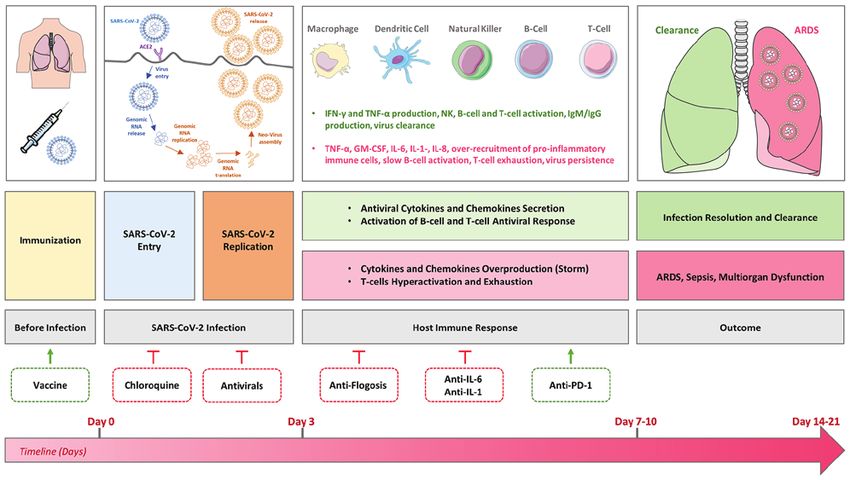
2               VIVARELLI et al: IMMUNE CHECKPOINT INHIBITORS FOR TREATMENT OF PATIENTS WITH COVID-19

pandemic threat on March 11, 2020 (1), leading all countries         2. SARS‑CoV‑2 immunopathology
to impose lockdowns with devastating socio‑economic conse‑
quences.                                                             SARS‑CoV‑2 is a single‑stranded positive‑sense RNA virus,
    The infection is caused by a highly pathogenic coronavirus,      whose RNA‑genome is approximately 30 Kb in length, with
the severe acute respiratory syndrome associated corona‑             a 5'‑CAP and a 3'‑poly‑A tail and containing 10 open reading
virus‑2 (SARS‑CoV‑2), which is the identified etiological agent of   frames (ORFs) (11,12). Genomic variations of SARS‑CoV‑2
the outbreak, named coronavirus disease 2019 (COVID‑19) (2).         have been observed; however, further studies are warranted
    Despite global lockdown measures adopted by most                 in order to determine whether differently virulent strains may
countries, as of October 11, 2020 over 37 million COVID‑19           be associated with the differential infection severity range
cases and 1 million deaths have been reported globally, with         observed in patients with COVID‑19 (13).
half of these cases and deaths reported in the regions of the            To enter the host cells, SARS‑CoV‑2, similar to SARS‑CoV,
Americas (3).                                                        binds to the extracellular enzymatic domain of the single pass
    SARS‑CoV‑2 transmission occurs through respira‑                  transmembrane angiotensin‑converting enzyme 2 (ACE2)
tory droplets, via direct person‑to‑person contact, with an          receptor (14). Physiologically, ACE2 negatively modu‑
incubation period of approximately 2 to 12 days following            lates blood pressure by degrading the vasoconstrictor
exposure (4). The symptoms of COVID‑19 caused by viral               angiotensin II peptide (15). SARS‑CoV‑2 binding is mediated
infection of the lower respiratory tract include dry cough,          by the viral glycoprotein spike (S), which also determines the
fever, fatigue hyposmia and hypogeusia. The infection may            crown‑shaped aspect of the virus (16). The difference between
rapidly degenerate to pneumonia, and it can be associated with       SARS‑CoV‑2 and SARS‑CoV is that SARS‑CoV‑2 possesses
mild complications, including dyspnea, lymphocytopenia,              a furin cleavage site within the S protein secondary sequence.
myalgia, or with more severe comorbidities, including signifi‑       This is likely to increase virus pathogenicity as cleavage of
cant hypoxia, associated with the appearance of the acute            the S domain during protein synthesis highly increases the
respiratory distress syndrome (ARDS) (5). Due to ARDS, a             affinity for the ACE2 receptor (17), subsequently promoting
non‑negligible proportion of COVID‑19‑affected patients may          SARS‑CoV‑2 endocytosis inside the target cell (18).
require oxygen therapy and admission to an intensive care                ACE2 receptor is expressed by alveolar epithelial cells,
unit (ICU), as the ARDS may be swiftly followed by septic            but also by lung endothelial cells, and, distally, by heart, lung,
shock and multi-organ dysfunction, thus resulting in a 1 to 4%       kidney, intestinal and neuronal cells. Whether SARS‑CoV‑2
case fatality rate (6).                                              is able to infect organs located far from the lungs (by directly
    Viral genome sequencing generated from samples of                binding ACE2 receptor in other cells distant from the primary
5 patients hospitalized with pneumonia led to the identifica‑        site) is currently under evaluation (19). Within the alveoli,
tion of this novel SARS‑CoV‑2 as part of the β‑coronaviridae         resident dendritic cells (DCs) and macrophages may also be
genera, exhibiting an 88% identity to the sequence of                invaded by the SARS‑CoV‑2, although it remains unclear as
2 bat‑derived CoVs. In fact, bats are considered reservoir hosts     to whether this occurs via primary viral infection or through
for CoVs and, it is considered that SARS‑CoV‑2 originated            the phagocytosis of epithelial and endothelial pre‑infected
from bats, later crossing species before infecting humans (7).       apoptotic cells (20).
Additionally, SARS‑CoV‑2 exhibits an 80 and 50% identity,                After entering host cells, SARS‑CoV‑2 uses the host
respectively, with the genome of 2 other clinically relevant         cellular replication machinery to synthesize the RNA nega‑
human‑β‑CoV, the SARS‑CoV and Middle East respiratory                tive strand and to produce up to 10 protein products, including
syndrome coronavirus (MERS‑CoV) (8). SARS‑CoV emerged                the 4 structural proteins S, envelope (E), nucleocapsid (N)
in 2002 in China, and caused an outbreak in 26 countries,            and membrane (M) proteins. The viral particles take shape
with a case fatality rate of 9.5%. MERS‑CoV originated               inside the endoplasmic reticulum‑Golgi intermediate cellular
in 2012 in Saudi Arabia, spreading to 27 countries, with a           compartment. Once formed, the host vesicles containing
case fatality rate of 34.4% (8). Therefore, SARS‑CoV‑2 is            the virus fuse with the plasma membrane, thereby releasing
considered the third highly pathogenic human‑adapted CoV of          multiple copies of the replicated SARS‑CoV‑2 into the inter‑
the 21st century, representing a threat for human health with a      cellular space, ready to spread to other cells (21).
heavy global impact (9).                                                 As SARS‑CoV‑2 is a cytopathic virus, infected cells
    To preserve humanity from this noxious CoV, biomedical           become deeply injured and may undergo cell death upon
research is proceeding at a rapid pace, with the intention of        infection. For this reason, viral infection can trigger extensive
developing a vaccine and/or a therapy to prevent and/or eradi‑       lung damage (22). In particular, within infected alveolar cells,
cate the virus (10).                                                 a peculiar type of inflammatory cell death termed pyroptosis
    The present review article reports up‑to‑date knowledge of       is observed. Pyroptosis is associated with a concurrent endo‑
SARS‑CoV‑2 and COVID‑19, with particular attention being             thelial leakage and triggers an acute pro‑inflammatory host
paid to novel research approaches aimed at identifying old and       immune response, mediated by the intracellular activation of
novel compounds for the treatment of COVID‑19, as well as            the NLRP3 inflammasome (23‑25).
all therapeutic options currently tested. Among these, the               Following SARS‑CoV‑2 infection, a high rate of immune
importance of modulating the host immune system is widely            inflammatory cell infiltration within the alveolar space
discussed, with particular emphasis on the clinical testing of       enhances the local synthesis and secretion of cytokines [inter‑
a drug currently used in oncology, the immune‑checkpoint             feron (IFN)‑α, IFN‑β, IFN‑γ, tumor necrosis factor (TNF)‑α,
inhibitor (ICI) anti‑programmed death‑1 (PD‑1) blocking              TGF β, granulocyte‑macrophage colony‑stimulating
antibody.                                                            factor (GM‑CSF), interleukin (IL)‑1β, IL‑6, IL‑8, IL‑12, IL‑18,
INTERNATIONAL JOURNAL OF ONCOLOGY 3
IL‑33, etc.] and chemokines (CCL2, CCL3, CCL5, CXCL8, assessed the increased expression of NKG2A and PD‑1 inhibi‑
etc.) by immune effector cells, thereby resulting in acute lung tory receptors on T- and NK cell surfaces of patients with
injury. If unresolved, this exacerbated production may lead to COVID‑19, suggesting that anticancer immunotherapy could
the so‑called ‘cytokine storm’ (CS) (26). In the near future, it be repurposed as a first line of defense to trigger SARS‑CoV‑2
would be crucial to better understand the full dynamics of the clearance (45). In general, SARS‑CoV‑2 infection may nega‑
cytokine storm kinetics (27). tively affect the host antiviral immunity at an early stage of
It has been demonstrated that the severity of COVID‑19 is the infection process. Importantly, both dysregulated innate
proportional to the pro‑inflammatory cytokine IL‑6 levels (28). and adaptive immune responses largely contribute to the
Moreover, the release of pro‑IL‑1β, subsequently cleaved to SARS‑CoV‑2 immunopathology (42). On the other hand,
produce mature IL‑1β, favors the onset of a pro‑inflammatory SARS‑CoV‑2 can evade the host immune system and use it to
milieu followed by pyroptosis in the lungs (29). Additionally, its own advantage. Therefore, the disruption of such immune
GM‑CSF if produced in excess, can lead to local tissue evasion may play an important role in the development of
damage (30). The CS, if uncontrolled, represents a lethal novel therapeutic approaches to eradicate SARS‑CoV‑2 infec‑
inflammatory response, which determines ARDS, respiratory tions (46‑48).
defeat and multiple organ failure, finally leading to death (27). COVID‑19 symptomatology is very heterogeneous and is
The CS itself may determine a high and unrestrained strictly dependent on the hosts' response: i) Mild, with upper
immune cell infiltration rate within the lungs, which may be respiratory tract infection and digestive tract impairment;
linked to thrombosis and pulmonary embolism, often observed ii) moderate, with lower respiratory tract infection, pneumonia,
in the severe forms of the COVID‑19 (31). Moreover, the no obvious hypoxemia with lung lesions; iii) severe, with pneu‑
subsequent endothelial cellular damage and death promoted monia and elevated hypoxemia; and iv) critical, characterized
by direct viral infection and by the activity of the immune cells by the appearance of ARDS, septic shock, myocardial and
recalled over the alveolar surface, can increase lung perme‑ kidney injuries, coagulation dysfunctions (49).
ability and promote systemic SARS‑CoV‑2 invasion (32). Such COVID‑19-infected patients who have recovered, even
damage is similar to that induced by other noxae, including when moderately affected, can develop chronic lung damage
natural or glass fibers, other viruses and bacteria, etc. (33‑35). at a diverse grade, as the injured tissue, once repaired, hardens.
A prospective autopsy cohort study on 21 deceased COVID‑19- These fibrotic scars, if accumulated into the alveoli, determine
infected patients demonstrated the existence of an extensive blood vessel blockage and a low oxygen absorbance (50).
systemic inflammatory response with diffused aggregates In patients with COVID‑19 who have succumbed to the
of neutrophils and platelets (36). Based on these results, the disease, the lung presents diffused bilateral alveolar damage
authors of that study suggested that COVID‑19 was associated and the formation of a hyaline membrane with the thickening
with a maladaptive immune response. Thus, immunomodula‑ of the alveolar wall (51). Moreover, in a number of cases, a
tory molecules may be used as therapeutic approach in severe strong accumulation of mononuclear cells and infiltrating
COVID‑19 (36). macrophages is also present, which occludes the air‑exposed
The rise of the CS is soon followed by the immune adap‑ surfaces. Following electron microscopy analyses, the
tive response which, in COVID‑19-infected patients with SARS‑CoV‑2 virus has been detected inside bronchial and
unresolved infection, is particularly low and thus unable to alveolar type 2 cells. Apart from the lungs, kidney hemor‑
effectively eradicate the virus (37). Specifically, SARS‑CoV‑2- rhage, inflamed liver and neuron degeneration have often been
infected cells may expose viral proteins (via mechanisms recorded in a number of patients (51).
that are not yet fully characterized) to antigen-presenting Additionally, in 70% of deaths, a disseminated intravascular
cells (APCs), thus promoting B‑cell activation and the subse‑ coagulation has also been observed (52). Although vascular
quent production of IgM and then IgG (38). Importantly, it has endothelial cells express ACE2 receptor, the exact mechanism
been observed that immunoglobulins are detectable within of such a coagulopathy has not yet been clarified. Moreover,
7 days from the beginning of infection and the increased IgG apart from being isolated from the lungs, SARS‑CoV‑2 viral
production is associated with the severity of COVID‑19 (39,40). particles have also been found in urine and stool samples (53). A
T‑cells also become activated, although the peripheral total of 70% of deaths of patients with COVID‑19 were caused
blood count of both CD4+ and CD8+ T‑cells in patients with by ARDS, whereas the other 30% by complications, including
COVID‑19 is extremely low (particularly CD8+ T‑cells). sepsis and multi-organ failure (possibly related to the CS) (54).
T‑cell activation is triggered during the acute phase of the Sepsis and septic shock have been reported in severe and
disease (41). Notably, in patients with severe and critical critical cases (55). In almost 80% of patients with COVID‑19
COVID‑19 infection, the T‑cell status shifts from extreme with sepsis, SARS‑CoV‑2 is the only etiological agent found in
activation at early stages to exhaustion. As a consequence, both blood and lower respiratory tract specimens, indicating
T‑cells exhibit a high prominence of surface markers typical that it is a viral sepsis. Viremia and sepsis are hypothesized to
of both activation (including CD69, CD38 and CD44), play a pivotal role in the dissemination of SARS‑CoV‑2 and the
as well as exhaustion (such as mucin‑3, PD‑1, NKG2A) (42,43). subsequent multi-organ damage (56,57).
Additionally, natural killer (NK) cells decrease in number The immune system of the host plays a fundamental role
in peripheral blood and express the exhaustion marker, in the individual outcome of SARS‑CoV‑2. Based on avail‑
NKG2A (44). able data from patients with COVID‑19, a recently published
Some studies have hypothesized that the exhaustion medical hypothesis postulated that in mild COVID‑19 cases,
of T‑cells may be a leading cause of the severe and critical lung resident macrophages initiated the anti‑viral inflamma‑
forms of COVID‑19. In particular, a recent preliminary report tory response. In turn, macrophages may contain the infection4 VIVARELLI et al: IMMUNE CHECKPOINT INHIBITORS FOR TREATMENT OF PATIENTS WITH COVID-19
Figure 1. Representation of the phases of SARS‑CoV‑2 infection and host immune response. Green boxes represent the infection dynamics in the case of a
good immune response and successful infection clearance. Red boxes indicate the infection dynamics in case of severe or critical complication and infection
persistence. All the preventive (vaccine) and therapeutic approaches currently tested clinically are reported (dotted lines) and placed in function of the infection
timeline. Green arrows indicate an activating effect of the intervention, red inhibitory arrows indicate an inhibitory effect of the intervention. SARS-CoV-2,
severe acute respiratory syndrome associated coronavirus‑2; TNF, tumor necrosis factor; GM-CSF, granulocyte‑macrophage colony‑stimulating factor; IL,
interleukin; ARDS, acute respiratory distress syndrome; PD-1, programmed death‑1.
and can trigger the innate and adaptive immune responses that with COVID‑19, confirming the overproduction of IgG, the
help to clear infection and induce recovery (58). presence of a dysregulated cytokine response, disrupted mono‑
On the contrary, in severe and/or critical COVID‑19 cyte and dendritic cell phenotype and selective cytopenia in
cases, due to the dysregulated immune system, the epithe‑ particular T-cell subsets. All these parameters are significantly
lial‑endothelial lung blood barrier is severely damaged, associated with a worse patient prognosis (63).
allowing SARS‑CoV‑2 to attack alveolar and endothelial lung Given all these factors, a therapy able to modulate the innate
resident cells, thus attracting additional immune cells into the immune response of the host and/or restore a positive and
alveoli (59). This accumulation, with a positive feedback loop, controlled anti‑viral adaptive immune reactivity may improve
exacerbates the local production of cytokines and chemokines, the outcome of patients with COVID‑19 (64‑66). A summary
resulting in an uncontrollable pro‑inflammatory state (the of the SARS‑CoV‑2 immunopathology is represented in Fig. 1.
CS), therefore worsening the lung insult and damage (60). CS
during SARS‑CoV‑2 infection has been ascribed mainly to 3. Management of COVID‑19: Prevention, diagnosis and
the rapid viral replication during the first phase of the infec‑ treatment
tion which triggers the pro-inflammatory response of the
host (60). Additionally, CoV‑derived molecules may induce In order to prevent the spread of COVID‑19, the WHO has
the accumulation of pro-inflammatory monocytes in the lungs elaborated standard guidelines, meant to reduce the overall
that, as a positive feedback loop, increase the local secretion of risk of SARS‑CoV‑2 transmission, which include: i) Hand
cytokines (61). Moreover, lymphocytes attracted to the lungs hygiene; ii) respiratory hygiene/cough etiquette; iii) the use of
soon become overactivated and finally exhausted, thereby personal protection equipment based on the risk assessment;
losing their ability to fight off the infection. Systemically, both and iv) environmental cleaning maintenance rules (67).
the development of a CS and the concurrent lymphopenia, Currently, there is no treatment approved by the European
negatively disrupt organ function, resulting in viral sepsis, Medicines Agency (EMA) or the US Food and Drug
microcirculation issues and multiorgan dysfunction (62). Administration (FDA) available for the prevention and cure of
An ongoing clinical study, whose preliminary results were COVID‑19 (68,69). Since July, 2020, there has been an open
published recently by Laing et al, is currently aiming to assess debate regarding the approval for the treatment of COVID‑19
the COVID‑19 immune signature, which is a current challenge using a specific inhibitor of viral RNA‑dependent RNA
given the patient heterogeneity (in terms of underlying comor‑ polymerase, Remdesivir has exhibited promising preclinical
bidities, sex, age, ethnicity etc.). The preliminary findings of efficacy (70). In spite of this, however, clinical trials have
the project, termed COVID‑IP (COVID‑immunophenotyping), yielded controversial results. The American FDA approved
revealed distinct features of the immune system in patients the use of Remdesivir for the treatment of COVID‑19, basedINTERNATIONAL JOURNAL OF ONCOLOGY 5
Table I. Current COVID-19 vaccines under clinical investigation, adapted from a previous study (74).
Vaccine Strategy Developer Associated Clinical Trials
ChAdOx1-S University of Oxford/AstraZeneca ISRCTN89951424, 2020-001228-32,
(non-replicating virus) 2020-001072-15
Adenovirus type 5 CanSino Biological/Beijing Institute ChiCTR2000031781,
(non-replicating virus) of Biotechnology ChiCTR2000030906
Nanoparticle-encapsulated viral RNA Moderna/NIAID NCT04405076, NCT04283461
Inactivated virus Wuhan Institute of Biological ChiCTR2000031809
Products/Sinopharm
Inactivated virus Beijing Institute of Biological ChiCTR2000032459
Products/Sinopharm
Inactivated virus Sinovac NCT04383574, NCT04352608
Nanoparticle-encapsulated Novavax NCT04368988
viral glycoprotein
Nanoparticle-encapsulated viral RNA BioNTech/Fosun Pharma/Pfizer 2020-001038-36, NCT04368728
Inactivated virus Institute of Medical Biology/ NCT04412538
Chinese Academy of Medical Sciences
DNA plasmid vaccine Inovio Pharmaceuticals NCT04336410
Adenovirus (non-replicating virus) Gamaleya Research Institute NCT04436471, NCT04437875
Nanoparticle-encapsulated viral RNA Imperial College London ISRCTN17072692
Viral RNA Curevac n.a.
COVID-19, coronavirus disease 2019; n.a., not available.
on compelling clinical evidence (i.e., ACTT‑1 trial, SIMPLE by the WHO for the diagnosis of infection with COVID‑19.
trials) (71). However, based on the results of the SOLIDARITY Finally, chest imaging (radiograph, computed tomography
WHO‑sponsored trial, which did not demonstrate any signifi‑ scan, ultrasound) may assist in diagnosis and may identify or
cant improvement in patients treated with such a molecule, exclude pulmonary complications (78).
the EMA disposed only a conditional authorization, while the According to a report from the EMA on September 24, 2020,
WHO presently discouraged the use of Remdesivir in patients 39 potential COVID‑19 vaccines and 163 potential COVID‑19
with COVID‑19 (72‑74). treatments are currently under investigation for their use
The management of patients with COVID‑19 is mainly in human subjects (81). Regarding vaccination, 13 out of 34
based on the provision of supportive care, including oxygen candidate vaccines have been so far approved for clinical eval‑
therapy, which represents the main treatment intervention. uation, based either on attenuated or inactivated viral particles
Although an approved anti‑SARS‑CoV‑2 drug is still lacking, or on viral‑derived nucleic acids or protein subunits (82,83).
therapy using anti‑inflammatories and antivirals is encour‑ A summary of the current candidate vaccines under clinical
aged (75‑77). A complete interim guidance produced by the evaluation, adapted from the WHO relative document (updated
WHO, last updated on May 27, 2020, is currently available, on June 22, 2020), is reported in Table I (84). Importantly,
with the main goals of drawing standard procedures to: as of October 11, 2020, preliminary data on two COVID‑19
i) Attenuate and stop transmission; ii) provide optimized care vaccines (one from AstraZeneca and another from BioNTech)
for all patients; and iii) minimize the impact of the pandemic are currently under evaluation by the EMA human medicines
on the health system and the socio‑economical activities (78). committee using a rapid rolling review procedure (81).
As regards the diagnosis of COVID‑19, the WHO docu‑ The heterogeneous development of COVID‑19 is strictly
ment recommends to proceed with the collection of upper dependent on the interaction between SARS‑CoV‑2 and the
respiratory tract specimens (nasopharyngeal and oropharyn‑ individual immune system. SARS‑CoV‑2 related factors are:
geal) for testing by reverse transcription‑polymerase chain The virus genetics, virulence and titer. Individual depending
reaction (RT‑PCR). Additionally, where clinical suspicion factors are: Genetics (e.g., HLA genes), sex, age, immune
remains, and tested specimens are negative, the advice is to system health, as well as the presence of comorbidities (85).
collect specimens from the lower respiratory tract for further With respect to the anti‑COVID‑19 potential treatments under
testing. More recently, novel high‑sensitive molecular methods investigation, the currently undergoing clinical trials include:
based on the usage of the high‑sensitive droplet digital i) Antiviral drugs; ii) antimalarial drugs; iii) anti‑inflammatory
PCR have been further proposed for the effective diagnosis molecules; and iv) monoclonal antibodies with activity against
of COVID‑19 patients with low viral load (79,80). On the components of the immune system. A detailed description of
contrary, SARS‑CoV‑2 antibody tests are not recommended all the available options (out of the scope of the present review)6 VIVARELLI et al: IMMUNE CHECKPOINT INHIBITORS FOR TREATMENT OF PATIENTS WITH COVID-19
Table II. Current COVID-19 repurposed drugs under clinical investigation.
Drug Primary pathology Mechanism of action Target
Hydroxychloroquine Malaria Cellular endocytosis inhibitor SARS-CoV-2
Chloroquine Malaria Cellular endocytosis inhibitor SARS-CoV-2
Remdesivir Ebola virus Viral RNA-dependent RNA SARS-CoV-2
polymerase inhibitor
Favipiravir Huma influenza virus; Viral RNA-dependent SARS-CoV-2
Ebola virus RNA polymerase inhibitor
Lopinavir/Ritonavir Human immunodeficiency virus Viral protease inhibitor SARS-CoV-2
Ribavirin Hepatitis C virus Viral RNA-dependent RNA polymerase SARS-CoV-2
inhibitor; RNA capping inhibitor
Thalidomide Cancer Immunomodulatory Host immune system
Human immunoglobulin Primary immunodeficiency Anti-inflammatory Host immune system
Heparin Coagulopathies Anticoagulant, anti-inflammatory Host immune system
Corticosteroids Inflammatory diseases Anti-inflammatory Host immune system
Tocilizumab Rheumatoid arthritis Monoclonal antibody against IL-6 receptor Host immune system
Sarilumab Rheumatoid arthritis Monoclonal antibody against IL-6 receptor Host immune system
Canakinumab Rheumatoid arthritis Monoclonal antibody against IL-1β Host immune system
Anakinra Rheumatoid arthritis Human IL-1 receptor antagonist protein Host immune system
Gimsilumab Inflammatory diseases; cancer Monoclonal antibody against GM-CSF Host immune system
Baricitinib Rheumatoid arthritis Janus kinase inhibitor; anti-inflammatory Host immune system
Ruxolitinib Myelofibrosis Janus kinase inhibitor; anti-inflammatory Host immune system
Nivolumab Cancer Monoclonal antibody against PD-1 Host immune system
COVID-19, coronavirus disease 2019; SARS-CoV-2, severe acute respiratory syndrome associated coronavirus‑2; IL, interleukin; GM-CSF,
granulocyte‑macrophage colony‑stimulating factor; PD-1, programmed death‑1.
is carefully reviewed elsewhere (86‑89). A summary of the platforms have been implemented to promote the public
main drugs currently under clinical investigation, together with availability of sequences and other complex data, as well as
their specific features is reported in Table II. A highlight of COVID‑19‑related literature, including the EBI COVID‑19
the leading preventive and therapeutic interventions currently Portal and the NIH SARS‑CoV‑2 Data Hub (92,93).
tested with respect to the COVID‑19 progression timeline, is In the USA, the White House Office of Science and
reported in Fig. 1. Technology Policy, the US Department of Energy and IBM
founded a public‑private partnership, termed the COVID‑19
4. Role of computational biology in the response to COVID‑19 High Performance Computing (HPC) Consortium, aimed
at providing researchers globally with massive computing
The interdisciplinary collaboration from diverse scientific resources, and counting already >100 active projects (94).
fields is truly relevant in these times. In general, not only is With the aid of computational modeling, a number of
computer science helping to increase the rate of COVID‑19 studies have recently been carried out to predict potential old
research, it also contributes to the management of severe and novel drug targets, but also candidate vaccines against
consequences of the SARS‑CoV‑2 threat, for healthcare COVID‑19. A Japanese study, recently published in the Journal
and socio‑economical systems, worldwide. A recent review of Human Genetics - Nature, developed a bioinformatics‑based
thoroughly described all the modern technologies which are predictive tool for the screening of potential T‑cell epitopes
currently helping to tackle the COVID‑19 pandemic (90). for SARS‑CoV‑2, useful for the better understanding of the
Computer science assists by efficiently monitoring and immunization dynamic (95). On the same line, a Chinese study
mapping the global spread of the disease, thereby contributing developed a virtual screening tool for enzymatic inhibitory
to a more rapid identification of newborn clusters, factors molecules, to predict their efficiency to block i) viral cellular
involved in the modulation of the infection rate, as well as to entrance, ii) viral replication in the cells, and iii) viral‑dependent
rapidly organize an effective public health response to novel immune evasion (96). Moreover, another Chinese study
outbreaks (91). developed a drug repurposing platform to assess the efficiency
Importantly, bioinformatics represents a fundamental tool of binding of known protease inhibitors to SARS‑CoV‑2
for researchers to speed up the process of the identification of enzyme, in order to identify potential effective molecules (97).
drugs and vaccines against COVID‑19. A number of web‑based Another multicenter study set up a computational method toINTERNATIONAL JOURNAL OF ONCOLOGY 7
track and map single cell RNA‑sequencing. The application COVID‑19 and of suffering severe complications (112). A
to COVID‑19 patient specimens proved that this tool may larger and more recent clinical study performed on 928 cancer
be applied for the identification of the molecular signatures patients affected by COVID‑19, confirmed that an increased
involved in pathogenesis of SARS‑CoV‑2, thereby assisting in 30‑day mortality was associated with age, male sex, smoking
the identification of novel druggable targets (98). For example, and an active cancer status (113). Importantly, that clinical
using network proximity analyses of drug targets and CoV‑host study, confirmed by several others, found that while the
interactions, Zhou et al identified potential repurposable drugs presence of comorbidities aggravated the mortality rate
against COVID‑19 (99). associated with COVID‑19, the provision of chemotherapy,
Predictive algorithms are also under rapid development, with targeted therapy, or immunotherapy was not associated with
the goal of identifying an effective SARS‑CoV‑2 vaccine. For an increased mortality in cancer patients (114‑118). Possibly,
example, an American study performed a bioinformatics anal‑ the effects of a more advanced malignant disease may explain
ysis to screen potential S protein features which may be highly the overall severity of infection, as well as the COVID‑19-
immunogenic (100). Computer simulation may also accelerate associated death toll (119).
the search for an effective vaccine. Importantly, a study carried The question remains of how to take care of cancer patients
out at the University of Catania, in Italy, led to the development to protect them from SARS‑CoV‑2. Recent recommendations,
of a useful platform to predict in silico, the efficiency of selected based on observations made on lung cancer patients, suggest to
anti‑SARS‑CoV‑2 monoclonal antibodies in generating an weigh the impact of interrupting any programmed cancer treat‑
adequate host immune response (101). The Universal Immune ment, using a case‑by‑case approach, as there is no universal
System Simulator (UISS) is able to simulate the dynamics of solution to oncological care during this pandemic. To note a
single entities of the immune system, following a stimulus or a warning from the authors was: ‘primum nil nocere’ (first do
therapeutic intervention, by using an agent‑based methodology. no harm) (120,121).
This methodology already provided useful prediction for the ICIs, including anti‑PD‑1, anti‑PD ligand-1 (PD‑L1) and
development of SARS‑CoV‑2 vaccines. This platform may be anti‑CTLA‑4 antibodies, represent an innovative therapy
applied in the future for the effective screening of novel and for the treatment of numerous solid tumors, as well as some
existing candidate vaccines against SARS‑CoV‑2 (101). On hematological malignancies (ie., certain lymphomas) (122) as
the same line, Kar et al from Bangalore University (India) they restore cellular‑mediated immunocompetence (123‑126).
used a computational approach to design a suitable candidate As said, chemotherapies and radiotherapies may induce
multi‑epitope vaccine against SARS‑CoV‑2 (102). In conclu‑ myelosuppression; therefore they lower the overall humoral,
sion, computational approaches and prediction platforms may as well as the cellular mediated immune response (127). On the
be applied for the effective screening of potential vaccination contrary, cancer patients treated with ICIs have been demon‑
and therapeutic strategies against SARS‑CoV‑2, with the aim of strated to be able to restore their immunocompetence during
remodulating the impaired immune system to in SARS‑CoV‑2 HIV, hepatitis B, or hepatitis C viral infection, suggesting that
infected individuals. these individuals may be highly immunocompetent compared
to the other cancer patients undergoing chemotherapy or
5. Immune‑checkpoint inhibitors against COVID‑19: A radiotherapy (128).
lesson learnt from cancer In rare cases, cancer patients who have undergone
ICI therapy, may develop lung immune‑related adverse
The outcome of COVID‑19 has been reported to be more severe events (irAEs), including pneumonia. This should be taken
in patients with co-existing pathologies, which are associated into consideration as a risk factor in cancer patients affected by
with an impaired immune system (6). For example, elderly COVID‑19 and under ICI therapeutic regimens (129). In spite
subjects or individuals with comorbidities, such as diabetes, of this, a recent analysis of a cohort of patients affected by lung
obesity, hypertension or cancer, possess an immune system cancer (TERAVOLT), suggested that ICI‑based therapy did not
that cannot efficiently contain and combat SARS‑CoV‑2 increase the overall mortality risk in cancer patients affected
infection. In these cases, COVID‑19, may rapidly degenerate by COVID‑19. Coherently, Gonzalez‑Cao et al observed,
towards a severe or critical status (6,103,104). through a retrospective analysis, of 50 cancer patients included
Of note, cancer is a multifactorial disease often associ‑ in the Spanish registry, that anticancer immunotherapy did not
ated with viral or bacterial infections. In particular, several significantly increase the risk of mortality by COVID‑19 in
studies have demonstrated a direct involvement of certain melanoma patients (130). However, while the therapy itself
viruses (HBV, HCV, HPV, etc.) in the pathogenesis of may not affect the infection risk, the fragility of cancer
tumors (105,106). On the other hand, cancer patients are highly patients represents an issue that needs to be assessed with a
vulnerable to infections, including SARS‑CoV‑2. They repre‑ greater attention being paid in terms of protective care for
sent fragile subjects, as the cancer itself may be associated these exposed individuals (110).
with an extensive immunosuppressive state (107) or as their ICIs may restore individual cellular‑mediated immuno‑
immunosuppression may be exacerbated by myelosuppressive competence and this lesson from cancer may be transferred to
therapies, such as chemotherapy or radiotherapy (108). non‑cancer COVID‑19 patients. ICIs have been already used
Given their immune‑compromised status, cancer patients beyond cancer for the treatment of, for example, sepsis‑induced
infected by SARS‑CoV‑2 may be at a higher risk of developing immunosuppression (131,132). Moreover, ICIs were safely
ARDS, septic shock and acute myocardial infarction (109‑111). administered to cancer patients vaccinated for influenza
An early nationwide study conducted in China demonstrated virus (133,134). Overall, ICIs are a promising tool to be studied
that cancer patients have a significantly higher risk of developing against infective diseases, including SARS‑CoV‑2 (135,136).8 VIVARELLI et al: IMMUNE CHECKPOINT INHIBITORS FOR TREATMENT OF PATIENTS WITH COVID-19
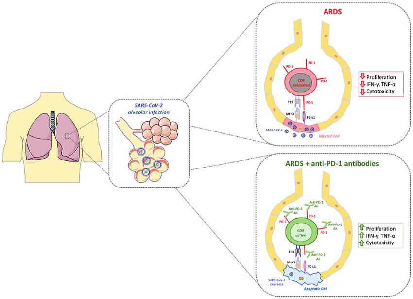
Figure 2. SARS‑CoV‑2 alveolar infection in severe cases may promote acute respiratory distress syndrome (ARDS; box on upper right, red color). Therapeutic
intervention with anti‑PD‑1 antibody may restore T‑cell cytotoxicity towards alveolar infected cells and help optimal viral clearance (box on lower right, green
color). SARS-CoV-2, severe acute respiratory syndrome associated coronavirus‑2; ARDS, acute respiratory distress syndrome; IFN, interferon; TNF, tumor
necrosis factor; PD-1, programmed death‑1; PD-L1, PD ligand-1.
The cytokine release syndrome, determining the CS, is an cytic choriomeningitis virus, enhanced the viral control and
important complication in patients with severe SARS‑CoV‑2 virus‑specific CD8+ T‑cell responses, demonstrating that the
infection, as it may lead to ARDS (27). As widely described in PD‑1 inhibitory pathway is of particular importance in exhausted
the section above, following a first hyper‑inflammatory phase T‑cell formation. Following the PD‑1/PD‑L1 blockage, T‑cells
triggered by the CS, SARS‑CoV‑2 prolonged infection may CD8+ restarted to proliferate in lymphoid and non‑lymphoid
induce T‑cell hyperactivation and finally exhaustion, associated tissues. Importantly, the effect seemed to be specific for
with concurrent lymphopenia in patient (137). High pro‑inflam‑ PD‑1/PD‑L1, as anti‑CTLA‑4 blocking antibody administration
matory IL‑6 levels, together with an elevated exhaustion and had no effect on either T‑cell function or viral control (140). In
reduced functional diversity of T‑cells in peripheral blood, line with these results, PD‑1 inhibition was also found relevant in
may predict poorer outcomes of patients with COVID‑19 (138). human viral disease. In fact, HIV-infected patients were shown
Although both CD4 + and CD8+ T‑cells in patients with to exhibit T‑cell exhaustion with a high PD‑1 expression. In such
COVID‑19 are produced, particularly against the SARS‑CoV‑2 subjects, PD1‑blocking antibodies restored both CD4+ and CD8+
S antigen, such cells are reduced in abundance and are less acti‑ T‑cell abundance and functionality (141).
vated in the case of severe SARS‑CoV‑2 infection. Therefore, Based on results obtained with other models of viral
the viral clearance may be delayed. Additionally, the excessive infections, in both mice and humans, the use of ICIs (and in
exhaustion of CD8+ T‑cells in patients with severe COVID‑19 particular anti‑PD‑1 antibody) has been suggested by several
may reduce their cellular‑mediated immune response to the authors for the treatment of SARS‑CoV‑2-infected subjects,
virus (138,139). In detail, peripheral T‑cells are impaired and alone or in combination with the concurrent blockage of the
express high levels of both the immunosuppressive markers, pro‑inflammatory IL‑6 pathway (142‑145). The rationale
mucin‑3 and PD‑1, which deeply impairs their effector behind this co‑administration is to contemporarily block both
functionality (137). In association with this T‑cell disruption, the detrimental humoral CS with anti‑IL‑6 pathway-targeting
patients with severe and critical COVID‑19 may develop viral antibodies, in association with restoring T‑cell cellular-mediated
sepsis (57). The identification of a therapy to restore the T‑cell immunity, by using the anti‑PD‑1 antibodies (146). These two
functionality in patients with COVID‑19 may be used to prevent treatments, if used in synergy, may re‑educate the defeated host
viral sepsis and therefore, the development of ARDS. immune system to finally wipe off the SARS‑Cov‑2 infection.
In a previous study, PD‑1/PD‑L1 blocking antibodies Importantly, it has also been suggested that an earlier anti‑PD‑1
administered to mice chronically infected with lympho‑ intervention in patients with COVID‑19 may block theINTERNATIONAL JOURNAL OF ONCOLOGY 9
development of ARDS, and thus minimize the need for further revolutionized the field of oncology over the past 10 years,
ICU support (143). Alternatively, anti‑PD‑1 administration may and may thus identify a cure for severe cases of COVID‑19. A
be associated with other modulators of the innate immunity, summary of the expected effects of anti‑PD‑1 administration
such as Toll‑like receptor (TLR) agonists/antagonists, as is presented in Fig. 2.
recently suggested (64).
Based on the reported observations, it seems reasonable to 6. Conclusions and future perspectives
suggest that ICIs may be used in both cancer and non‑cancer
patients affected by COVID‑19 (147). Indeed, 5 clinical studies Over the past 10 months, the threat of COVID‑19 has led to a
are currently registered at clinicaltrials.gov with the common marked increase in both research and medical efforts internation‑
goal of studying the potentialities of administering anti‑PD‑1 ally, with the collective aim of identifying a successful cure with
antibody to cure COVID‑19. which to eradicate SARS‑CoV‑2. Although standard care proto‑
The first one is being conducted on metastatic and cols and guidelines have been elaborated in order to ameliorate
advanced cancer patients, affected by COVID‑19 and which the outcomes of patients with COVID‑19 and to contain the global
are not eligible for a transfer to an ICU. It is a French, phase II, spread of the disease, a cure has not yet been identified. Research
prospective, controlled, randomized study, which has already has moved forward at a rapid pace and, regarding the vaccine,
enrolled 384 patients since its opening. The study will assess as of December 2, 2020 the UK gave emergency authorization
the difference in the efficacy to eradicate SARS‑CoV‑2 to the Pfizer and BioNTech’s vaccine. As of December 8, 2020,
infection between COVID‑19 patients treated with anti‑PD‑1 the UK began the vaccination campaign, which will be soon
antibody nivolumab in association with standard care protocol, followed by Canada and other nations.
versus standard care protocol offered alone (NCT04333914). A number of drugs have been adapted from other patholo‑
Notably, the other 4 registered studies will be enrolling and gies to either block viral entry and intracellular replication
testing the safety and the efficacy of anti‑PD‑1 administration or to redirect the impaired immune system of compromised
on NON‑cancer COVID‑19-infected patients. The first one is severe and critical COVID‑19-infected patients.
a Chinese, phase II, interventional randomized study, aimed As regards the host immune system remodulation, two
at recruiting a total of 120 COVID‑19-infected patients. Given clinical approaches are currently tested in patients. On the one
that PD‑1 is a key mediator of T‑cell depletion and viral sepsis hand, tests are ongoing to identify molecules able to block the
in patients with COVID‑19, the investigators would like to exacerbated CS that is responsible for extensive multi-organ
assess the clinical efficacy of PD‑1 blockade (administering a damage and ultimately death. This category of molecules
PD‑1 blocking antibody), in association with standard of care includes monoclonal antibodies and small molecules targeting
treatments, in COVID‑19-infected patients with pneumonia both the IL‑6 and IL‑1 pro‑inflammatory pathways, which may
and lymphocytopenia (NCT04268537). reduce unrestrained cytokine production. On the other hand,
A second, Hong Kong, phase II, interventional, open‑label, novel trials are exploring specific ICIs able to re‑activate the
controlled pilot study on 15 adult patients with COVID‑19 exhausted T‑cell immune response, and thus prevent important
has been set up to evaluate: i) The efficacy of anti‑PD1 anti‑ COVID‑19-related complications, including viral septicemia.
body (nivolumab) in clearing the SARS‑CoV‑2 infection; and For example, it is hoped that in COVID‑19-infected patients,
ii) the safety of the anti‑PD1 treatment in COVID‑19-infected anti‑PD‑1 monoclonal antibodies, which are already being used
patients. In that study, nivolumab will be administered in in cancer patients to re‑activate the immune system against
association with the optimal standard supportive care for cancer cells may restore the cytotoxic activity of T‑cells, partic‑
COVID‑19 (NCT04356508). ularly CD8+ cytotoxicity against the SARS‑CoV‑2 pathogen.
A third French, phase II, interventional randomized study It may be possible in the future to extend the study of ICIs by
will recruit about 100 COVID‑19-infected patients which will also blocking PD‑L1 expressed by infected cells; however,
be treated with standard of care with or without nivolumab additional translational studies will be required.
administration. As in previous studies, the primary goal is The vast number of clinical trials on COVID‑19-infected
to assess the time required for clinical improvement and the patients that have begun at the onset of 2020 will undoubtedly
overall efficacy of administering anti‑PD‑1 antibody to patients uncover a huge quantity of novel results, hopefully leading to
with COVID‑19 (NCT04343144). the identification of valid therapies and vaccines that may help
A fourth, French, phase II, randomized study, is enrolling humanity to overcome this testing and troublesome time in
120 obese COVID‑19-infected patients, to evaluate the history.
efficacy of anti‑PD‑1 nivolumab in treating severe forms of
COVID‑19 in such high‑risk class of infected subjects. Obesity Acknowledgements
markedly increases the risk of developing a severe, or even
critical, form of COVID‑19 (103). Obese individuals develop a The authors would like to acknowledge the Italian League
chronic meta‑inflammatory status associated with a dysregu‑ Against Cancer (LILT) for its support. The authors would
lated immune system. This obesity‑related status is called like to specially thank Dr Golnar Kolahgar, University of
inflammaging (148). That study will be divided into 2 arms. Cambridge, for her support with the English language revision
All participants will receive routine standard of care for and editing of this manuscript.
COVID‑19, while one arm will receive anti‑PD‑1 nivolumab
in combination (NCT04413838). Funding
These clinical studies hold immense potential in that they
may soon uncover the hidden capability of ICIs, that have No funding was received.10 VIVARELLI et al: IMMUNE CHECKPOINT INHIBITORS FOR TREATMENT OF PATIENTS WITH COVID-19
Availability of data and materials 9. da Costa VG, Moreli ML and Saivish MV: The emergence of
SARS, MERS and novel SARS‑2 coronaviruses in the 21st
century. Arch Virol 165: 1517‑1526, 2020.
The original contributions presented in the study are publicly 10. Arab‑Zozani M and Hassanipour S: Features and limitations
available. These data can be found at: www.pubmed.com; of LitCovid hub for quick access to literature about COVID‑19.
Balkan Med J 37: 231‑232, 2020.
www.clinicaltrials.gov. 11. Khailany RA, Safdar M and Ozaslan M: Genomic character‑
ization of a novel SARS‑CoV‑2. Gene Rep 19: 100682, 2020.
Authors' contributions 12. Cohen J: New coronavirus threat galvanizes scientists.
Science 367: 492‑493, 2020.
13. Lokman SM, Rasheduzzaman M, Salauddin A, Barua R,
ML conceptualized and supervised the drafting of the Tanzina AY, Rumi MH, Hossain MI, Siddiki AMAMZ,
manuscript and provided critical revisions. SV assisted in the Mannan A and Hasan MM: Exploring the genomic and proteomic
variations of SARS‑CoV‑2 spike glycoprotein: A computational
conceptualization of the manuscript and wrote the first draft biology approach. Infect Genet Evol 84: 104389, 2020.
of the manuscript, including the drawing of the 2 figures and 14. Wan Y, Shang J, Graham R, Baric RS and Li F: Receptor
the preparation of the both tables. LF, FT, GS, RB, DAS and recognition by the novel coronavirus from Wuhan: an analysis
based on decade‑long structural studies of SARS coronavirus. J
GRu contributed to the writing of the contents presented in Virol 94: e00127-20, 2020.
Chapters 1, 2 and 5 and both tables of the manuscript. GRa 15. Ziegler CGK, Allon SJ, Nyquist SK, Mbano IM, Miao VN,
and FP contributed to the writing of the contents presented Tzouanas CN, Cao Y, Yousif AS, Bals J, Hauser BM, et al; HCA
Lung Biological Network. Electronic address: lung‑network@
in Chapters 3 and 4 of the manuscript. All authors contrib‑ huma ncellatlas.org; HCA Lung Biologica l Network:
uted to manuscript revision, and read, and approved the final SARS‑CoV‑2 receptor ACE2 is an interferon‑stimulated gene
manuscript. in human airway epithelial cells and is detected in specific cell
subsets across tissues. Cell 181: 1016‑1035.e19, 2020.
16. Shang J, Wan Y, Luo C, Ye G, Geng Q, Auerbach A and Li F: Cell
Ethics approval and consent to participate entry mechanisms of SARS‑CoV‑2. Proc Natl Acad Sci USA 117:
11727‑11734, 2020.
17. Hoffmann M, Kleine‑Weber H and Pöhlmann S: A Multibasic
Not applicable. cleavage site in the spike protein of SARS‑CoV‑2 is essential for
infection of human lung cells. Mol Cell 78: 779‑784.e5, 2020.
Patient consent for publication 18. Devaux CA, Rolain J‑M and Raoult D: ACE2 receptor poly‑
morphism: susceptibility to SARS‑CoV‑2, hypertension,
multi‑organ failure, and COVID‑19 disease outcome. J Microbiol
Not applicable. Immunol Infect 53: 425‑435, 2020.
19. Zou X, Chen K, Zou J, Han P, Hao J and Han Z: Single‑cell
RNA‑seq data analysis on the receptor ACE2 expression reveals
Competing interests the potential risk of different human organs vulnerable to
2019‑nCoV infection. Front Med 14: 185‑192, 2020.
DAS is the Editor‑in‑Chief for the journal, but had no personal 20. Xiao L, Sakagami H and Miwa N: ACE2: The key molecule
for understanding the pathophysiology of severe and critical
involvement in the reviewing process, or any influence in conditions of COVID‑19: Demon or Angel? Viruses 12: 491, 2020.
terms of adjudicating on the final decision, for this article. 21. Romano M, Ruggiero A, Squeglia F, Maga G and Berisio R: A
The other authors declare that the review was conducted in Structural view of SARS‑CoV‑2 RNA replication machinery:
RNA synthesis, proofreading and final capping. Cells 9: 1267,
the absence of any commercial or financial relationships that 2020.
could be construed as a potential conflict of interest. 22. Kaye M, Druce J, Tran T, Kostecki R, Chibo D, Morris J,
Catton M and Birch C: SARS‑associated coronavirus replication
in cell lines. Emerg Infect Dis 12: 128‑133, 2006.
References 23. Yap JKY, Moriyama M and Iwasaki A: Inflammasomes and
pyroptosis as therapeutic targets for COVID‑19. J Immunol 205:
307‑312, 2020.
1. Chan JFW, Yuan S, Kok KH, To KK, Chu H, Yang J, Xing F, 24. Li S, Jiang L, Li X, Lin F, Wang Y, Li B, Jiang T, An W, Liu S,
Liu J, Yip CC, Poon RW, et al: A familial cluster of pneumonia Liu H, et al: Clinical and pathological investigation of patients
associated with the 2019 novel coronavirus indicating with severe COVID‑19. JCI Insight 5: e138070, 2020.
person‑to‑person transmission: A study of a family cluster. Lancet 25. Shah A: Novel coronavirus‑induced NLRP3 Inflammasome
395: 514‑523, 2020. activation: a potential drug target in the treatment of COVID‑19.
2. Lu R, Zhao X, Li J, Niu P, Yang B, Wu H, Wang W, Song H, Front Immunol 11: 1021, 2020.
Huang B, Zhu N, et al: Genomic characterisation and epide‑ 26. Coperchini F, Chiovato L, Croce L, Magri F and Rotondi M: The
miology of 2019 novel coronavirus: Implications for virus origins cytokine storm in COVID‑19: An overview of the involvement
and receptor binding. Lancet 395: 565‑574, 2020. of the chemokine/chemokine‑receptor system. Cytokine Growth
3. World Health Organization (WHO): Coronavirus disease Factor Rev 53: 25‑32, 2020.
(COVID-19): Situation reports. https://www.who.int/emer‑ 27. Ye Q, Wang B and Mao J: The pathogenesis and treatment of the
gencies/diseases/novel-coronavirus-2019/situation-reports. ‘Cytokine Storm’ in COVID‑19. J Infect 80: 607‑613, 2020.
4. Su S, Wong G, Shi W, Liu J, Lai ACK, Zhou J, Liu W, Bi Y and 28. Liu F, Li L, Xu M, Wu J, Luo D, Zhu Y, Li B, Song X and
Gao GF: Epidemiology, genetic recombination, and pathogenesis Zhou X: Prognostic value of interleukin‑6, C‑reactive protein,
of coronaviruses. Trends Microbiol 24: 490‑502, 2016. and procalcitonin in patients with COVID‑19. J Clin Virol 127:
5. Li X and Ma X: Acute respiratory failure in COVID‑19: Is it 104370, 2020.
‘typical’ ARDS? Crit Care 24: 198, 2020. 29. Ong EZ, Chan YFZ, Leong WY, Lee NMY, Kalimuddin S, Haja
6. Renu K, Prasanna PL and Valsala Gopalakrishnan A: Mohideen SM, Chan KS, Tan AT, Bertoletti A, Ooi EE, et al: A
Coronaviruses pathogenesis, comorbidities and multi‑organ dynamic immune response shapes COVID‑19 progression. Cell
damage ‑ A review. Life Sci 255: 117839, 2020. Host Microbe 27: 879‑882.e2, 2020.
7. Cui J, Li F and Shi Z‑L: Origin and evolution of pathogenic 30. McKechnie JL and Blish CA: The innate immune system:
coronaviruses. Nat Rev Microbiol 17: 181‑192, 2019. fighting on the front lines or fanning the flames of COVID‑19?
8. Ja i mes JA, A nd ré N M, Chappie JS, M i l let J K a nd Cell Host Microbe 27: 863‑869, 2020.
Whittaker GR: Phylogenetic analysis and structural modeling 31. Thachil J and Srivastava A: SARS‑2 coronavirus‑associated
of SARS‑CoV‑2 spike protein reveals an evolutionary distinct hemostatic lung abnormality in COVID‑19: is it pulmonary
and proteolytically sensitive activation loop. J Mol Biol 432: thrombosis or pulmonary embolism? Semin Thromb Hemost 46:
3309‑3325, 2020. 777‑780, 2020.INTERNATIONAL JOURNAL OF ONCOLOGY 11
32. Huang C, Wang Y, Li X, Ren L, Zhao J, Hu Y, Zhang L, Fan 53. Chen Y, Chen L, Deng Q, Zhang G, Wu K, Ni L, Yang Y, Liu B,
G, Xu J, Gu X, et al: Clinical features of patients infected with Wang W, Wei C, et al: The presence of SARS‑CoV‑2 RNA in
2019 novel coronavirus in Wuhan, China. Lancet 395: 497‑506, the feces of COVID‑19 patients. J Med Virol 92: 833‑840, 2020.
2020. 54. Zaim S, Chong JH, Sankaranarayanan V and Harky A: COVID‑19
33. Rapisarda V, Loreto C, Ledda C, Musumeci G, Bracci M, and multiorgan response. Curr Probl Cardiol 45: 100618, 2020.
Santarelli L, Renis M, Ferrante M and Cardile V: Cytotoxicity, 55. Singer M, Deutschman CS, Seymour CW, Shankar‑Hari M,
oxidative stress and genotoxicity induced by glass fibers on Annane D, Bauer M, Bellomo R, Bernard GR, Chiche JD,
human alveolar epithelial cell line A549. Toxicol In Vitro 29: Coopersmith CM, et al: The Third International Consensus
551‑557, 2015. Definitions for Sepsis and Septic Shock (Sepsis‑3). JAMA 315:
34. Armstrong SM, Wang C, Tigdi J, Si X, Dumpit C, Charles S, 801‑810, 2016.
Gamage A, Moraes TJ and Lee WL: Influenza infects lung 56. Yi Y, Lagniton PNP, Ye S, Li E and Xu RH: COVID‑19: What
microvascular endothelium leading to microvascular leak: Role has been learned and to be learned about the novel coronavirus
of apoptosis and claudin‑5. PLoS One 7: e47323, 2012. disease. Int J Biol Sci 16: 1753‑1766, 2020.
35. Ohmura T, Tian Y, Sarich N, Ke Y, Meliton A, Shah AS, 57. Li H, Liu L, Zhang D, Xu J, Dai H, Tang N, Su X and Cao B:
Andreasson K, Birukov KG and Birukova AA: Regulation of lung SARS‑CoV‑2 and viral sepsis: Observations and hypotheses.
endothelial permeability and inflammatory responses by prosta‑ Lancet 395: 1517‑1520, 2020.
glandin A2: Role of EP4 receptor. Mol Biol Cell 28: 1622‑1635, 58. Lescure F‑X, Bouadma L, Nguyen D, Parisey M, Wicky PH,
2017. Behillil S, Gaymard A, Bouscambert‑Duchamp M, Donati F,
36. Schurink B, Roos E, Radonic T, Barbe E, Bouman CSC, Le Hingrat Q, et al: Clinical and virological data of the first cases
de Boer HH, de Bree GJ, Bulle EB, Aronica EM, Florquin S, et al: of COVID‑19 in Europe: A case series. Lancet Infect Dis 20:
Viral presence and immunopathology in patients with lethal 697‑706, 2020.
COVID‑19: A prospective autopsy cohort study. Lancet 59. Endeman H, van der Zee P, van Genderen ME, van den Akker JPC
Microbe 1: e290‑e299, 2020. and Gommers D: Progressive respiratory failure in COVID‑19:
37. Ahmadpoor P and Rostaing L: Why the immune system fails to A hypothesis. Lancet Infect Dis 20: 1365, 2020.
mount an adaptive immune response to a COVID‑19 infection. 60. de la Rica R, Borges M and Gonzalez‑Freire M: COVID‑19: In
Transpl Int 33: 824‑825, 2020. the Eye of the Cytokine Storm. Front Immunol 11: 558898, 2020.
38. Wang B, Wang L, Kong X, Geng J, Xiao D, Ma C, Jiang XM and 61. Channappanavar R and Perlman S: Pathogenic human coro‑
Wang PH: Long‑term coexistence of SARS‑CoV‑2 with antibody navirus infections: Causes and consequences of cytokine storm
response in COVID‑19 patients. J Med Virol 92: 1684‑1689, 2020. and immunopathology. Semin Immunopathol 39: 529‑539, 2017.
39. Zhang B, Zhou X, Zhu C, Song Y, Feng F, Qiu Y, Feng J, Jia Q, 62. Lin L, Lu L, Cao W and Li T: Hypothesis for potential patho‑
Song Q, Zhu B and Wang J: Immune phenotyping based on genesis of SARS‑CoV‑2 infection‑a review of immune changes
neutrophil‑to‑lymphocyte ratio and IgG predicts disease severity in patients with viral pneumonia. Emerg Microbes Infect 9:
and outcome for patients with COVID‑19. Front Mol Biosci 7: 157, 727‑732, 2020.
2020. 63. Laing AG, Lorenc A, Del Molino Del Barrio I, Das A,
40. Zhao J, Yuan Q, Wang H, Liu W, Liao X, Su Y, Wang X, Fish M, Monin L, Muñoz‑Ruiz M, McKenzie DR, Hayday
Yuan J, Li T, Li J, et al: Antibody responses to SARS‑CoV‑2 in TS, Francos‑Quijorna I, et al: A dynamic COVID‑19 immune
patients with novel coronavirus disease 2019. Clin Infect Dis 71: signature includes associations with poor prognosis. Nat Med 26:
2027‑2034, 2020. 1623‑1635, 2020.
41. Fathi N and Rezaei N: Lymphopenia in COVID‑19: Therapeutic 64. Onofrio L, Caraglia M, Facchini G, Margherita V, Placido S and
opportunities. Cell Biol Int 44: 1792‑1797, 2020. Buonerba C: Toll‑like receptors and COVID‑19: a two‑faced
42. Diao B, Wang C, Tan Y, Chen X, Liu Y, Ning L, Chen L, Li M, story with an exciting ending. Future Sci OA 6: FSO605, 2020.
Liu Y, Wang G, et al: Reduction and functional exhaustion of 65. Florindo HF, Kleiner R, Vaskovich‑Koubi D, Acúrcio
T Cells in patients with coronavirus disease 2019 (COVID‑19). RC, Carreira B, Yeini E, Tiram G, Liubomirski Y and
Front Immunol 11: 827, 2020. Satchi‑Fainaro R: Immune‑mediated approaches against
43. De Biasi S, Meschiari M, Gibellini L, Bellinazzi C, Borella R, COVID‑19. Nat Nanotechnol 15: 630‑645, 2020.
Fidanza L, Gozzi L, Iannone A, Lo Tartaro D, Mattioli M, et al: 66. Skalny AV, Rink L, Ajsuvakova OP, Aschner M, Gritsenko VA,
Marked T cell activation, senescence, exhaustion and skewing Alekseenko SI, Svistunov AA, Petrakis D, Spandidos DA,
towards TH17 in patients with Covid‑19 pneumonia. Nat Aaseth J, et al: Zinc and respiratory tract infections: Perspectives
Commun 11: 3434, 2020. for COVID-19 (Review). Int J Mol Med 46: 17‑26, 2020.
44. Yaqinuddin A and Kashir J: Innate immunity in COVID‑19 67. World Health Organization (WHO): Standard precautions in
patients mediated by NKG2A receptors, and potential treatment health care. https://www.who.int/publications/i/item/standard-
using Monalizumab, Cholroquine, and antiviral agents. Med precautions-in-health-care. Accessed September 30, 2007
Hypotheses 140: 109777, 2020. 68. Nitulescu GM, Paunescu H, Moschos SA, Petrakis D,
45. Carvelli J, Demaria O, Vely F, Batista L, Benmansour NC, Nitulescu G, Ion GND, Spandidos DA, Nikolouzakis TK,
Fares J, Carpentier S, Thibult ML, Morel A, André P, et al: Drakoulis N and Tsatsakis A: Comprehensive analysis of
Association of COVID-19 inflammation with activation of the drugs to treat SARS‑CoV‑2 infection: Mechanistic insights
C5a‑C5aR1 axis. Nature 588, 146‑150, 2020. into current COVID‑19 therapies (Review). Int J Mol Med 46:
46. Cully M: Immune status could determine efficacy of COVID‑19 467‑488, 2020.
therapies. Nat Rev Drug Discov 19: 431‑434, 2020. 69. Dehelean CA, Lazureanu V, Coricovac D, Mioc M, Oancea R,
47. Yang L, Liu S, Liu J, Zhang Z, Wan X, Huang B, Chen Y Marcovici I, Pinzaru I, Soica C, Tsatsakis AM and Cretu
and Zha ng Y: COV I D ‑19: I m munopathogenesis and O: SARS‑CoV‑2: Repurposed drugs and novel therapeutic
Immunotherapeutics. Signal Transduct Target Ther 5: 128, 2020. approaches‑insights into chemical structure‑biological activity
48. Mortaz E, Tabarsi P, Varahram M, Folkerts G and Adcock IM: and toxicological screening. J Clin Med 9: 2084, 2020.
The Immune Response and Immunopathology of COVID‑19. 70. Beigel JH, Tomashek KM, Dodd LE, Mehta AK, Zingman BS,
Front Immunol 11: 2037, 2020. Kalil AC, Hohmann E, Chu HY, Luetkemeyer A, Kline S, et al;
49. Yuki K, Fujiogi M and Koutsogiannaki S: COVID‑19 patho‑ ACTT‑1 Study Group Members: Remdesivir for the treatment of
physiology: A review. Clin Immunol 215: 108427, 2020. Covid‑19 ‑ Final report. N Engl J Med 383: 1813‑1826, 2020.
50. Yuen KS, Ye ZW, Fung SY, Chan CP and Jin DY: SARS‑CoV‑2 71. FDA: FDA Remdesivir update, 2020. https://www.fda.gov/
and COVID‑19: The most important research questions. Cell news-events/press-announcements/coronavirus-covid-19-update-fda-
Biosci 10: 40, 2020. authorizes-drug-combination-treatment-covid-19. Accessed
51. Carsana L, Sonzogni A, Nasr A, Rossi RS, Pellegrinelli A, December 1, 2020.
Zerbi P, Rech R, Colombo R, Antinori S, Corbellino M, et al: 72. World Health Organization (WHO): WHO recommends against
Pulmonary post‑mortem findings in a series of COVID‑19 cases the use of remdesivir in COVID‑19 patients. https://www.who.int/
from northern Italy: A two‑centre descriptive study. Lancet news-room/feature-stories/detail/who-recommends-against-the-
Infect Dis 20: 1135‑1140, 2020. use-of-remdesivir-in-covid-19-patients. Accessed November 20,
52. Lodigiani C, Iapichino G, Carenzo L, Cecconi M, Ferrazzi P, 2020.
Sebastian T, Kucher N, Studt JD, Sacco C, Bertuzzi A, et al; 73. European Medicines Agency (EMA): Update on remdesivir -
Humanitas COVID‑19 Task Force: Venous and arterial throm‑ EMA will evaluate new data from Solidarity trial. https://www.
boembolic complications in COVID‑19 patients admitted to an ema.europa.eu/en/news/update-remdesivir-ema-will-evaluate-
academic hospital in Milan, Italy. Thromb Res 191: 9‑14, 2020. new-data-solidarity-trial. Accessed November 20, 2020.You can also read