Nanoparticle Toxicology - Annual Review of Pharmacology and Toxicology - Wilhelm Lab
←
→
Page content transcription
If your browser does not render page correctly, please read the page content below
Annual Review of Pharmacology and Toxicology
Nanoparticle Toxicology
Wen Yang,1 Lin Wang,1 Evan M. Mettenbrink,1
Paul L. DeAngelis,2 and Stefan Wilhelm1,3,4
Access provided by 2600:1700:5270:f8e0:1131:433c:95c:42eb on 01/08/21. For personal use only.
Annu. Rev. Pharmacol. Toxicol. 2021.61:269-289. Downloaded from www.annualreviews.org
1
Stephenson School of Biomedical Engineering, University of Oklahoma, Norman,
Oklahoma 73019, USA; email: stefan.wilhelm@ou.edu
2
Department of Biochemistry and Molecular Biology, University of Oklahoma Health Sciences
Center, Oklahoma City, Oklahoma 73104, USA
3
Institute for Biomedical Engineering, Science, and Technology (IBEST), Norman,
Oklahoma 73019, USA
4
Stephenson Cancer Center, Oklahoma City, Oklahoma 73104, USA
Annu. Rev. Pharmacol. Toxicol. 2021. 61:269–89 Keywords
First published as a Review in Advance on
nanoparticles, toxicology, physicochemical properties, anti-PEG antibody,
August 25, 2020
protein corona, ROS
The Annual Review of Pharmacology and Toxicology is
online at pharmtox.annualreviews.org Abstract
https://doi.org/10.1146/annurev-pharmtox-032320-
Nanoparticles from natural and anthropogenic sources are abundant in the
110338
environment, thus human exposure to nanoparticles is inevitable. Due to
Copyright © 2021 by Annual Reviews.
this constant exposure, it is critically important to understand the potential
All rights reserved
acute and chronic adverse effects that nanoparticles may cause to humans.
In this review, we explore and highlight the current state of nanotoxicology
research with a focus on mechanistic understanding of nanoparticle toxic-
ity at organ, tissue, cell, and biomolecular levels. We discuss nanotoxicity
mechanisms, including generation of reactive oxygen species, nanoparticle
disintegration, modulation of cell signaling pathways, protein corona for-
mation, and poly(ethylene glycol)-mediated immunogenicity. We conclude
with a perspective on potential approaches to advance current understanding
of nanoparticle toxicity. Such improved understanding may lead to mitiga-
tion strategies that could enable safe application of nanoparticles in humans.
Advances in nanotoxicity research will ultimately inform efforts to estab-
lish standardized regulatory frameworks with the goal of fully exploiting the
potential of nanotechnology while minimizing harm to humans.
269
1. INTRODUCTION
                                                                                                      Nanoparticles comprise a class of materials with dimensions in the 1–100-nm range that exhibit
                                                                                                      unique physical, chemical, and biological properties, making them distinct from their corre-
                                                                                                      sponding bulk materials or small molecules (1, 2). Owing to their unique material characteristics,
                                                                                                      nanoparticles are broadly used in a range of applications and products, including industrial
                                                                                                      catalytic processes, energy conversion and storage, and image display technologies as well as
                                                                                                      cosmetics, medical devices, and therapeutics and diagnostics (3–5). In addition to such rationally
                                                                                                      designed nanoparticles, we are constantly surrounded by substantial amounts of naturally and
                                                                                                      incidentally formed particles, such as corrosion- or erosion-derived nanoparticles in water and
                                                                                                      airborne nanoparticles from traffic and industrial combustion (6, 7). The abundance of nanopar-
                                                                                                      ticles in the environment and in everyday consumer products makes human exposure inevitable.
Access provided by 2600:1700:5270:f8e0:1131:433c:95c:42eb on 01/08/21. For personal use only.

                                                                                                      However, the potential acute and chronic health risks that nanoparticles may pose to humans are
 Annu. Rev. Pharmacol. Toxicol. 2021.61:269-289. Downloaded from www.annualreviews.org

                                                                                                      poorly investigated and understood.
                                                                                                          In this review, we introduce the major nanoparticle classes and explore how their corre-
                                                                                                      sponding physicochemical properties affect toxicity. Our discussion of the main nanotoxicity
                                                                                                      mechanisms provides an overview of how nanoparticles interact with the body at organ, tissue,
                                                                                                      cell, and biomolecular levels. Such mechanistic understanding is enabled by diverse experimental
                                                                                                      and theoretical methods that have been developed and applied for the assessment and evaluation
                                                                                                      of nanotoxicity. We conclude our discussion with a perspective on potential strategies to mitigate
                                                                                                      nanotoxicity, with the goal of exploiting the full potential of nanotechnology for safe applications
                                                                                                      in humans. We hope that our review serves as a valuable resource that covers the current
                                                                                                      landscape of nanotoxicity research and that it will inspire new studies focused on expanding our
                                                                                                      understanding of nanoparticle toxicity. Improved understanding of nanotoxicity may ultimately
                                                                                                      inform and guide the development of regulatory frameworks to minimize potential harm to
                                                                                                      humans.

                                                                                                      2. NANOPARTICLE CLASSIFICATION AND PHYSICOCHEMICAL
                                                                                                      PROPERTIES
                                                                                                      Nanoparticles can be grouped into three main classes: (a) natural, (b) incidental, or (c) engineered
                                                                                                      nanoparticles (Figure 1a–c). The first class, natural nanoparticles, is ubiquitous in the environ-
                                                                                                      ment and generated via normal physical, chemical, and biological processes. Examples of such
                                                                                                      natural nanoparticles include inorganic metal-based nanoparticles, e.g., naturally formed silver
                                                                                                      (Ag) nanoparticles, and organic nanoparticles, e.g., virus nanoparticles and exosomes (Figure 1a).
                                                                                                      The second class, incidental nanoparticles, is generated unintentionally as byproducts of both
                                                                                                      industrial and nonindustrial processes such as corrosion, combustion, and cooking. Examples in-
                                                                                                      clude inorganic and organic combustion products such as metal- and carbon-based nanoparticles,
                                                                                                      respectively (Figure 1b). The third class, engineered nanoparticles, is intentionally designed and
                                                                                                      fabricated for specific industrial and/or medical applications. Examples include zinc oxide (ZnO)
                                                                                                      and titanium dioxide (TiO2 ) nanoparticles in sunscreen and liposomes for drug delivery appli-
                                                                                                      cations (8–10) (Figure 1c). An alternative terminology for natural and incidental nanoparticles
                                                                                                      is ultrafine particles (UFPs). These UFPs are airborne particulates of less than 100 nm in aero-
                                                                                                      dynamic diameter. While incidental and engineered nanoparticles are typically of anthropogenic
                                                                                                      origin, i.e., caused and/or prepared by human activity, natural nanoparticles are generated without
                                                                                                      human intervention (1, 6).
                                                                                                          Beyond classification by origin, the various nanoparticle types can be further differentiated by
                                                                                                      their physicochemical properties (Figure 1d). Physicochemical properties such as nanoparticle

                                                                                                270   Yang et al.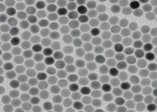
a b c
Natural nanoparticles Incidental nanoparticles Engineered nanoparticles
Inorganic
50 nm 50 nm 100 nm
Access provided by 2600:1700:5270:f8e0:1131:433c:95c:42eb on 01/08/21. For personal use only.
Annu. Rev. Pharmacol. Toxicol. 2021.61:269-289. Downloaded from www.annualreviews.org
Organic
40 nm 100 nm 100 nm
Physicochemical properties of nanoparticles
d COMPOSITION SIZE
Au Fe3O4 Ag
1 nm 100 nm
Liposome Micelle
SURFACE CHEMISTRY AND CHARGE SHAPE
e
Po
tiv
sit
ga
iv
Ne
e
Sphere Cube Polyhedron
ni r-
Ne
io itte
ut
c
Surface ligands
Zw
ra
l
Rod Disc
Figure 1
Nanoparticle classification and physicochemical properties. Nanoparticles can be broadly organized into three different classes:
(a) natural, (b) incidental, or (c) engineered nanoparticles. Nanoparticles from all of these classes can be made from inorganic or organic
materials. In contrast to the majority of natural and incidental nanoparticles, engineered nanoparticles typically exhibit narrow size
distributions as well as defined shapes and surface properties. Panels a–c display transmission electron micrographs of different
inorganic nanoparticles [(a,b, top) silver (Ag) nanoparticles, (c, top) upconversion (NaYF4 /Yb,Er) nanoparticles] and organic
nanoparticles [(a, bottom) cowpea mosaic virus–like nanoparticles, (b, bottom) carbon black nanoparticles, (c, bottom) doxorubicin-loaded
liposomes]. (d) Schematic of various nanoparticle physicochemical properties, including different nanoparticle compositions, sizes,
surface chemistries, and shapes. Panel a adapted with permission from References 138 and 140, panel b adapted with permission from
References 139 and 141, and panel c adapted with permission from References 110 and 142.
www.annualreviews.org • Nanoparticle Toxicology 271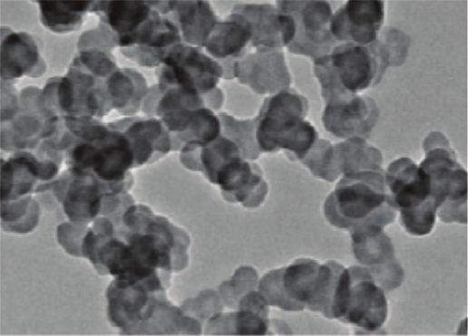
Exposure pathways Organ/tissue interactions Cell interactions Biomolecular interactions
a Ocular b c d Nanoparticle
Inhalation
Biomolecules
Oral Nanoparticles
Nanoparticles
Dermal
Surface
Intravenous Cell adsorption
Chemical, physical, and biological effects
e i ROS generation ii Disintegration iii Signaling pathways
Access provided by 2600:1700:5270:f8e0:1131:433c:95c:42eb on 01/08/21. For personal use only.
Annu. Rev. Pharmacol. Toxicol. 2021.61:269-289. Downloaded from www.annualreviews.org
Singlet oxygen ( 1O 2)
Nanoparticle Nanoparticle Nanoparticle
Superoxide radical (O2•– ) Cell receptor
Oxygen radical (O• )
Cell
Reactive oxygen Peroxide ion (O22– )
species (ROS) MAPK PKC
Hydrogen peroxide (H2O2 )
Metal ions Nrf-2 NF-κB
Hydroxyl radical (OH• )
Organic species HIF Src
Cellular toxicity Organ/tissue toxicity
f g
Cell Apoptosis
Inflammation
Damage
Necrosis
Damage/failure of tissues and organs:
lungs, liver, brain, spleen, kidneys, and skin
Damage/lysis of intracellular organelles
Damage to nucleic acids
Damage to plasma membrane Inflammation
Damage to membrane receptors and ion channels Allergic reactions and anaphylactic shock
Figure 2
Schematic representation of nanoparticle adverse effects and nanotoxicity. (a) Nanoparticle exposure pathways. Upon exposure,
nanoparticles can interact with (b) organs/tissues, (c) cells, and (d) biomolecules. Major nanoparticle toxicity mechanisms include
(e, i) the generation of reactive oxygen species (ROS), (e, ii) nanoparticle disintegration and release of metal ions and organic species,
and (e, iii) nanoparticle-mediated activation of cell signaling pathways. Nanoparticle adverse effects and toxicity can lead to ( f ) cell
apoptosis and necrosis and (g) tissue/organ damage, inflammation, and anaphylactic shock.
composition, size, surface chemistry, and shape are key factors that govern nanoparticle interac-
tions with biological systems and biomolecules. These interactions can affect biomolecular and
cellular signaling, biological kinetics and transport, nanoparticle biodistribution, immunogenicity,
and toxicity (11) (Figure 2). Compared to samples of engineered nanoparticles, natural and in-
cidental nanoparticles tend to exhibit more heterogeneous physicochemical properties, with sub-
stantial variations in nanoparticle composition, size, surface chemistry, and shape (Figure 1a–c).
This heterogeneity complicates the assessment and understanding of nanoparticle biological
interactions, adverse effects, and toxicity.
272 Yang et al.3. NANOTOXICOLOGY
The study of nanoparticle adverse effects and toxicity is commonly referred to as nanotoxicol-
ogy (12). Upon exposure, all three classes of nanoparticles, i.e., natural, incidental, and engineered
nanoparticles, may interact with organs, tissues, cells, and biomolecules (Figure 2). Consequently,
nanoparticle exposure may induce undesirable and harmful nano-bio interactions and other down-
stream mechanisms that can potentially result in adverse effects and nanotoxicity.
Nanoparticle toxicity may occur as a function of exposure route, dose, concentration, time,
and/or frequency. Traditionally, these fundamental toxicity factors are relevant for the assessment
of small-molecule drugs and other compounds. In the evaluation of nanotoxicology, these pa-
rameters are also widely used. However, beyond these traditional toxicology parameters, other
important factors that may affect nanoparticle toxicity need to be considered, including nanopar-
Access provided by 2600:1700:5270:f8e0:1131:433c:95c:42eb on 01/08/21. For personal use only.
ticle physicochemical properties such as material composition, size, surface chemistry, and shape
Annu. Rev. Pharmacol. Toxicol. 2021.61:269-289. Downloaded from www.annualreviews.org
(Figure 1d). Compared to small molecules, these additional physicochemical variables make nan-
otoxicity assessment complex, and evaluation of nanoparticle toxicity on a case-by-case basis may
be required. For example, slight variations in nanoparticle surface chemistry can result in signifi-
cantly different toxicity, biodistribution, and elimination profiles, even if the nanomaterial core is
the same (13–15).
To fully evaluate nanoparticle toxicity, nanoparticle structure and corresponding physicochem-
ical properties need to be completely characterized and understood. In this way, observed toxic
effects can be better attributed to certain nanoparticle properties for establishing specific
nanoparticle structure-activity/toxicity functional relationships. As nanoparticle structural
properties significantly affect toxicity, it is even more challenging to evaluate the safety of
nanoparticles that exhibit large variations in physicochemical properties, as is often seen in
natural and incidental nanoparticles (Figure 1a,b). Therefore, it is challenging to draw general
conclusions about nanoparticle toxicity, as nanotoxicity is dependent on complex interactions
between different physicochemical properties and the corresponding biological environment.
Based on this complexity, it is important to establish well-defined, standardized methodologies
for the systematic evaluation of nanotoxicity under relevant conditions to achieve comparable
toxicological data sets. However, this level of standardization has not yet been achieved, which
makes it difficult to provide general trends of nanotoxicity for acute (4
months) exposure regimens (16).
To provide examples of the broad range of potential nanoparticle adverse effects and toxicity,
including neurotoxicity, pulmonary toxicity, vascular dysfunction, genotoxicity, and immunotox-
icity, we have summarized studies that assessed the nanotoxicity of different nanoparticle classes
and types in human subjects (Table 1). Table 1 also includes studies that evaluated the toxic-
ity of relevant engineered nanoparticles, such as Ag and ZnO nanoparticles, on human subjects
but without any reported clinical or pathological findings, implying that the tested nanoparticles
were safe and without noticeable adverse effects under the specific testing conditions. For con-
text, Ag and ZnO nanoparticles are used in over-the-counter consumer products such as antiviral,
antibacterial, and anti-inflammatory products and compounds as well as sunscreen (17–19). We
want to emphasize that detailed reports and systematic clinical studies of nanoparticle toxicity in
humans for various nanoparticle types are limited. Most published reports focus on the assessment
of nanoparticle toxicity in cell culture and animal models. However, these models do not fully re-
capitulate nanoparticle toxicity responses in humans and are therefore limited in their predictive
power of possible hazards to humans (20).
To emphasize the importance of composition and other physicochemical properties on
nanoparticle adverse effects and toxicity, we highlight a study by Mills et al. (7) that assessed adverse
www.annualreviews.org • Nanoparticle Toxicology 273Table 1 Examples of nanoparticle toxicity in human subjects
Exposure route,
dose, duration,
Nanoparticle number of human
Adverse effect class and type subjects Toxicity mechanism Toxicity assessment Reference
Neurotoxicity Natural NA Abnormal, age-associated Quantitative 143
Fe3 O4 NA biomineralization of magnetometry;
(10 inflammation and nasal lavage samples and
complex times background systemic oxidative analysis of 8-OH-dG
mixtures levels) stress with generation and creatinine in human
(10–80 nm) Acute (6 h/day for of proinflammatory urine samples
3 days) cytokines
17 human subjects
Vascular Incidental Inhalation Increased systolic blood Measurement of forearm 7
dysfunction Diesel exhaust 1.2 × 10⁶ NPs/cm3 pressure and attenuated blood flow and blood
nanoparticles Acute (up to 14 days) vasodilation due to pressure and biomarker
(vascular side effects in 16 healthy human subjects exposed to combustion-derived nanoparticles
from diesel exhaust over an acute exposure duration of 14 days. Impaired vascular function in study
subjects was observed due to oxidative stress caused by inhalation of diesel exhaust nanoparticles.
In contrast, when study subjects were exposed to filtered exhaust, i.e., exhaust without nanopar-
ticles, or air containing pure carbon nanoparticles, vascular impairment was not observed. These
findings indicate that nanoparticle composition and other physicochemical properties play key
roles in nanotoxicity.
Other important nanoparticle physicochemical properties that affect nanotoxicity include size,
surface chemistry, and shape (Figure 1d). For more detailed information on how nanoparticle
physicochemical properties affect nano-bio interactions, adverse effects, and toxicity, we refer in-
terested readers to excellent review articles by the Chan (11) and Howard groups (12).
Access provided by 2600:1700:5270:f8e0:1131:433c:95c:42eb on 01/08/21. For personal use only.
Nanoparticle size is a physicochemical parameter that has been reported to affect cellular up-
Annu. Rev. Pharmacol. Toxicol. 2021.61:269-289. Downloaded from www.annualreviews.org
take efficiency and cytotoxicity (21). A study by Pan et al. (22) reported size-dependent cytotoxicity
of gold nanoparticles with identical surface chemistry in fibroblasts, epithelial cells, macrophages,
and melanoma cells in cell culture. The researchers reported that nanoparticles with a diameter
of 1.4 nm exhibited the highest cytotoxicity, while nanoparticles with a diameter of 15 nm had no
reported toxicity. As potential reasons for the observed differences in cytotoxicity, the researchers
listed size-dependent nanoparticle cell uptake kinetics and interactions with the cell plasma mem-
brane promoting cell apoptosis and necrosis.
In addition to nanoparticle size, surface chemistry is another important parameter that directly
affects nanotoxicity. For example, a study by Bozich et al. (23) concluded that gold nanoparticles
with an overall positive surface charge exhibited greater toxicity on Daphnia magna model or-
ganisms compared to negatively charged gold nanoparticles of the same core size. Similarly, Lee
et al. (24) reported that positively charged gold nanoparticles caused an ∼50% reduction in cell
viability compared to identical neutrally charged particles in cultured mouse breast cancer 4T1
cells. In comparison to neutral nanoparticles, there was a substantial reduction in cell viability of
∼50% for positively charged nanoparticles. Potential reasons for the increased toxicity of posi-
tively charged nanoparticles include a higher electrostatic attraction of nanoparticles to negatively
charged cell surfaces and overall increased nanoparticle cellular uptake, potentially leading to in-
creases in oxidative stress and reactive oxygen species (ROS) (25, 26). As the nanoparticle surface
interacts directly with biomolecules and biological systems, it is a driver of cellular uptake and
intracellular transport kinetics (21). In addition, surface chemistry and surface charge are key fac-
tors of nanoparticle agglomeration and aggregation, which are additional variables that need to
be considered in the assessment of nanotoxicity (27, 28).
Besides size and surface chemistry, nanoparticle shape may significantly affect nanotoxicity
(26). For example, a study by Zhao et al. (29) reported increased cytotoxicity of needle- and plate-
shaped nanosized hydroxyapatite compared to sphere- and rod-shaped nanoparticles in human
lung BEAS-2B epithelial cells. A potential reason for the increased cytotoxicity may be that needle-
and spike-like nanoparticle shapes potentially puncture cellular membranes, leading to compro-
mised cellular integrity and cell death. The shape properties of micro- and nanoparticles can also
induce physical activation of innate immunity. As reported by Wang et al. (30), TiO2 microparti-
cles exhibiting nanospikes can exert mechanical stress on cells, which can lead to potassium efflux
and inflammasome activation in macrophages and dendritic cells. These findings highlight the
potential of nanoparticle shape as a means to tune nanoparticle immunogenicity by physical cues,
which could potentially be attractive for more efficient and effective vaccination and immunother-
apy approaches.
Nanoparticle physicochemical properties not only affect cellular interactions but may also de-
termine biodistribution, clearance, and elimination (31–33). For example, nanoparticles with sizes
www.annualreviews.org • Nanoparticle Toxicology 275smaller than 5.5 nm will be eliminated rapidly into urine via the kidneys (34). Nanoparticles larger
than the renal cutoff size are often efficiently sequestered by cells in the liver and spleen, including
Kupffer cells, B cells, T cells, and endothelial cells (33), and may be eliminated to varying extents
via the hepatobiliary pathway (15). Understanding how nanoparticle physicochemical properties
affect nano-bio interactions will provide an opportunity to control nanoparticle fate and toxic-
ity inside the body. Such control may ultimately lead to more potent nanoparticle-based medi-
cal treatments and diagnostics with reduced side effects and toxicity for patients. An example of
this control is the application of liposomes to encapsulate the small-molecule cancer drug dox-
orubicin. Compared to treatment with the free (i.e., unencapsulated) drug, US Food and Drug
Administration–approved doxorubicin liposomes (i.e., Doxil) can reduce cardiotoxicity and other
adverse effects to improve the quality of life for cancer patients (35).
Access provided by 2600:1700:5270:f8e0:1131:433c:95c:42eb on 01/08/21. For personal use only.
Annu. Rev. Pharmacol. Toxicol. 2021.61:269-289. Downloaded from www.annualreviews.org
4. NANOTOXICITY MECHANISMS
There are a number of different pathways by which nanoparticles can enter the body. These path-
ways include inhalation, oral ingestion, ocular exposure, application and deposition on skin, and
intravenous administration (10, 36–39) (Figure 2a). The inhalation of airborne nanoparticles is
a major exposure pathway that allows nanoparticles to enter and deposit in lung tissues and the
alveolar region (40) (Figure 2b). Accumulation of nanoparticles in the lung can lead to oxidative
stress–mediated lung inflammation at both acute and chronic stages (41, 42). Inhalation may also
lead to the accumulation of nanoparticles in the brain. Maher et al. (43) reported that airborne
magnetite nanoparticles can enter the brain via the olfactory bulb. Accumulation of magnetite
nanoparticles in the brain that are abundant in airborne particulate matter pollution can lead
to enhanced production of ROS, which is causally linked to neurodegenerative diseases such as
Alzheimer’s disease (43, 44).
After entering the body, nanoparticles may interact with the initially encountered organ or
tissue. Nanoparticles may also subsequently translocate and enter the bloodstream (for example,
from lungs to the capillary network to bigger vessels) to access distant organs/tissues via sys-
temic transport (45, 46) (Figure 2b). Within organs, tissues, and blood, nanoparticles can interact
with cells and intracellular organelles to potentially cause toxicity at cellular and subcellular lev-
els (Figure 2c). It is important to point out that, upon entry into the body, nanoparticles interact
with a variety of different biomolecules, including proteins, carbohydrates, lipids, and nucleic acids
(Figure 2d). These interactions result in the formation of a biomolecular nanoparticle surface
corona, often referred to as the protein corona. Protein corona (or biomolecular corona) forma-
tion may change nanoparticle surface chemistry or stimulate the complement system substantially
and ultimately affect nanotoxicity or the efficacy of nanomedicine (47–49). The damage of pro-
teins to nanoparticle surfaces may also lead to protein unfolding (50, 51). This process may induce
the loss of protein function and may cause immunotoxicity (52, 53). In addition, the protein con-
figuration change can lead to adverse effects and toxicity via cell signaling pathway activation (51),
enzyme function loss (54), nanoparticle aggregation (50), new antigenic site formation (55), and
protein fibrillation (56).
At the cellular level, direct interaction between nanoparticles and cells may result in physical
damage of cell membrane structures (57, 58). For example, graphene nanoparticles have been re-
ported to cause physical damage, cytoskeletal dysfunction, and abnormal morphological stretching
in different cell types as a result of the blade-like shape of these materials (57, 58). In addition,
nanoparticles may be able to block cell membrane receptors and membrane ion channels, which
may interrupt normal cellular biofunctions and homeostasis (59). Leifert et al. (59) reported that
1.4-nm gold nanoparticles were able to block voltage-gated potassium channels in vitro, which
may lead to unwanted cardiac malformation in mice.
276 Yang et al.A major nanotoxicity mechanism is the generation of ROS such as singlet oxygen, super-
oxide anion radicals, oxygen radicals, peroxide ions, hydrogen peroxide, and hydroxyl radicals
(Figure 2e). ROS generation can occur in different ways. One route is through one-electron ox-
idative reactions with transition metals or nanoparticle surface groups (60, 61). It is important to
note that a nanoparticle exhibits a relatively large surface area compared to the particle volume.
An increase in surface area is typically accompanied by an increase in chemical reactivity poten-
tially leading to increased ROS production. Another ROS generation mechanism is via mitochon-
drial respiration and subsequent ROS release into the cytoplasm through pores in mitochondrial
membranes created by nanoparticles (60). In healthy cells, an equilibrium is maintained between
intracellular antioxidants and ROS. However, intracellular nanoparticles can directly damage mi-
tochondria, causing an increase in intracellular ROS and oxidative stress (62). Enhanced intracel-
Access provided by 2600:1700:5270:f8e0:1131:433c:95c:42eb on 01/08/21. For personal use only.
lular ROS levels may stimulate further ROS release from mitochondria through a process called
Annu. Rev. Pharmacol. Toxicol. 2021.61:269-289. Downloaded from www.annualreviews.org
ROS-induced ROS release. This process can substantially increase intracellular ROS levels and
amplify the oxidative imbalance (63). High levels of ROS can cause oxidative stress and damage
to cellular organelles, DNA, cell membranes, ion channels, and cell surface receptors, leading to
adverse effects and toxicity.
Metal or metal oxide nanoparticles are used in preclinical and clinical applications such as imag-
ing, photothermal therapy, and biosensors (64). However, corrosive tissue microenvironments and
lysosomal degradation may disintegrate nanoparticles to release potentially harmful metal ions
(Figure 2e). For many nanoparticles, including Ag, cadmium selenide (CdSe), ZnO, and ferroso-
ferric oxide nanoparticles, released metal ions may generate high levels of oxidative stress and
are primary sources of nanotoxicity. For example, Ag(I) ions released from Ag nanoparticles can
cause DNA damage, ROS generation, and cell membrane destruction as reported from cell cul-
ture studies (65). We want to emphasize that nanotoxicity results obtained in cell culture studies
do not necessarily recapitulate the nanoparticle toxicity potential in animal models or human sub-
jects. For example, CdSe quantum dots were found to be toxic in cell culture; however, no toxicity
was observed in animal models under the specific reported testing conditions (66–69).
The generation of high levels of ROS and the release of harmful metal ions from nanoparticles
have been reported to affect a variety of cell signaling pathways such as nuclear factor kappa-light-
chain enhancer of activated B cells (NF-κB), mitogen-activated protein kinase (MAPK), Akt, and
Src (70–73) (Figure 2e). Activation and modulation of these signaling pathways can affect cell
proliferation, differentiation, and cell survival. Nyga et al. (74) reported that cobalt nanoparti-
cles can stabilize hypoxia-inducible factor (HIF) protein and upregulate HIF gene expression in
human macrophages. HIF pathway activation can affect cell growth, cell survival, apoptosis, and
metabolic adaptation (75). Importantly, there can be interplay and synergistic effects between ROS
generation, cell signaling modulation, and nanoparticle disintegration. For example, nanoparticle
disintegration may lead to modulation of signaling pathways and/or induce ROS generation (76),
which can activate numerous signaling pathways or cause nanoparticle disintegration; in turn, dif-
ferent cell signaling pathways can subsequently induce ROS generation (77). These mechanisms
may cause damage to cell membranes, intracellular organelles, and nucleic acids and eventually
lead to cell apoptosis or necrosis (Figure 2f ). Loss of functional cells may compromise organ
function and result in organ damage or inflammatory responses (Figure 2g). Moreover, cell apop-
tosis, necrosis, and pyroptosis may lead to the release of large amounts of intracellular content to
potentially cause local inflammation or systemic immune responses (78–80) (Figure 2g).
In addition to the nanoparticle core material, surface components may also contribute sig-
nificantly to nanoparticle adverse effects and toxicity. For example, researchers coat nanopar-
ticle surfaces with polymers such as dextran or poly(ethylene glycol) (PEG) to reduce adsorp-
tion of proteins and other biomolecules and to prolong nanoparticle blood circulation times
www.annualreviews.org • Nanoparticle Toxicology 277Anti-PEG immune response Anti-PEG antibody nanoparticle interactions
a i Anti-PEG IgM b
Core
PEG
Spleen ii Anti-PEG IgG
Anti-PEG IgM nanoparticle Anti-PEG IgG nanoparticle
Pseudoallergic infusion reaction cascade and ABC phenomenon
c Histamine
Access provided by 2600:1700:5270:f8e0:1131:433c:95c:42eb on 01/08/21. For personal use only.
Cell
Annu. Rev. Pharmacol. Toxicol. 2021.61:269-289. Downloaded from www.annualreviews.org
TXA2
C Release PAF
i CARPA Generation of
Activation via anaphylatoxins Tryptase
classical pathway LT-2,4
IL-6
Cell Histamine
Blood level (% ID)
TXA2 iii ABC phenomenon
1st injection
Fc Release PAF Liver
ii CIPA
Tryptase
Fcγ
receptor LT-2,4 2nd injection
IL-6 Anti-PEG IgM–mediated
Time (h)
Figure 3
Anti-poly(ethylene glycol) (PEG) immunogenicity–induced mechanisms of nanoparticle pseudoallergic infusion reaction cascade and
accelerated blood clearance (ABC) phenomenon. (a) Anti-PEG immune responses can be stimulated by intravenous administration of
PEGylated nanoparticles. These nanoparticles can stimulate spleen marginal zone B cells and plasma B cells to produce anti-PEG
immunoglobulin (Ig)M and IgG, respectively. (b) Anti-PEG IgM and IgG can bind efficiently to PEGylated nanoparticles. (c, i) The
complement (C) activation–related pseudoallergy (CARPA) mechanism is initiated by anti-PEG IgM binding to the nanoparticle
surface. In a subsequent step, the complement system is activated via the classical pathway. This activation leads to the generation of
anaphylatoxins that stimulate different types of innate immune and blood cells, including macrophages, mast cells, basophils, and
granulocytes, to release secondary mediators of pseudoallergy, such as histamine, TXA2, PAF, tryptase, LT-2 and 4, and IL-6. (c, ii) The
complement-independent pseudoallergy (CIPA) is characterized by anti-PEG IgG binding to nanoparticles. The fragment
crystallizable (Fc) portion of the anti-PEG IgG can bind to Fcγ receptors on macrophages, mast cells, and basophils to release
secondary mediators of pseudoallergy. (c, iii) The rapid blood clearance of PEGylated nanoparticles upon repeated administration is
referred to as the ABC phenomenon and is mediated in part by anti-PEG IgM opsonization, leading to efficient nanoparticle
phagocytosis and accumulation in cells and organs of the mononuclear phagocyte system, including the liver. The second dose of
PEGylated medicine (red) has a shorter half-life than the initial dose (purple) due to the patient’s immune reaction.
(81, 82). Therefore, PEG is widely used in preclinical and clinical studies for surface modification
of nanomedicines. However, PEG may induce hypersensitivity reactions and anaphylaxis mediated
by anti-PEG antibodies in humans (83–85).
After an initial systemic administration of PEGylated nanoparticles, anti-PEG immunoglobu-
lins (Igs; IgM initially, then IgG) may be generated by marginal zone spleen B cells (Figure 3a,b).
The anti-PEG IgM then targets PEGylated nanoparticles during subsequent administrations,
causing complement activation via the classical pathway (48, 86–88) (Figure 3c). Upon activation
of the complement system, anaphylatoxins will be released, including platelet-activating factor,
histamine, or cytokines, resulting in hypersensitivity reactions (89). Kozma et al. (90) documented
the causal relationship between complement activation by anti-PEG IgM and hypersensitivity
278 Yang et al.reactions in pig models. Although the study was conducted in pigs, it provides valuable insights
into potential PEG-related toxicity mechanisms in humans. Other reported studies indicate that
hypersensitivity to nanoparticle surface components may be induced by complement activation
via the alternative pathway (47, 91). This pathway does not depend on anti-PEG antibodies and
is not limited to PEGylated nanoparticles (47, 91). More in-depth studies are needed to fully
elucidate the underlying mechanisms of hypersensitivity reactions. In addition to complement
activation–related pseudoallergy reactions, complement-independent pseudoallergy is caused by
anti-PEG IgG (Figure 3c). Mechanistically, a PEGylated nanoparticle is bound by anti-PEG IgG
forming the nanoparticle-IgG complex, which subsequently can interact with the Fcγ receptors
on mast cells, basophils, and neutrophils, resulting in the release of platelet-activating factor,
histamine, or cytokines, to induce hypersensitivity reactions (92–94).
Access provided by 2600:1700:5270:f8e0:1131:433c:95c:42eb on 01/08/21. For personal use only.
Hypersensitivity reactions often occur during second- and later-stage administration of
Annu. Rev. Pharmacol. Toxicol. 2021.61:269-289. Downloaded from www.annualreviews.org
PEGylated nanoparticles. However, hypersensitivity reactions have also been observed during
the first dosage in human subjects. A potential rationale for this side effect is the abundance of
preexisting anti-PEG antibodies in some of the patient population (93). Many humans who had
never received PEGylated materials or drugs still possess preexisting anti-PEG IgM and IgG in
various amounts, which is likely due to exposure to PEG-containing over-the-counter medication
(e.g., daily multigram doses in some laxatives), cosmetics, and other everyday consumer products
(89, 95–97). According to a study by Yang et al. (96), anti-PEG antibodies were detected in 72% of
human samples collected after 1999, while 56% of historical samples from the previous 30 years
(1970–1999) exhibited anti-PEG antibodies. In other words, a large number of humans exhibit de-
tectable levels of anti-PEG IgG and IgM, and these numbers are expected to increase in humans
in the future as a result of wider exposure to products that contain PEG.
Besides hypersensitivity reactions, complement activation can also lead to a direct attack of the
lipid membrane of drug-carrying nanoparticles, such as doxorubicin liposomes, to prematurely
release encapsulated chemotherapy drugs. Such premature drug release can affect the therapeu-
tic effect of nanomedicines and could potentially contribute to additional nanotoxicity concerns
(98). Anti-PEG immunity may also contribute to the so-called accelerated blood clearance (ABC)
phenomenon (49, 99) (Figure 3c). Upon repeated administration of PEGylated nanoparticles,
anti-PEG IgM opsonization may trigger efficient nanoparticle phagocytosis. As a result, nanopar-
ticles may accumulate to a large extent in cells and organs of the mononuclear phagocyte system,
including the liver, after their first administration, and thus not arrive at the intended target lo-
cation such as a tumor. Besides the observed decrease in nanoparticle therapeutic efficacy upon
ABC, acute and chronic nanotoxicity of sequestered nanoparticles are substantial concerns.
5. NANOTOXICITY ASSESSMENT
Toxicity assessments are used to evaluate the safety of nanoparticles. In Table 2, we have sum-
marized examples of commonly used cell culture and animal toxicity tests used for nanotoxic-
ity assessment. Cell culture studies enable nanotoxicity evaluation of various model animal and
human cell lines and are beneficial due to their simplicity, scalability, low cost, and throughput.
However, cell culture studies, in contrast to animal models, lack complex physiology and are lim-
ited in their predictive power of nanotoxicity for other species and humans. Animal model testing
can account for complex physiological environments during nanotoxicity assessment but may be
limited in predicting toxic responses and adverse effects in humans. Computational nanotoxic-
ity methods can assist with bridging the gaps between cell culture, animal models, and human
subjects. If the underlying assumptions and models are not flawed, then these methods should
substantially assist nanotoxicity modelling and prediction in the future for broad and routine
www.annualreviews.org • Nanoparticle Toxicology 279Table 2 Examples of nanoparticle toxicity assessment tools
Toxicity tests Assessment tool(s) Reference(s)
Cell culture level
Cell membrane integrity LDH assay 147
Cell morphology Microscopy 148
Cell necrosis and apoptosis Flow cytometry 149
Cell viability and cell death MTT assay, live/dead assay, flow cytometry, trypan blue, WST 150
DNA damage and gene expression Comet assay with Fpg treatment 151
Gene expression levels monitored by qPCR
Hemoglobin release Hemolysis assay 152
Inflammation and immune responses ELISA 153
Access provided by 2600:1700:5270:f8e0:1131:433c:95c:42eb on 01/08/21. For personal use only.
Ion channel disruption Patch-clamp experiment 154
Annu. Rev. Pharmacol. Toxicol. 2021.61:269-289. Downloaded from www.annualreviews.org
Mitochondrial damage Mitochondrial membrane potential measurements 155
Protein structure CD, DSC, FTIR, cryo-EM 156
ROS generation DCFH assay, fluorescence lifetime imaging microscopy 157, 158
Animal and human level
Biochemistry Tissue-damaging enzymes (ALP, LDH, ALAT), cytokine analysis 120
Hematology Hemoglobin content, total protein, total erythrocyte and leukocyte 159
counts
Histopathology Tissue sections (hematoxylin/eosin, immunohistochemistry) 120, 160
Pharmacokinetics and pharmacodynamics MRI, PET, SPECT, CT, ICP-MS, fluorescence, biodistribution, 39, 161
clearance, and elimination
Skin test Skin penetration and skin allergic reactions 10
Survival studies Kaplan-Meier analysis, survival curves, median survival, LC50 , 159
LD50
Clinical trials (phase I–IV) Safety and toxicity data on human subjects 162
Abbreviations: ALAT, alanine aminotransferase; ALP, alkaline phosphatase; CD, circular dichroism; cryo-EM, cryogenic electron microscopy; CT, X-ray
computed tomography; DCFH, 2 7-dichlorodihydrofluorescein; DSC, differential scanning calorimetry; ELISA, enzyme-linked immunosorbent assay;
Fpg, formamidopyrimidine-DNA glycosylase; FTIR, Fourier transform infrared spectroscopy; ICP-MS, inductively coupled plasma mass spectrometry;
LC50 , lethal concentration 50%; LD50 , lethal dose 50%; LDH, lactate dehydrogenase; MRI, magnetic resonance imaging; MTT, methyl tetrazolium;
PET, positron emission tomography; qPCR, quantitative polymerase chain reaction; SPECT, single-photon emission computed tomography; WST,
water-soluble tetrazolium salt.
applications (100). Computational studies can reduce the need, cost, and time required for an-
imal and cell nanotoxicity testing (101–103). However, due to the lack of standardized protocols
for nanotoxicity testing, published studies exhibit substantial heterogeneities in terms of nanopar-
ticle characterization, dose metrics, experimental methods, and data completeness, reducing the
overall statistical power and accuracy of computational models for nanotoxicity predictions (104,
105).
6. STRATEGIES TO MITIGATE NANOTOXICITY
By understanding the underlying toxicity mechanisms, researchers can start to devise strategies for
mitigating nanoparticle adverse effects and nanotoxicity. While few studies focus on manipulating
the nanoparticle core composition, most reported approaches center around the modification of
nanoparticle surface chemistry and surface properties. For example, silica coating and polymer
encapsulation strategies can be used to control nanoparticle disintegration and ion release
kinetics of metal and metal oxide nanoparticles to mitigate metal ion–induced toxicities and ROS
280 Yang et al.production (106–110). Another popular approach is to passivate the nanoparticle surface with
PEG for reducing biomolecular corona formation and for camouflaging nanoparticles. However,
due to the immunogenic potential of PEG, other nanoparticle surface–coating technologies are
urgently needed that provide similar camouflaging properties as PEG but without side effects for
patients, including hypersensitivity, allergic reactions, and anaphylactic shock. A creative approach
to address this challenge is to wrap nanoparticles in cell plasma membranes such as membranes
derived from erythrocytes (111). Red blood cell membrane–coated nanoparticles exhibit minimal
protein corona formation, toxicity, and immunogenicity. This camouflage strategy can be effective
in mitigating the potential nanotoxicity of engineered nanoparticles to provide safer and more
potent nanomedicines in the future and to use nanoparticles for safe applications in consumer
products and industrial processes. However, it is difficult to apply such a strategy to natural and
Access provided by 2600:1700:5270:f8e0:1131:433c:95c:42eb on 01/08/21. For personal use only.
incidental nanoparticles as well as mitigate concerns for contaminating adventitious agents. The
Annu. Rev. Pharmacol. Toxicol. 2021.61:269-289. Downloaded from www.annualreviews.org
nanotoxicity of these natural and incidental nanoparticles may be mitigated by antioxidant therapy
or by reducing human exposure to these nanoparticles via respiratory protection, including masks
and other protective equipment (106, 112).
7. PERSPECTIVES AND CONCLUSION
One of the most pressing questions in conversations about nanoparticles is whether nanoparticles
are toxic. The question has no direct answer due to the multiparameter nature of nanoparticle
toxicity. Great caution is required to not generalize nanoparticle safety and toxicity concerns. In
its current state, the evaluation of nanoparticle toxicity requires careful case-by-case assessment,
because biological and pathological effects are determined by a number of variables, including
nanoparticle physiochemical properties, exposure route, dose, and duration, to name a few. This
opinion is in line with recent thoughtful editorials, viewpoints, and correspondences on nanopar-
ticle risk assessment and nanosafety (113–118). Nanotoxicity is a highly important and timely re-
search area, as human exposure to different nanoparticle classes and types will continue to increase
in the future.
One of the main barriers to advancing progress in nanotoxicology is the lack of unified and
standardized procedures for nanoparticle characterization, risk assessment methods, and report-
ing (105, 113, 119). For example, nanoparticle dose metrics are used and reported differently in
different research studies, including mass-, surface area–, and nanoparticle number–based metrics
(12). Standardization of nanoparticle dose metrics in experimental design and data reporting will
be critical in the facilitation of data mining and development of computational approaches, such
as the NanoSolveIT project, that use multiscale physics-based and data-driven models, including
toxicogenomics and biokinetics, for integrated in silico nanoparticle risk assessment (100). In ad-
dition to dose metrics, standardized experimental design and data reporting will be required in all
aspects of nanoparticle toxicity testing in order to train predictive computational models.
These data sets are typically obtained from conventional nanotoxicity studies that are based on
in vitro cell culture and/or in vivo animal model experiments. There are ongoing debates within
the research community about the potential and power of these models for predicting nanotoxicity
in humans (101, 103, 104, 120). A limitation of nanoparticle risk assessment in those simplified
models is that it is difficult to model chronic long-term exposure in laboratory animals that exhibit
significantly shorter life spans than humans. However, data on chronic, low-dose nanoparticle
exposure might provide valuable new insights on the long-term toxic effects of nanoparticles.
Great attention and care should be placed on evaluating and understanding the mechanisms of
nanotoxicity in biologically and physiologically relevant models. This may require new models of
toxicity evaluation that exploit high-throughput screening methods (121, 122), machine learning
www.annualreviews.org • Nanoparticle Toxicology 281approaches (101), and the development of new three-dimensional microfluidic-based tissue chips
and organoids that are aimed at better recapitulating human physiology (123–125). Such advanced
tissue models combined with advanced optical imaging and single-cell analytical methods, such
as single-cell RNA sequencing and single-cell elemental quantification, could provide powerful
tools to assess nanotoxicity at the individual cell level (126–131).
Since nanoparticles may trigger immunogenicity and immunotoxicity, as observed in some
cases with PEGylated nanomedicines, the formulation of nonimmunogenic nanoparticles is re-
quired. This goal may be achieved by coating nanoparticles with PEG alternatives such as zwit-
terionic polymers, cell-derived plasma membranes, or self (i.e., identical to human) carbohydrate
polymers such as hyaluronan, polysialic acid, or heparosan (132–136). Recently, Lazarovits et al.
(137) reported a method to create a new class of size- and shape-tunable nanoparticles that are
Access provided by 2600:1700:5270:f8e0:1131:433c:95c:42eb on 01/08/21. For personal use only.
made entirely from (human) patient-derived proteins. These nanoparticles are biodegradable and
Annu. Rev. Pharmacol. Toxicol. 2021.61:269-289. Downloaded from www.annualreviews.org
have not been observed to activate innate or adaptive immunity following systemic administration
in animal models.
Nanotoxicology research will greatly benefit from the convergence of disciplines such as ma-
terials science, chemistry, engineering, biology, data science, medicine, and toxicology to answer
pressing questions regarding the conditions required for the safe application and exposure of
nanoparticles in humans. Ultimately, such concerted research will inform regulatory agencies and
catalyze the generation of frameworks to exploit the full potential of safe nanoparticle exposure
and application in humans.
DISCLOSURE STATEMENT
P.L.D. has a financial stake in Caisson Biotech, LLC, which is commercializing heparosan for drug
delivery.
ACKNOWLEDGMENTS
The authors thank N. Donahue, V. Sheth, and S. Quine for fruitful discussions and assistance with
illustrations. S.W. and P.L.D. acknowledge funding support from the University of Oklahoma
IBEST-OUHSC Seed Grant for Interdisciplinary Research and OCAST (HR20-106).
LITERATURE CITED
1. Br. Stand. Inst. 2007. Terminology for nanomaterials. Publicly Availab. Specif., Br. Stand. Inst., London
2. Li N, Georas S, Alexis N, Fritz P, Xia T, et al. 2016. A work group report on ultrafine particles (Ameri-
can Academy of Allergy, Asthma & Immunology): why ambient ultrafine and engineered nanoparticles
should receive special attention for possible adverse health outcomes in human subjects. J. Allergy Clin.
Immunol. 138(2):386–96
3. Santos AC, Morais F, Simões A, Pereira I, Sequeira JAD, et al. 2019. Nanotechnology for the develop-
ment of new cosmetic formulations. Expert Opin. Drug Deliv. 16(4):313–30
4. Hajba L, Guttman A. 2016. The use of magnetic nanoparticles in cancer theranostics: toward handheld
diagnostic devices. Biotechnol. Adv. 34(4):354–61
5. Liu Y, Bhattarai P, Dai Z, Chen X. 2019. Photothermal therapy and photoacoustic imaging via nanoth-
eranostics in fighting cancer. Chem. Soc. Rev. 48(7):2053–108
6. Hochella MF, Mogk DW, Ranville J, Allen IC, Luther GW, et al. 2019. Natural, incidental, and engi-
neered nanomaterials and their impacts on the Earth system. Science 363(6434):eaau8299
7. Mills NL, Miller MR, Lucking AJ, Beveridge J, Flint L, et al. 2011. Combustion-derived nanoparticulate
induces the adverse vascular effects of diesel exhaust inhalation. Eur. Heart J. 32(21):2660–71
282 Yang et al.8. Narum SM, Le T, Le DP, Lee JC, Donahue ND, et al. 2020. Passive targeting in nanomedicine: fun-
damental concepts, body interactions, and clinical potential. In Nanoparticles for Biomedical Applications,
ed. EJ Chung, L Leon, C Rinaldi, pp. 37–53. Amsterdam: Elsevier
9. Sindhwani S, Syed AM, Ngai J, Kingston BR, Maiorino L, et al. 2020. The entry of nanoparticles into
solid tumours. Nat. Mater. 19:566–75
10. Mohammed YH, Holmes A, Haridass IN, Sanchez WY, Studier H, et al. 2019. Support for the safe use of
zinc oxide nanoparticle sunscreens: lack of skin penetration or cellular toxicity after repeated application
in volunteers. J. Investig. Dermatol. 139(2):308–15
11. Albanese A, Tang PS, Chan WCW. 2012. The effect of nanoparticle size, shape, and surface chemistry
on biological systems. Annu. Rev. Biomed. Eng. 14:1–16
12. Elsaesser A, Howard CV. 2012. Toxicology of nanoparticles. Adv. Drug Deliv. Rev. 64(2):129–37
13. Muhr V, Wilhelm S, Hirsch T, Wolfbeis OS. 2014. Upconversion nanoparticles: from hydrophobic to
hydrophilic surfaces. Acc. Chem. Res. 47(12):3481–93
Access provided by 2600:1700:5270:f8e0:1131:433c:95c:42eb on 01/08/21. For personal use only.
Annu. Rev. Pharmacol. Toxicol. 2021.61:269-289. Downloaded from www.annualreviews.org
14. Elci SG, Jiang Y, Yan B, Kim ST, Saha K, et al. 2016. Surface charge controls the suborgan biodistribu-
tions of gold nanoparticles. ACS Nano 10(5):5536–42
15. Poon W, Zhang Y-N, Ouyang B, Kingston BR, Wu JLY, et al. 2019. Elimination pathways of nanopar-
ticles. ACS Nano 13(5):5785–98
16. Liu J, Feng X, Wei L, Chen L, Song B, Shao L. 2016. The toxicology of ion-shedding zinc oxide nanopar-
ticles. Crit. Rev. Toxicol. 46(4):348–84
17. Sun D, Zhang W, Mou Z, Chen Y, Guo F, et al. 2017. Transcriptome analysis reveals silver nanoparticle-
decorated quercetin antibacterial molecular mechanism. ACS Appl. Mater Interfaces 9(11):10047–60
18. El-Rafie HM, Hamed MA-A. 2014. Antioxidant and anti-inflammatory activities of silver nanoparticles
biosynthesized from aqueous leaves extracts of four Terminalia species. Adv. Nat. Sci. Nanosci. Nanotechnol.
5(3):035008
19. Holmes AM, Song Z, Moghimi HR, Roberts MS. 2016. Relative penetration of zinc oxide and zinc ions
into human skin after application of different zinc oxide formulations. ACS Nano 10(2):1810–19
20. Rivera Gil P, Oberdörster G, Elder A, Puntes V, Parak WJ. 2010. Correlating physico-chemical with
toxicological properties of nanoparticles: the present and the future. ACS Nano 4(10):5527–31
21. Donahue ND, Acar H, Wilhelm S. 2019. Concepts of nanoparticle cellular uptake, intracellular traffick-
ing, and kinetics in nanomedicine. Adv. Drug Deliv. Rev. 143:68–96
22. Pan Y, Neuss S, Leifert A, Fischler M, Wen F, et al. 2007. Size-dependent cytotoxicity of gold nanopar-
ticles. Small 3(11):1941–49
23. Bozich JS, Lohse SE, Torelli MD, Murphy CJ, Hamers RJ, Klaper RD. 2014. Surface chemistry, charge
and ligand type impact the toxicity of gold nanoparticles to Daphnia magna. Environ. Sci.: Nano 1(3):260–
70
24. Lee JC, Donahue ND, Mao AS, Karim A, Komarneni M, et al. 2020. Exploring maleimide-based
nanoparticle surface engineering to control cellular interactions. ACS Appl. Nano Mater. 3:2421–29
25. Fröhlich E. 2012. The role of surface charge in cellular uptake and cytotoxicity of medical nanoparticles.
Int. J. Nanomed. 7:5577–91
26. Sukhanova A, Bozrova S, Sokolov P, Berestovoy M, Karaulov A, Nabiev I. 2018. Dependence of nanopar-
ticle toxicity on their physical and chemical properties. Nanoscale Res. Lett. 13(1):44
27. Tripathy N, Hong T-K, Ha K-T, Jeong H-S, Hahn Y-B. 2014. Effect of ZnO nanoparticles aggregation
on the toxicity in RAW 264.7 murine macrophage. J. Hazard. Mater. 270:110–17
28. Albanese A, Chan WCW. 2011. Effect of gold nanoparticle aggregation on cell uptake and toxicity. ACS
Nano 5(7):5478–89
29. Zhao X, Ng S, Heng BC, Guo J, Ma L, et al. 2013. Cytotoxicity of hydroxyapatite nanoparticles is shape
and cell dependent. Arch. Toxicol. 87(6):1037–52
30. Wang J, Chen H-J, Hang T, Yu Y, Liu G, et al. 2018. Physical activation of innate immunity by spiky
particles. Nat. Nanotechnol. 13(11):1078–86
31. Dai Q, Wilhelm S, Ding D, Syed AM, Sindhwani S, et al. 2018. Quantifying the ligand-coated nanopar-
ticle delivery to cancer cells in solid tumors. ACS Nano 12(8):8423–35
32. Wilhelm S, Tavares AJ, Dai Q, Ohta S, Audet J, et al. 2016. Analysis of nanoparticle delivery to tumours.
Nat. Rev. Mater. 1(5):16014
www.annualreviews.org • Nanoparticle Toxicology 28333. Tsoi KM, MacParland SA, Ma X-Z, Spetzler VN, Echeverri J, et al. 2016. Mechanism of hard-
nanomaterial clearance by the liver. Nat. Mater. 15(11):1212–21
34. Du B, Yu M, Zheng J. 2018. Transport and interactions of nanoparticles in the kidneys. Nat. Rev. Mater.
3(10):358–74
35. Tahover E, Patil YP, Gabizon AA. 2015. Emerging delivery systems to reduce doxorubicin cardiotoxicity
and improve therapeutic index: focus on liposomes. Anticancer Drugs 26(3):241–58
36. Westerhoff P, Atkinson A, Fortner J, Wong MS, Zimmerman J, et al. 2018. Low risk posed by engineered
and incidental nanoparticles in drinking water. Nat. Nanotechnol. 13(8):661–69
37. Zhu S, Gong L, Li Y, Xu H, Gu Z, Zhao Y. 2019. Safety assessment of nanomaterials to eyes: an impor-
tant but neglected issue. Adv. Sci. 6(16):1802289
38. Liou S-H, Tsou T-C, Wang S-L, Li L-A, Chiang H-C, et al. 2012. Epidemiological study of health
hazards among workers handling engineered nanomaterials. J. Nanopart. Res. 14(8):878
39. Ye L, Yong K-T, Liu L, Roy I, Hu R, et al. 2012. A pilot study in non-human primates shows no adverse
Access provided by 2600:1700:5270:f8e0:1131:433c:95c:42eb on 01/08/21. For personal use only.
Annu. Rev. Pharmacol. Toxicol. 2021.61:269-289. Downloaded from www.annualreviews.org
response to intravenous injection of quantum dots. Nat. Nanotechnol. 7(7):453–58
40. Qiao H, Liu W, Gu H, Wang D, Wang Y. 2015. The transport and deposition of nanoparticles in
respiratory system by inhalation. J. Nanomater. 2015:394507
41. Zhang H, Ji Z, Xia T, Meng H, Low-Kam C, et al. 2012. Use of metal oxide nanoparticle band gap
to develop a predictive paradigm for oxidative stress and acute pulmonary inflammation. ACS Nano
6(5):4349–68
42. Adamcakova-Dodd A, Stebounova LV, Kim JS, Vorrink SU, Ault AP, et al. 2014. Toxicity assessment of
zinc oxide nanoparticles using sub-acute and sub-chronic murine inhalation models. Part. Fibre Toxicol.
11:15
43. Maher BA, Ahmed IAM, Karloukovski V, MacLaren DA, Foulds PG, et al. 2016. Magnetite pollution
nanoparticles in the human brain. PNAS 113(39):10797–801
44. Lin Y, Hu C, Chen A, Feng X, Liang H, et al. 2020. Neurotoxicity of nanoparticles entering the brain
via sensory nerve-to-brain pathways: injuries and mechanisms. Arch. Toxicol. 94(5):1479–95
45. Choi HS, Ashitate Y, Lee JH, Kim SH, Matsui A, et al. 2010. Rapid translocation of nanoparticles from
the lung airspaces to the body. Nat. Biotechnol. 28(12):1300–3
46. Raftis JB, Miller MR. 2019. Nanoparticle translocation and multi-organ toxicity: a particularly small
problem. Nano Today 26:8–12
47. Moghimi SM, Simberg D. 2017. Complement activation turnover on surfaces of nanoparticles. Nano
Today 15:8–10
48. Szebeni J. 2014. Complement activation-related pseudoallergy: a stress reaction in blood triggered by
nanomedicines and biologicals. Mol. Immunol. 61(2):163–73
49. Abu Lila AS, Kiwada H, Ishida T. 2013. The accelerated blood clearance (ABC) phenomenon: clinical
challenge and approaches to manage. J. Control. Release 172(1):38–47
50. Dominguez-Medina S, Kisley L, Tauzin LJ, Hoggard A, Shuang B, et al. 2016. Adsorption and unfolding
of a single protein triggers nanoparticle aggregation. ACS Nano 10(2):2103–12
51. Deng ZJ, Liang M, Monteiro M, Toth I, Minchin RF. 2011. Nanoparticle-induced unfolding of fibrino-
gen promotes Mac-1 receptor activation and inflammation. Nat. Nanotechnol. 6(1):39–44
52. Saptarshi SR, Duschl A, Lopata AL. 2013. Interaction of nanoparticles with proteins: relation to bio-
reactivity of the nanoparticle. J. Nanobiotechnol. 11(1):26
53. Neagu M, Piperigkou Z, Karamanou K, Engin AB, Docea AO, et al. 2017. Protein bio-corona: critical
issue in immune nanotoxicology. Arch. Toxicol. 91(3):1031–48
54. Nel A, Xia T, Mädler L, Li N. 2006. Toxic potential of materials at the nanolevel. Science 311(5761):622–
27
55. Cedervall T, Lynch I, Lindman S, Berggård T, Thulin E, et al. 2007. Understanding the nanoparticle-
protein corona using methods to quantify exchange rates and affinities of proteins for nanoparticles.
PNAS 104(7):2050–55
56. Linse S, Cabaleiro-Lago C, Xue W-F, Lynch I, Lindman S, et al. 2007. Nucleation of protein fibrillation
by nanoparticles. PNAS 104(21):8691–96
57. Akhavan O, Ghaderi E. 2010. Toxicity of graphene and graphene oxide nanowalls against bacteria. ACS
Nano 4(10):5731–36
284 Yang et al.You can also read