The role of vegetative cell fusions in the development and asexual reproduction of the wheat fungal pathogen Zymoseptoria tritici
←
→
Page content transcription
If your browser does not render page correctly, please read the page content below
Francisco et al. BMC Biology     (2020) 18:99
https://doi.org/10.1186/s12915-020-00838-9

 RESEARCH ARTICLE                                                                                                                                    Open Access

The role of vegetative cell fusions in the
development and asexual reproduction of
the wheat fungal pathogen Zymoseptoria
tritici
Carolina Sardinha Francisco1*, Maria Manuela Zwyssig1 and Javier Palma-Guerrero1,2*

  Abstract
  Background: The ability of fungal cells to undergo cell-to-cell communication and anastomosis, the process of
  vegetative hyphal fusion, allows them to maximize their overall fitness. Previous studies in a number of fungal
  species have identified the requirement of several signaling pathways for anastomosis, including the so far best
  characterized soft (So) gene, and the MAPK pathway components MAK-1 and MAK-2 of Neurospora crassa. Despite
  the observations of hyphal fusions’ involvement in pathogenicity and host adhesion, the connection between cell
  fusion and fungal lifestyles is still unclear. Here, we address the role of anastomosis in fungal development and
  asexual reproduction in Zymoseptoria tritici, the most important fungal pathogen of wheat in Europe.
  Results: We show that Z. tritici undergoes self-fusion between distinct cellular structures, and its mechanism is
  dependent on the initial cell density. Contrary to other fungi, cell fusion in Z. tritici only resulted in cytoplasmic
  mixing but not in multinucleated cell formation. The deletion of the So orthologous ZtSof1 disrupted cell-to-cell
  communication affecting both hyphal and germling fusion. We show that Z. tritici mutants for MAPK-encoding
  ZtSlt2 (orthologous to MAK-1) and ZtFus3 (orthologous to MAK-2) genes also failed to undergo anastomosis,
  demonstrating the functional conservation of this signaling mechanism across species. Additionally, the ΔZtSof1
  mutant was severely impaired in melanization, suggesting that the So gene function is related to melanization.
  Finally, we demonstrated that anastomosis is dispensable for pathogenicity, but essential for the pycnidium
  development, and its absence abolishes the asexual reproduction of Z. tritici.
  Conclusions: We demonstrate the role for ZtSof1, ZtSlt2, and ZtFus3 in cell fusions of Z. tritici. Cell fusions are
  essential for different aspects of the Z. tritici biology, and the ZtSof1 gene is a potential target to control septoria
  tritici blotch (STB) disease.
  Keywords: Cell-to-cell communication, Anastomosis, Vegetative growth, Melanization, Asexual reproduction

* Correspondence: carolina.sardinha@usys.ethz.ch; javier.palma-
guerrero@rothamsted.ac.uk
1
 Plant Pathology Group, Institute of Integrative Biology, ETH Zürich, 8092
Zürich, Switzerland
Full list of author information is available at the end of the article

                                         © The Author(s). 2020 Open Access This article is licensed under a Creative Commons Attribution 4.0 International License,
                                         which permits use, sharing, adaptation, distribution and reproduction in any medium or format, as long as you give
                                         appropriate credit to the original author(s) and the source, provide a link to the Creative Commons licence, and indicate if
                                         changes were made. The images or other third party material in this article are included in the article's Creative Commons
                                         licence, unless indicated otherwise in a credit line to the material. If material is not included in the article's Creative Commons
                                         licence and your intended use is not permitted by statutory regulation or exceeds the permitted use, you will need to obtain
                                         permission directly from the copyright holder. To view a copy of this licence, visit http://creativecommons.org/licenses/by/4.0/.
                                         The Creative Commons Public Domain Dedication waiver (http://creativecommons.org/publicdomain/zero/1.0/) applies to the
                                         data made available in this article, unless otherwise stated in a credit line to the data.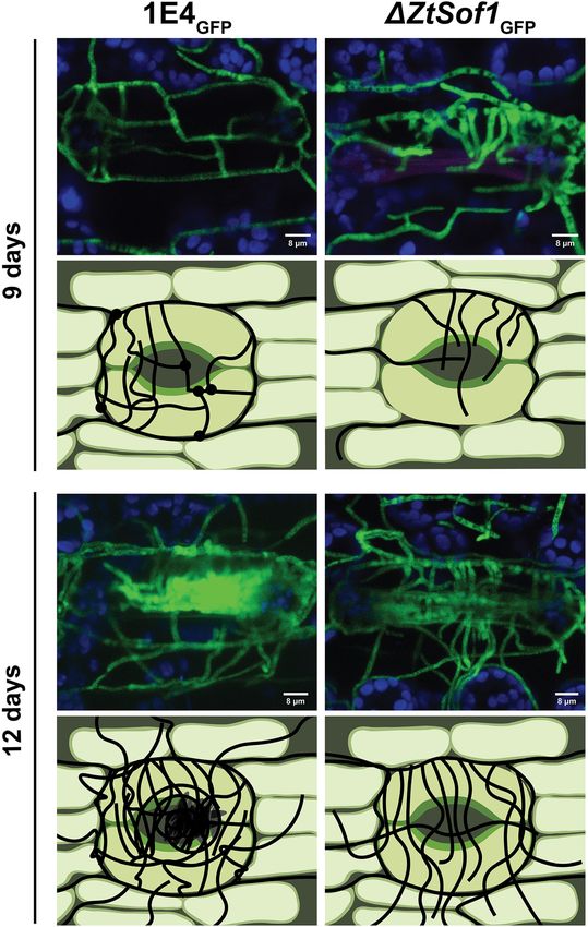
Francisco et al. BMC Biology   (2020) 18:99                                                                       Page 2 of 16

Background                                                        broaden their host specificity [19–21]. Albeit non-self-
Communication is a ubiquitous primitive characteristic            anastomoses are described [22, 23], this might be a rare
developed by all living species. The ability to communicate       event in nature.
effectively may affect mating, predation, competition,               In the last decades, different studies about the molecu-
dominance hierarchy, signal modalities, and survival [1–          lar mechanisms underlying cell fusion identified several
3]. This complex mechanism starts when a given organism           mutants defective in anastomosis, revealing that fungal
(the sender) secretes in the environment a self-produced          communication and fusion are complex mechanisms
molecular signal (the message) that alters the behavior of        that encompass several signaling pathways [6]. The best-
another organism (the receiver) [1, 3]. Communication             characterized mutant is the soft (So) gene of Neurospora
also happens at the cellular level. This so-called cell-to-       crassa [24]. So, it is proposed to be the scaffold protein
cell communication creates a complex signaling network            for some mitogen-activated protein kinase (MAPK) from
that involves different extracellular signals and distinct cell   the cell wall integrity (CWI) signaling pathway impli-
types that regulate several pathways [4–6]. Inter- and in-        cated in the regulation of different fungal processes [25–
traspecies cell-to-cell communication has been widely             27]. For instance, So contributes to septal plugging dur-
studied in fungi to address biological functions including        ing hyphal injury or damage caused by environmental
the secretion of pheromones to attract the opposite sexual        stresses [28, 29]. Nevertheless, So gene has an essential
partner, the production of quorum sensing molecules con-          role in the hyphal anastomosis, presumably by regulating
trolling the expression of virulence factors or morpho-           the secretion or perception of an undefined chemo-
logical changes, and the regulation of cell fusions during        attractant in an oscillatory manner with the Fus3 (ortho-
vegetative growth [6–9].                                          logous to MAK-2 in N. crassa) from the MAPK
   The fungal mycelium is formed by three integrated              pheromone response pathway, as demonstrated for N.
processes, including hyphal extension, branching, and             crassa [11]. Beyond N. crassa, the So was also character-
vegetative hyphal fusion (VHF) (also known as anasto-             ized in other model organisms, plant pathogens, and
mosis) [10]. In this last-mentioned process, two growing          endophytic fungi [15–17, 29, 30]. Though all So mutants
cells with identical vegetative compatibility loci engage         fail to undergo hyphal fusions, the distinct effects on
in cell-to-cell communication, which is thought to in-            pathogenicity reported for those null mutants suggest
volve the secretion of unknown diffusible molecules and           that the biological contribution of anastomosis might de-
results in re-direction of polarized hyphal growth toward         pend on the infection strategies developed by different
each other. After physical contact, the cell walls are re-        fungal pathogen species.
modeled, the plasma membranes fuse, and the two inter-               Zymoseptoria tritici is an apoplastic pathogen with a
connected cells exchange cytoplasm and organelles [11].           hemibiotrophic lifestyle and considered the most dam-
When the anastomosed individuals are vegetatively in-             aging pathogen of wheat in Europe [31]. This fungus has
compatible, the two fused cells rapidly collapse following        the ability to undergo morphological transitions in re-
DNA degradation by programmed cell death, or they are             sponse to the environment, switching between hyphal
severely inhibited in their growth [12]. It is widely ac-         growth and yeast-like growth [32–34]. Hyphae formed
cepted that mycelial network formed through VHF facil-            from either germinated ascospores (sexual spore), pycni-
itates the intra-hyphal communication, translocation of           diospores (asexual spores), or blastospores (asexual
water and nutrients, and signal molecules, which im-              yeast-like spores produced by budding) are essential for
prove the general homeostasis and spatial expansion of            penetrating wheat leaves through the stomata and
the fungal colony [13, 14]. In some fungi, hyphal fusion          colonization of the apoplastic space. After a long
is required for pathogenicity and host adhesion [15–17].          asymptomatic phase (which varies depending on the
Cell fusions can also occur between germinating conidia,          wheat genotype and fungal strain combination) [35–
the asexual spores of many fungi. The process of fusion           37], the onset of the necrotrophic phase is followed
between germinating conidia involves the formation and            by the appearance of lesions, disintegration of host
interaction of specialized hyphae, so-called conidial             tissue, and formation of asexual fruiting bodies.
anastomosis tubes (CATs). CATs are thinner and                    Though Z. tritici is among the top 10 most studied
shorter than vegetative hyphal fusions (VHFs), and its            phytopathogens [38], there is little known about vege-
induction is dependent on nutrient deprivation and ini-           tative cell fusion in this organism. To date, it was
tial cell density [18]. Cell fusions may serve to improve         shown that the deletion of the β-subunit of the het-
colony establishment, as well as to increase the genetic          erotrimeric G protein MgGpb1 or ZtWor1, a tran-
variability by facilitating heterokaryosis and parasexual         scriptional regulator of genes located downstream of
recombination [18]. Gene or chromosome transfers by               the cyclic adenosine monophosphate (cAMP) pathway,
cell fusions between individuals of the same or different         results in germ tubes that undergo extensive anasto-
species allow certain fungi to acquire pathogenicity or to        mosis [39, 40].
Francisco et al. BMC Biology   (2020) 18:99                                                                       Page 3 of 16

   In this study, we aimed to determine whether vegeta-          the generation of multinucleated cells in this uninu-
tive cell fusions play essential biological roles in the life-   cleated fungus.
style of Z. tritici. We showed that the ubiquitous ability
of Z. tritici to undergo self-fusion was disrupted by the        The mutual perception or response to genetically
deletion of ZtSof1, affecting both hyphal and germling           identical fusion partners requires the ZtSof1 gene and
fusions. The characterization of mutants lacking the             different MAPK pathways
MAPK-encoding ZtSlt2 or ZtFus3 indicates a conserved             So has an essential role in self-anastomosis [24]. To de-
role of the CWI and pheromone response pathways on               termine whether this gene plays the same role in Z. tri-
fungal anastomosis. We found that ZtSof1 contributes to          tici, we identified the So orthologous (ZtSof1) in the Z.
vegetative growth and is required for melanization, but          tritici genome (Mycgr3G74194 or Zt09_7_00503), which
not to maintain the fungal cellular integrity. We discov-        consists of a 3794-bp open reading frame that encodes a
ered that anastomoses are dispensable for pathogenicity,         polypeptide of 1227 amino acid and is widely distributed
but they are essential for asexual fruiting body develop-        within the Dothideomycetes (Additional file 3: Fig. S3).
ment. In the absence of cell fusions, Z. tritici does not        We phenotyped the ΔZtKu70, ΔZtSof1, and ΔZtSof1-
undergo asexual reproduction. These findings illustrate          comp mutants generated on the 1E4 strain genetic back-
the impact of ZtSof1 for fungal development and the im-          ground for the presence of interconnected individuals
portance of vegetative cell fusions for fungal fitness.          through anastomoses. Fusion bridges between blasto-
                                                                 spore germlings and filamentous hyphae were only ob-
Results                                                          served for those strains possessing the ZtSof1 gene, but
Cell fusions in Z. tritici allow the bidirectional transfer of   none between ΔZtSof1 mutant cells (Fig. 2a and b), dem-
cytoplasmic content but do not enable multinucleated             onstrating that ZtSof1 plays an essential role in anasto-
cell formation                                                   mosis between genetically identical Z. tritici strains.
We co-inoculated either blastospores or pycnidiospores              To ensure the failure of cytoplasmic exchange on
of both 1E4GFP and 1E4mCh fluorescent strains onto               those individuals lacking the ZtSof1 gene, we mixed blas-
water agar (WA—1% agar in water), a hyphal-inducing              tospores of each tested strain with 1E4GFP blastospores
medium, to investigate the ability of Z. tritici to undergo      in a 1:1 ratio and traced the spores up to 40 h. Hyphal
self-fusions. Though Z. tritici produces blastospores and        fusions and the continuous streaming of cytoplasmic
pycnidiospores as asexual spores instead of conidium,            green fluorescence coming from the fusion with the
we used the CAT terminology to define the fusion be-             1E4GFP strain were observed for all tested combinations
tween germinating spores. CATs formed between blasto-            (Additional file 4: Fig. S4, and Additional file 5: Fig. S5),
spores or pycnidiospores germlings happened at high              except for the combination of ΔZtSof1 and 1E4GFP
cell density (107 blastospores/mL), starting after 4 h of        spores (Additional file 6: Fig. S6). These findings con-
incubation, but they were frequently observed after 17 h         firmed that ZtSof1 is required in both fusion partners for
of incubation, resulting in cells co-expressing both fluor-      a mutual recognition previous to the anastomosis.
escent proteins (Fig. 1b). On the other hand, vegetative            To determine whether the CWI and pheromone re-
hyphal fusions (VHFs) from germinated blastospores or            sponse pathways also contribute to anastomoses in Z.
pycnidiospores were noticed at 40 h after incubation             tritici, we incubated the knocked-out ZtSlt2 (ortholo-
(hai) only at low cell density (106 blastospores/mL)             gous to MAK-1 in N. crassa) and ZtFus3 (orthologous to
(Fig. 1c, d). Multiple interconnections via fusion bridges       MAK-2 in N. crassa) mutants on WA plates. No hyphal
were observed in all tested morphotypes (Fig. 1b–d).             fusion was observed for both mutants. Unlikely, fusion
The co-infection of wheat plants using either blasto-            bridges were regularly found between IPO323 wild-type
spores or pycnidiospores of 1E4GFP and 1E4mCh strains            or ΔZtSlt2-complemented fungal cells (Additional file 7:
also resulted in VHFs on the wheat leaf surface (Add-            Fig. S7). These results indicate the recruitment of both
itional file 1: Fig. S1). Self-fusions and cytoplasmic mix-      CWI and pheromone response MAPK signaling cascades
ing occurred in the first 48 hai (Additional file 1a-b: Fig.     for the regulation of self-fusion in Z. tritici, as it has
S1a-b). To monitor for nucleus movement enabling mul-            been observed in N. crassa [11].
tinucleated cell formation, we used the IPO323 ZtHis1-
ZtGFP strain [41]. As previously reported, only one nu-          Deletion of ZtSof1 affects growth and melanization
cleus per cell was observed in this strain. No multinucle-       To assess whether vegetative cell fusions affect fungal
ated septal compartments were observed between the               development, we determined the radial growth of
two interconnected hyphae at the fusion bridges or out-          ΔZtKu70, ΔZtSof1, and ΔZtSof1-comp on different nutri-
lying of the fusion points (Additional file 2: Fig. S2).         tional environments inducing different morphotypes.
These findings suggest that cell fusions culminate in            We observed a slightly reduced growth of ΔZtSof1 col-
cytoplasmic mixing in Z. tritici, but it does not lead to        onies when grown on WA plates, a condition that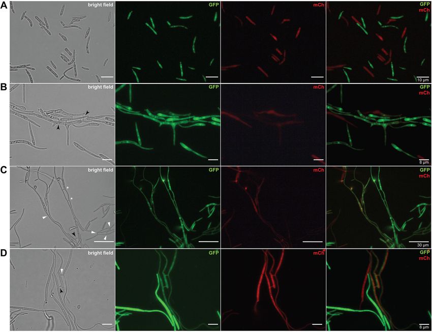
Francisco et al. BMC Biology       (2020) 18:99                                                                                               Page 4 of 16

 Fig. 1 Vegetative cell fusions of Zymoseptoria tritici. a Blastospores of a GFP- or mCh-tagged 1E4 strain appeared only in a one-color channel of the
 fluorescence microscope. b High initial cell density (1 × 10⁷ blastospores/mL) induced conidial anastomosis tubes (CATs) that were frequently observed
 after 17 h of incubation. c, d Vegetative hyphal fusions (VHFs) from germinating blastospores or pycnidiospores of a GFP- or mCh-tagged 1E4 strain
 were induced at lower initial cell density (1 × 10⁶ blastospores/mL) and were noticed after 40 h of incubation. Cell fusions may result in the continuous
 mixing of cytoplasmic content of individuals expressing GFP or mCh fluorescent proteins. Black arrows or white triangles point to the CATs or VHFs,
 respectively. White asterisks point to the fusion bridges formed between individuals expressing identical fluorescent proteins

induces hyphal growth, compared to those possessing                            comp were respectively 32% and 16% lower than the
the ZtSof1 gene (Additional file 8a: Fig. S8a). On average,                    ΔZtSof1 mutant. No morphological differences were de-
the colony radii were 5.31 ± 0.12 for ΔZtKu70, 4.87 ±                          tected between blastospores produced by the tested
0.10 for ΔZtSof1, and 5.50 ± 0.09 for ΔZtSof1-comp [ra-                        strains (Additional file 8d: Fig. S8d). The data suggest
dial growth (mm) ± standard error] (Additional file 8c:                        that ZtSof1 mutation may affect fungal growth in a
Fig. S8c). Nevertheless, ΔZtKu70 and ΔZtSof1-comp                              morphotype-dependent manner. Consistent with these
generated colonies with highly hyphal dense margins,                           findings, we found that the deletion of ΔZtSof1 increased
while ΔZtSof1 exhibited sparse filamentations at the col-                      blastosporulation in a nutrient-rich liquid medium. At
ony periphery (Additional file 8b: Fig. S8b). Unlikely, the                    48 and 72 hai, the ΔZtSof1 mutant produced a signifi-
ΔZtSof1 mutant grew significantly faster than the                              cantly higher amount of blastospores than ΔZtKu70 and
ΔZtKu70 and ΔZtSof1-comp on the nutrient-rich PDA                              ΔZtSof1-comp strains (Additional file 9: Fig. S9).
medium, a condition that induces blastosporulation                               Interestingly, no melanin accumulation was observed
(yeast-like growth) (Additional file 8e: Fig. S8e). Over                       in ΔZtSof1 mutant colonies (Fig. 3), demonstrating the
time, the relative growth rate of ΔZtKu70 and ΔZtSof1-                         impact of ZtSof1 deletion on Z. tritici pigmentation. We
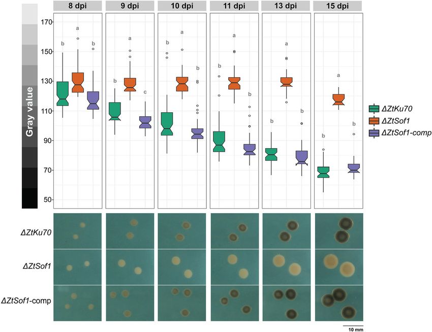
Francisco et al. BMC Biology      (2020) 18:99                                                                                            Page 5 of 16

 Fig. 2 ZtSof1 is required for vegetative cell fusion in Zymoseptoria tritici. All mutant lines derived from the 1E4 strain. a Fusion bridges between
 blastospore germlings were observed at high cell density for ΔZtKu70 and ΔZtSof1-comp strains but not in the ΔZtSof1 mutant. Black arrows
 indicate CATs. b VHFs were noticed at low cell density and only for those strain possessing the ΔZtSof1 gene. White triangles point to the fusion
 bridges between two fused hyphal cells

 Fig. 3 Disruption of ZtSof1 impacts the melanization of Zymoseptoria tritici. The defective fusion mutant was significantly less melanized than the
 ΔZtKu70 and ΔZtSof1-comp strains, which exhibited higher melanin accumulation over time. Bars represent standard errors of the mean gray
 values on at least 40 colonies. Different letters on the top of the bars indicate a significant difference among the tested strains according to the
 analysis of variance (ANOVA). The notch displays a 95% confidence interval of the median. Open circles represent the outlier values of each strain.
 Pictures shown below the bar plot represent the melanization level of ΔZtKu70, ΔZtSof1, and ΔZtSof1-comp strains. Gray value scale (0 = black
 and 255 = white) is shown on the left
Francisco et al. BMC Biology      (2020) 18:99                                                                                            Page 6 of 16

 Fig. 4 ZtSof1 is dispensable for cellular integrity. A serial dilution of blastospore suspensions of ΔZtKu70, ΔZtSof1, and ΔZtSof1-comp strains were
 exposed for 5 days to nine different stress conditions, including different temperatures (18 °C and 27 °C), oxidative stress (0.5 and 1 mM of
 hydrogen peroxide (H2O2)), osmotic stresses (1 M sodium chloride (NaCl) and 1 M sorbitol), cell wall stresses (2 mg/mL Congo red and 10 μg/mL
 Calcofluor white (CFW)), and plasma membrane stress (0.01% sodium dodecyl sulfate (SDS)). The tested strains do not vary on their tolerance to
 different cellular stressors

postulated that the ΔZtSof1 mutant could either display                      Hyphal fusions are essential for the development of
cellular integrity defects and/or being susceptible to en-                   asexual fruiting bodies
vironmental stresses. We tested nine different abiotic                       We inoculated a susceptible wheat cultivar with the
stressors, such as temperature, oxidative, osmotic, cell                     tested Z. tritici strains to assess the biological role of
wall, and cell membrane stresses. Overall, no variability                    vegetative cell fusion during the pathogen lifecycle in
in stress responses was noticed among the strains (Fig. 4).                  planta. Typical symptoms caused by Z. tritici infections
However, ΔZtSof1 formed slightly bigger colonies than                        were visible after 11 days post-infection (dpi), and the
those from ΔZtKu70 and ΔZtSof1-comp, corroborating                           disease progression was similar among the plants inocu-
the increased growth rate observed for this mutant on                        lated with ΔZtKu70, ΔZtSof1, or ΔZtSof1-comp strains
nutrient-rich medium (Additional file 8e: Fig. S8e).                         (Fig. 5a and Additional file 10: Fig. S10). This result indi-
Therefore, we found no evidence that the ZtSof1 gene is                      cates that ZtSof1 is neither required for host penetration
involved in the maintenance of the cell wall integrity of                    nor for the asymptomatic or necrotrophic phases of the
Z. tritici.                                                                  fungus. The asexual fruiting bodies (pycnidia) were
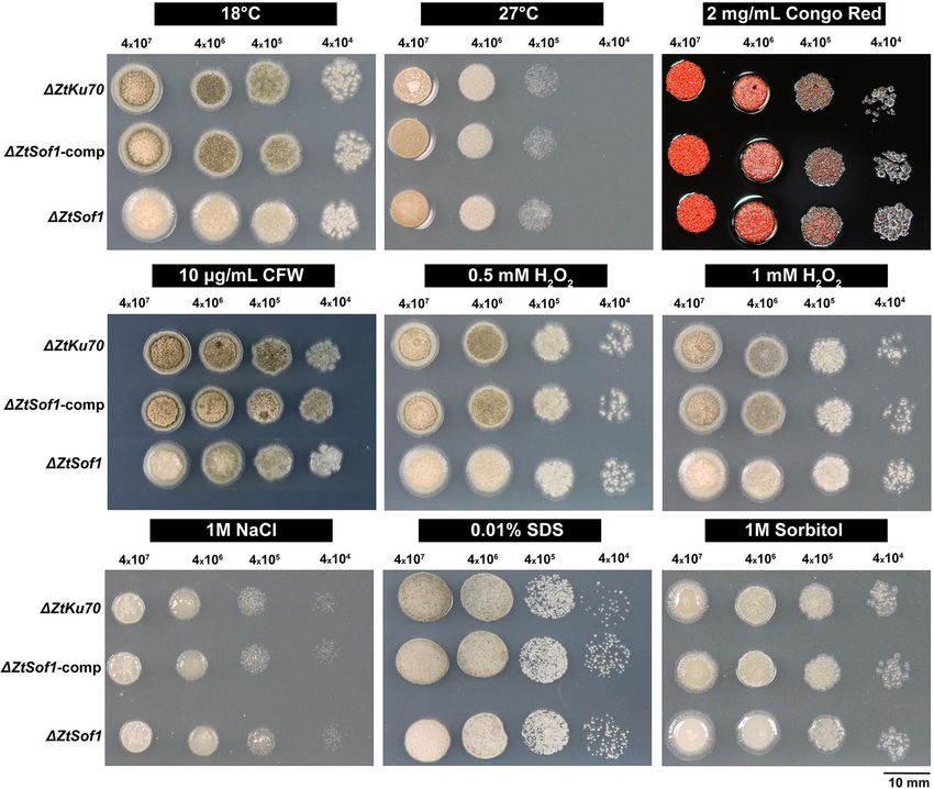
Francisco et al. BMC Biology      (2020) 18:99                                                                                           Page 7 of 16

 Fig. 5 The role of vegetative hyphal fusions for disease progression and pycnidial development. a Susceptible wheat cultivar Drifter inoculated
 with ΔZtKu70, ΔZtSof1, or ΔZtSof1-comp strains were evaluated up to 21 days post-infection (dpi). All tested strains exhibited similar disease
 progression, including the onset of the necrotrophic phase at 11 dpi. b Pycnidia production on wheat extract agar. After 20 days of incubation,
 ΔZtKu70 and ΔZtSof1-comp strains produced brown pycnidium-like structures exuding a whitish liquid similar to the oozed cirrhus-containing
 pycnidiospores spores observed for Z. tritici-infected wheat plants. In contrast, ΔZtSof1 mutant formed mycelial knots, but those structures never
 developed in mature asexual fruiting bodies

visible on plants inoculated with those strains possessing                   structures formed by ΔZtKu70 or ΔZtSof1-comp strains
the ZtSof1 gene at 14 days post-inoculation (dpi). In con-                   were exuding a whitish liquid containing pycnidiospores
trast, plants infected with ΔZtSof1 never developed pyc-                     (Fig. 5b). Next, we evaluated the pycnidium formation in
nidia. The failure to undergo asexual reproduction was                       planta. The wheat plants infected with 1E4GFP or
observed in a broad range of wheat genotypes (Add-                           1E4GFPΔZtSof1 strains were monitored using confocal
itional file 11: Fig. S11).                                                  microscopy up to 12 dpi. We observed mainly host
   To distinguish whether pycnidium formation would be                       penetration, initial intercellular hyphal extension, and
a consequence of the lack of hyphal fusions or suscepti-                     sub-stomatal colonization at the earlier stages of the
bility of ΔZtSof1 cells to defense compounds produced                        plant infection (Additional file 12: Fig. S12 – 6 and 7
by the plant, we used a wheat extract agar medium to                         dpi). No difference was noticed in fungal development;
induce pycnidium formation in vitro. ΔZtSof1 aggregated                      however, hyphal fusions established during epiphytic
into mycelial knots, which is the initial developmental                      host colonization were only detected for 1E4GFP strain.
stage of the pycnidium, but no asexual reproductive                          At 8 and 9 dpi, we observed that the primary intercellu-
structures were further developed. The pycnidium-like                        lar hyphae surrounding the stomatal guard cells
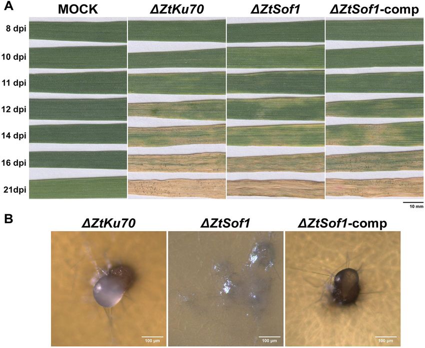
Francisco et al. BMC Biology     (2020) 18:99                                                                                           Page 8 of 16

 Fig. 6 Confocal microscopy images and schematic demonstration of pycnidial development during plant infection. Susceptible wheat cultivar
 Drifter inoculated with the fluorescent 1E4GFP (wild-type, left) or ΔZtSof1GFP (right) strains were monitored by confocal microscopy at different
 days post-infection (dpi). For 1E4GFP, the primary intracellular hyphae surrounding the stomatal guard cells produced specialized knots from
 where secondary hyphae emerge and elongate. After 9 days of infection, these secondary hyphae fuse with another nearby hypha (represented
 by black circles), creating an interconnected network in the sub-stomatal cavity. The combination of sub-stomatal hyphal accumulation and
 anastomoses generates the pre-pycnidium at 12 days, which later supports the asexual reproduction of Z. tritici. For the ΔZtSof1GFP mutant, the
 filamentous hyphae kept extending as individual hyphae, and no fusion points were observed. The lack of anastomosis stops the developmental
 process of the pycnidium formation. For earlier time points (6, 7, and 8 dpi), please see Additional file 12: Fig. S12

produced specialized knots from where secondary hy-                         Fig. S12). The combination of sub-stomatal hyphal accu-
phae emerged and elongated (Fig. 6 and Additional file 12:                  mulation and anastomoses generates the mature pycnid-
Fig. S12). These secondary hyphae fused with other                          ium, which supports the asexual reproduction of Z.
nearby hyphae, creating an interconnected network in                        tritici. On the other hand, the lack of anastomosis
the sub-stomatal cavity (e.g., for the 1E4GFP strain) or                    stopped the symphogenous development by affecting the
they kept extending as individual hyphae (e.g., for the                     interweaving of hyphal branches through hyphal fusion,
1E4GFPΔZtSof1 mutant) (Fig. 6 and Additional file 12:                       a crucial mechanism for the formation of pycnidia, and
Francisco et al. BMC Biology   (2020) 18:99                                                                      Page 9 of 16

hence, impairing the asexual cycle of the fungus. There-         question of whether anastomosis would enable multinu-
fore, we concluded that hyphal fusions are decisive for          cleated cell formation in Z. tritici. We showed that the
the pycnidial development, and the disturbance of this           new cell compartment is occupied by a migrating nu-
mechanism ceases the asexual reproduction of Z. tritici.         cleus coming from a neighboring nucleus divided by mi-
                                                                 tosis after the fusion bridge between two identical Z.
Discussion                                                       tritici cells. However, cells containing multiple nuclei
The evolution of the fungal language allowed a fine-tune         were not observed neither near nor far from the anasto-
coordination of signal senders and receivers driving their       mosis point, indicating that Z. tritici may have evolved
ecological diversifications and, consequently, the regula-       to limit the spread of genetic elements and restrict the
tion of complex signaling networks. Here, we explored            formation of multinucleated cells. Hence, we cannot dis-
the functional relationship of a gene involved in cell-to-       card that multinucleated, homokaryotic, or heterokary-
cell communication and its biological contribution to            otic cells can be formed at low frequency in nature.
the development and fitness of a fungal plant pathogen.          These properties could impact the evolution of the
   Vegetative cell fusion is one of the most important de-       pathogen, and therefore, additional studies will be
velopmental processes of a mycelial fungal colony [10,           needed to underlie the roles played by the genetic ex-
42]. The cytoplasmic continuity generated by cell fusion         change during vegetative cell fusions in Z. tritici.
provides adaptative advantages to the interconnected                In the last decades, the identification of fusion-
mycelial network by allowing resource sharing and intro-         defective mutants has contributed to the understanding
gression of genetic material [19, 42, 43]. CATs forming          of the molecular mechanisms underlying cell communi-
at earlier stages of the vegetative growth have been re-         cation and fusion, especially by unveiling the interplay of
ported in several filamentous fungi [18]. We observed            the mitogen-activated protein kinase (MAPK) pathways
that these specialized fusion bridges require a specific         during chemotropic interactions [11]. MAPK pathways
cell density, indicating that Z. tritici may induce CATs         are involved in extracellular signal perception and regu-
at a critical concentration of a self-produced molecule,         lation of diverse genes essential for mating, filamenta-
presumably a quorum-sensing molecule. For instance,              tion, pathogenicity, cell integrity, and stress responses
Fusarium oxysporum secretes and senses both a- and α-            [50–55]. Therefore, the deletion of MAPK-encoding
pheromone, and their perception regulates spore ger-             genes results in pleiotropic phenotypes due to their in-
mination in a cell density-dependent manner [44]. On             volvement in multiple biological processes. Though fun-
the other hand, the induction of VHFs was often ob-              gal communication and fusion require the regulation of
served at lower cell density, indicating that VHFs may be        several genes [6], the cross-talk between cell wall integ-
induced at low self-produced molecule concentration or           rity (CWI) and pheromone response, two conserved
some other environmental signal. Z. tritici is a pleo-           MAPK signaling pathways, is essential to produce, se-
morphic fungus changing growth morphology according              crete, and sense the chemoattractant molecule produced
to the environment. Nutrient-limited conditions induce           during cell fusion [11]. The deletion of Slt2 or Fus3
both hyphal growth and fusions. Plant pathogens typic-           orthologous genes disrupts the signaling cascade affect-
ally experience nutrient limitations while growing on            ing self-anastomosis in filamentous fungi. We used the
leaves, and the perception of a nutrient-limited environ-        ZtSlt2 and ZtFus3 to demonstrate the functional conser-
ment may act as a stimulus to induce interconnected              vation of these MAPK pathways in Z. tritici. ZtSlt2 and
mycelial formation in foliar plant pathogens, as observed        ZtFus3 are known as essential genes for the pathogen-
for the causal agent of anthracnose disease, Colletotri-         icity and developmental processes of Z. tritici, including
chum lindemuthianum [45]. Both germinating blasto-               vegetative growth, melanization, and pycnidium forma-
spores or pycnidiospores underwent CATs and VHFs                 tion [56, 57]. Here, we showed that both ΔZtSlt2 and
in vitro and in planta, supporting our previous observa-         ΔZtFus3 mutants were unable to undergo anastomosis.
tion [34]. Therefore, we demonstrated that vegetative            Our results indicate that cell fusions in Z. tritici follow
cell fusions are a ubiquitous cellular process, which may        the same signaling mechanism described for N. crassa
have an essential contribution to the lifestyle of Z. tritici.   [11, 58], where the signal sending and receiving are co-
   Nucleus transfer between two encountering hyphae              ordinated by genes associated with the CWI and phero-
occurs in some fungal species [46, 47]. The conse-               mone response pathways. Besides, this is the first report
quences of genetic exchange include the formation of vi-         of those pathways regulating cell communication in Z.
able heterokaryons and the risk of introgression of              tritici.
pathogenic elements or virulence genes [20, 48, 49]. Un-            To evaluate the impact of vegetative cell fusion in the
like the majority of fungal species that form a multinu-         biology of a hemibiotrophic fungus, we used the ZtSof1
cleated hyphal network [47], Z. tritici has only one             orthologous of the N. crassa So gene. Contrary to the
nucleus per septal compartment [41], which raises the            MAPK-related genes, the characterization of So
Francisco et al. BMC Biology (2020) 18:99 Page 10 of 16 orthologs results in low pleiotropy [15, 16, 24]. Filament- [60]. Thereby, we cannot discard that the deletion of ous fungi lacking So gene were impaired in self- ZtSof1 may affect the cross-talk between CWI with other anastomosis [15–17, 24], including Z. tritici, in which signaling cascades impairing fungal melanization. Fur- the deletion of ZtSof1 abolished vegetative cell fusion. ther investigations are needed to elucidate both We characterized the impact of the fusion deficiency at hypotheses. different developmental stages of Z. tritici. For instance, Melanin is postulated to contribute to fungal protec- the ΔZtSof1 mutant exhibited a more asymmetrical hy- tion against fungicide and environmental stresses in Z. phal extension in a nutrient-poor medium inducing hy- tritici [59, 60], and its regulation depends on environ- phal growth than the fusion-competent individuals. It mental cues and colony development [60], though it is was demonstrated that the direction of the nutrient dis- not yet a fully understood mechanism. We used nine dif- tribution occurs from the central part of the mycelium ferent cellular stressors to evaluate whether (i) the defect to outwards, and its streaming speed is driven by anasto- in melanin accumulation or (ii) the deletion of ZtSof1, mosis [42]. Thereby, the reduced colony extension ob- the scaffold protein for the MAPK genes from the CWI served for the ΔZtSof1 may be a consequence of the pathway, affects pathogen stress tolerance. Since the irregular distribution of cytoplasmic content throughout non-melanized ΔZtSof1 mutant displayed the same de- the mycelial colony. In contrast, the induced blastospor- gree of stress sensitivity than ΔZtKu70 and ΔZtSof1- ulation and larger yeast-like colonies exhibited by the comp strains, we concluded that ZtSof1 does not act as a fusion-defective mutant in nutrient-rich environments scaffold protein for all CWI pathway functions. Consist- may be a consequence of the disruption of the CWI ent with our findings, the model fungus Sordaria macro- pathway or its cross-talk with other MAPK pathways in- spora, the PRO40 (orthologous to ZtSof1), operates as a volved in fungal growth. The genetic relationship be- scaffold for the CWI-encoding genes during fungal de- tween vegetative growth and the ZtSof1 gene remains to velopment, hyphal fusion, and stress response, but not be elucidated. for growth under cell wall stress agents [25]. Our results We showed that the ΔZtSof1 colonies do not accumu- showed that melanin accumulation of Z. tritici does not late melanin, resulting in whiter and larger colonies than benefit fungal survival in harsh environments, at least those formed by ΔZtKu70 and ΔZtSof1-comp strains. for the stressful conditions tested in this study. These findings suggest a possible trade-off between en- We demonstrated that VHFs are dispensable for the ergy cost for pigment production and growth. The re- pathogenicity of Z. tritici. The fusion-defective ΔZtSof1 duction of fungal growth caused by the higher mutant displayed a similar host damage progression than accumulation of melanin was reported before for Z. tri- those individuals possessing the gene. This finding ex- tici [59]. Melanins are dark-pigmented secondary metab- emplifies the distinct effects of cell fusions on fungal olites often associated with the fungal cell walls. Though pathogenicity. For instance, for the soil-borne Fusarium fungi can produce different kinds of melanins, it is sug- oxysporum, VHF-impaired mutants exhibited only a gested that melanization of Z. tritici is only controlled slightly reduced virulence, whereas for the necrotrophic by the polyketide synthase (PKS) gene cluster containing plant pathogen Alternaria alternata, VHFs are necessary catalytic enzymes and transcription regulators of the 1,8- for the full virulence of the fungus [15, 16]. Though the dihydroxynaphthalene (DHN) melanin [59, 60]. It was deletion of ZtSof1 is not essential for host penetration, demonstrated for plant pathogenic fungi that the dele- colonization, or for the onset of the necrotrophic phase tion of CWI-associated genes inhibited pigmentation by per se, it is during hyphal accumulation in the sub- reducing the expression of DHN melanin biosynthetic stomatal cavity that the fusion defect impacts Z. tritici genes [61–64]. Therefore, the deletion of ZtSof1 may im- fitness. The infection process during wheat colonization pair the CWI-regulatory cascade and, consequently, the was microscopically detailed before for Z. tritici [70]; regulation of DHN-melanin production, resulting in the however, the study did not explore the contribution of lack of pigmentation observed for both ΔZtSof1 and VHFs. Here, we showed that the primary intercellular ΔZtSlt2 [56] mutants. On the other hand, the CWI, hyphae surrounding the stomatal guard cells produced high-osmolarity glycerol (HOG), cyclic adenosine mono- specialized knots from where secondary hyphae emerge phosphate (cAMP), and the pheromone response path- and elongate to fuse with other adjacent hyphae. Hence, ways can interact regulating melanization in a the preliminary hyphal network creates the basis for a cooperative manner [65–67]. Consistent with the find- symphogenous development that builds the concave- ings in other fungi, mutants in these signaling pathways, shaped pycnidial wall of the mature pycnidium. On the including ΔZtFus3, display altered pigmentation in Z. other hand, the inability to undergo anastomosis ceases tritici [33, 39, 56, 57, 68, 69]. Additionally, a QTL map- the development of the asexual fruiting bodies at this ping study identified ZtSlt2, ZtHog1, ZtGpa1, and stage, and therefore abolishes fungal reproduction. The ZtFus3, contributing to the melanization of this fungus accumulation of hyphae observed in the sub-stomata
Francisco et al. BMC Biology   (2020) 18:99                                                                     Page 11 of 16

chamber by the ΔZtSof1 mutant confirms that a specific          strain was stored in glycerol at − 80 °C until required
signal triggers sub-stomatal hyphal aggregation inde-           and then recovered in yeast-sucrose broth (YSB)
pendent of the putative chemoattractant molecule se-            medium (10 g/L yeast extract, 10 g/L sucrose, 50 μg/mL
creted by the fungus to induce hyphal fusion. In line           kanamycin sulfate; pH 6.8) incubated at 18 °C for 4 days.
with this hypothesis, a recent study showed that the dis-
ruption of the transcription factor ZtStuA impairs hy-          Plant infection to obtain fluorescent pycnidiospores
phal aggregation, resulting in the lack of pycnidium            Wheat seedlings from the susceptible wheat cultivar
formation in vitro and in planta [68]. Therefore, we sug-       Drifter were grown for 16 days in the greenhouse at
gest that ZtStuA acts regulating genes necessary for the        18 °C (day) and 15 °C (night) with a 16-h photoperiod
pre-pycnidium stages (e.g., hyphal aggregation) [68],           and 70% humidity. Blastospore suspensions of 1E4GFP or
while ZtSof1 controls the hyphal fusions required for the       1E4mCh were obtained after 4 days of growth in the YSB
formation of the mature pycnidium. The initiation of the        medium. Spore suspensions were adjusted to a final con-
asexual sporulation may depend on the regulatory genes,         centration of 106 blastospores/mL in 30 mL of sterile
such as ZtBrlA2 and ZtFlbC, as described by Tiley et al.        water supplemented with 0.1% (v/v) Tween and applied
Moreover, the inability of ΔZtFus3 to undergo VHFs              to run-off using a sprayer, and the plants were kept for
and produce pycnidia [57] confirms the interplay be-            3 days in sealed plastic bags, followed by 21 days in a
tween the CWI and pheromone response pathways dur-              greenhouse. Leaves with pycnidia were harvested and
ing hyphal fusion, a developmental process essential for        transferred to a 50-mL Falcon tube containing sterile
the asexual reproduction of Z. tritici. Fleissner et al. [24]   water and gently shaken to harvest the pycnidiospores.
demonstrated that the deletion of So in N. crassa also          Fluorescent pycnidiospores were used to assess the cell
affects female fertilization, blocking the sexual               fusion events during in vitro and in vivo growth. Pycni-
reproduction of the mutant. Further experiments need            diospore suspension-tagged GFP or mCherry were also
to be performed to address this question for Z. tritici,        adjusted to a final concentration of 106 pycnidiospores/
but, likely, the ZtSof1 gene may also play a crucial role       mL, and a new batch of plants was inoculated as de-
in the sexual reproduction of this pathogen.                    scribed above. Plants co-infected by both 1E4GFP and
                                                                1E4mCh strains were used to observe VHFs on the wheat
Conclusion                                                      leaf surface.
The characterization of the ZtSof1 gene demonstrated its
fundamental role in fungal biology. Beyond the impact           Characterization of cell fusion events in vitro and in vivo
of ZtSof1 for self-fusion, we show the contribution of          The ability of Z. tritici to undergo cell fusions was evalu-
this gene for fungal development, including vegetative          ated using blastospores and pycnidiospores of 1E4GFP
growth and melanization. Besides, we demonstrated that          and 1E4mCh. Cell concentrations were adjusted to 3.3 ×
VHFs are dispensable for pathogenicity, but essential for       107 blastospores/mL or 3.3 × 10⁶ blastospores/mL to in-
pycnidial development, and these mechanisms might be            duce CATs or VHFs, respectively. Three hundred micro-
controlled by the interplay between the CWI and the             liters of each morphotype and fluorescence was plated
pheromone response pathways. Our data show how cell             on WA to create a ratio of 1:1 and to provide a final
fusion affects Z. tritici fitness and provides a new gene       concentration of 107 blastospores/mL or 106 blasto-
target to control septoria tritici blotch (STB) disease.        spores/mL. A section of about 1 cm2 of agar was aseptic-
                                                                ally cut and placed on a microscope slide. The mixing of
Methods                                                         both cytoplasm contents through CATs or VHFs was
Strains and growth conditions                                   checked up to 40 hai using a Leica DM2500 fluorescent
The Swiss Z. tritici strain ST99CH_1E4 (abbreviated as          microscope with LAS v.4.6.0 software. GFP excitation
1E4), described by Zhan et al. [71], and mutant lines de-       and emission was at 480/40 nm and 527/30 nm, respect-
rived from this strain were used in this study. 1E4 strains     ively, whereas mCherry was excited at 580/20 nm and
expressing cytoplasmic GFP (1E4GFP) or mCherry                  detected at 632/60 nm.
(1E4mCh) were provided by Andrea Sanchez-Vallet after              VHFs during spore germination on wheat leaf surfaces
being generated by Sreedhar Kilaru and Gero Steinberg.          were obtained via confocal images using a Zeiss LSM
The knocked-out ΔZtSlt2 [56] and ΔZtFus3 [57] mu-               780 inverted laser scanning microscope with ZEN Black
tants were provided by Marc-Henri Lebrun (National In-          2012 software. An argon laser at 500 nm was used to ex-
stitute of Agricultural Research – INRA, France).               cite GFP fluorescence and chloroplast autofluorescence,
Because the MAPK mutants were generated in the gen-             while mCherry excitation was at 588 nm. The emission
etic background of IPO323 [72], this strain was also used       wavelength was 490–535, 624–682, and 590–610 nm for
as a control. The strain IPO323 ZtHis1-ZtGFP [41] was           GFP, chloroplast autofluorescence, and mCherry, re-
provided by Gero Steinberg (Exeter University). Each            spectively. Plants co-inoculated with blastospores or
Francisco et al. BMC Biology (2020) 18:99 Page 12 of 16
pycnidiospores of 1E4GFP and 1E4mCh strains were medium as mentioned earlier and incubated in the dark
checked up to 48 hai. at 18 °C. At least 40 colonies formed in five independent
PDA plates were photographed from the bottom using a
Plasmid constructions and transformations standardized camera setting [60] at 8, 9, 10, 11, 13, and
Primers used for cloning, sequencing, and knock-out 15 days post-incubation (dpi). Digital images were proc-
confirmations are listed in Table S1. DNA assemblies essed using a macro developed in the ImageJ software
were conducted with the In-Fusion HD Cloning Kit [74], which scores the area of individual colonies in the
(Takara BIO) following the manufacturer’s instructions. images. Fungal growth was obtained by converting the
The plasmid constructions to generate the three mutants colony area into radial growth (mm) based on the for-
rffiffiffiffiffi
in the 1E4 genome background and used in this study A
(ΔZtKu70, ΔZtSof1, and ΔZtSof1-comp) are described in mula ¼ . Radial growth values were plotted in a box-
π
Additional file 13: Fig. S13. The pES1-ΔZtSof1 construc- plot graphic using the ggplot2 package from R [75].
tion was also used to knock out the ZtSof1 gene in the Analysis of variance (ANOVA) was performed to deter-
1E4GFP genome background, enabling the visualization mine the differences in fungal growth among the strains
of the GFP-tagged mutant during host infection. using the agricolae package in R [76]. The radial growth
Z. tritici 1E4 strain was transformed by Agrobacterium rate (mm/day) for each strain was measured by plotting
tumefaciens-mediated transformation (ATMT) accord- the colony radius over time, which fitted to a linear
ing to Meile et al. [73]. The knock-out of the target model (Pearson’s correlation coefficient value (r2 ≥
genes was verified by a PCR-based approach using a for- 0.98)). The relative growth rate was calculated by divid-
ward primer specific to the upstream sequence of the ing the slope of the regression line of ΔZtSof1 by the
disrupted gene and a reverse primer specific to bind in slope of ΔZtKu70 or ΔZtSof1-comp strains. To measure
the resistance cassette (Additional file 14: Table S1). We mycelial growth on WA, we calculated the mycelial
determined the copy number of the transgene by quanti- diameter from digital images of at least 40 colonies
tative PCR (qPCR) on genomic DNA extracted with the formed in five independent Petri dishes at 15 dpi and
DNeasy Plant Mini Kit (Qiagen). We used qPCR target using ImageJ software [74]. The mycelial diameter values
gene as the selection marker and the 18S rDNA as the were divided by two to generate the radial growth (mm)
reference gene (Additional file 14: Table S1). Lines with values.
a single insertion were selected for further experiments. The effect on the blastosporulation was assessed by
adding each tested Z. tritici strain at an initial concen-
Phenotypic characterizations tration of 104 blastospores/mL into YSB and incubated
For all phenotypic analyses, ΔZtKu70 was considered the at 18 °C. Aliquots of each flask were taken every 24 h for
wild-type (WT) strain. To pinpoint the role of the ZtSof1 4 days, and the blastospore concentration was calculated
gene on the vegetative cell fusion, we added blastospore using a KOVA cell chamber system (KOVA Inter-
suspension cells of ΔZtKu70, ΔZtSof1, and ΔZtSof1- national Inc., USA).
comp to a final concentration of 106 blastospores/mL or The degree of melanization was estimated from at least
107 blastospores/mL into WA and incubated at 18 °C to 40 colonies formed on PDA plates. We used a macro de-
induce CATs or VHFs, respectively. For the MAPK veloped in ImageJ [74], which scores the mean gray value
ΔSlt2 and ΔFus3, IPO323 and ΔSlt2-complemented of each colony. Gray values range from 0 to 255, with 0
strains, WA plates were inoculated only at a final con- representing black and 255 representing white. The mean
centration of 106 blastospores/mL. Cell fusion events gray values of each strain over time were plotted in a box-
were monitored up to 40 hai by light microscopy. Be- plot using ggplot2 package from R [75].
cause fusion bridges were not observed between individ- The impact on the cell integrity caused by the deletion
uals lacking ΔZtSof1, we mixed 150 μL of 106 of ΔZtSof1 was verified by exposing the blastospores of
blastospores/mL of ΔZtKu70, ΔZtSof1, or ΔZtSof1-comp ΔZtKu70, ΔZtSof1, and ΔZtSof1-comp to nine different
strains with the same concentration of 1E4GFP blasto- stress conditions, including different temperatures (18 °C
spores in a ratio of 1:1 to confirm the failure of cytoplas- and 27 °C), oxidative stress (0.5 and 1 mM of hydrogen
mic streaming. At least 50 spores of each sample peroxide (H2O2)), osmotic stress (1 M sodium chloride
combination were monitored. (NaCl) and 1 M sorbitol), cell wall stress (2 mg/mL
To test for altered fungal growth, we used PDA (39 g/ Congo red and 10 μg/mL Calcofluor white (CFW)), and
L potato dextrose agar, 50 μg/mL kanamycin sulfate) or plasma membrane stress (0.01% sodium dodecyl sulfate
WA media to induce blastospore or hyphal growth, re- (SDS)). Spore suspensions of each strain were serial di-
spectively. Two hundred microliters of spore suspension luted to 4 × 10⁴, 4 × 10⁵, 4 × 10⁶, and 4 × 10⁷ blasto-
of ΔZtKu70, ΔZtSof1, and ΔZtSof1-comp was plated at a spores/mL, and drops of 3.5 μL were plated on five
final concentration of 2 × 10² blastospores/mL on eachFrancisco et al. BMC Biology (2020) 18:99 Page 13 of 16
independent PDA plates amended with the mentioned concentration of 106 blastospores/mL and incubated at 18°C. After 72
stresses and incubated at 18 °C. Colony phenotypes were hours of incubation, none septum containing more than one nucleus
assessed by digital images taken at 5 dpi. was observed neither at hyphal bridges nor distant of the fusion point.
Black triangles indicate the septal compartment containing only one nu-
cleus at the fusion bridges.
Virulence assay and pycnidium formation in vitro Additional file 3: Figure S3. Scheme showing the phylogenetic
Seeding, greenhouse and plant growth conditions, inocu- relationship of the So gene within Ascomycete species. The so gene
sequence from Neurospora crassa (XM_958983.3) was blasted against the
lum preparation, and plant inoculation followed the pro- Z. tritici genome (https://genome.jgi.doe.gov/Mycgr3/Mycgr3.home.html)
cedures described by Meile et al. [73]. To estimate the to identify its orthologous in this fungus. The Z. tritici So orthologous
percentage of leaf covered by lesions (PLACL) and pyc- protein sequence was used for a Blastp analysis against the NCBI
database (National Center of Biotechnology Information). Blastp searches
nidium formation, we harvested the second leaves of at expected value homology cut-off of 1e-10 were included as positive. A
Drifter plants inoculated with ΔZtKu70, ΔZtSof1, or dataset containing So orthologous proteins of different Ascomycete spe-
ΔZtSof1-comp strains at 8, 10, 11, 12, 14, 16, and 21 dpi. cies were used for phylogenetic analysis. Protein sequences were aligned
using the AliView program [78]. The best-fit model of amino acid evolu-
Leaves were mounted on a paper sheet, scanned with a tion was the LG+G, determined by Mega6 software [79]. Amino acid se-
flatbed scanner, and analyzed using automated image quences were aligned using Muscle, followed by maximum likelihood
analysis [77]. Data analysis and plotting were performed (ML) phylogeny reconstruction using 1,000 bootstraps and performed
with the software Mega6 [79]. (A) The illustration demonstrates the ZtSof1
using the ggplot2 package [75]. gene locus and its protein sequence containing the Atrophin 1, WW, and
The defect in pycnidium formation was confirmed by PhoD, as protein domains. Comparison of ZtSof1 protein sequence with
plating blastospores of ΔZtKu70, ΔZtSof1, and ΔZtSof1- its orthologs showed 53% identity with Epichloe festucae; 54% identity
with Neurospora crassa and Sordaria macrospora; 55% identity with Fusar-
comp strains onto wheat extract agar medium (50 g/L ium oxysporum; 60% identity with Aspergillus oryzae; and 63% identity
blended 21-day-old wheat leaves cultivar Drifter, 10 g/L with Alternaria brassicicola. (B) The alignment of the WW protein-protein
agar) and incubated under UV-A light (16:8 light:dark interaction domain, including the PPLP motif of 41 different fungal spe-
cies. Red boxes surround the two conserved tryptophan residues spaced
cycle) up to 40 days at 18 °C, following the protocol opti- by 22 amino acids apart. (C) Phylogenetic analysis grouped the orthologs
mized by Tiley et al. [68]. of the ZtSof1 gene onto three groups based on fungal Classes (Dothideo-
mycetes, Sordariomycetes, and Chaetothyriomycetes together with Euro-
tiomycetes), independently whether they were parasites, mutualists or
Confocal laser scanning microscopy of infected wheat saprotrophs. Three members of Basidiomycetes were used as an out-
leaves group to root the tree.
To assess the impact of ZtSof1 deletion on fungal fitness Additional file 4: Figure S4. Cytoplasmic streaming between ΔZtKu70
and the GFP-tagged 1E4 strain. Blastospores of ΔZtKu70 and 1E4GFP were
during host colonization, we inoculated wheat plants co-inoculated on water agar (WA) plates, a hyphal fusion-inducing condi-
with 1E4GFP and other two independent GFP-tagged tion. After 40 hours of incubation, fusion bridges were observed between
ΔZtSof1 mutants. Infected leaves were harvested at 6, 7, ΔZtKu70 and 1E4GFP strains. The detection of the green fluorescent pro-
tein in the cytoplasm of the recipient hypha ΔZtKu70 confirms the cyto-
8, 9, 10, 11, and 12 dpi and checked for developmental plasmic streaming between the two fused individuals (panel 1). Black
stages of asexual fruiting bodies. Microscopy was con- asterisk points to the non-fluorescent ΔZtKu70 spore before hyphal fu-
ducted using Zeiss LSM 780 inverted laser scanning sion. White triangle indicates the fusion point between the ΔZtKu70 and
1E4GFP strains.
microscope with ZEN Black 2012 software. An argon
Additional file 5: Figure S5. Cytoplasmic streaming between ΔZtSof1-
laser at 500 nm was used to excite GFP fluorescence and comp and GFP-tagged 1E4 strain. Blastospores of ΔZtSof1-comp and
chloroplast autofluorescence with an emission wave- 1E4GFP were co-inoculated on water agar (WA) plates, a hyphal fusion-
length of 490–535 nm and 624–682 nm, respectively. inducing condition. After 40 hours of incubation, fusion bridges were ob-
served between ΔZtSof1-comp and 1E4GFP strains. The detection of the
Analyses, visualization, and processing of image z-stacks green fluorescent protein in the cytoplasm of the recipient hyphae
were performed using ImageJ software [74]. ΔZtSof1-comp confirms the cytoplasmic streaming between the fused in-
dividuals (panel 1). Black asterisks point to the non-fluorescent ΔZtSof1-
comp spore before hyphal fusion. White triangles indicate the fusion
Supplementary information points between the ΔZtSof1-comp and 1E4(GFP) strains.
Supplementary information accompanies this paper at https://doi.org/10.
Additional file 6: Figure S6. Co-inoculation of ΔZtSof1 and GFP-tagged
1186/s12915-020-00838-9.
1E4 strain confirms the failure of the ΔZtSof1 mutant to undergo hyphal
fusion. Blastospores of ΔZtSof1 and 1E4GFP were co-inoculated on water
Additional file 1: Figure S1. Vegetative hyphal fusion occurs during agar (WA) plates, a hyphal fusion-inducing condition. After 40 hours of in-
epiphytic growth on wheat leaves. Co-infection of wheat plants with cubation, fusion bridges were only observed between 1E4GFP germinat-
blastospores (A) or pycnidiospores (B) from the 1E4 strain expressing ei- ing spores (panel 1). Fluorescent green protein was never detected on
ther the cytoplasmic green fluorescent protein (GFP) or the red- the cytoplasm of ΔZtSof1 cells (panels 1, 2, and 3). The filamentous of the
fluorescent protein (mCherry) resulted in hyphal fusions and cytoplasmic ΔZtSof1 mutant grew in parallel with those hyphae from the 1E4GFP, but
exchange after 48 hours of infection. Hyphal fusion during epiphytic they never undergo hyphal fusion (panels 2 and 3), demonstrating that
colonization may assist the fungus to create an interconnected network ZtSof1 is required in both fusion partners to establish the fungal commu-
supporting its establishment on the leaf surface before host penetration. nication required for perception or response during cell fusion. Black as-
Additional file 2: Figure S2. Hyphal fusion does not lead to the terisks point to the non-fluorescent ΔZtSof1 spores.
generation of multinucleated cells in Zymoseptoria tritici. Blastospores of Additional file 7: Figure S7. MAPK-encoding ZtSlt2 and ZtFus3 genes
the IPO323 ZtHis1-ZtGFP strain, which has the GFP as a fluorescent are required for anastomosis in Zymoseptoria tritici. (A) Hyphal fusions
marker labeling the nucleus, were plated on WA plates at a final were regularly found in the wild-type strain (IPO323). The deletion ofFrancisco et al. BMC Biology (2020) 18:99 Page 14 of 16 ZtSlt2 (orthologous to MAK-1) or ZtFus3 (orthologous to MAK-2) resulted Additional file 12: Figure S12. Confocal microscopy images and in fusion-defective mutants, probably due to the disruption of the oscilla- schematic demonstration of hyphal penetration, substomatal tory recruitment of both MAPK modules required for cell-to-cell commu- colonization, and initial stages of pycnidial development. Susceptible nication and fusion, as described for Neurospora crassa [11]. The defective wheat cultivar Drifter was inoculated with the fluorescent 1E4GFP (wild- phenotype was restored in the complemented ΔZtSlt2-comp strain. type) and ΔZtSof1GFP strains and monitored by confocal microscopy at White triangles point to self-fusion events. (B) Deletion of the MAPK Slt2 different days post-infection (dpi). At 6 dpi, the epiphytic filamentous hy- or Fus3 is dispensable for the cellular integrity of Z. tritici. A serial dilution phae penetrate the host tissue through stomatal openings. At 7 dpi, the of blastospore suspensions of IPO323, ΔZtSlt2, ΔZtSlt2-comp, and ΔZtFus3 fungus initiated the intracellular hyphal colonization of the substomatal strains were exposed for five days to cell wall stresses (2 mg/mL Congo chamber. The filamentous surrounding the stomatal guard cells produce red - CR and 10 μg/mL Calcofluor white - CFW). The tested strains do not specialized knots from where secondary hyphae emerge and germinate. vary on their tolerance to the cellular stressors. Up to this point, none morphological difference of hyphal extension or Additional file 8: Figure S8. ZtSof1 impact the vegetative growth in a intracellular hyphal colonization was noticed between 1E4GFP and morphotype-depending manner in Zymoseptoria tritici. A nutrient-poor ΔZtSof1GFP strains. At 8 dpi, the secondary hyphae fuse with another medium (WA), inducing hyphal growth, and a nutrient-rich medium nearby hypha (represented by black circles) in the 1E4GFP strain, creating (PDA), inducing blastosporulation, were used to assess the effect of ZtSof1 an interconnected network in the sub-stomatal cavity. Unlike, the second- deletion on fungal radial growth. (A) The ΔZtSof1 mutant exhibited a ary hyphae of the ΔZtSof1GFP mutant kept extending as individual fila- similar colony morphology than ΔZtKu70 and ΔZtSof1-comp strains on mentous. No anastomosis was observed until this developmental stage. WA plates. (B) Light microscopy of colony edges showed a dense hyphal- For later time points (9 and 12 dpi), please see Figure 6. thickened margin for ΔZtKu70 and ΔZtSof1-comp colonies, whereas the Additional file 13: Figure S13. Description of functional ΔZtSof1 mutant exhibited only a few filamentous at the colony periphery. characterizations performed in this study. (A) To increase the Dashed squares point to the localization of microscope images. (C) homologous recombination efficiency, we first inactivated the ZtKu70 Thought no morphological differences were observed for the tested Z. (Mycgr3G85040 or Zt09_3_00215) gene via homologous recombination in tritici strains, the fusion defective ΔZtSof1 mutant had a slight, but signifi- the 1E4 wild-type (WT) strain using the plasmid pGEN-YR-ΔZtKu70 [80], cant reduction of its radial growth (mm) compared to ΔZtKu70 and containing a geneticin resistance gene cassette (also known as G418), as ΔZtSof1-comp when grown on a nutrient-limited medium. At least 40 a selectable marker. To disrupt the Z. tritici So gene (ZtSof1), 1 Kb size of colonies of each tested strains were evaluated. Two and three stars indi- both flanking regions were amplified from the 1E4-WT genomic DNA. cate a p-value
You can also read