Polyphyletic origin, intracellular invasion, and meiotic genes in the putatively asexual agamococcidians (Apicomplexa incertae sedis) - Nature
←
→
Page content transcription
If your browser does not render page correctly, please read the page content below

www.nature.com/scientificreports
OPEN Polyphyletic origin, intracellular
invasion, and meiotic genes
in the putatively asexual
agamococcidians (Apicomplexa
incertae sedis)
Tatiana S. Miroliubova1,2*, Timur G. Simdyanov3, Kirill V. Mikhailov4,5, Vladimir V. Aleoshin4,5,
Jan Janouškovec6, Polina A. Belova3 & Gita G. Paskerova2
Agamococcidians are enigmatic and poorly studied parasites of marine invertebrates with unexplored
diversity and unclear relationships to other sporozoans such as the human pathogens Plasmodium
and Toxoplasma. It is believed that agamococcidians are not capable of sexual reproduction, which
is essential for life cycle completion in all well studied parasitic apicomplexans. Here, we describe
three new species of agamococcidians belonging to the genus Rhytidocystis. We examined their cell
morphology and ultrastructure, resolved their phylogenetic position by using near-complete rRNA
operon sequences, and searched for genes associated with meiosis and oocyst wall formation in two
rhytidocystid transcriptomes. Phylogenetic analyses consistently recovered rhytidocystids as basal
coccidiomorphs and away from the corallicolids, demonstrating that the order Agamococcidiorida
Levine, 1979 is polyphyletic. Light and transmission electron microscopy revealed that the
development of rhytidocystids begins inside the gut epithelial cells, a characteristic which links them
specifically with other coccidiomorphs to the exclusion of gregarines and suggests that intracellular
invasion evolved early in the coccidiomorphs. We propose a new superorder Eococcidia for early
coccidiomorphs. Transcriptomic analysis demonstrated that both the meiotic machinery and oocyst
wall proteins are preserved in rhytidocystids. The conservation of meiotic genes and ultrastructural
similarity of rhytidocystid trophozoites to macrogamonts of true coccidians point to an undescribed,
cryptic sexual process in the group.
The order Agamococcidiorida Levine, 1979 is a small group of Apicomplexa initially established for rhyti-
docystids—insufficiently investigated coccidian-like parasites of p olychaetes1. Their life cycles are enigmatic.
The Leuckart’s triad (gametogony, merogony, and sporogony) typical of the sporozoan life cycle2 seems to be
broken in rhytidocystids: no gametogony (sexual reproduction) and merogony (asexual reproduction) have
been documented to date. Only sporozoites, large trophozoites embedded in the host intestinal epithelium, and
coccidian-like oocysts with numerous sporocysts have been observed. Originally identified as gregarines from
the family Monocystidae, rhytidocystids were later classified as c occidians1,3,4. Currently, the Agamococcidiorida
comprises two families: Rhytidocystidae with five named species and Gemmocystidae with a single species
Gemmocystis cylindrus originally described from eight species of Caribbean scleractinian corals. This parasite
1
Severtsov Institute of Ecology and Evolution, Russian Academy of Sciences, Leninsky pr. 33, Moscow 119071,
Russian Federation. 2Department of Invertebrate Zoology, Faculty of Biology, Saint Petersburg State University,
Universitetskaya emb. 7/9, 199034 Saint Petersburg, Russian Federation. 3Faculty of Biology, u1. Leninskiye
Gory, Lomonosov Moscow State University, 1c12, 119991 Moscow, Russian Federation. 4Belozersky Institute for
Physico-Chemical Biology, Lomonosov Moscow State University, u1. Leninskiye Gory, 1c40, 119992 Moscow,
Russian Federation. 5Kharkevich Institute for Information Transmission Problems, Russian Academy of Sciences,
Bolshoy Karetny per. 19c1, 127051 Moscow, Russian Federation. 6Department of Pharmacy, University of Oslo,
Sem Sælands vei 2C, 0371 Oslo, Norway. *email: provorosenok@gmail.com
Scientific Reports | (2020) 10:15847 | https://doi.org/10.1038/s41598-020-72287-x 1
Vol.:(0123456789)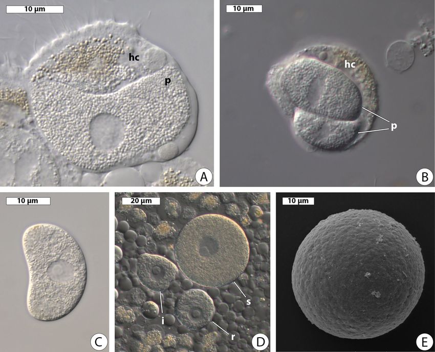
www.nature.com/scientificreports/
has been preliminarily assigned to agamococcidians because gamonts and other developmental stages beside
sporozoites and oocysts (without sporocysts, unlike in Rhytidocystis) are not known5.
Until recently, only a couple of studies on molecular phylogeny of rhytidocystids have been published. Those
phylogenies were based on 18S ribosomal RNA gene (rDNA) and did not conclusively resolve the rhytidocystid
position6,7. No molecular data on Gemmocystis are available but, based on similarities in cell size, coccidian-like
morphology, and localization in their hosts (mesenterial filaments), it was suggested that G. cylindrus belongs to
corallicolids8. The corallicolids, previously called “genotype N” or “apicomplexans Type-N” in nuclear ribosomal
DNA (rDNA) p hylogenies9,10 and ARL-V in plastid rDNA p hylogenies11,12, are related to e ucoccidians8,10. In the
most recent higher rank classification of p rotists , the agamococcidians are assigned to the Apicomplexa incertae
13
sedis. Lately, transcriptomes of two unclassified rhytidocystids were s equenced14, one of which corresponds to
Rhytidocystis pertsovi sp. n. described in the present paper. The multiprotein phylogeny showed a basal position of
rhytidocystids in the coccidiomorph c lade14 but included only sparse species sampling without coralicollids and
closest known relatives of Rhytidocystis such as Margolisiella islandica15. Clarifying the position of rhytidocystids
in densely sampled phylogenies is therefore essential for the further development of apicomplexan systematics.
In this study, we sequenced three new species of rhytidocystids from the White Sea and created phylogenies of
Apicomplexa based on SSU rDNA alone and concatenated SSU, 5.8S, and LSU rDNAs. We here describe these
new species as Rh. nekhoroshkovae sp. n. and Rh. dobrovolskiji sp. n. with light and scanning electron microscopy
and Rh. pertsovi sp. n. with light, scanning and transmission electron microscopy. To assess whether the “asexual”
rhytidocystids potentially retain the capacity for meiotic recombination, we surveyed the available transcriptomic
data of Rh. pertsovi and yet undescribed Rhytidocystis sp. from Travisia forbesii for meiosis-specific genes. We
also searched for the homologs of the coccidian oocyst wall proteins (OWPs) in rhytidocystids.
Results
Occurrence and morphology of new rhytidocystid species. Rhytidocystis nekhoroshkovae sp. n.
Parasites were found in all 30 examined polychaetes Pectinaria (Cistenides) hyperborea collected in the vicinity
of Educational and Research Station “Belomorskaya” of Saint Petersburg State University (ERS SPbU, see Meth-
ods for details). Infected midguts showed plenty of white dots on the outside surface, which corresponded to
rhytidocystids, generally located at the basal part of the midgut epithelium. The polychaetes were usually heavily
infected (hundreds of parasites per host).
Spindle-shaped zoites were observed inside the host enterocytes (Fig. 1A). As zoites grew, they lost their
elongated shape and transformed into trophozoites (Fig. 1B). Early development took place intracellularly and
young trophozoites were located inside parasitophorous vacuoles (Fig. 1C). Both young and adult trophozoites
had near-round or irregular shape with a slightly uneven border and measured 13.0–68.0 µm in maximal dimen-
sion (av. 47.4 ± 2.54 µm, n = 23). The trophozoites’ cytoplasm was filled with granules of storage carbohydrate
(presumably, amylopectin), and smaller cells were more transparent than larger ones (Fig. 1D). Live parasites
had a spherical nucleus located centrally and measured 6.6–19.2 µm in diameter (av. 14.64 ± 0.41 µm, n = 17). A
single medium-sized spherical nucleolus was eccentric. Adult trophozoites were outside host cells close to the
basal lamina of the midgut epithelium. No pathological changes were observed in infected tissue: the neighboring
enterocytes had an appearance of active digestive cells with numerous phagosomes (Fig. 1E). Parasites isolated
from the host gut were immotile. Their cell surface was rugose with longitudinal and transverse grooves, little
creases, and small depressions (Fig. 1F). Numerous micropores on the parasite surface were arranged in curved
rows, which merged with each other (Fig. 1G).
Rhytidocystis dobrovolskiji sp. n. Parasites were found in 34 out of 70 (48.6%) examined polychaetes Ophelia
limacina collected in the vicinity of ERS SPbU (see Methods for details). Parasites were embedded in the host
midgut epithelium and visible as white dots from the inside and outside the gut. The hosts contained between
several and several dozens of parasites.
The early trophozoite stages were crescent-shaped and measured 21.6–41.0 µm long (av. 32.9 ± 2.37 µm,
n = 10) (Fig. 2A). Occasionally, trophozoites were found tightly packed inside the host cells (Fig. 2B). Maturing
trophozoites measured 36.3–67.0 µm in maximal dimension (av. 51.0 ± 1.13 µm, n = 33), were irregular or round-
ish (Fig. 2C) and became spherical in a short time after the release from the host tissue (Fig. 2D). All forms had
a spherical nucleus with a relatively large eccentric or centric nucleolus, usually located in the central part of the
cell and measured 3.6–10.0 µm in diameter (av. 6.8 ± 0.61 µm, n = 10) in crescent-shaped forms and 12.1–25.0 µm
(av. 18.2 ± 0.83 µm, n = 18) in maturing ones. The cell surface of spherical trophozoites was smooth (Fig. 2E).
Only once we observed young oocysts released from the host intestinal epithelium. They were spherical and
covered by a thick transparent envelope (Fig. 2F). The nuclei were not clearly visible in all of them (Fig. 2G).
Rhytidocystis pertsovi sp. n. Parasites were found in 73 out of 106 (68.9%) examined polychaetes Ophelia limac-
ina collected at the White Sea Biological Station of Lomonosov Moscow State University (WSBS MSU, see Meth-
ods for details). Similar to Rh. dobrovolskiji, parasites were located in the host midgut epithelium. The infected
polychaetes contained from a few up to hundreds of parasites.
Intracellular crescent-shaped trophozoites measured 14.3–25.3 µm long (av. 19.86 ± 1.36 µm, n = 7) were
found inside the host enterocytes (Fig. 3A). Crescent-shaped trophozoites were sometimes located in pairs
inside a one host cell (Fig. 3B). We observed several crescent-shaped trophozoites slowly becoming bean-shaped
after the releasing from the host midgut epithelium (Fig. 3C). A spherical nucleus measuring 4.0–6.6 µm in
diameter (av. 5.1 ± 0.51 µm, n = 5) was usually located in the central part of the cell and had a spherical eccentric
nucleolus. Larger trophozoites, 21.3–59.2 µm in maximal dimension (av. 49.15 ± 1.66 µm, n = 32), were irregu-
lar or roundish in shape (Fig. 3D). They had a spherical centric nucleus measuring 8.0–20.0 µm in diameter
Scientific Reports | (2020) 10:15847 | https://doi.org/10.1038/s41598-020-72287-x 2
Vol:.(1234567890)
www.nature.com/scientificreports/

                            Figure 1.  Morphology of Rhytidocystis nekhoroshkovae n. sp. (A) Spindle-shaped zoite on squash preparation
                            of the host intestinal epithelium; hcr the host cell residue. DIC. (B) Growing zoite; squash preparation. DIC. (C)
                            Histological section of P. hyperborea intestinal epithelium with young trophozoite inside parasitophorous vacuole
                            (pv). LM. (D) Trophozoites (arrowheads) on squash preparation of the host intestinal epithelium. DIC. (E)
                            Histological section of P. hyperborea intestinal epithelium with adult parasite (p); bl basal lamina, ph phagosome. LM.
                            (F) Adult trophozoite. SEM. (G) Adult trophozoite’s cell surface with numerous micropores (arrowheads). SEM.

                            (av. 15.79 ± 0.79 µm, n = 17) with a relatively large eccentric or centric nucleolus. Being released from the host
                            tissue, they became spherical in a short time. The cell surface of spherical trophozoites had small depressions,
                            but no folds (Fig. 2E).

Scientific Reports |   (2020) 10:15847 |                https://doi.org/10.1038/s41598-020-72287-x                                               3

                                                                                                                                            Vol.:(0123456789)
www.nature.com/scientificreports/
Figure 2. Morphology of Rhytidocystis dobrovolskiji n. sp. (A) Crescent-shaped trophozoite. DIC. (B) Tightly
packed parasite (p) inside the host cell (hc). DIC. (C) Irregular trophozoite. DIC. (D) Spherical trophozoite.
DIC. (E) Spherical trophozoite. SEM. (F,G) Young oocysts; cyst oocyst envelope, N nucleus. DIC.
Transmission electron microscopy of the parasitized host intestine showed intracellular trophozoites both
in the apical parts of enterocytes (Fig. 4A–D) and deep in the epithelium (Fig. 4E-F). Apical parts of infected
enterocytes were extended into the intestinal lumen. Some parasites were in direct contact with the host cell
cytoplasm (Fig. 4A,B,F,G) and surrounded by many small (Fig. 4A,F) or several large vacuoles (Fig. 4B). Other
parasites were inside a parasitophorous vacuole (Fig. 4A,C). The general ultrastructure of the trophozoites was
similar to that of intracellular coccidians (Fig. 4F–J). The tegument was represented by a trimembrane pellicle,
which consisted of the plasma membrane and the inner membrane complex (Fig. 4I). Micropores were rarely
present in the sections (Fig. 4J). The cytoplasm of trophozoites was filled with cisternae of the endoplasmic
reticulum, dictyosomes, mitochondria having tubular cristae and mainly located under the pellicle, lipid droplets,
granules of storage carbohydrate (probably, amylopectin) and numerous dense bodies of mostly oval or round
shape and resembling oocyst wall forming bodies of some coccidians (Fig. 4H,I).
Scientific Reports | (2020) 10:15847 | https://doi.org/10.1038/s41598-020-72287-x 4
Vol:.(1234567890)www.nature.com/scientificreports/
Figure 3. Morphology of Rhytidocystis pertsovi n. sp. (A) Host enterocyte with a crescent-shaped trophozoite
inside; hc host cell, p parasite; squash preparation. DIC. (B) Host cell (hc) containing two parasites (p). DIC.
(C) Bean-shaped trophozoite. DIC. (D) Squash preparation of the host intestinal epithelium with irregular (i),
roundish (r) and spherical (s) trophozoites. DIC. (E) Spherical trophozoite. SEM.
Molecular phylogeny of Rh. nekhoroshkovae, Rh. dobrovolskiji, and Rh. pertsovi. The near-
complete sequences of rRNA operon (SSU rDNA, ITS1, 5.8S rDNA, ITS2, and LSU rDNA) were obtained for
Rh. nekhoroshkovae (5,410 bp), Rh. dobrovolskiji (5,387 bp), and Rh. pertsovi (5,281 bp). Despite parasitizing the
same hosts and being very similar in appearance, Rh. dobrovolskiji and Rh. pertsovi were different genetically.
Both Bayesian (BI) and Maximum Likelihood (ML) analyses produced almost identical tree topologies except
for the position of blastogregarines, which were either the sister lineage to coccidiomorphs (ML; not shown)
or placed among gregarines (BI; Fig. 5). The Bayesian tree inferred from the concatenated rDNA dataset of 99
taxa and 4,517 sites (Fig. 5) showed the monophyly of major alveolate lineages with high posterior probabilities
(PP), but with moderate or low ML supports (bootstrap percentages, BP) in the apicomplexan part of the tree.
The sequences of the three new Rhytidocystis species had relatively short branches and grouped into a
common rhytidocystid clade with the other representatives of the genus (Rh. cyamus and Rh. polygordiae), an
undescribed parasite of the European oyster Ostrea edulis, and several environmental sequences from oceanic
sediments with PP = 1 and BP = 88%. The rhytidocystids then formed a robust higher-order clade (PP = 1.0
and BP = 100%) with an unidentified parasite of Tridacna croecia, the coccidians Margolisiella islandica and
Pseudoklossia pectinis, and an environmental sequence from a sulfidic karst spring in Slovenia (KT072247). The
robust Rhytidocystis-Pseudoklossia-Margolisiella clade was the most early-branching lineage of coccidiomorphs,
including coccidians and haematozoans (PP = 1, BP = 83%). We suggested the name “Eococcidia” for this clade
(Fig. 5, also see “Discussion”).
Analysis of meiosis‑specific and oocyst wall protein transcripts. We examined the available tran-
scriptomic data of rhytidocystids for transcripts of meiosis-specific genes to estimate whether meiotic recombi-
nation is possible in the reportedly asexual rhytidocystids. Homology searches identified seven meiosis-specific
Scientific Reports | (2020) 10:15847 | https://doi.org/10.1038/s41598-020-72287-x 5
Vol.:(0123456789)www.nature.com/scientificreports/
Figure 4. Transmission electron microscopy of intracellular stages Rhytidocystis pertsovi n. sp. (A–D) parasites
in apical parts of enterocytes projected into the intestinal lumen. (E) Parasite in the middle of intestinal
epithelium. (F–H) The same individual as in (E) under higher magnification; hc host cell, hcN host cell
nucleus. (I,J) The same individual as in (C) under higher magnification. Parasites are in direct contact with
the host cytoplasm (A,B,E,F) surrounded by small or large vacuoles (v) or inside a parasitophorous vacuole
(pv) (C,D). (G) No parasitophorous vacuole membrane has been observed between the tegument of the
parasite (the pellicle, pe) and adjacent plasma membranes (arrowheads) of two host cells: parasitized (hc 1) and
neighboring (hc 2). (H) The trophozoite cytoplasm; am amylopectin granules, d dictyosomes, db dense bodies,
er endoplasmic reticulum, hc host cell, l lipid droplets, m mitochondria, pe pellicle. (I,J) The trophozoite pellicle;
imc inner membrane complex, m mitochondrion, mp micropore, pe pellicle, pm plasma membrane of parasite.
Scientific Reports | (2020) 10:15847 | https://doi.org/10.1038/s41598-020-72287-x 6
Vol:.(1234567890)www.nature.com/scientificreports/
Figure 5. Bayesian inference tree of alveolates obtained by using the GTR + Г + I model from the dataset of 99 concatenated SSU, 5.8S,
and LSU rDNA sequences (4,517 sites). Missing data on 5.8.S or/and LSU rDNA are marked by “–” in place of GenBank accessions;
data assembled from transcriptomes are marked by “A”. Numbers at the nodes indicate Bayesian posterior probabilities (numerator)
and ML bootstrap percentage (denominator). Black dots on the branches indicate Bayesian posterior probabilities and bootstrap
percentages of 0.95 and 90% (respectively) and higher. The newly obtained sequences of Rhytidocystis spp. are on black background.
The Eococcidia clade is highlighted by gray. Polyphyletic agamococcidians are marked by asterisks.
Scientific Reports | (2020) 10:15847 | https://doi.org/10.1038/s41598-020-72287-x 7
Vol.:(0123456789)www.nature.com/scientificreports/
Figure 6. Occurrences of meiosis-specific genes in alveolates. Rhytidocystis spp. represents combined
transcriptomic data of Rh. pertsovi and Rhytidocystis sp. ex Travisia forbesii. Pie charts show completeness scores
of underlying genomic or transcriptomic data as estimated by BUSCO; filled boxes correspond to complete
or fragmented orthologs. The Spo11 box includes either Spo11-1, Spo11-2, or both orthologs; non-meiotic
Spo11-3/Top6A orthologs were not considered. Homologs reported in the Rec8 category include any findings
of the Rad21/Rec8 family, as specific orthology of the proteins is difficult to determine (see also Supplementary
Figs. S1–S9).
Figure 7. Phylogenetic reconstructions of oocyst wall proteins. (A) Maximum likelihood tree of COWP
and TgOWP1-7 family homologs reconstructed with IQ-TREE (WAG + R5 evolutionary model selected by
ModelFinder). Branches are colored according to the species legend (bottom left). Well-supported clusters
(over 90% bootstrap support) of species-specific sequences are collapsed into triangles with side lengths
proportional to the shortest and longest branches in the cluster. Branches with over 90% bootstrap support
are marked with a black dot. The characterized OWPs of Cryptosporidium and Toxoplasma are labeled in the
tree. Rhytidocystis spp. sequences represent homologs from the combined transcriptomic data of Rhytidocystis
pertsovi and Rhytidocystis sp. ex Travisia forbesii. (B) Maximum likelihood tree of TgOWP8-12 family
homologs reconstructed with IQ-TREE (DCMut + F + R5 evolutionary model selected by ModelFinder); all tree
specifications are as in (A) (see also Supplementary Figs. S10,S11).
genes in the transcriptomic data of Rh. pertsovi and Rhytidocystis sp. ex Travisia forbesii (Fig. 6). Nearly all
gene transcripts were partial or incompletely spliced in the assemblies, indicating low transcript presence in the
sequencing libraries. Phylogenetic analyses grouped the rhytidocystid meiotic genes with other apicomplexans,
ruling out that they could be contaminating sequences (Supplementary Figs. S1–S9). The rhytidocystid tran-
scriptomes lack two genes of the core meiotic gene set—Msh4 and Msh5, but retain meiotic helicase Mer3 and a
member of the Rad21/Rec8 cohesin family, which are both absent in sexual coccidians.
The discovery of young oocysts in Rh. dobrovolskiji and the presence of putative wall-forming bodies in Rh.
pertsovi prompted us to search the rhytidocystid transcriptomes for genes encoding oocyst wall proteins previ-
ously described in Toxoplasma and Cryptosporidium. We found over one hundred transcripts related to the api-
complexan oocyst wall family proteins COWP and TgOWP1-716,17 in the transcriptomes of the two Rhytidocystis
species. Similarly to other apicomplexans, the rhytidocystid sequences are characterized by the N-terminal signal
peptide followed by a series of cysteine-containing repeats. The tree reconstruction of apicomplexan COWP and
TgOWP1-7 family proteins groups rhytidocystid sequences into 19 divergent clusters (Fig. 7A). The rhytidocystid
OWP clusters are distributed evenly in the tree, pointing to their large diversity, with several clusters showing
potential orthology to the characterized OWPs of Cryptosporidium and Toxoplasma: COWP4, COWP5, COWP9,
Scientific Reports | (2020) 10:15847 | https://doi.org/10.1038/s41598-020-72287-x 8
Vol:.(1234567890)www.nature.com/scientificreports/
TgOWP3, and TgOWP4 sequences. Another abundant family of candidate OWPs in the two rhytidocystid
species (over 200 of total identified sequences) is homologous to the more recently described TgOWP8-1218.
Similarly to the TgOWP1-7 family, the proteins contain periodical cysteine residues and a signal peptide. The
TgOWP8-12 family was significantly expanded in rhytidocystids compared to the coccidians (Fig. 7B). However,
that the majority of TgOWP1-7 and TgOWP8-12 homologs in the rhytidocystid transcriptomes are incomplete,
which confounds their classification, especially considering the repetitive structure.
Discussion
In the present study, we described three new species of Rhytidocystis agamococcidians, defined their phylogenetic
position and surveyed for molecular markers of their sexual reproduction and oocyst wall formation. The new
data reveal several key points on rhytidocystid biology and evolution.
Rhytidocystids are most likely distributed worldwide and prefer to parasitize opheliid poly-
chaetes. Previously described Rhytidocystis species were found on the Western and Eastern coasts of the
North Atlantic and in the North-Eastern Pacific O cean3,4,6,7. The rhytidocystids also parasitize polychaetes in
the Arctic Ocean: the three new species described here come from the White Sea. In addition, environmental
sequences belonging to the rhytidocystid clade (Fig. 5) were derived from the Yellow Sea (KC851785), the Car-
ibbean Sea (GU823445) and the South Pacific (shallow water hydrothermal system near Papua New Guinea;
JQ244474; JQ242635; JQ242218). Three out of the five previously described rhytidocystids parasitize polychaetes
from the family Opheliidae: Rh. opheliae, Rh. henneguyi, and Rh. cyamus3,4,7. Here we described two new rhyti-
docystids from Ophelia limacina—Rh. dobrovolskiji and Rh. pertsovi. The third new species, Rh. nekhoroshkovae,
parasitizes the polychaete from Pectinaria, the genus, which is relatively close to Ophelia19,20. Dissecting O. ver-
rilli, Riser21 had also observed putative rhytidocystids (described as “coccidians”), which were located within
projected apical ends of host enterocytes, similar to Rh. pertsovi. Apparently, most of the real biodiversity of
rhytidocystids remains undiscovered.
Eococcidia: the sister group of coccidians and haematozoans. Previous phylogenetic studies of
SSU rDNA sequences did not resolve the position of r hytidocystids6,7, making it unclear whether they could be
related to Gemmocystis cylindrus, the other agammococcidian now represented by the “genotype-N” and corali-
collid sequences. In some phylogenies, Rh. cyamus and Rh. polygordiae were the sister group of the coccidians
Margolisiella islandica and Pseudoklossia pectinis15,22,23, but their common clade was never strongly affiliated
with gregarines, cryptosporidians, or coccidiomorphs. Reasons for the lack of resolution might be two-fold: Rh.
cyamus and Rh. polygordiae form long tree branches which may cause the long branch attraction artifact, and the
earlier studies used SSU rDNA phylogenies only, which have inferior resolution to the whole rRNA operon24–27.
In the current study, we enlarged a broadly and evenly sampled alveolate dataset with environmental sequences
and short-branching sequences of our new species. We also sequenced the first rhytidocystid LSU rDNA and ana-
lyzed their concatenated rDNA phylogeny in both Maximum likelihood and Bayesian frameworks. The resulting
phylogenies have a resolution superior to earlier studies and resolve rhytidocystid and sporozoan relationships
in two important ways. Firstly, rhytidocystids are unrelated to corallicolids which include Gemmocystis cylindrus
(Fig. 5), demonstrating that the order Agamococcidiorida is polyphyletic. Secondly, the analyses unambiguously
combined rhytidocystids with coccidiomorphs (coccidians and haematozoans). This finding matches the more
sparsely-sampled but multiprotein phylogeny of apicomplexans based on 296 concatenated markers14, which
recovered rhytidocystids as basal coccidiomorphs. Margolisiella islandica infecting the Iceland scallop Chlamys
islandica is the closest described relative of rhytidocystids with complete Leukart’s triad in monoxenous life
cycle (Fig. 5)15,22,23. Pseudoklossia pectinis is a putatively heteroxenous parasite of Pecten maximus: gamogony
and sporogony occur in the great scallop, and a merogonic phase is supposed to be in some other h ost28. Other
members of the Rhytidocystis-Pseudoklossia-Margolisiella clade are poorly-studied (unnamed parasites of Tri-
dacna croecia and Ostrea edulis), and the whole clade lacks obvious shared morphological characteristics (syna-
pomorphies). Nevertheless, since this robust clade has been recovered in several analyses and discovered as basal
group of all coccidiomorphs, including coccidians and haematozoans, we suggest establishing the new taxon
Eococcidia for basal coccidiomorphs mainly parasitizing marine invertebrates. The term “Eococcidia” refers to
Eos—the goddess of the dawn in Greek mythology, who rose in the morning from the Oceanus; the prefix “eo-”
is used in geology and biology for the designation of something to be early (Eococcidia—early coccidians). Coc-
cidian genera Merocystis and Aggregata are also candidate members to Eococcidia since they were recovered as
close relatives of Rhytidocystis, Pseudoklossia, and Margolisiella23,29.
Intracellular parasitism links rhytidocystids with other coccidiomorphs. The phylum Apicompl-
exa contains both extracellular and intracellular parasites. Development of many gregarines runs extracellularly,
whereas coccidians and haematozoans invade host c ells30,31. Four out of five previously described rhytidocystids
have trophozoites in the host intestinal e pithelium3,4,6,7, but their intracellular stages were never detected. Here
we provide evidence of intracellular localization of three new rhytidocystid species. The findings of putative
sporozoites inside host cells and young trophozoites within parasitophorous vacuoles in Rh. nekhoroshkovae,
and trophozoites tightly packed inside host cells in Rh. dobrovolskiji suggest that the development of both spe-
cies begins intracellularly. The trophozoites of Rh. pertsovi on squash preparations of the host enterocytes and
in TEM sections were undoubtedly located under the plasma membrane of the host cell. Perhaps, the develop-
ment of other rhytidocystid may also start with intracellular forms. Rh. polygordiae was the only species previ-
ously described with TEM, and its trophozoites were observed in the interstitial space between adjacent host
enterocytes6. However, the structure purported to be “the plasma membrane of the adjacent epithelial cell”
Scientific Reports | (2020) 10:15847 | https://doi.org/10.1038/s41598-020-72287-x 9
Vol.:(0123456789)www.nature.com/scientificreports/
(Fig. 19 in6) has the thickness (a little more than 20 nm) and appearance of two membranes with the intercel-
lular space between them, and most likely represents two plasma membranes of two adjoining enterocytes.
Furthermore, “the interstitial space” (Fig. 19 in6) is filled with structures resembling endoplasmic reticulum and
apparently is the host cell cytoplasm. We therefore suppose that at least some trophozoites of Rh. polygordiae
were located intracellularly. Overall, evidence for intracellular development strongly links rhytidocystids with
other coccidiomorphs such as coccidians and haematozoans to the exclusion of gregarines and cryptosporid-
ians, which are chiefly extracellular or epicellular. Congruent with their phylogeny (Fig. 5)14, and intracellular
stages in Margolisiella15, this distribution suggests that intracellular invasion evolved in the common coccidi-
omorph ancestor. Unlike other coccidiomorphs, however, rhytidocystid trophozoites grow up to a relatively
large size, destroy infected cells, and end up lying extracellularly within the host tissue.
Intracellular apicomplexans may be either embedded in a parasitophorous vacuole (PV) made of compo-
nents of host origin or host and parasite origin, or be in direct contact with the host cell cytoplasm32–34. In the
first case a zoite penetrates the host cell membrane, induces the PV formation and becomes surrounded by PV
since the beginning of its intracellular development35,36. Unexpectedly, younger forms of Rh. pertsovi were not
within a parasitophorous vacuole in our TEM sections (Fig. 4A,B), whereas several larger ones were (Fig. 4C,D).
This result does not correspond to typical PV development, so a thorough TEM investigation of rhytidocystid
intracellular development is needed.
Morphology of trophozoite and oocyst stages. The trophozoite cell shape of earlier described Rhyti-
docystis species varies from oblong Rh. polygordiae and bean-shaped Rh. cyamus to flat oval cells of Rh. opheliae
and Rh. henneguyi3,4,6,7. In the case of Rh. pertsovi and Rh. dobrovolskiji, it seems to be that crescent-shaped,
bean-shaped, irregular, roundish and spherical forms represent successive stages of development. A zoite
invades the host cell and transforms into a trophozoite. Presumably, during the growth inside the limited space
of the host cell, the young trophozoite bends and becomes crescent-shaped, then it loses peaked cell poles and
becomes bean-shaped; over time, it undergoes marked growth and becomes tightly packed under the host cell
membrane. Released from the host cell to the interstitial space of the tissue, a trophozoite unbends, becomes
irregular, then roundish and spherical eventually. Spherical trophozoites, apparently, represent the transitional
form to the oocyst stage. Young oocysts Rh. dobrovolskiji look like spherical trophozoites covered by a thick
transparent envelope and resemble young oocysts of Rh. sthenelais37.
The cytoplasm of intracellular trophozoites Rh. pertsovi looks typical of sporozoans and possesses all general
structures. Rh. pertsovi retains an active a picoplast14 but none was observed in this TEM study. Instead, we found
numerous dense bodies of oval or round shape, which could be homologous to wall-forming bodies (WFBs) in
coccidian and cryptosporidian m acrogamonts38–41. The WFBs mediate the formation of the oocyst wall, which
more than 90% is made up of p roteins42. The presence of oocysts and putative WFBs has prompted us to search
in rhytidocystid transcriptomes for homologs of oocyst wall proteins (OWPs), some of which are proven to be
located in W FBs40,43. The analysis revealed an astounding diversity of transcripts related to COWP, TgOWP1-7
and TgOWP8-12 family proteins, supporting the presence of WFBs in rhytidocystids and a common mechanism
for their oocyst wall formation with coccidia and cryptosporidia.
Discovery of meiotic genes in “asexual” rhytidocystids. Since rhytidocystids are closely related to
M. islandica, which is an eucoccidian-like protist, whose life cycle includes all three types of sporozoan reproduc-
tion: gametogony, merogony, and sporogony (Leuckart’s triad), and which, hence, produces sexual gamonts15,
the supposed absence of sexual life stages in rhytidocystids raises the possibility that they recently lost sexual
reproduction. Generally conserved across the eukaryotes, the core meiosis machinery includes nine proteins
(Spo11, Hop1, Hop2, Mnd1, Dmc1, Mer3, Msh4, Msh5, Rec8), with functions spanning sister chromatid cohe-
sion, induction of double-strand breaks, heteroduplex DNA and synaptonemal complex formation, and Hol-
liday junction resolution44,45. In rhytidocystids, the core meiotic gene set is short of two genes for Msh4 and
Msh5, unlike the chrompodellid Vitrella brassicaformis, which retains a full set of core meiosis-specific genes
and where sexual process has been p roposed46. The absence of the heterodimer-forming Msh4 and Msh5 in
rhytidocystids is consistent with their absence in other coccidiomorphs’ genomes (Fig. 6): they are involved in
the stabilization of Holliday junctions and meiotic crossover interference in model organisms47 but apparently
dispensable in a picomplexans48. Notably, the closely related Msh2 family, which is involved in DNA repair, has
expanded in Rhytidocystis sp. ex Travisia forbesii. Unlike many sporozoans, rhytidocystids retain the meiotic
helicase Mer3 and a member of the Rad21/Rec8 cohesin family. Thus, the inventory of meiosis-specific genes in
rhytidocystids does not display evidence of reduction in relation to the same gene set of sexual coccidians. The
presence of these genes alone, however, does not constitute conclusive evidence of sexual reproduction in the
family. Meiosis-specific genes, contrary to their designation, were reported to have functions outside of meiosis,
specifically in homologous recombination and DNA repair49. The preservation of these genes weighs in favor of
meiotic recombination in rhytidocystids, but more direct evidence would be necessary to verify the existence of
sexual process.
In terms of appearance, rhytidocystid trophozoites are similar to macrogamonts of their closest relatives—
sexual coccidians: they develop intracellularly, amass a supply of nutrients, produce wall-forming bodies and
eventually become the oocysts. The presence of meiosis-specific transcripts challenges the long held belief that
rhytidocystids lack gamogony and inspires search for their cryptic sexual process, which has remained hidden
for over a hundred years. Future research on early rhytidocystid development and genetics are awaited to con-
tribute to this matter.
Scientific Reports | (2020) 10:15847 | https://doi.org/10.1038/s41598-020-72287-x 10
Vol:.(1234567890)www.nature.com/scientificreports/
Taxonomic summary. Here we use the most recent Adl et al.13 system for higher-ranks as phylum and
class, despite the inconsistency of this system to the actual phylogeny of Apicomplexa (Fig. 5)14,26,50, and due to
the absence of a correct system. The eococcidians are as close to coccidians as to haematozoans (Aconoidasida)
in our phylogeny. However, we classify Eococcidia into Conoidasida and Coccidia because of the findings of the
apical complex in their zoites6,37,51. We suggest the order Pseudoklossiida for Pseudoklossia and Margolisiella (for-
mer Eimeriorina) as P. pectinis and M. islandica were recovered strongly within the eococcidians, but not within
the eimeriids (Fig. 5)15,23. We keep the rhytidocystids into the order Agamococcidiida as no microgamonts and
microgametes were found, only trophozoites that potentially may be macrogamonts. We do not include Gemmo-
cystis to the Agamococcidiida as we consider it belonging to the corallicolids. The mature sporulated oocysts of
the newly described rhytidocystids were not observed, but we use the characteristics of the previously described
rhytidocystid oocysts for the taxon d iagnosis3,37. We still lack distinct morphological synapomorphies both for
Eococcidia and for Pseudoklossiida; therefore, the establishment of these taxa is mainly based on molecular data.
Phylum Apicomplexa Levine, 1970.
Class Conoidasida Levine, 1988.
Subclass Coccidia Leuckart, 1879.
Superorder Eococcidia superordo novus.
Coccidia. Homoxenous and heteroxenous parasites of marine invertebrates, predominantely polychaetes and
mollusks. Molecular data: earlier robust sister clade to coccidians and haematozoans in rDNA and multiprotein
phylogenies. Etymology: from “eo-”, a prefix meaning the earliest appearance, and “coccidia”.
Order Pseudoklossiida ordo novus.
Eococcidia. Homoxenous or heteroxenous parasites of marine molluscs (definitive hosts in heteroxenous); life
cycle—complete Leuckart’s triad; development and merogony intracellular, sexuality intracellular or extracellular,
fecundation intracellular or extracellular. Margolisiella, Pseudoklossia.
Order Agamococcidiida (Levine, 1979) emend.
Eococcidia. Merogony, microgamonts and microgametes not reported. Rhytidocystis.
Family Rhytidocystidae Levine, 1979.
Agamococcidiida. Early development intracellular; adult trophozoites extracellular in the host intestinal epi-
thelium or coelom; large oocysts with many tens of sporocysts in the host intestinal epithelium or coelom; in
annelids.
Genus Rhytidocystis Henneguy, 1907[= Dehornia Porchet-Henneré, 1972].
Rhytidocystis nekhoroshkovae sp. n. Miroliubova, Simdyanov, Janouškovec, Paskerova, 2020.
Description. Trophozoites 13.0–68.0 µm, flattened, almost round or irregular; immotile; young stages intracel-
lular, adults extracellular. Spherical centric nucleus 6.6–19.2 µm with a spherical centric nucleolus. Cell surface
of adult trophozoites rugose with longitudinal and transverse grooves, little creases and small depressions and
numerous micropores in the curved rows, which merged with each other.
Molecular data. Partial rDNA (SSU rDNA, ITS1, 5.8S rDNA, ITS2, and LSU rDNA), GenBank accession number
MT231950.
Type locality. White Sea, Kandalaksha Gulf, Chupa Bay, Podpakhta Strait (66.301129 N, 33.622062 E), a depth
of ~ 20 m.
Type habitat. Marine.
Type host. Pectinaria (Cistenides) hyperborea Malmgren, 1866 (Polychaeta: Pectinariidae).
Location in host. Midgut epithelium.
Type (syntype) material. A platinum sputter-coated SEM stub with several protists, slides with histological sec-
tions, specimen of parasite cells and host material fixed in ethanol have been deposited in the collection of
Scientific Reports | (2020) 10:15847 | https://doi.org/10.1038/s41598-020-72287-x 11
Vol.:(0123456789)www.nature.com/scientificreports/
Department of Invertebrate Zoology, Saint Petersburg State University; extracted DNA used for obtaining of
rDNA sequences deposited in the collection of Department of evolutionary biochemistry, Belozersky Institute
for Physico-Chemical Biology, Lomonosov Moscow State University; Fig. 1 (this publication) shows some of
the syntypes.
Zoobank registration. LSID urn:lsid:zoobank.org:act:A80C2D4C‑238E‑4E30‑A6D5‑E43DF2D379A2.
Etymology. This species was named in honor of Svetlana Nekhoroschkova, PhD, an inspiring ecology lecturer
in the Ecological and Biological Lyceum (Arkhangelsk, Russia), dear teacher of the author Tatiana Miroliubova.
Rhytidocystis dobrovolskiji sp. n. Miroliubova, Simdyanov, Janouškovec, Paskerova, 2020.
Description. Young intracellular trophozoites 21.6–41.0 µm, crescent-shaped. Larger trophozoites 36.3–67.0 µm;
irregular, roundish then spherical and smooth; extracellular. Spherical centric nucleus with relatively big eccen-
tric or centric nucleolus 3.6–10.0 µm in young and 12.1–25.0 µm in larger forms. Young oocyst within thick
transparent envelope.
Molecular data. Partial rDNA (SSU rDNA, ITS1, 5.8S rDNA, ITS2, and LSU rDNA), GenBank accession number
MT231947.
Type locality. White Sea, Kandalaksha Gulf, Chupa Bay, Yakovleva Inlet (66.315649 N, 33.836669 E) at a depth
of ~ 1–15 m.
Type habitat. Marine.
Type host. Ophelia limacina Rathke, 1843 (Polychaeta: Opheliidae).
Location in host. The midgut epithelium.
Type (syntype) material. A platinum sputter-coated SEM stub with several protists, specimen of parasite cells and
host material fixed in ethanol have been deposited in the collection of Department of Invertebrate Zoology, Saint
Petersburg State University; extracted DNA used for obtaining of rDNA sequences deposited in the collection
of Department of evolutionary biochemistry, Belozersky Institute for Physico-Chemical Biology, Lomonosov
Moscow State University; Fig. 2 (this publication) shows some of the syntypes.
Zoobank registration. LSID urn:lsid:zoobank.org:act:6B80FBE6‑63E7‑4CCF‑A054‑8D76E142320E.
Etymology. This species was named after Dr Andrej Alexandrovitch Dobrovolskij (1939–2019), Professor of
the Department of Invertebrate Zoology at Saint Petersburg State University, a recognized authority in the field
of Invertebrate Zoology and Parasitology, the Great Teacher of many generations of Russian zoologists, who
devoted himself utterly to science and students. In particular, he taught and inspired the authors TSM, TGS,
and GGP.
Rhytidocystis pertsovi sp. n. Miroliubova, Simdyanov, Janouškovec, Belova, Paskerova, 2020.
Description. Young intracellular trophozoites 14.3–25.3 µm; crescent-shaped then bean-shaped with a spherical
centric nucleus 4–6.6.0 µm and a spherical eccentric nucleolus. Larger trophozoites extracellular, 21.3–59.2 µm;
irregular, roundish then spherical; with a spherical centric nucleus 8.0–20.0 µm and a relatively big eccentric or
centric nucleolus. The cell surface of spherical trophozoites has no folds.
Molecular data. Partial rDNA (SSU rDNA, ITS1, 5.8S rDNA, ITS2, and LSU rDNA), GenBank accession number
MT231948; transcriptome shotgun assembly, GenBank accession number GHVQ00000000.1.
Type locality. White Sea, Kandalaksha Gulf (66.556758 N, 33.106862 E), a depth of ~ 1–15 m.
Type habitat. Marine.
Type host. Ophelia limacina Rathke, 1843 (Polychaeta: Opheliidae).
Location in host. The midgut epithelium.
Type (syntype) material. A platinum sputter-coated SEM stub with several protists, specimen of parasite cells and
host material fixed in ethanol have been deposited in the collection of Department of Invertebrate Zoology, Saint
Petersburg State University. Resin blocks and fixed slides containing pieces of infected host intestine deposited
in the collection of the author TGS, Department of Invertebrate Zoology, Lomonosov Moscow State University;
extracted DNA used for obtaining of rDNA sequences deposited in the collection of Department of evolutionary
Scientific Reports | (2020) 10:15847 | https://doi.org/10.1038/s41598-020-72287-x 12
Vol:.(1234567890)www.nature.com/scientificreports/
biochemistry, Belozersky Institute for Physico-Chemical Biology, Lomonosov Moscow State University; Figs. 3,
4 (this publication) show some of the syntypes.
Zoobank registration LSID. urn:lsid:zoobank.org:act:71B99A74‑6B81‑40C0‑87CA‑5D0FF8D33F39.
Etymology. This species was named after Nickolay Pertsov (1924–1987), a long-time director and an eminent
innovator at the White Sea Biological Station of Lomonosov Moscow State University, where this species was
found and sampled.
Materials and methods
Polychaete hosts were collected from the sublittoral zone in different sites in Kandalaksha Gulf, White Sea in
2015–2018. The bristle worms Pectinaria (Cistenides) hyperborea Malmgren, 1866 (Polychaeta: Pectinariidae)
were found in the vicinity of Educational and Research station “Belomorskaya” of the Saint Petersburg State
University (ERS SPbU), Chupa Bay, Podpakhta Strait (66.301129 N, 33.622062 E), at a depth of ~ 20 m, under
the thermocline. Bristle worms Ophelia limacina Rathke, 1843 (Polychaeta: Ophellidae) were collected from
two distant locations: White Sea Biological Station Lomonosov Moscow State University (WSBS MSU), Velikaja
Salma, Kandalaksha Bay, White Sea (66.556758 N, 33.106862 E) and in the vicinity of ERS SPbU, Yakovleva Inlet,
Keret’ Archipelago, Chupa Bay, Kandalaksha Bay, White Sea (66.315649 N, 33.836669 E) at a depth of ~ 1–15 m.
The O. limacina worms from these different locations were stored and processed independently. The coccidian
individuals and pieces of the infected host intestine were isolated with fine tip needles under Olympus SZ40
(Olympus, Japan) or MBS-10 (LOMO, Russia) stereomicroscopes.
Light microscopy. Fragments of the infected host intestine were fixed with Bouin solution, rinsed two
times with distilled water and dehydrated in ethanol series. Using paraffin-celloidin m ethod52 4 μm thick sec-
tions were made with the Leica RM-2265 microtome and stained with Ehrlich’s Hematoxylin. Separate alive par-
asites and squash preparations of the host intestine fragments containing parasites as well as histological sections
were photographed with the help of Leica DM 2500 light microscopes equipped with differential interference
contrast (DIC) optics and Plan-Apo objective lenses and connected to a Leica DFC295 or a Nikon DS-Fi1 digital
cameras (Leica Microsystems, Germany; Nikon Corporation, Japan). All in vivo microphotographs were taken
with the use of DIC technique, microphotographs of histological sections—by means of bright-field microscopy
(LM). Maximal dimensions of protist cells were measured with the ImageJ program (rsb.info.nih.gov/ij/). Aver-
age values and standard errors were calculated.
Electron microscopy. The cell surface of the isolated parasites was studied with scanning electron micros-
copy (SEM). Trophozoites Rhytidocystis pertsovi were also studied with transmission electron microscopy
(TEM). For both methods, the individual trophozoites or small fragments of the infected host intestine were
fixed with 2.5% (v/v) glutaraldehyde in 0.05 M cacodylate buffer (pH 7.4) containing 1.28% (w/v) NaCl in an ice
bath in the dark. The fixative was once replaced with fresh fixative after 1 h, and the total fixation time was 2 h.
The fixed samples were rinsed three times with cacodylate buffer and post-fixed with 2% (w/v) osmium tetroxide
in the cacodylate buffer (ice bath, 2 h). After fixation, samples were dehydrated in an ethanol series.
For SEM dehydrated samples were critical point dried in liquid CO2 and then sputter-coated with platinum.
The samples were investigated with a Tescan MIRA3 LMU scanning electron microscope (TESCAN, Czech
Republic), and FEI Quanta 250 (Thermo Fisher Scientific, Netherlands).
For TEM study dehydrated samples were transferred to an ethanol/acetone mixture 1:1 (v/v), rinsed twice in
pure acetone, and embedded in Epon resin using a standard procedure. Ultrathin sections obtained using LKB-III
(LKB-produkter, Sweden) or Leica EM UC6 (Leica Microsystems, Germany) ultramicrotomes were contrasted
with uranyl acetate and lead c itrate53 and examined under a JEM 1011 electron microscope (JEOL, Tokyo, Japan).
DNA extraction, PCR amplification, and sequencing of rDNA. Isolated trophozoites (up to 50 cells
from each location) were washed three times in filtered sea water and deposited into 1.5 ml microcentrifuge
tubes. Samples of Rh. nekhoroshkovae and Rh. pertsovi were fixed with 96% ethanol, the sample of Rh. dobro-
volskiji was fixed with RNA-later (Life Technologies, USA). Extraction of DNA from fixed cells was performed
using the NucleoSpin Tissue kit (Macherey–Nagel GmbH & Co. KG, Germany). Whole Genome Amplification
(WGA) was performed for Rh. nekhoroshkovae and Rh. dobrovolskiji samples using REPLI-g Midi Kit (Qiagen,
UK). The contiguous nucleotide sequences (SSU, ITS1, 5.8S, ITS2 and LSU rDNAs) were assembled from a
series of overlapping fragments obtained by PCR with different pairs of primers, followed by Sanger sequencing
(see26,27,50 for the general approach). The rDNA fragments were amplified with Encyclo PCR kit (Evrogen, Rus-
sia) in a total volume of 20 µl using a DNA Engine Dyad thermocycler (Bio-Rad) and a T100 Thermal Cycler
(Bio-Rad). General scheme of PCR protocol as follows: lid temperature 100 °C; initial denaturation at 95 °C for
2.5 min; 40 cycles of 95 °C for 30 s (denaturation); 48–55 °C for 30 s (annealing); 72 °C for 1.5 (elongation) and a
final extension at 72 °C for 10 min. Table 1 shows the lengths of amplified and overlapping fragments, sequences
of used oligonucleotides and exact annealing temperatures.
Assembling of rRNAs sequences from transcriptomic data. The rDNAs of Pterospora schizosoma,
Monocystis agilis, Lecudina tuzetae, and Heliospora caprellae were assembled from the transcriptome sequenc-
ing data generated by Mathur et al.56. The rDNAs of Lankesteria abbotti were assembled from the transcrip-
tome sequencing data of The Marine Microbial Eukaryote Transcriptome Sequencing Project57. The sequenc-
ing data were obtained from the NCBI Sequence Read Archive (https://www.ncbi.nlm.nih.gov/sra; accessions:
Scientific Reports | (2020) 10:15847 | https://doi.org/10.1038/s41598-020-72287-x 13
Vol.:(0123456789)www.nature.com/scientificreports/
Object, length of contig, and its accession Primers: forward (F) and reverse (R);
number Amplified fragment Length Overlap annealing temperature used in the PCRs
(F) ͣ 5′-GTATCTGGTTGATCCTGCCAGT-3’
(I) SSU rDNA (part) ~ 1,780 bp 98 bp (R) 5′-GGAAACCTTGTTACGACTTCTC-3’
t° = 48 °C
(F) 5′-GTACACACCGCCCGTCGCTC-3’
(II) SSU rDNA (part), ITS1, 5.8S rDNA, ITS2,
~ 1,200 bp 619 bp (R) 5′-CCTTGGTCCGTGTTTCAAGAC-3’
LSU rDNA (part)
t° = 50 °C
Rhytidocystis nekhoroshkovae (F)b 5′-ACCCGCTGAAYTTAAGCATAT-3’
5,410 bp (III) LSU rDNA (part) ~ 2,170 bp 892 bp (R)b 5′-ACATTCAGAGCACTGGGCAG-3’
MT231950 t° = 50 °C
(F)b 5′-TCCGCTAAGGAGTGTGTAACAAC-3’
(IV) LSU rDNA (part) ~ 1,350 bp 397 bp (R)b 5′-CCGCCCCAGYCAAACTCCC-3′
t° = 53 °C
(F)b 5′-GATTTCTGCCCAGTGCTCTG-3′
(V) LSU rDNA (part) ~ 1,090 bp (R)b 5′-MRGGCTKAATCTCARYRGATCG-3′
t° = 55 °C
(F) 5′-TMYCYGRTTGATYCTGYC-3′
(I) SSU rDNA (part) ~ 1,760 bp 63 bp (R) 5′-GGAAACCTTGTTACGACTTCTC-3′
t° = 48 °C
(F)c 5′-GGTCCGGTGAATTAACCAGATT-3′
(II) SSU rDNA (part), ITS1, 5.8S rDNA, ITS2,
~ 1,230 bp 647 bp (R)c 5′-CCTTGGTCCGTGTTTCAAGAC-3′
LSU rDNA (part)
t° = 50 °C
Rhytidocystis dobrovolskiji (F)b 5′-ACCCGCTGAAYTTAAGCATAT-3′
5,387 bp (III) LSU rDNA (part) ~ 1,950 bp 636 bp (R)b 5′-AGCCAATCCTTWTCCCGAAGTTAC-3′
MT231947 t° = 53 °C
(F)b 5′-TCCGCTAAGGAGTGTGTAACAAC-3′
(IV) LSU rDNA (part) ~ 1,350 bp ∼395 bp (R)b 5′-CCGCCCCAGYCAAACTCCC-3′
t° = 53 °C
(F)b 5′-GATTTCTGCCCAGTGCTCTG-3′
(V) LSU rDNA (part) ~ 1,010 bp (R)b 5′-MRGGCTKAATCTCARYRGATCG-3′
t° = 55 °C
(F) 5′-TMYCYGRTTGATYCTGYC-3′
(I) SSU rDNA (part) ~ 1,680 bp 101 bp (R) 5′-GGAAACCTTGTTACGACTTCTC-3′
t° = 48 °C
(F) 5′-GTACACACCGCCCGTCGC-3′
(II) SSU rDNA (part), ITS1, 5.8S rDNA, ITS2,
~ 1,170 bp 601 bp (R) 5′-CCTTGGTCCGTGTTTCAAGAC-3′
Rhytidocystis pertsovi LSU rDNA (part)
t° = 50 °C
5,281 bp
MT231948 (F)b 5′-ACCCGCTGAAYTTAAGCATAT-3′
(III) LSU rDNA (part) ~ 2,150 bp 891 bp (R)b 5′-ACATTCAGAGCACTGGGCAG-3′
t° = 50 °C
(F)b 5′-TCCGCTAAGGAGTGTGTAACAAC-3′
(IV) LSU rDNA (part) ~ 2,020 bp (R)b 5′-MRGGCTKAATCTCARYRGATCG-3′
t° = 53 °C
Table 1. Main characteristics of the rhytidocystid sequences obtained in this study. a The primer sequence was
based on Medlin et al.54. b The primer sequences were based on Van der Auwera et al.55. b The primer sequence
was tailored specially for Rh. dobrovolskiji.
SRR8980200-SRR8980205, SRR8980208-SRR8980213, SRR1300212), and the assemblies were performed with
SPAdes58 utilizing k-mer size 127 and Trinity59 programs. Assembled rDNA contigs were aligned using MAFFT60
and inspected by eye for assembly errors and chimeric sequences.
Molecular phylogenetic analyses. The alignment of concatenated SSU, 5.8S, and LSU rDNAs (54
sequences, 4,618 sites) was prepared for phylogenetic analyses. The taxon sampling was designed in order to
maximize the phylogenetic diversity of Apicomplexa and completeness of sequences in alignments, by prefer-
entially selecting taxa having both SSU and LSU rDNA sequences. However, since the taxon samplings of 5.8S
and LSU rDNAs are limited by available sequences, we then expanded the dataset by adding SSU rDNA-alone
sequences of previously published Rhytidocystis spp., their closest relatives (several environmental sequences,
Pseudoklossia pectinis and Margolisiella islandica), two adeleid coccidians (additionally to Hepatozoon canis),
and major gregarine lineages not represented in 5.8S and LSU rDNA databases (Stylocephaloidea eugregarines
and archigregarine lineage IV from terebellid polychaetes). The missing nucleotide sites of 5.8S and LSU rDNAs
were marked as "N" in the concatenated dataset. The rDNAs’ alignments were generated in MUSCLE 3.661 under
default parameters and then manually adjusted and concatenated with BioEdit 7.0.9.062; columns containing few
nucleotides or hypervariable regions were removed. Representatives of stramenopiles and Rhizaria were used as
outgroups. The final dataset included 99 taxa with 4,517 unambiguously aligned sites.
Maximum-likelihood (ML) analyses were performed with RAxML 8.2.963 under the GTR + Г model and
CAT approximation (25 rate categories per site). The procedure included 100 alternative runs of the ML analysis
and 1,000 replicates of multiparametric bootstrap. Bootstrap percentages were merged on the user trees (both
ML and BI) with the same program. Bayesian inference (BI) analyses were done in MrBayes 3.2.664 under
GTR + Г + I model with 12 discrete categories of gamma distribution. The following parameters were used: nst = 6,
Scientific Reports | (2020) 10:15847 | https://doi.org/10.1038/s41598-020-72287-x 14
Vol:.(1234567890)www.nature.com/scientificreports/
ngammacat = 8, rates = invgamma; parameters of Metropolis Coupling Markov Chains Monte Carlo (mcmc):
nruns = 2, nchains = 4, temp = 0.2, ngen = 10,000,000, samplefreq = 1,000, burninfrac = 0.5. The average standard
deviation of split frequencies at the end of computations was 0.001441.
Analysis of meiosis‑specific and oocyst wall protein transcripts. Searches for meiosis-specific and
oocyst wall protein families in the transcriptomes of Rhytidocystis species were carried out with H MMER65
using profiles constructed from protein family alignments. The corresponding protein families were identified
with OrthoFinder66 clustering with predicted protein sequences in a set of 70 eukaryotic genomes; the protein
family alignments were generated using MAFFT60. We utilized the following sources for genomic data: Genome
database of NCBI (https://www.ncbi.nlm.nih.gov/genome), Genome Portal of DOE JGI (https://genome.jgi.doe.
gov/portal/), Ensembl Protists resources (https://protists.ensembl.org/), genome projects of Marine Genomics
Unit (https://marinegenomics.oist.jp/), and genomic resources of multicellgenome lab (https://multicellgenome
.com/). The transcriptomic data for Rhytidocystis pertsovi and Rhytidocystis sp. ex Travisia forbesii were obtained
from GenBank transcriptome shotgun assembly projects GHVQ00000000.1 and GHVS00000000.1. The tran-
scriptome assemblies of Rhytidocystis species were processed with TransDecoder67 utilizing BLAST68 and
HMMER searches against the UniProtKB/Swiss-Prot69 and P fam70 databases for ORF prediction. Orthology of
proteins discovered by HMMER profile searches was verified by reciprocal BLAST searches against OrthoFinder
orthogroups: proteins with best hit outside of the queried protein family were excluded from the set of find-
ings. The findings satisfying reciprocal BLAST search criterion were added to the protein family alignments
using MAFFT60, and the family membership was further inspected by reconstructing phylogenies. The trees for
meiosis-specific protein families were reconstructed by IQ-TREE71 using the LG + C10 + F + G4 profile mixture
model, and ultrafast bootstrap a pproximation72 with 1,000 replicates for estimation of branch support; IQ-TREE
reconstructions for oocyst wall protein families utilized ModelFinder73 to automatically select the best-fit model.
The trees were visualized using MEGA74 and iTOL75. The completeness estimates for genomic and transcrip-
tomic data were performed with BUSCO76 using the eukaryota_odb9 dataset.
Received: 18 May 2020; Accepted: 28 August 2020
References
1. Levine, N. D. Agamococcidiorida ord. n. and Rhytidocystidae fam. n. for the coccidian genus Rhytidocystis Henneguy, 1907. J.
Protozool. 26, 167–168 (1979).
2. Krylov, M. V. & Dobrovolskij, A. A. Macrosystem and phylogeny of the Sporozoa. Proc. ZIN 94, 62–74 (1980).
3. De Beauchamp, P. Recherches sur les Rhytidocystis parasities des Ophélies. Arch. Protistenkd. 31, 138–168 (1913).
4. Henneguy, L. F. Sur une Grégarine parasite des Ophélies. C. R. Assoc. Fr. Avanc. Sci. 36, 633–636 (1907).
5. Upton, S. J. & Peters, E. C. A new and unusual species of coccidium (Apicomplexa: Agamococcidiorida) from Caribbean sclerac-
tinian corals. J. Invert. Pathol. 47, 184–193 (1986).
6. Leander, B. S. & Ramey, P. A. Cellular identity of a novel small subunit rDNA sequence clade of apicomplexans: description of the
marine parasite Rhytidocystis polygordiae n. sp. (host: Polygordius sp., Polychaeta). J. Eukaryot. Microbiol. 53, 280–291 (2006).
7. Rueckert, S. & Leander, B. S. Phylogenetic position and description of Rhytidocystis cyamus sp. n. (Apicomplexa, Rhytidocystidae):
a novel intestinal parasite of the north-eastern Pacific ‘stink worm’ (Polychaeta, Opheliidae, Travisia pupa). Mar. Biodiv. 39, 227–234
(2009).
8. Kwong, W. K., del Campo, J., Mathur, V., Vermeij, M. J. A. & Keeling, P. J. A widespread coral-infecting apicomplexan with chlo-
rophyll biosynthesis genes. Nature 568, 103–107 (2019).
9. Toller, W., Rowan, R. & Knowlton, N. Genetic evidence for a protozoan (phylum Apicomplexa) associated with corals of the
Montastraea annularis species complex. Coral Reefs 21, 143–146 (2002).
10. Šlapeta, J. & Linares, M. C. Combined amplicon pyrosequencing assays reveal presence of the apicomplexan “type-N” (cf. Gem-
mocystis cylindrus) and Chromera velia on the Great Barrier Reef. Australia. PLOS ONE 8, e76095 (2013).
11. Janouškovec, J., Horák, A., Barott, K. L., Rohwer, F. L. & Keeling, P. J. Global analysis of plastid diversity reveals apicomplexan-
related lineages in coral reefs. Curr. Biol. 22, R518–R519 (2012).
12. Janouškovec, J. et al. Environmental distribution of coral-associated relatives of apicomplexan parasites. ISME J. 7, 444–447 (2013).
13. Adl, S. M. et al. Revisions to the classification, nomenclature, and diversity of Eukaryotes. J. Eukaryot. Microbiol. 66, 4–119 (2019).
14. Janouškovec, J. et al. Apicomplexan-like parasites are polyphyletic and widely but selectively dependent on cryptic plastid orga-
nelles. eLife 8, e49662 (2019).
15. Kristmundsson, Á, Helgason, S., Bambir, S. H., Eydal, M. & Freeman, M. A. Margolisiella islandica sp. nov. (Apicomplexa: Eimeri-
dae) infecting Iceland scallop Chlamys islandica (Müller, 1776) in Icelandic waters. J. Invert. Pathol. 108, 139–146 (2011).
16. Possenti, A. et al. Molecular characterisation of a novel family of cysteine-rich proteins of Toxoplasma gondii and ultrastructural
evidence of oocyst wall localisation. Int. J. Parasitol. 40, 1639–1649 (2010).
17. Templeton, T. J. et al. The Cryptosporidium oocyst wall protein is a member of a multigene family and has a homolog in Toxoplasma.
Infect. Immun. 72, 980–987 (2004).
18. Salman, D. et al. Evaluation of novel oocyst wall protein candidates of Toxoplasma gondii. Parasitol. Int. 66, 643–651 (2017).
19. Struck, T. H. et al. The evolution of annelids reveals two adaptive routes to the interstitial realm. Curr. Biol. 25, 1993–1999 (2015).
20. Zrzavý, J., Říha, P., Piálek, L. & Janouškovec, J. Phylogeny of Annelida (Lophotrochozoa): total-evidence analysis of morphology
and six genes. BMC Evol. Biol. 9, 189 (2009).
21. Riser, N. W. Observations on the genus Ophelia (Polychaeta: Opheliidae) with the description of a new species. Ophelia 28, 11–29
(1987).
22. Janouškovec, J. et al. Factors mediating plastid dependency and the origins of parasitism in apicomplexans and their close relatives.
Proc. Natl. Acad. Sci. USA 112, 10200–10207 (2015).
23. Kristmundsson, Á & Freeman, M. A. Harmless sea snail parasite causes mass mortalities in numerous commercial scallop popula-
tions in the northern hemisphere. Sci. Rep. 8, 7865. https://doi.org/10.1038/s41598-018-26158-1 (2018).
24. Jamy, M. et al. Long-read metabarcoding of the eukaryotic rDNA operon to phylogenetically and taxonomically resolve environ-
mental diversity. Mol. Ecol. Resour. 20, 429–443 (2020).
Scientific Reports | (2020) 10:15847 | https://doi.org/10.1038/s41598-020-72287-x 15
Vol.:(0123456789)You can also read