The multilevel organismal diversity approach deciphers difficult to distinguish nudibranch species complex
←
→
Page content transcription
If your browser does not render page correctly, please read the page content below
www.nature.com/scientificreports
OPEN The multilevel organismal diversity
approach deciphers difficult
to distinguish nudibranch species
complex
Tatiana A. Korshunova1, Floor M. F. Driessen2,3, Bernard E. Picton4,5 &
Alexander V. Martynov6*
Species identification is a key procedure for broad-scoped ecological, phylogeographic and
evolutionary studies. However, to perform a taxonomic study in the molecular era is a complicated
task that has many pitfalls. In the present study we use particular examples of common but difficult
to distinguish European species within the genus of Polycera (Nudibranchia, Mollusca) to discuss
the general issues of the “cryptic species” problem that has broad biological and interdisciplinary
importance and can significantly impede ecological, evolutionary, and other biodiversity-related
research. The largest dataset of molecular and morphological information for European nudibranchs
ever applied encompasses a wide geographical area and shapes a robust framework in this study.
Four species are recognized in the species complex, including a new one. It is shown that a lack of
appropriate taxonomic analysis led recently to considerable errors in species identity assessment of
this complex. Chromatic polymorphism for each species is mapped in a periodic-like framework and
combined with statistical analysis of the diagnostic features that considerably facilitates identification
of particular species in the complex for biologists and practitioners. The present study evidently shows
that “cryptic” and “non-cryptic” components are present within the same species. Therefore, this
species complex is well suited for the exploring and testing of general biological problems. One of the
main conclusions of this study is that division of biological diversity into “cryptic” and “non-cryptic”
components is counterproductive. We propose that the central biological phenomenon of a species
can instead be universally designated as multilevel organismal diversity thereby provide a practical set
of methods for its investigation.
The species concept is a central biological p roblem1–4. Polymorphism became a key notion for a species concept
in the mid-twentieth c entury5,6. The problem of a hidden species (when among polymorphic lineages several
difficult-to-distinguish but separate species do exist) had already emerged in the pre-molecular phylogenetic e ra7.
When application of molecular data became a routine, any potentially hidden lineages were labelled as “cryptic
species”8. Recently several studies independently showed that this counterproductively disrupts organismal
diversity into “cryptic” and “non-cryptic” s pecies9–12.
Particularly, to attempt to make a distinction between ‘non-cryptic species’ and ‘cryptic/sibling’ species we
need a number of intermediate terms, which could grow indefinitely12. However, despite that, there are continu-
ous recent attempts to putatively distinguish the terms “cryptic” and “non-cryptic” species, commonly with the
presentation of some morphological distinguishing characters that, by definition, mean that the newly separated
species cannot be considered as “cryptic”13–15. Hidden diversity therefore is an important problem, especially in
the context of the polymorphism16,17, which can appear as a parallel-like pattern of characters in two or more
closely related species18,19. Because within already fine-scale delineated species, it is still possible to uncover more
diversity12, the commonest European nudibranchs Polycera are investigated in this study in order to further
1
Koltzov Institute of Developmental Biology RAS, 26 Vavilova Str., 119334 Moscow, Russia. 2Bureau Waardenburg
BV, Aquatic Ecology, Varkensmarkt 9, 4101 CK Culemborg, The Netherlands. 3Royal Netherlands Institute for
Sea Research (NIOZ), PO Box 59, 1790 AB Den Burg, The Netherlands. 4National Museums Northern Ireland,
Holywood, Northern Ireland BT18 0EU, UK. 5Queen’s University, Belfast, Northern Ireland, UK. 6Zoological
Museum, Moscow State University, Bolshaya Nikitskaya Str. 6, 125009 Moscow, Russia. *email: martynov@
zmmu.msu.ru
Scientific Reports | (2021) 11:18323 | https://doi.org/10.1038/s41598-021-94863-5 1
Vol.:(0123456789)
www.nature.com/scientificreports/
test the reliability of the currently widely discussed “cryptic species” concept. It was specially highlighted in a
recent study that the “cryptic species problem” represents a significant importance for the most general species
problem20 and therefore needs a further detailed exploration.
The species of the genus Polycera are also important for broad-scoped evolutionary and ecological
studies, in environmental monitoring, conservation, and are well known to the researchers from various
fields and practitioners21–25. Particularly, Polycera species became the focus of a study with broad interdis-
ciplinary importance of evolution of the warning c olouration26. Furthermore, a Polycera species already
became an indicator of global warming since it is among few species of European nudibranch which was
recently found in the Arctic Barents Sea, but importantly, it was never indicated in historical records for
this area27. Climate changes are among key modern challenges for protection of the world biodiversity28.
Therefore this species complex is very well suited for the broad-scaled exploring and testing of many bio-
logical problems, including the key cryptic species problem because on one hand this is a very common
marine organism which inhabits the whole European region, but on the other hand the species within
this complex exhibit substantial polymorphism, which is crucial for study of the general species problem.
In the present study the largest molecular dataset ever available for European nudibranchs at species
level is used (in total ca. 200 specimens). The present dataset encompasses a broad area geographically
from the subarctic regions of Northern Europe to the Mediterranean Sea in the south and includes data
from several European countries and is comprehensive in coverage of morphological, molecular and geo-
graphic parameters. It allowed us to exhaustively investigate potential “hidden lineages” within a given
species complex and align it with the fine-scale morphological characters. This study also considerably
illuminates the problem where an immense external polymorphism is nevertheless restrained by the
presence of internal well-diagnosable c haracters 29. Ideally, however, a species can be distinguished and
diagnosed using external features. This is especially important for quick identification in the field during
ecological monitoring. The amount of molecular data presented here further allows mapping of the big
range of the polychromatic diversity into several periodically arranged chromatic variants. Afterwards,
using statistical methods, the range of variability of the key diagnostic characters are checked inside of
these chromatic variants. Thus, a broad integration of molecular and morphological data as well as phy-
logenetic, phylogeographical and ecological patterns, and statistical analysis will be used in the present
study. Application of this approach enables the presentation of diagnosable taxonomic units (important
for broad-scoped ecological, phylogeographic and evolutionary studies) in a more efficient way than it is
employed currently, and therefore shows that the used distinction between “cryptic” and “non-cryptic”
diversity is an artificial one.
Materials and methods
Sampling. Material for this study was obtained by scuba diving at widely separated locations in
Europe: Denmark, Germany, Ireland, the Netherlands, Norway, Spain, Sweden, Portugal, and the United
Kingdom. Specimens were photographed underwater and measured. Magnesium sulphate (7%) has a
relaxing effect on muscles, therefore each specimen was immediately stored in a dilution with seawater
(1:1) at 4 °C overnight, before preserving in 96% ethanol. The specimens were deposited in the Zoological
Museum of Lomonosov Moscow State University (ZMMU), National Museums Northern Ireland and in
Bureau Waardenburg BV, Aquatic ecology, the Netherlands (FD).
Nomenclatural acts. The electronic version of this article in Portable Document Format (PDF)
will represent a published work according to the International Commission on Zoological Nomencla-
ture (ICZN), and hence the new name contained in the electronic version is effectively published under
that Code from the electronic edition alone. This published work and the nomenclatural acts it con-
tains have been registered in ZooBank, the online registration system for the ICZN. The ZooBank LSIDs
(Life Science Identifiers) can be resolved and the associated information viewed through any standard
web browser by appending the LSID to the prefix http://zooban k.org/. The LSID for this publication is:
[urn:lsid:zoobank.org:pub:15279E86-D7E2-4765-BF22-C0EB01E48E3D]. The online version of this work
is archived and available from the following digital repositories: PubMed Central and CLOCKSS.
Morphological analysis. The external and internal morphology was studied using a stereomicro-
scope and digital cameras (Nikon D-810, Nikon D-7000, Nikon D-600 and Nikon D-80). The buccal
masses were extracted and processed in 10% sodium hypochlorite solution to extract the radula and the
jaws. Reproductive systems were examined using the stereomicroscope. The jaws were analyzed under a
stereomicroscope and then photographed. The radulae and jaws were rinsed in water and 70% ethanol,
then dried, mounted on stubs using carbon tape, coated with gold and palladium and finally examined
using scanning electron microscopes (CamScan Series II and JSM 6380). The images were captured using
a maximum quality mode (4) in CamScan II and a 80-s capturing mode in JSM 6380.
Statistical analysis. External morphology features (body length; number of frontal veil appendages,
rhinophoral lamellae, and gills) were evaluated statistically using nonparametric Mann–Whitney rank
sum test.
Molecular analysis. A total of 102 Polycera specimens of various colour patterns and one Palio
dubia were successfully sequenced in the Netherlands and Russia for the mitochondrial cytochrome c
oxidase subunit I (COI) gene, and the ribosomal 16S RNA gene (see Supplementary information S1 for
Scientific Reports | (2021) 11:18323 | https://doi.org/10.1038/s41598-021-94863-5 2
Vol:.(1234567890)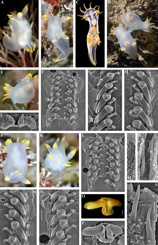
www.nature.com/scientificreports/
DNA extraction procedure, and PCR amplification options). All new sequences were deposited in Gen-
Bank (Supplementary information, Table S1, highlighted in bold). Original data and publicly available
sequences were aligned with the MAFFT algorithm30. Separate analyses were conducted for COI (658 bp),
16S (463 bp), and concatenated data (1121 bp). Evolutionary models for each data set were selected using
MrModelTest 2.331. The GTR + I + G model was chosen. Two different phylogenetic methods, Bayesian
inference (BI) and Maximum Likelihood (ML), were used to infer evolutionary relationships. Bayesian
estimation of posterior probability was performed in MrBayes 3.232. Four Markov chains were sampled
at intervals of 500 generations. Analysis was started with random starting trees and 5 × 10⁶ generations.
ML analysis was performed using RAxML 7.2.833 with 1000 bootstrap replicates. Final phylogenetic tree
images were rendered in FigTree 1.4.2. To evaluate the genetic distribution of the different haplotypes, a
haplotype network for the COI molecular data was reconstructed using Population Analysis with Reticu-
late Trees (PopART, http://p opart.otago.ac.nz) with the TCS-network method. The program M ega734 was
used to calculate uncorrected p-distances between all sequences. Additionally, Automatic Barcode Gap
Discovery (ABGD)35 was used to group the sequence data into operational taxonomic units. The align-
ment from the COI marker for all Polycera specimens under consideration was submitted and processed
in ABGD using the Jukes–Cantor (JC69) and Kimura (K80) models with default settings.
Building the periodic‑like rows of chromatic variants. Similar chromatic variants within each
potential species were aligned using calibration by the degree of light to dark surface pigmentation and
transparency of body tissue, including presence of black stripes and brownish/dark pigmentation on the
dorsal and lateral sides of the body. These similar forms establish horizontal rows of similar looking
specimens within the species (Fig. 1). In total, eight chromatic variants (horizontal rows I–VIII) are rec-
ognized: I—body semi-transparent white, orange–yellow spots or brownish/dark colouration completely
absent on the dorsal and lateral sides (orange–yellow colouration restricted to the frontal veil appendages,
tips of rhinophores, gills, and postbranchial lobes); II—body semi-transparent white, few or indistinct
orange–yellow spots present on the dorsal and lateral sides; III—orange–yellow (sometimes with reddish
hue) spots are distinct and tend to form lines; IV—in addition to orange–yellow spots blackish or brown-
ish spots or weak lines appeared (in case of lines, mostly in the anterior part of the body); V—in addition
to orange–yellow spots, blackish stripes appear (but do not form continuous lines throughout dorsal side),
blackish/brownish spots become evident and closer each other; VI—blackish stripes become evident and
form continuous lines throughout dorsal side, blackish/brownish spots begin to blend together and form
faint stripe-like pattern, orange–yellow spots evident in striped morphs, and less evident or almost absent
in spotted morphs; VII—blackish stripes or a brownish (with a greenish hue) colouration pattern become
dominant, orange–yellow spots are distinct in striped morphs, and less distinct or almost absent in spot-
ted morphs. VIII—the blackish stripes pattern remains evident or merged, whereas orange–yellow spots
(often with more intense reddish colouration) are merged into distinct lines. The proposed scheme of the
chromatic variants has a biological basis since it was shown for P. quadrilineata s.l. that during the earlier
post larval ontogenetic development orange and blackish colouration is weak or almost absent, and dis-
tinct spots, lines and stripes appear only towards later ontogenetic s tages36 and this coincides well with
general patterns of the ontogeny among dorid nudibranchs, when colourless or white forms appear first,
and intense colouration is added later.
Results
Morphological and statistical analyses. The scheme of description of the chromatic polymor-
phism among the monophyletic group of closely related species of Polycera which inhabit European waters
is consistently applied to each of the four potential species in this complex (Fig. 1, vertical columns).
Among specimens whose external and internal morphology correspond to the descriptions of Polycera
quadrilineata and P. capitata, specimens with the colour patterns which correspond to the rows II–VIII
were detected (Fig. 1, horizontal rows show eight different color patterns). In P. faeroensis specimens
with colour patterns for the rows I–III and VIII were found, whereas for P. kernowensis sp. nov. (holo-
type ZMMU Op-755, ZooBank registration: urn:lsid:-zoobank.org:act: 5C821EFD-FB12-49D5-A9C3-
026A325F6D21) specimens with colour patterns corresponding only to row I were found. Afterwards,
an additional morphological study in order to detect morphological differences between similar speci-
mens of I–III rows (semitransparent white body and absence/presence yellow-orange spots/lines, that
was observed in 42% among P. quadrilineata specimens, used for statistical analysis, 47% P. capitata,
88% P. faeroensis, and 100% of P. kernowensis sp. nov.) (Fig. 1). In the present study it is shown that
these similar white-and-yellow specimens of all four species reveal significant statistical differences in
number of rhinophoral lamellae (p = 0.023 between P. quadrilineata and P. kernowensis sp. nov.; p < 0.001
between all other species). For the number of frontal veil appendages there were no significant statistical
differences between P. quadrilineata and P. capitata. However, such differences were revealed between P.
faeroensis and P. kernowensis sp. nov. (p < 0.001); P. faeroensis compared with P. quadrilineata or P. capitata
(p < 0.001); P. kernowensis sp. nov. compared with P. quadrilineata or P. capitata (p < 0.001). There were no
significant statistical differences in number of gills for P. quadrilineata, P. capitata, and P. faeroensis. But P.
kernowensis sp. nov. shows statistically significant fewer gills compared with P. quadrilineata, P. capitata,
and P. faeroensis (p < 0.001) (Fig. 2A, Table 1, Table S2). It is unmistakably visible that P. faeroensis com-
monly possesses more than one pair of postbranchial lobes (Fig. 2B). Significant statistical differences
in external morphology were revealed for specimens of all colour patterns (Supplementary information,
Fig. S1). Mean P. faeroensis body length is statistically significantly bigger than mean body length of P.
Scientific Reports | (2021) 11:18323 | https://doi.org/10.1038/s41598-021-94863-5 3
Vol.:(0123456789)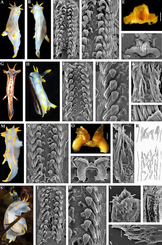
www.nature.com/scientificreports/

                                            Figure 1.  Periodic-like presentation of chromatic variation patterns among species within European Polycera,
                                            represented as vertical rows. Eight main periods (horizontal rows, roman numerals) are presented whereas
                                            spotless body/colourless forms are at the bottom and forms with a maximal number of spots/coloured are at the
                                            top. Non-observed forms for each particular species are indicated as “unkn” = “unknown”.

          Scientific Reports |   (2021) 11:18323 |              https://doi.org/10.1038/s41598-021-94863-5                                              4

Vol:.(1234567890)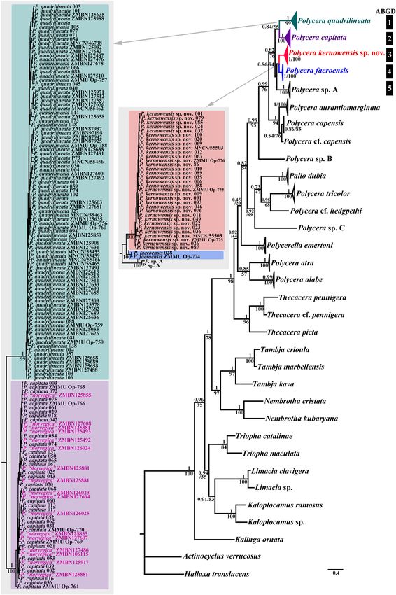
www.nature.com/scientificreports/

                                  Figure 2.  Statistical analysis (A) of the external diagnostic characters (indicated by arrows) among white and
                                  orange colour variants of detected species within European Polycera (mean ± the standard error of the mean),
                                  and presentation of the internal diagnostic features (B). Scale bars: all radulae – 100 μm, all spines − 10 μm.

Scientific Reports |   (2021) 11:18323 |               https://doi.org/10.1038/s41598-021-94863-5                                                   5

                                                                                                                                             Vol.:(0123456789)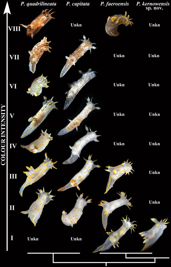
www.nature.com/scientificreports/
P. quadrilineata P. capitata P. faeroensis P. kernowensis sp. nov
Length (mm) Max. 45 Max. 16 Max. 45 Max. 20
Reported chromatic variants II–VIII II–VII I–III, VIII I
Few or numerous blackish or
Few or numerous blackish stripes brownish (with greenish hue) Chromatic variants IV–VII not Chromatic variants II–VIII had
Chromatic differences
in chromatic variants IV–VII spots present in chromatic variants known not known
IV–VII
Number of frontal veil appendages Mean 4–5, max. 7 Mean 4–5, max. 6 Mean 8–9, max. 14 Mean 7–8, max. 9
Number of lamellae of rhino-
Mean 11–12, max. 17 Mean 8, max. 11 Mean 18–19, max. 25 Mean 14–15, max. 22
phores
Number of gills Mean 6–7, max. 11 Mean 6–7, max. 9 Mean 6–7, max. 11 gills Mean 4–5, max. 7 gills
Compound (2–5 pairs plus smaller
tubercles; if simple-looking lobes,
Postbranchial lobes Simple (rarely bifurcated), one pair Simple, one pair Simple, one pair
several smaller tubercles present
in addition)
With moderately narrow shoulders With moderately narrow shoulders With broad, rounded shoulders, With moderately narrow
Jaws and strong, distinct wing-like and distinct wing-like expansions, without distinct wing-like expan- shoulders, and distinct wing-like
expansions delicate sions expansions
9–20× 9–10× 11–16× 7–11×
Reported radula formula
0–5.1.1.0.1.1.0–5 0–5.1.1.0.1.1.0–5(6?) 0–3.1.1.0.1.1.0–3 0–4.1.1.0.1.1.0–4
First teeth differ in shape from First teeth differ in shape from First teeth similar in shape and First teeth differ in shape from
First and second inner lateral teeth
second one, usually larger second one, usually larger size to second one second one, usually larger
Outer lateral teeth number Up to 5 (5 is more common) Up to 5(6?) (4 is more common) Up to 3 Up to 4
Distinct in majority of radular Distinct in majority of radular Evident in the posterior rows of
Middle cusp of first lateral teeth Not evident
teeth teeth radula
Relatively large, conspicuously Relatively small, somewhat Relatively large, conspicuously Relatively small, not evidently bent
Ampulla
bent in middle part enlarged in the proximal part bent in middle part in middle part
Bursa copulatrix Large, elongate Medium-sized, widened, oval Very large, elongate Large, elongate
Relatively long, distinctly widened Relatively long, not widened Relatively long, not widened
Vas deferens Very long, not widened distally
distally distally distally
Long, straight or slightly bent
Needle-shaped, straight or winding Short hooks or short to elongate spines or shorter more elongate
Copulative spines Elongate, somewhat hooked cones
or shorter cones cones cones with a peculiar base with
a hole
Table 1. Verified and updated comparison of key diagnostic characters of the European Polycera species
complex based on the multilevel organismal diversity approach. The characters are given for adults or subadults
individuals.
quadrilineata, P. capitata, and P. kernowensis sp. nov. Mean P. capitata and P. kernowensis sp. nov. body
length is statistically significantly smaller than mean body length of P. quadrilineata and P. faeroensis
(p < 0.001, Fig. S1, Table S2). In addition, differences in internal morphology were revealed (Fig. 2B). P.
capitata have clear differences from the other species in shape of the copulative spines. P. faeroensis have
clear differences in shape and size of the lateral teeth of radula.
Molecular phylogenetic analysis. Phylogenetic analysis was performed using 197 specimens of
Polycera, including data for 178 P. quadrilineata species complex (102 of which were preliminarily divided
into four groups using the methods described above and data for 76 downloaded from GenBank), and
24 outgroup specimens. The dataset consisted of 356 nucleotide sequences including COI and 16S genes.
Bayesian Inference (BI) and Maximum Likelihood (ML) analyses based on the combined dataset for the
COI, and 16S genes yielded similar results (Fig. 3). To define species, we use a set of m ethods12,37 includ-
ing phylogenetic tree topologies, ABGD analysis, pairwise distances and the haplotype network contain-
ing phylogeographic data rendered using PopART (Fig. 4, Table 2). The results of this study confidently
confirmed the presence of four species among P. quadrilineata similar specimens that coincided with
species detected before the molecular study: P. quadrilineata, P. capitata (that do not show any differences
from the recently described P. norvegica), P. faeroensis, and P. kernowensis sp. nov. A clade containing P.
quadrilineata (n = 92, PP = 1, BS = 99) has the closest position to the clades containing P. capitata (n = 34)
combined with former P. norvegica (n = 17, PP = 1, BS = 100), P. faeroensis (n = 2, PP = 1, BS = 100), P. ker-
nowensis sp. nov. (n = 33, PP = 1, BS = 100), and two Polycera sp. A from South Africa (PP = 1, BS = 100).
P. quadrilineata and P. capitata (combined with former P. norvegica) clustered in two distinct and well
separated sister clades that formed the sister group to P. faeroensis, P. kernowensis sp. nov., and P. sp. A,
which are clustered together in a separate clade, wherein P. faeroensis has sister position to P. kernowensis
sp. nov. The ABGD analysis of the COI data set run with two different models are fully concordant with
the clades in the molecular phylogenetic analysis (Fig. 3). Results obtained by PopART showed a network
of haplotypes that clearly clustered into four distinct groups coincident with P. quadrilineata, P. capitata
(combined with former P. norvegica), P. faeroensis, and P. kernowensis sp. nov. (Fig. 4). While no correla-
tion was found between the molecular characteristic for each of the four species and the geographical
Scientific Reports | (2021) 11:18323 | https://doi.org/10.1038/s41598-021-94863-5 6
Vol:.(1234567890)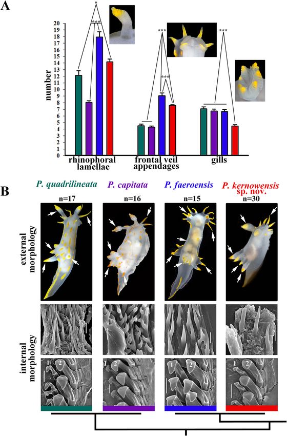
www.nature.com/scientificreports/

                                  Figure 3.  Phylogenetic relationships of Polycera specimen based on COI + 16S concatenated dataset inferred
                                  with Bayesian (BI) inference. Numbers above branches represent the posterior probabilities from BI. Numbers
                                  below branches indicate the bootstrap values for Maximum Likelihood (ML).

Scientific Reports |   (2021) 11:18323 |              https://doi.org/10.1038/s41598-021-94863-5                                                7

                                                                                                                                         Vol.:(0123456789)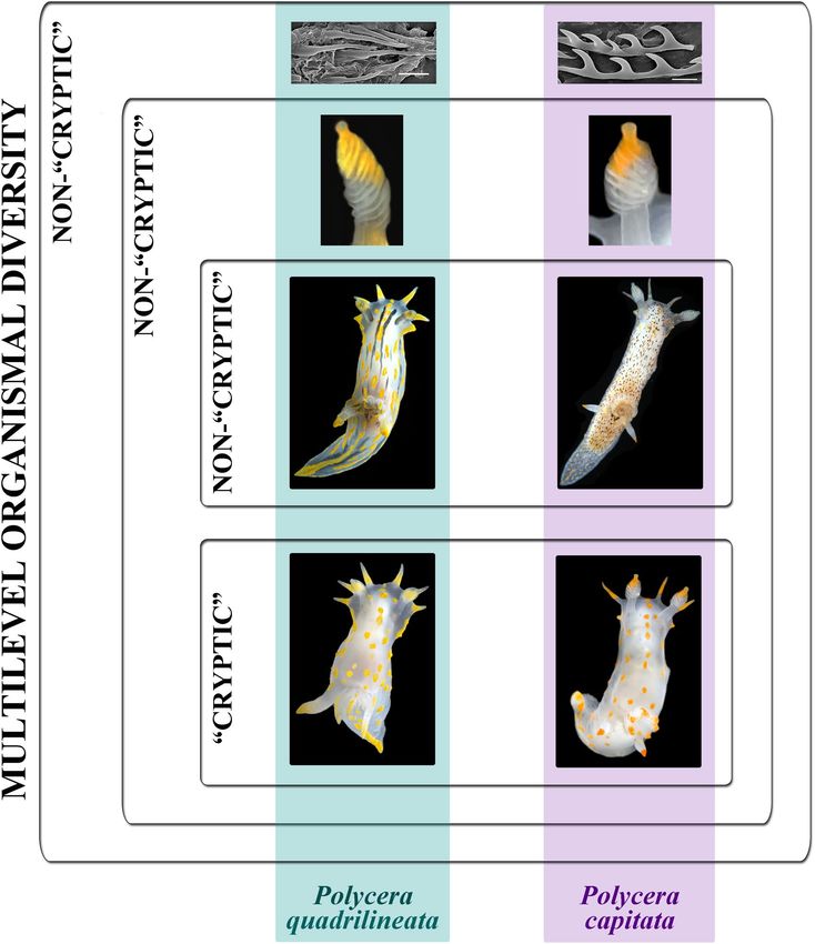
www.nature.com/scientificreports/
Figure 4. The haplotype network for Polycera specimens based on cytochrome c oxidase subunit I (COI)
molecular data (642 bp). Phylogeographic data are represented within a broad geographic framework. Each
circle represents one haplotype, the circle area indicates the occurrence rate. Each cluster (species) is coloured
individually (P. quadrilineata in green, P. capitata in purple, “P. norvegica” in pink, P. faeroensis in blue and
P. kernowensis sp. nov. in red.). DM Denmark, GM Germany, IR Ireland, NL Netherlands, NW Norway, PG
Portugal, SP Spain, SW Sweden, UK United Kingdom.
distribution of each of the four species from this complex, P. capitata and “P. norvegica” recently described
from Norway have a widespread distribution in England, Ireland and Norway, and do not show separate
clustering. Regarding the supposedly fast-evolving COI marker, uncorrected p-distances within the P.
quadrilineata clade range 0–3.36%. Whereas minimal uncorrected p-distances between the P. quadriline-
ata clade and P. capitata (combined with P. norvegica), P. faeroensis, and P. kernowensis sp. nov. clades are
9.44%, 11.09%, and 10.79% respectively. Uncorrected COI p-distances within the P. capitata (combined
with P. norvegica) clade range from 0.15 to 3.04%. Minimal uncorrected p-distances between the P. capi-
tata (combined with P. norvegica) clade and P. faeroensis, and P. kernowensis sp. nov. clades are 8.52%,
and 8.66% respectively. Uncorrected COI p-distances within the P. faeroensis clade are 0.61%; within the
P. kernowensis sp. nov. clade range 0–2.33%. Uncorrected COI p-distances between P. faeroensis and P.
kernowensis sp. nov. clades range from 5.47 to 6.38% (Table 2).
Polycera aurantiomarginata García-Gómez & Bobo, 1984, which was described from Spain and which
is also distributed on the West African coasts22 belongs to a different Polycera clade, considerably differ-
ent morphologically, and hence is not part of the complex of the European species closely related to P.
quadrilineata. Therefore P. aurantiomarginata is included in the present molecular phylogenetic analysis,
Scientific Reports | (2021) 11:18323 | https://doi.org/10.1038/s41598-021-94863-5 8
Vol:.(1234567890)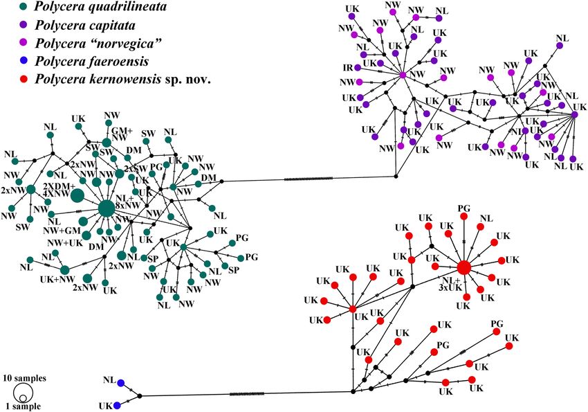
www.nature.com/scientificreports/
P. capitata (including P.
P. quadrilineata norvegica syn.nov.) P. faeroensis P. kernowensis sp. nov P. sp. A
P. quadrilineata 0–3.36 9.44–12.68 11.09–13.13 10.79–14.53 10.42–12.5
P. capitata (including P. nor-
9.44–12.68 0.15–3.04 8,52–9.97 8.66–11.99 9.95–11.21
vegica syn.nov.)
P. faeroensis 11.09–13.13 8.52–9.97 0.61 5.47–6.38 8.09
P. kernowensis sp. nov 10.79–14.53 8.66–11.99 5.47–6.38 0–2.33 8.71–10.42
P. sp. A 10.42–12.5 9.95–11.21 8.09 8.71–10.42 1.4
Table 2. Intragroup (highlighted in bold) and Intergroup genetic distances (%) for COI in the European
Polycera species complex.
but not considered in this study in detail (Fig. 3). An undescribed species (“Polycera sp. A”) which was
included to a molecular phylogenetic study without any morphological d ata14 is basal to the clade of the
European Polycera (Fig. 3) but occurs exclusively in South Africa and is out of scope in the present study.
For Polycera marplatensis Franceschi, 1 928 38 which is partly similar to P. quadrilineata molecular data
are not available, but this species occurs exclusively in South America and therefore is also out of scope
of the present study.
The morphological analysis data were confirmed by molecular phylogenetic results. P. quadrilineata, P.
capitata, P. faeroensis, and P. kernowensis sp. nov. are four separate species in the genus Polycera. P. capitata
and former P. norvegica are the same species.
Difficult to distinguish European species within the genus Polycera: recognition of the
involved species. We obtained a very robust framework of four closely related species from Euro-
pean waters: Polycera quadrilineata, P. capitata, P. faeroensis and P. kernowensis sp. nov. (Figs. 3, 4, for
detailed systematic account of all four species see Supplementary information S2). Polycera quadrilineata
and P. capitata are the two common European species and usually present in the shallow marine waters at
depths easily accessible for diving (ca. 5–40 m), making these species always a focus of attention of vari-
ous environmental associations, ecological studies and currently also citizen scientists. Therefore it is of
high general importance to present a framework for morphological identification of these species complex
based on robust molecular data (Tables 1, 2).
Throughout the history of nudibranch studies in Europe, a species similar, but distinct from P. faeroensis has
been confused with the latter and was never taxonomically r ecognized14,39,40. In the present study this species
using robust molecular framework based on a broad geographical sampling and significant external and internal
morphological differences (Figs. 1, 2, 9; Table 1) it is for the first time recognized and described as a new species,
Polycera kernowensis sp. nov. (see details in Supplementary information S2). This species has significantly more
rhinophoral lamellae (14–15) than in P. capitata (8) and P. quadrilineata (11–12) and same is true for the frontal
veil appendages (7–8 vs. 4–5) including specimens of similar sizes (Figs. 2, Supplementary information Fig. S1).
Polycera quadrilineata can reach a similar large size as P. faeroensis (commonly the former species is smaller), but
the mean number of rhinophoral lamellae in P. quadrilineata is significantly smaller (11–12), than in P. faeroensis
(18–19) (Figs. 2, Fig. S1) and even in largest specimens of P. quadrilineata the number of rhinophoral lamellae is
smaller than in P. faeroensis. In addition, the number of the frontal veil appendages differs with a high support
among these three species (Fig. 2, Supplementary information S2, Fig. S1).
Remarkably, in this study we also recovered two closely related species P. quadrilineata and P. capitata as sister
species (Fig. 3), in contrast to a previous s tudy14, because taxon selection was previously not exhaustive. True
P. faeroensis is a more rarely encountered species than P. quadrilineata and P. capitata (at least in the relatively
shallow water environments) and verified molecular data which were aligned with the fine-scale morphological
data are presented in this study for the first time (Figs. 1, 2, 3, 4, 5, 6, 7, 8, 9, 10). Previously real P. faeroensis were
misidentified14,40 with its new sister species, here described as P. kernowensis sp. nov. (Figs. 8, 9). Our verified
data on the morphology of the radula matched well with the original description of P. faeroensis from the Faeroe
Islands41 and a morphological redescription from S weden42.
Scientific Reports | (2021) 11:18323 | https://doi.org/10.1038/s41598-021-94863-5 9
Vol.:(0123456789)www.nature.com/scientificreports/
Figure 5. Polycera quadrilineata (O.F. Müller, 1776). External and internal morphology, light and scanning
electron microscopy and comparison with data from Thompson and Brown40 and Bergh46. (A) Neotype from
Norway, Gulen, dorsal view, 25 mm in length, live (ZMMU Op-750). (B) Same, ventral view. (C) Radula,
neotype, scanning electron microscopy (SEM). (D) Same, radula details. (E) Jaws, neotype, light microscopy.
(F) Same, SEM. (G) Specimen from United Kingdom, Skomer Island, depicted in Thompson and Brown (40:
pl 18, a). H. Specimen from United Kingdom, Cornwall, Porthkerris, dorsolateral view, live (ZMMU Op-758).
(I) Radula, SEM (ZMMU Op-758). (J) Same, radula details. (K) Copulative spines, SEM (ZMMU Op-758).
(L) Same, details. (M) Specimen from Portugal, Setubal, dorsal view, live (ZMMU Op-760). (N) Radula, SEM
(ZMMU Op-760). (O) Jaws, light microscopy (ZMMU Op-760). (P) Same, SEM. (Q) Copulative spines, SEM
(ZMMU Op-760). (R) Spines of a specimen from Norway depicted in Bergh (46, not in copyright), essentially
similar to our present data. (S) Specimen from United Kingdom, Cornwall, Porthkerris, dorsolateral view, live
(ZMMU Op-756). (T) Radula, SEM (ZMMU Op-756). (U) Same, radula details. (V) Copulative spines from
the tip of everted part (shorter than common ones), SEM (ZMMU Op-756). (W) Copulative spines (common
type), light microscopy (FD 041). (X) Same, SEM. Scale bars: c, e, f—500 μm, d, i, j, o, p, t—200 μm, k, q, v, w,
x—10 μm, l—5 μm, n, u—100 μm. Photographs: Tatiana Korshunova: (a, b); F.M.F. Driessen (h), (s); Bernard
Picton (m). Reproduction of figure from Thompson and B rown40 with permission of Gregory Brown, original
artist and copyright holder of the images. SEM micrographs and light microscopy photographs: Alexander
Martynov.
Scientific Reports | (2021) 11:18323 | https://doi.org/10.1038/s41598-021-94863-5 10
Vol:.(1234567890)www.nature.com/scientificreports/
Figure 6. Schemes of the reproductive systems. (A) Polycera quadrilineata. (B) P. capitata. (C) P. faeroensis. (D)
P. kernowensis sp. nov. am ampulla, bc bursa copulatrix, fgm female gland mass, go genital opening, pr prostate,
psh penial sheath, rs receptaculum seminis, ud uterine duct, vd vas deferens, vg vagina. Scale bars: 1 mm.
Scientific Reports | (2021) 11:18323 | https://doi.org/10.1038/s41598-021-94863-5 11
Vol.:(0123456789)www.nature.com/scientificreports/
Figure 7. Polycera capitata (Alder & Hancock, 1854)49. External and internal morphology, light and scanning
electron microscopy and comparison with data from Thompson and Brown40. (A) Specimen from United
Kingdom, Cornwall, Porthkerris, dorsal view, live (ZMMU Op-766). (B) Radula, SEM (ZMMU Op-766).
(C) Same, radula details. (D) Jaws, SEM. (E) Copulative spines, SEM (ZMMU Op-766). (F) Same, details.
(G) Specimen from United Kingdom, Cornwall, Porthkerris, dorsal view, live (ZMMU Op-764). (H) Same,
lateroventral view. (I) Radula, SEM (ZMMU Op-764). (J) Same, details. (K) Jaws, SEM (ZMMU Op-764). (L)
Copulative spines, SEM (ZMMU Op-764). (M) Specimen from United Kingdom, Lundy Island, depicted in
Thompson and Brown (40: pl 18, c). (N) Specimen from United Kingdom, Cornwall, Porthkerris, ventral view,
live (ZMMU Op-765). (O) Same, ventral view. (P) Radula, details, SEM (ZMMU Op-765). (Q) Radula (ZMMU
Op-765). (R) Copulative spines, SEM (ZMMU Op-765). (S) Same, light microscopy. (T) Specimen from Ireland,
Mullaghmore, Sligo, dorsal view (ZMMU Op-770). (U) Same, ventral view. (V) Radula, details, SEM (ZMMU
Op-770). (W–Y) Copulative spines, details, SEM (ZMMU Op-770). (Z) Same, light microscopy. Scale bars: b–d,
j, k, p, v—100 μm, i, q—200 μm, e, f, l, s, w–z—10 μm, r—5 μm. Photographs: F.M.F. Driessen (a), (g), (h), (n),
(o); Bernard Picton (t), (u). Reproduction of figure from Thompson and Brown40 with permission of Gregory
Brown, original artist and copyright holder of the images. SEM micrographs and light microscopy photographs:
Alexander Martynov, Tatiana Korshunova.
Scientific Reports | (2021) 11:18323 | https://doi.org/10.1038/s41598-021-94863-5 12
Vol:.(1234567890)www.nature.com/scientificreports/
Figure 8. Polycera faeroensis Lemche, 192941. External and internal morphology, light and scanning electron
microscopy and comparison with data from Thompson and B rown40. (A, B) Specimen from United Kingdom,
Belfast Lough, Northern Ireland, dorsal views, live (ZMMU Op-774). (C) Specimen from United Kingdom,
Strangford Lough, Northern Ireland, dorsolateral view, live. (D) Another specimen from Strangford Lough,
Northern Ireland, lateral view, live. (E) Specimen from the United Kingdom, Skomer Island, Pembrokeshire,
dorsal view, live. (F) Juvenile specimen from the Strangford Lough, dorsal view, live. (G) Copulating specimens
from Skomer Island, Pembrokeshire, Wales, dorsal views. (H) Specimen from United Kingdom, Isle of Man,
depicted in Thompson and Brown (40: pl 18, g). (I) Schemes of jaws (above) and radula teeth (below) of
holotype from the Faeroe Islands. (J) Radula, SEM (ZMMU Op-774). (K, L) Same, details of radula. (M) Jaws,
light microscopy (ZMMU Op-774). (N) Same, details, SEM. (O, P) Copulative spines, SEM (specimen from
Belfast Lough). (Q) Same, light microscopy. Scale bars: j–l—100 μm, m, n—200 μm, o–q—10 μm. Photographs:
Bernard Picton (a–g). Reproduction of figure from Thompson and Brown40 with permission of Gregory Brown,
original artist and copyright holder of the images. SEM micrographs and light microscopy photographs:
Alexander Martynov.
Scientific Reports | (2021) 11:18323 | https://doi.org/10.1038/s41598-021-94863-5 13
Vol.:(0123456789)www.nature.com/scientificreports/
Figure 9. Polycera kernowensis sp. nov. External and internal morphology, light and scanning electron
microscopy and comparison with data from Thompson and B rown40. (A) Paratype from United Kingdom,
Cornwall, Porthkerris, dorsal view, live (FD 01). (B) Paratype from United Kingdom, Cornwall, Porthkerris,
dorsal view, live (FD 093). (C) Specimen from United Kingdom, Skomer Island, depicted in Thompson and
Brown (40: pl 18, e) identified as P. faeroensis but which has some features that more similar to P. kernowensis sp.
nov. (D) Copulating specimens from the Great Britain, dorsal view, live. (E) Paratype from Portugal, Setubal,
lateral view, live (ZMMU Op-775). (F) Radula, SEM (ZMMU Op-775). (G, H) Radula details, SEM (ZMMU
Op-775). (I) Jaws, SEM (ZMMU Op-775). (J) Holotype from United Kingdom, Cornwall, Porthkerris, dorsal
view, 5.6 mm, live (ZMMU Op-755). (K) Same, frontal view, live. (L) Radula of holotype, SEM. (M, N) Radula
details (holotype), SEM. (O) Jaws (holotype), light microscopy. (P) Jaws (holotype), SEM. (Q) Copulative spines
(holotype), light microscopy. (R, S) Copulative spines (holotype), SEM. Scale bars: f–i, l–p—100 μm, q—10 μm,
r, s—5 μm. Photographs: F.M.F. Driessen (a), (b), (e), (j), (k); (d) Bernard Picton. Reproduction of figure from
Thompson and Brown40 with permission of Gregory Brown, original artist and copyright holder of the images.
SEM micrographs and light microscopy photographs: Alexander Martynov.
Scientific Reports | (2021) 11:18323 | https://doi.org/10.1038/s41598-021-94863-5 14
Vol:.(1234567890)www.nature.com/scientificreports/
Figure 10. Presentation of the multilevel organismal diversity approach using the example of closely related
Polycera quadrilineata and P. capitata: presence of conditional “non-cryptic” and putative “cryptic” components
among the same species and provision of fine-scale morphological diagnostic characters even among the
“cryptic component” within a single species. Scale bars—10 μm.
The results of the statistical test in key diagnostic external features (number of rhinophoral lamellae, frontal
veil appendages and gills) in the Polycera species complex (Fig. 2, Supplementary information S2), showed that
easily accessible external morphological features are distinguishing factors among adult individuals between the
species in the complex, with high statistical support (Figs. 2, Supplementary information S2, Fig. S1). Particularly,
the number of the rhinophoral lamellae differs with high support among all four species (Figs. 2, 10, Supplemen-
tary information S1). It must be noted that differences in rhinophoral lamellae are partly related to the animal
length, due to the fact that larger specimens may have larger numbers of rhinophoral lamellae, starting from 0
(i.e. smooth rhinophores) and reaching a mean number of lamellae towards mature s tages43,44. However, even
most closely related sister species P. quadrilineata and P. capitata were revealed as having smaller, but statistically
significant differences in the mean number of the rhinophoral lamellae (mean 8 in P. capitata and 11–12 in P.
quadrilineata) including specimens of similar sizes (Figs. 2, 10, Supplementary information S2).
The significant differences can be further used for the fine-scale diagnostics of these species. This is a very
important result for further assessment of the general reliability of the “cryptic species” concept.
Scientific Reports | (2021) 11:18323 | https://doi.org/10.1038/s41598-021-94863-5 15
Vol.:(0123456789)www.nature.com/scientificreports/
Discussion
Importance of the true, and not a formal integration of the molecular, morphological, and
taxonomic data for applications in ecological and evolutionary studies. The present study is very
relevant for the investigation of the general species concept and so-called “cryptic species” concept and also
for the currently commonly claimed “integration” of molecular and morphological data. It was already high-
lighted that the challenge represented by cryptic species has great importance for general biological problems
and represents “a window into the paradigm shift of the species concept”20. We therefore present in this study
the largest currently possible molecular and morphological dataset on a particular nudibranch species complex,
which is widely distributed over the vast European region. Molecular data of 178 specimens belonging to four
species were involved in the molecular phylogenetic analysis (Fig. 3). Furthermore, for the practitioners, and
for researchers who perform environmental monitoring in the European marine waters, it is very important
to trustworthily distinguish the four species, including the most difficult to distinguish P. quadrilineata and P.
capitata, in the field and, above all, without complicated, time-consuming and expensive molecular analysis. This
is especially important since P. quadrilineata complex represents an indicator of biodiversity shifts in relation to
climatic changes27.
In order to develop a robust framework to make it possible to identify all four species using only easily
accessible external characters and applying in the majority of cases, we hereby provide a summary of an
exhaustive analysis of molecular and morphological data for all European Polycera species (Figs. 1, 2, 3, 4,
5, 6, 7, 8, 9, Tables 1, 2), i.e. every newly available specimen or sequence from European waters belonging
to each of the four recognized species (Fig. 3, Tables 1, 2) and not to any other potential “hidden” lineages.
Polycera quadrilineata (O. F. Müller, 1776) is one of the oldest species names amongst European nudi-
branch m olluscs 45,46. Since then there were numerous attempts to separate more species similar to P.
quadrilineata (see the Supplementary information S1 for details on the systematics of P. quadrilineata),
however all of them without a detailed morphological analysis were later considered to be synonyms
of P. quadrilineata 40. Using molecular data, Driessen et al. 21 showed two lineages among European “P.
quadrilineata” for the first time. Recently one of these lineages has been formally described as Polycera
norvegica14. The description proposed the species to occur exclusively in Norway. According to our data,
applying a very broad geographic sampling in frames of the present study (see e.g., Figs. 3, 4) the recently
named “Polycera norvegica” definitely is not distributed solely in Norway, but is a species widely distrib-
uted in other European countries, such as the United Kingdom and Ireland (Supplementary information,
Table S1). Furthermore, in that publication, the numerous synonyms of P. quadrilineata were neither listed
nor investigated, and its synonymy was just referred to the internet database M olluscaBase47.
In the present study we carefully investigated all existing available synonyms of P. quadrilineata (Figs. 1,
2, 3, 4, 5, 6, 7, Table 1, Supplementary information Fig. S1). Therefore, being part of the difficult to dis-
tinguish P. quadrilineata complex, for which more than ten synonyms have been suggested, and having a
broad distribution in Europe, “P. norvegica” could have already been described. If “P. norvegica” had no
clear recognizable chromatic variants which immediately distinguished it from P. quadrilineata it would
be difficult to assess a potential synonym. However, “darker” chromatic variants (Fig. 1IV–VII) are clearly
different between P. quadrilineata and “P. norvegica”, which makes a solid base for the search for such
previously described variants within taxonomic synonyms. During this study we identified two avail-
able taxonomic names that contain chromatic variants with darker colouration without evident stripes,
and those can be thus excluded from the striped variants of true P. quadrilineata (Figs. 1, 2, 5, 7). These
available names are Polycera ornata d’Orbigny, 1837 and Thecacera capitata Alder & Hancock, 1854. For
Polycera ornata a main morph with red–orange lines was d escribed48 and in addition a morph with weaker
orange and black spots was mentioned. Because of a main morph with orange-reddish lines in Polycera
ornata, and having only an old painting published, it is difficult to surely distinguish the morph from P.
quadrilineata. Furthermore, for P. ornata a “tiger-like” colouration was indicated for the darker m orph48,
that may imply stripes, as in true P. quadrilineata. Thus, to assign Polycera ornata to this species would be
ambiguous. Also, the type material of Polycera ornata was not saved.
Instead, for Thecacera capitata in the original description by Alder & Hancock (1854) the coloura-
tion was solely indicated as “freckled with brownish greenish” 49 that immediately allows exclusion of any
morphs with evident black stripes, that are present only in true P. quadrilineata (Figs. 1, 5). Thecacera
capitata was later confirmed as belonging to the genus Polycera and not to Thecacera, including study
of the type m aterial 40; presence of rhinophoral sheaths for P. capitata was thus indicated m istakenly 49.
Polycera capitata was partly redescribed with aid of the type material regarding external features and the
external colouration40 and exactly matched the chromatic variant V in P. norvegica but not any chromatic
variants of P. quadrilineata (Figs. 1, 7M). In this study we additionally studied the saved radula from the
type specimen of Polycera capitata as was figured in the original d escription49. The radula has only four
outer lateral teeth and thus fits well to the most rigorous assessment of the radular characters in this spe-
cies, including “P. norvegica”14.
This combination of the external and internal data excludes the possibility that Polycera capitata is a
synonym of P. quadrilineata but instead conforms closely to the characters that were recently described
for P. norvegica 14. We have involved numerous specimens from the United Kingdom and Ireland—that
matched well morphologically to the original description of P. capitata (Figs. 1, 7)—in the present molecu-
lar analysis. The present study shows that darker chromatic variants V–VII without distinct stripes are
characteristic solely for P. capitata (Figs. 1, 7, 10). Furthermore, these darker chromatic variants of P.
capitata are very common throughout the United Kingdom from Cornwall in the south to the Scotland in
the north, including the type locality at St. Ives, Cornwall according to the available photographic d ata50,51.
Scientific Reports | (2021) 11:18323 | https://doi.org/10.1038/s41598-021-94863-5 16
Vol:.(1234567890)www.nature.com/scientificreports/
These data were already available, but an essential external similarity of “P. norvegica” to P. capitata was
orway14. “P. norvegica” syn. nov. thus
missed and it was incorrectly stated that the species is restricted to N
becomes a junior synonym of Polycera capitata. Additionally, (with permission) we present a copy of plate
18c from Thompson & Brown (1984) 40 where the external features illustrated for Polycera capitata cor-
respond exactly with our morphological data for P. capitata (Fig. 7M). Therefore, our present taxonomic
assignment of P. capitata is robustly supported by the abundant morphological (including statistical study),
molecular and distributional data (Figs. 1, 2, 3, 4, 5, 6, 7, Table 1, Supplementary information, Table S1,
Fig. S1). In the present study we therefore restore the species P. capitata and P. norvegica becomes its
junior synonym.
Importantly, what appears to be a particular taxonomic problem, has in reality general importance
for an array of biological fields since it clearly shows that without true integration of an “old” taxonomic
knowledge, that tends to be neglected currently, with modern morphological and molecular data, an
appropriate study of world biodiversity cannot be performed. The “cryptic species problem” therefore does
not emerge only recently, but was always part of taxonomy since the Linnean era. A formal integration
of the molecular and morphological data and inaccurate claim for “cryptic species” led to omission of a
synonym for a common, species which is important for ecological monitoring. Therefore, the importance
of true and not just a formal integration of molecular, morphological and taxonomic data are here specially
highlighted and a practical set of methods is proposed below.
Periodic‑like framework for the recognition diagnosable characters in the species com-
plexes. Periodic-like as well as parallel-like patterns in biological applications have been discussed
for a long time and successfully applied for protein s tructure52, but they are still not widely used practi-
cal tools for taxonomy and phylogeny. Several recent studies on different groups, such as rodents53 and
f ishes54,55 robustly confirmed the existence of periodic patterns during development of morphological
characters at a genomic level, particularly concerning chromatic variants and thus are directly connected
with the present Polycera case. Notably, the ontogenetic periodicity is based on periodicity of homeobox
and other developmental gene systems in animals and plants56,57, thus approaching chemical periodic-
ity. Recently an evident periodicity was revealed for a higher-level organism group using an ontogenetic
phylotypic periods/stages approach, that consistently links the genomic and morphological l evels44. There
were attempts to describe chromatic variants within another nudibranch family Chromodorididae under
the term “colour groups”58 or as different colour morphs in frames of a phylogeny59,60, but not in a periodic
framework. However, when similar morphs of different closely related species are mapped in the same
horizontal sections, the partial periodicity can be clearly r evealed18.
During individual development, this was investigated for Polycera quadrilineata 36, the darker coloura-
tion appeared during later stages of the ontogeny, and similar patterns of the ontogeny of the sister species
P. capitata must present in parallel, which makes the biological grounds for the periodic-like approach.
Such an approach is a practical one and helps to reveal fine distinguishing details among apparently very
similar morphs (e.g. chromatic variants) and also potentially not yet discovered morphs of the closely
related species. Currently we have no information about which particular genes underlie any common
genetic basis in the polychromatic nudibranchs 61, but it must inevitably imply similar developmental
genes basis and it was recently used for delineation of a very difficult nudibranch species complex of
the genus Amphorina 18. A North Pacific species, Polycera atra MacFarland, 1905, provides evidence for
the existence of the underlying similar genomic basis, that appears in parallel in various phylogenetic
lineages. According to the present analysis it represents a taxon which is only distantly related to the P.
quadrilineata complex (Fig. 3), and potentially may belong to a separate genus (a general revision of the
family Polyceridae is pending), but nevertheless exhibits similar chromatic v ariants62. As in the P. quad-
rilineata complex, about eight chromatic variants of P. atra are recognized. These are used in the present
study to align the chromatic variants found within the European Polycera species (Fig. 1). Such parallel
appearance of the similar chromatic variations in relatively distantly related clades is well matched to the
parallel appearance of the shell morphs within gastropod molluscs 63, for which a similar genomic base
has already been c onfirmed64.
The present Polycera chromatic polymorphism is a clear case of periodic appearance of similar col-
our morphs among phylogenetically related but different species which has robust support from a large
molecular dataset (Figs. 1, 3). Polycera quadrilineata also possesses stripe patterns in their colouration
(Fig. 1V–VIII), and similar patterns were recently showed as underlain by ontogenetic periodicity in vari-
ous g roups54,55. Within P. capitata instead darker forms (Fig. 1V–VIII) appear in parallel, without distinct
stripes, but with remnants of a faint stripe-like pattern (Fig. 1VI). In both P. faeroensis and P. kernowensis
sp. nov. the chromatic variants IV–VII are not yet discovered (Fig. 1). This makes the method a practical
tool for revealing a diagnosable character within putative “cryptic species complexes”. While biologists
from non-taxonomic fields or experienced practitioners have the task to identify Polycera in the field,
they will be guided by a periodic-like mapping (Fig. 1) which is based on accurate taxonomy and robust
molecular phylogenetic data (Figs. 3, 4). It will be easier to exclude these variants that do not occur (as
striped or heavily spotted variants are not yet found in P. faeroensis and P. kernowensis sp. nov.), and
instead carefully investigate similar chromatic variants II and III in two sister species P. quadrilineata
and P. capitata (Fig. 1).
When such periodic-like mapping becomes a routine part of biodiversity studies, the accumulated
data will allow the presentation of small, fine-scale distinguishing characters even between such highly
similar orange-spotted chromatic variants that are present in both P. quadrilineata and P. capitata. At
Scientific Reports | (2021) 11:18323 | https://doi.org/10.1038/s41598-021-94863-5 17
Vol.:(0123456789)www.nature.com/scientificreports/
present, they remain difficult to distinguish, but using available data we can preliminarily conclude that
in P. capitata orange spots are more commonly smaller and more rarely form lines, than in P. quadrilineata
(Figs. 1, 2, 5, 7). A statistical test of the diagnostic value of the external characters, that were mapped in
the periodic-like framework, revealed that number of the rhinophoral lamellae, though it may overlap in
juvenile specimens, in adult specimens is different with a statistical high support including among these
difficult to distinguish chromatic variants II and III between P. capitata and P. quadrilineata (Fig. 2, Sup-
plementary information S1, Fig. S1). Thus, the number of the rhinophoral lamellae (a very easy to check
character even on the photographs), can be used as an additional verification in case of similar chromatic
variants II and III between P. capitata (commonly less than ten rhinophoral lamellae in adults) and P.
quadrilineata (commonly more than ten rhinophoral lamellae in adults).
The periodic-like mapping of the chromatic variants revealed that orange dots on the body sometimes occur,
as well as potentially darker morphs in true P. faeroensis (Fig. 1II–VIII), but patterns and quantity differ between
P. quadrilineata and P. capitata (Fig. 1II–VIII). Previously all four species of the European Polycera complex have
been confused with each other40,65 also because these chromatic variants were previously never accurately mapped
with each other, but instead it was commonly noted that similar to P. quadrilineata and P. capitata yellow–orange
dots were sometimes present on the body in P. faeroensis, which immediately misled practitioners that have had
tasks to identify animals in the field. Further, this profound confusion among identification of European Polycera
persists even using modern molecular tools as P. faeroensis until the present study was not distinguished from
P. kernowensis sp. nov., despite the presence of robust external, internal, and molecular differences (Figs. 1, 2, 3,
4, 8, 9). Thus, the periodic-like mapping of the polychromatic variants in combination with statistical analysis
of the diagnostic characters is an important tool in addition to the molecular phylogenetic analysis. It will be
particularly important since P. quadrilineata and P. capitata are two of the most common nudibranch species in
Europe, and particularly in the UK the further testing of their chromatic variance already attracted a ttention51
and further attraction of various environmental and educational organizations and citizen scientists is expected.
Our present study builds a major framework for further broader testing of the colour polymorphism and other
distinguishing features in the Polycera species complex that will have importance for the development of docu-
menting the fine-scale diversity not only in other nudibranchs, but in the variety of multicellular organisms.
Practical guidelines how to perform a taxonomic study in the molecular era. The pre-
sent study evidently shows that “cryptic” and “non-cryptic” components are present within the same
species. This further significantly undermines the “cryptic species” concept, which recently was already
questioned4,10–12. The presence of both “cryptic” and “non-cryptic” components is demonstrated here
for sister species P. quadrilineata and P. capitata sharing externally similar chromatic variants II and III
(Fig. 1), which however can be distinguished by the morphological features of another level (Figs. 5, 7).
Particularly, the internal features of the shape and size of copulative spines allow in 100% cases to con-
firm the identity of both species (compare Figs. 5K, L, Q, R, V–X and 7E, F, L, R, S, W–Z), even without
aid of molecular data this undermines the proposal that these species are morphologically “cryptic”. In
our recent s tudy12 we already showed how the underestimation of the taxonomic and morphological data
resulted in a long-term omission of reliable multilevel differences in the nudibranch Trinchesia species
complex. Recently, there are proposals that taxonomy should be an integrative study66–68. However, in
current common practice “integration” unfortunately mainly means performing a molecular study on
some selection of specimens and then morphological features listed “in addition”, rather as an auxiliary
information. The best confirmation of this is the recent Polycera study14, when crucial previous taxonomic
information was omitted, and while some morphological diagnostic features were revealed, both their
stability and variability (and hence, their usefulness for taxonomic diagnostics) were over- or underesti-
mated that subsequently led to an incorrect statement about “general crypticity” of the involved species.
To avoid this, practical guidelines proposing how to perform a taxonomic study in the molecular era are
outlined here: (1) make a selection of a taxonomic group and appropriate specimens; (2) make a relevant
morphological study in a given group, including for example scanning electron microscopy of previously
commonly used diagnostic characters in the given group; (3) ensure that ontogenetic information is con-
sidered during taxonomic assessment, because adult diagnostic characters can be considerably transformed
at different ontogenetic stages, whereas adult paedomorphic characters can be easily misidentified with
juvenile transitive f eatures44,69–71; (4) make an appropriate bibliographic study, to exhaustively study the
synonymy of a studied taxa/species group, importantly, not just as a reference to a taxonomic data base,
but to perform a real study of original sources; (5) molecular study selected and taxonomically checked
specimens with commonly used genetic markers in a given group; (6) compare the results of the morpho-
logical (step 2) and taxonomic investigation (steps 3, 4) with the molecular (step 5) results; (7) in case
of finding discrepancies between previously commonly assessed diagnostic characters in a given species
group and results of molecular phylogeny, respective diagnostic features of specimens in question should
be presented in a periodic-like framework of the parallel rows, that will enable their detailed comparison
and further search for fine-scale differences between given rows for each of the closely related species; (8)
in case difficult to distinguish variants are present among the same parallel rows, a statistic study of the
relevant diagnostic features should be performed in order to reveal fine-scale differences among closely
related species; (9) a complete study at a given time and using current research possibilities should result in
fine-scale taxonomic diagnoses for all closely related species in a given taxa/complex (including new taxa);
(10) test the established framework by further investigation of a given group with new materials and data.
Applying the above described methodology and using a large dataset we were able to exhaustively
investigate the European Polycera species complex (Figs. 1, 2, 3, 4) which was recently specially termed
Scientific Reports | (2021) 11:18323 | https://doi.org/10.1038/s41598-021-94863-5 18
Vol:.(1234567890)You can also read