Dynamic role of LMW-hyaluronan fragments and Toll-like receptors 2,4 in progression of bleomycin induced lung parenchymal injury to fibrosis
←
→
Page content transcription
If your browser does not render page correctly, please read the page content below
Pandey et al. The Egyptian Journal of Bronchology
https://doi.org/10.1186/s43168-021-00073-y
                                                                 (2021) 15:27
                                                                                                                                      The Egyptian Journal
                                                                                                                                      of Bronchology

 RESEARCH                                                                                                                                             Open Access

Dynamic role of LMW-hyaluronan
fragments and Toll-like receptors 2,4 in
progression of bleomycin induced lung
parenchymal injury to fibrosis
Apoorva Pandey1,2, Ritu Kulshrestha1*                      and Surendra Kumar Bansal2

  Abstract
  Background: Pulmonary fibrosis (PF) is a progressive and lethal lung disease of elderly whose incidence has been
  increasing following the Covid-19 pandemic caused by severe acute respiratory syndrome corona virus 2 (SARS-
  CoV-2). PF immunopathogenesis involves progressive alveolar epithelial cell damage, release of damage-associated
  molecular patterns (DAMPs), and extracellular matrix (ECM) injury. We assessed the dynamic role of LMW-
  hyaluronan (LMW-HA) as DAMP in initiation of host immune TLR-2,4 responses and as determinant in progression
  of ECM injury to fibrosis. Male Wistar rats were divided into Group I (saline control, n = 24) and Group II
  (intratracheal bleomycin, 7 U/kg/animal, n = 24). Animals were euthanized on 0, 7, 14, and 28 days. The time course
  of release of LMW-HA, TLR-2,4 mRNA and protein levels, and NF-κB-p65 levels after bleomycin injury were
  correlated with the development of parenchymal inflammation, remodelling, and fibrosis.
  Results: Acute lung injury caused by bleomycin significantly increases the pro-inflammatory LMW-HA levels and
  elevates TLR-2,4 levels on day 7. Subsequently, TLR-2 upregulation, TLR-4 downregulation, and NF-κB signalling
  follow on days 14 and 28. This results in progressive tissue inflammation, alveolar and interstitial macrophage
  accumulation, and fibrosis.
  Conclusions: LMW-HA significantly increases in PF caused by non-infectious and infectious (Covid-19) etiologies.
  The accumulating HA fragments function as endogenous DAMPs and trigger inflammatory responses, through
  differential TLR2 and TLR4 signalling, thus promoting inflammation and macrophage influx. LMW-HA are reflective
  of the state of ongoing tissue inflammation and may be considered as a natural biosensor for fibrotic lung diseases
  and as potential therapeutic targets.
  Keywords: Pulmonary fibrosis, Toll-like receptor 2, 4, LMW-hyaluronan, NF-κB

Background                                                                               arises from repetitive sub-lethal insults caused by oxida-
Pulmonary fibrosis is a progressive lung disease charac-                                 tive stress, radiation, chemotherapeutic agents, etc.
terized by aberrant tissue repair, excessive accumulation                                These varied etiologies show common underlying patho-
of extracellular matrix (ECM), and scarring. It is a recog-                              genesis, alveolar epithelial cell (AEC) injury, epithelial–
nized sequelae in genetically predisposed individuals                                    mesenchymal transition (EMT), and persistent ECM
undergoing age-related fibroproliferative diseases. It                                   production [1]. Abnormal hyperactive and dysregulated
                                                                                         innate immune mechanisms are initiated as a conse-
* Correspondence: ritukumar71@yahoo.com                                                  quence of release of inflammatory cytokines; IL-1β, IL-6,
1
 Deparment of Pathology, V.P. Chest Institute, University of Delhi, Delhi                and TNF-α “cytokine storm” and result in (i) acute lung
110007, India
Full list of author information is available at the end of the article                   epithelial injury, (ii) release of DAMPs such as low

                                         © The Author(s). 2021 Open Access This article is licensed under a Creative Commons Attribution 4.0 International License,
                                         which permits use, sharing, adaptation, distribution and reproduction in any medium or format, as long as you give
                                         appropriate credit to the original author(s) and the source, provide a link to the Creative Commons licence, and indicate if
                                         changes were made. The images or other third party material in this article are included in the article's Creative Commons
                                         licence, unless indicated otherwise in a credit line to the material. If material is not included in the article's Creative Commons
                                         licence and your intended use is not permitted by statutory regulation or exceeds the permitted use, you will need to obtain
                                         permission directly from the copyright holder. To view a copy of this licence, visit http://creativecommons.org/licenses/by/4.0/.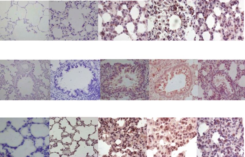
Pandey et al. The Egyptian Journal of Bronchology   (2021) 15:27                                                    Page 2 of 13

molecular weight-hyaluronan (LMW-HA), heat-shock                   levels of HA in blood are indicative of both local lung
proteins, high mobility group box protein-1 (HMGB1),               injury and sequential organ failure. Thus, suggesting the
etc., (iii) induction of HA synthase 2 (HAS2) in endothe-          potential utility of HA estimation in identifying local and
lium, lung alveolar epithelial cells, and fibroblasts [2],         systemic organ dysfunction in acute respiratory distress
(iv) dysregulated release of matrix metalloproteinases             syndrome (ARDS) patients [24].
and ECM remodelling, (v) acute respiratory disease syn-               The present study focuses on the pathogenetic path-
drome (ARDS), (vi) epithelial–mesenchymal transition,              way of progression of lung tissue inflammation to fibro-
and (vii) pulmonary fibrosis. The availability of only two         sis after release of LMW-HA. Elevated LMW-HA
antifibrotic drugs till date has highlighted the need to           release pro-inflammatory cytokines, IL-1β,6, TNF-α [26,
identify the potential clinical and laboratory biomarkers          27], and chemokines and facilitates leukocyte access to
that can predict the subgroup of patients that are going           the injury site.This results in cell proliferation [28], mi-
to deteriorate or develop lung fibrosis.                           gration [29], dendritic cell activation [17], and sterile in-
   The ongoing Covid-19 pandemic caused by severe                  flammation [30]. During stage of chronic inflammation,
acute respiratory syndrome corona virus 2 (SARS-CoV-               LMW-HA transcribes matrix metalloproteinases (MMP-
2) has further increased the occurrence of pulmonary fi-           1,3,9,10,13), collagen, and cytokines, TGF-β, IL-12, and
brosis since 2020. Diffuse alveolar damage (DAD) caused            IGF-I [30–32], resulting in attenuation or progression of
by SARS-CoV-2 can progress to fibrosis even after virus            ECM remodelling. Further, the LMW-HA fragments act
clearance [3]. Hyaluronan (HA), a highly hygroscopic               as endogenous ligands for Toll-like receptor (TLR-2 and
ECM molecule with the ability to absorb water up to                TLR-4) leading to lung inflammation and injury [14].
1000 times its molecular weight, is found in lung alveoli          LMW-HAs engage TLR-2 and activate the macrophage
in severe Covid-19 and can promote edema [4]. Since                inflammatory response [28]. On the other hand, LMW-
the hyaluronan in cadaveric COVID-19 lung tissue com-              HAs engage TLR-4 and protect type-II AECs against
prises low molecular weight fragments [5], recent studies          oxidant-mediated injury. TLR-4 induction maintains ap-
have suggested estimation of serum and sputum levels               propriate anti-apoptotic response [33] leading to AEC
of HA at admission to distinguish critically ill patients          self-renewal and limiting the extent of fibrosis [34]. The
with Covid-19 infection [5, 6] as well as prove to be a            ECM participates in progressive fibrotic scarring of lung
potential therapeutic target [7].                                  by (i) activating a profibrotic feedback loop [35], (ii) ab-
   The ECM comprises of fibrous proteins, collagen and             normal ECM cross-linking resulting in enhanced fibro-
elastin, residing in a milieu of glycoproteins, proteogly-         blast growth and preventing normal ECM turnover in
cans, glycosaminoglycans, growth factors, cytokines/che-           IPF [36]. However, the specific ECM-HA-induced TLR
mokines, proteases, etc [8]. ECM contributes as an                 signalling resulting in progression of fibrosis continues
active or passive player to diverse cellular processes; dif-       to remain an enigma [37].
ferentiation, proliferation, adhesion, migration, and                 We propose that the differential host immune re-
apoptosis [9]. ECM disruption releases hyaluronidases              sponse to ECM injury and LMW-HA fragments is the
[10], reactive oxygen species [11], and degrades en-               critical determinant of epithelial injury/repair pro-
dogenous HA into LMW-HA and HMW-HA fragments                       cesses outcome after both infectious and non-
[12, 13]. These HA fragments are recognized by cell sur-           infectious injurious stimuli. These generate feedback
face receptors; TLR-2,4, CD44, CD168, layilin, RHAMM               signals, leading to either (i) alveolar macrophage
[14–18], on the basis of their size and correlate with na-         priming, increased TLR-2/4 ratio, basal nuclear
ture and extent of injury. The LMW-HA vary from a                  factor-kappa B (NF-κB) activation, inflammation, and
few disaccharides up to over 700 kDa [19] and function             progression of parenchymal fibrosis, or (ii) reducing
as pro-inflammatory DAMPs [20], while the high mo-                 oxidative stress, decreased TLR-2/4 ratio, type-II AEC
lecular weight HA (HMW-HA) (> 5000 kDa) signal the                 protection, and renewal and repair of lung injury. We
resolution of inflammation and injury [14, 21]. There-             elaborate the differential activation of TLRs-2,4 and
fore, the type of HA fragments (HMW/LMW) predom-                   macrophage influx during bleomycin-induced paren-
inating in the tissue after injury act as natural biosensors       chymal remodelling.
for the state of tissue integrity [22]. The HA fragments
differentially trigger an inflammatory immune response             Methods
during acute lung infection, and chronic injury/repair             Chemicals
[23]. Elevated LMW-HA levels have been reported in                 Bleomycin sulfate (Bleocip, Cipla), ketamine hydrochlor-
sputum of Covid-19 patients [5] and in bronchoalveolar             ide, xylocaine, anti-goat-IgG (SAB3700288, Sigma Life
lavage (BAL) fluid of asthma, sarcoidosis, ARDS [24], al-          Science), TLR-2 (Sc-10739, Santa Cruz, USA), TLR-4
veolar proteinosis, IPF patients [25]. BALF elevation of           (Sc-16240, Santa Cruz), CD-68 (ab125212, Abcam), NF-
HA is associated with local lung injury while raised               κB-p65 (Sc-109, Santa Cruz), ExtrAvidin® Peroxidase
Pandey et al. The Egyptian Journal of Bronchology   (2021) 15:27                                                 Page 3 of 13

(Extra-2,3, Sigma), NovaRED (SK-4800, Vector labs,                 immunoassay technique (Hyaluronan Quantikine ELISA
USA), Meyer’s hematoxylin, TRIzol® (Invitrogen                     Kit DHYAL0, R&D systems). Lung tissue (500 mg) was
15596018), chloroform, isopropanol, MMLV (M0253S,                  homogenized in lysis buffer (0.5% TritonX-100, 150
NEB), RNase (M0314, NEB), dNTPs (N0447S, NEB),                     mMNaCl, 15 mM Tris, 1 mM CaCl2, 1 mM MgCl2, pH
random primers (S1330S, NEB), SYBR Green (S4438,                   7.40) and centrifuged at 12,000g (4 °C, 20 min). Fifty-
Sigma), protease inhibitor (Sigma), hyaluronan quanti-             milliliter aliquots of supernatant sample were pipetted
kine ELISA (LMW-HA < 35–950 kDa, DHYAL0, R&D                       into the pre-coated wells. After binding and washing
Systems, USA), and Lamin-A/C (612162, BD Biosci-                   steps, 100 μL of enzyme-linked polyclonal antibodies
ences, India) were used.                                           specific for LMW-hyaluronan was added to the wells.
                                                                   The plates were incubated for 2 h at 37 °C. The unbound
Animals                                                            antibody-enzyme reagent was removed by washing and a
Male Wistar rats (150–250 g, n = 48) were obtained from            chromogen substrate solution was added. The plates
the animal house, V.P.Chest Institute. The experimental            were incubated at room temperature for 30 min. The re-
protocol was approved by institutional animal ethical              action was terminated with 100 μl of diluted hydro-
committee and written consent for use of animals was ob-           chloric acid solution per well and read at 450 nm in an
tained from IAEC. The animals were divided into two                ELISA reader.
groups, group I: saline control, group II: bleomycin. Both
the groups contained 6 animals on each day 0, 7, 14, and
                                                                   Gene expression
28. Animals were provided with standard rodent diet and
                                                                   Total RNA was extracted from lung using guanidinium
water ad libitum. Animal care was as per guidelines laid
                                                                   thiocyanate-phenol-chloroform extractionand reverse-
down by Indian National Science Academy, New Delhi.
                                                                   transcribed to cDNA. cDNA was amplified: PCR activa-
The experiments were performed in the Animal house of
                                                                   tion (95 °C, 5 min); 35 cycles of denaturation @ 95 °C
the V.P. Chest Institute. No randomization method and
                                                                   (30 s), annealing @ 60 °C (35 s), extension @ 72 °C (30
strategy control potential confounders were used.
                                                                   s); final extension @ 72 °C (7 min). Quantitative real-
                                                                   time PCR was performed using Mastercycler, Eppendorf,
Induction of lung fibrosis
                                                                   and primers: TLR-2: Forward-Primer-ATGGCAGCTC
Animals were anesthetized with ketamine hydrochloride
                                                                   CAGGTCTTTC, Reverse-Primer-TTCCGCTGGACTCC
(50 mg/kg-b.w, I.M) and local anesthesia with 1% ligno-
                                                                   AATGTC, TLR-4: Forward-Primer-TCAAGCCCAA
caine. The skin was incised under aseptic precautions
                                                                   GCCTTTCAGG, Reverse-Primer-TTCTCCCAAGATC
and trachea was exposed. In control animals, 100 μl of
                                                                   AACCGATGG, β-actin: Forward-Primer-GACCTTCA
0.9% normal saline was instilled intratracheally. Experi-
                                                                   ACACCCCAGCCA,           Reverse-Primer-GTCACGCACG
mental animals received single intratracheal instillation
                                                                   ATTTCCCTCTC. Relative gene expression was calcu-
of bleomycin (7 units/kg-bw) in 100 μl saline, as previ-
                                                                   lated, using ΔΔCt method.
ously described [38]. After instillation, incision was su-
tured and betadine and antibiotic ointment was applied.
Animals were euthanized 0, 7, 14, and 28 days after                TLR protein
intratracheal bleomycin administration, by using over-             Immunohistochemistry was performed on lung sections
dose of ketamine hydrochloride. The lungs were ligated             which were deparaffinized and rehydrated through
at the trachea and removed en bloc. The lungs were                 graded alcohols. Endogenous peroxidase was quenched
immersed in 10% neutral buffered formalin for fixation             by treatment with 0.3% hydrogen peroxide in methanol
and processed through a graded series of alcohols and              for 3 min. Sections were incubated with the primary
xylene prior to paraffin embedding. Five-micrometer                antibodies—TLR-2, TLR-4, CD68. The bound antigen
sections of the lungs were deparaffinized and stained              was then visualized with the avidin-biotinylated peroxid-
with hematoxylin and eosin stain. The time course of re-           ase technique using DAB substrate. Sections were coun-
lease of LMW-HA fragments, TLR-2,4 mRNA and pro-                   terstained with Harris’ hematoxylin, dehydrated, cleared
tein, NF-κB-p65, macrophage influx, and CD68                       in xylene, and mounted with DPX. Immunostaining was
expression after bleomycin injury were correlated with             quantified using a Nikon-90i microscope and NIS-Ar
development of parenchymal inflammation and fibro-                 image analysis software as per previously described
sis.There were no exclusions in analysis of control and            method [39]. Briefly, 10 fields (× 40) were randomly se-
experimental groups.                                               lected and chromogen-positive cells measured. The in-
                                                                   tensity of positively stained cells was subtracted from
LMW-HA                                                             250 (maximum intensity of RGB image) to obtain recip-
LMW-hyaluronan levels (< 35–950 kDa) were quanti-                  rocal intensity which is directly proportional to protein
tated by using the quantitative sandwich enzyme                    expression.
Pandey et al. The Egyptian Journal of Bronchology (2021) 15:27 Page 4 of 13
NF-κB-p65 fibronectin [44], heparan sulfate [45], and HMGB-1 [46].
NF-κB-p65 was assessed in lung tissue nuclear extracts These ligands induce innate and adaptive immune re-
by Western blot as per previously described method sponse through induction of costimulatory molecules in
[40]. Then, 200 mg tissue was homogenized in buffer-A antigen-presenting cells [47] and propagate parenchymal
(150 mM NaCl, 0.5 mM PMSF, 1 mM EDTA, 10 mM inflammation [48
HEPES, 0.6% NP-40). The nuclear pellet was resus- The present study shows significant increase in levels
pended in solution-B (25% glycerol, 20 mM HEPES, 420 of LMW-HA fragments in lung tissue on day 7 (322 ±
mM NaCl, 1.2 mM MgCl2, 0.2 mM EDTA, 0.5 mM 14.0 pg/mL), after bleomycin as compared to control
PMSF, 0.5 mM DTT). Total nuclear proteins were quan- (162.2 ± 3.79 pg/mL) (Fig. 1). LMW-HA increased per-
tified using Bradford assay [41]. Proteins were resolved sistently up to day 14 (264 ± 16.65 pg/mL) and de-
on 12% SDS-PAGE and transferred onto PVDF mem- creased in fibrotic phase (day 28, 128 ± 13.15 pg/mL).
branes. Membranes were blocked with 5% skimmed milk This is similar to previous study by Teder et al. in mouse
in TBST buffer and incubated with 10 μl of primary anti- model, who observed massive accumulation of HA (5.4
bodies (1:1000), NF-κB-p65, and laminin-A/C (1:1000) × 105 MW), on day 7, in alveolar spaces and intersti-
for 2 h at room temperature. Membrane was washed tium, following bleomycin, as compared to control (14.4
thrice with TBST and then incubated with biotinylated × 105 MW). They reported that a vast majority of HA
secondary antibody (1:2000) of goat anti-Rabbit IgG fragments are cleared from the lung within 14 days after
for 2 h. Following washing, membrane was incubated injury and impaired clearance is followed by collagen de-
with extravidin (1:5000) for 2 h and visualized using position and fibrosis [49]. LMW-HA clearance occurs
NovaRED in Gel documentation system (Bio-Rad). after their internalization [50] by receptors such as
Membrane was then blocked again with 5% skimmed TLR2, TLR4, and CD4 [51, 52]. Persisting HA has pro-
milk in TBST at 4 °C overnight and re-probed with inflammatory effects and perpetuates tissue inflamma-
housekeeping protein (Lamin-A/C, 612162, BD Biosci- tion and injury [53]. In a recent autopsy study, hyaluro-
ences, India). Densitometry was performed using nan staining confirmed prominent HA exudates in
Image lab software-2.0, and values were normalized alveolar spaces of Covid-19 lungs, suggesting its role in
to Lamin-A/C. ARDS caused by SARS-CoV-2 [7].
Statistics Bleomycin-induced lung inflammation
Statistical analysis was done by GraphPad prism-5.0, Bleomycin causes oxidative damage to AECs DNA, lead-
using one-way ANOVA with Newman Keule’s post hoc ing to an initial neutrophil influx, followed by infiltration
test and expressed as Mean ± SEM (standard error of of lymphocytes and macrophages from day 3 onwards
mean). P value < 0.05 was considered significant. [54]. In the present study, we demonstrated chronic
interstitial inflammation comprising of lymphocytes and
Results macrophages on days 7 and 14 after bleomycin (Fig. 2d,
Bleomycin-induced LMW-HA e) as compared to control (Fig. 2a, b). This was associ-
Bleomycin-induced lung injury is characterized by HA ated with increase in LMW-HA (Fig. 1). This is similar
fragmentation that act as endogenous ligands for TLRs to previous studies, where LMW-HA expression coin-
[17]. Other endogenous ligands of TLRs include fibrino- cides with recruitment of circulating monocytes [55] and
gen [42], surfactant protein-A [43], extradomain-A of early macrophage accumulation at site of lung injury
Fig. 1 Hyaluronan fragments in lung tissue before and after bleomycin instillation. Significant increase in levels of HA fragments levels on day 7
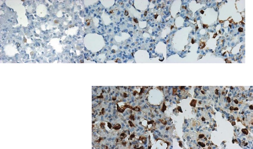
that persist on day 14 and reduce to baseline levels on day 28. ***p < 0.0001 group II B and group II C vs. group II A and group IPandey et al. The Egyptian Journal of Bronchology (2021) 15:27 Page 5 of 13 Fig. 2 a Macrophage infiltration in lung parenchyma before and after bleomycin instillation. Weak CD68 positive macrophages were seen in saline control lungs and on day 0 bleomycin (a, b) by immunohistochemistry. c Note the increase in CD68 expression and influx of macrophages on day 7 after bleomycin instillation (c) that persists up to days 14 and 28 (d, e). f Quantification of CD68 protein expression by macrophages shows significant increase in CD68 protein intensity from day 7 onwards up to day 28. ***p < 0.0001 group II B, group II C, group II D vs. group II A, group I. g A significant net influx of macrophages/high power field (× 400) is seen after bleomycin instillation by morphometry on day 14 that persisted up to day 28. ***p < 0.0001 group II C, group II D vs. group II A, group I; ^^^p < 0.0001 group II C, group II D vs. group II B [56]. These CD68-positive macrophages localize to peri- MyD88 pathway [19] leading to IRAK, TRAF6, and NF- vascular sites of injury on day 7 after bleomycin [57] and κB activation [28]. These accumulating macrophages undergo proliferation, M1/M2 polarization, and release and their associated hyperactive and dysregulated innate profibrotic cytokines like TGF-β1. TGF-β1 activates fi- immune response need to be explored as biomarkers of broblasts, causing EMT and ascending grade of paren- disease activity and progression [38]. The innate and chymal fibrosis [58]. In the present study, the adaptive immune imbalance results in unbridled produc- parenchymal remodelling on day 28 was characterized tion of pro-inflammatory cytokines and chemokines and by reduced cellularity with persistence of macrophages contributes to “cytokine storm” and severity of Covid-19 (Fig. 2f) even after LMW-HA levels declined (Fig. 1). patients [60, 61]. LMW-HA and TLR-2,4-induced macrophage macro- phage influx and accumulation [14] is suggested to be Bleomycin-induced TLR-2 response key component in progression of lung fibrosis [59]. During inflammation, HA fragments differentially en- However, LMW-HAs can also stimulate macrophages gage TLRs, based on their size. HA fragments bind to independently of CD44 and TLR-4 via the TLR-2/ TLR-2 on alveolar macrophages, trigger NF-κB
Pandey et al. The Egyptian Journal of Bronchology   (2021) 15:27                                                   Page 6 of 13

activation, provide a supportive environment for the im-           cytokine storm and multiple organ failure [73]. TLR-4
mune cells, and promote inflammation [62]. On the one              deficiency increases the inflammatory response elicited
hand, the TLR-NF-κB pathway is central in promoting                by LMW-HA [74] resulting in elevated cytokine and
infection-induced lung injury while on the other hand,             chemokine levels [71], which skew towards a Th2/Th1
increased uptake of HA by macrophages can help in re-              response and increased fibrosis.
ducing inflammation and promoting repair; therefore,                 In the present study, increased TLR-4 mRNA (Fig. 4g,
the exact role of TLRs, as a friend or foe in pathogenesis         FC-9.4, **p < 0.001) and protein expression was seen in
of lung fibrosis, remains to be elaborated [63].                   AECs, BECs, and macrophages (Fig. 4c) on day 7, after
   In present study, significant increase in TLR-2 mRNA            bleomycin as compared to control (Fig. 4a, f). On day
(Fig. 3g, fold change (FC)-3.8, **p < 0.001) and protein           14, TLR-4 mRNA levels decreased (Fig. 4g, FC-2.29, p =
expression (Fig. 3c) was seen in AECs, perivascular in-            ns), while TLR-4 protein expression persisted in AECs,
flammatory cells, and macrophages, on day 7, after bleo-           BECs, and macrophages (Fig. 4d) up to day 28 (Fig. 4e).
mycin, as compared to control (Fig. 3a). On day 14,                TLR-4 mRNA downregulation correlated with the pro-
TLR-2 mRNA levels remained elevated (Fig. 3g, FC-4.8,              gression of fibrosis (Fig. 4g, e). TLR-4 protects against
***
    p < 0.0001) and correlated with its enhanced protein           oxidant-mediated lung injury by maintaining anti-
expression in all above cell types (Fig. 3d). The signifi-         apoptotic responses [75], promoting alveolar stem cell
cantly increased TLR-2 mRNA levels on days 7 and 14                renewal [33] and epithelial self-defense mechanisms
correlated with elevated LMW-HA levels on these days               through TLR-4-dependent basal activation of NF-κB
(Fig. 1). Upregulated TLR-2 mediates production of                 [34]. Studies in bleomycin challenged TLR-4 knockout
TGF-β1 and interleukins, IL-6,12,23 [9, 64], and initiates         mice have found them to develop stronger inflammatory
the Th2-lymphocyte response [65]. From the resulting               response [71] with significantly lower type-I collagen
chemokine production, M2 macrophage polarization                   mRNA levels as compared to WT mice [76]. The basal
leads to cellular phase of bleomycin-induced pneumon-              TLR-4 activity is critical for resolution of acute and
itis [66]. On day 28, TLR-2 mRNA levels decreased as               chronic inflammation in pulmonary fibrosis [77]. Our
compared to control (Fig. 3g, FC-1.65); however, TLR-2             group has previously demonstrated reduction of
protein expression persisted in AECs and macrophages               caveolin-1 levels in bleomycin-instilled lungs [78]. Thus,
(Fig. 3e,f) and was associated with persistent M2 macro-           the TLR-4 mRNA downregulation and accompanying
phage polarization and progression of tissue fibrosis              caveolin deficiency [78] contribute to progression to fi-
[67]. HA-TLR2 binding activates NF-κB, MAPKs, p38,                 brosis during lung injury [79].
and JNK pathways and releases pro-inflammatory and
profibrotic cytokines such as interleukin-1, MIP-1,                Bleomycin induced NF-κB signalling
PDGF, and TGF-β1 [68]. Previously, our group has dem-              NF-κB activation is induced by HA fragments [80] and
onstrated an increased expression of TGF-β1 in type-II             TLR-2,4 activation and results in downstream stimula-
AECs, EMT cells, alveolar macrophages, and interstitial            tion of TNF-α, TGF-β, and IFN-γ [81]. In the present
fibroblasts from day 7 up to day 35 after bleomycin [69].          study, a significant increase of NF-κB-p65 levels were
Thus, LMW-HA-TLR-2 interactions are not only critical              observed from day 7 onwards up to day 28 after bleo-
as pro-inflammatory signalling cascade but are also asso-          mycin as compared to control (Fig. 5a, b). This is similar
ciated with increased TGF-β1 expression [69]. Blocking             to a previous study which found maximal nuclear trans-
this pathway may attenuate lung inflammation and fi-               location of NF-κB-p65 on day 7 after bleomycin instilla-
brosis by altering the pulmonary immune microenviron-              tion [82]. NF-κB-p65 upregulation correlated with
ment [70].                                                         perivascular lymphocytes and interstitial macrophage in-
                                                                   filtration, in the cellular phase. These alveolar macro-
Bleomycin-induced TLR-4 response                                   phages function as the “first responders,” resulting in the
LMW-HA are mainly TLR-4 dependent [15] and upreg-                  production of cytokines that then activate NF-κB in
ulate CD68 expression in macrophages in a TLR-4-                   other cell types [83]. After nuclear translocation, the
dependent manner similar to bacterial lipopolysacchar-             NF-κB transcriptionally regulates (i) TGF-β1 resulting in
ide [71] and interferon-γ [72]. The activated macro-               fibroblast proliferation [84], (ii) matrix metalloprotein-
phages use HA as a substrate to aid in migration                   ases (MMPs) [85] and their inhibitors, tissue Inhibitor of
towards site of injury, and HA binding helps in retaining          Matrix       Metalloproteinases(TIMPs),     resulting    in
the activated cells at the sites of inflammation. The              protease-antiprotease imbalance, ECM deposition, and
SARS-CoV-2 spike protein strongly interacts with the               matrix remodelling. In the previous study by our group,
Toll-like receptor 4 (TLR4) pathway producing pro-                 we have demonstrated that it is the shift in the balance
inflammatory cytokines such as interleukin-6 (IL-6) and            of MMP-9/TIMP-1,3 ratio to less than 1 that primes the
tumor necrosis factor-alpha (TNF-α) culminating in                 inflammatory response and its progression to fibrosis
Pandey et al. The Egyptian Journal of Bronchology (2021) 15:27 Page 7 of 13 Fig. 3 Toll-like receptor-2 (TLR-2) mRNA and protein expression in lungs before and after bleomycin instillation: As compared to saline control and bleomycin day 0 (a, b), on day 7 and 14 after bleomycin instillation, an increased TLR-2 expression is seen in AECs, perivascular inflammatory cells, alveolar and interstitial macrophage by immunohistochemistry (c, d respectively). e On day 28, in fibrotic phase, TLR-2 protein expression persisted in AECs, alveolar and interstitial macrophages of lung parenchyma. f Quantification of the intensity of TLR-2 protein expression in the lung parenchyma. Significant increase in TLR-2 protein expression was seen from day 7 that persisted up to day 28. ***p < 0.0001 group II B, group II C, group II D vs. group II A and group I. g TLR-2 mRNA levels were significantly upregulated on day 7 and day 14 and returned to baseline on day 28. **p < 0.001 group II B vs. group II A; ***p < 0.0001 group II C vs. group II A
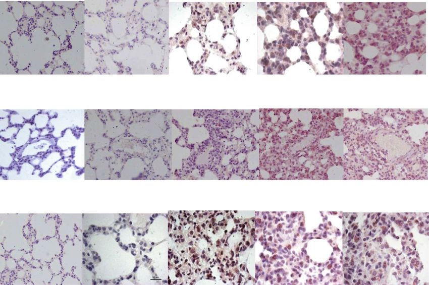
Pandey et al. The Egyptian Journal of Bronchology         (2021) 15:27                                                                       Page 8 of 13

 Fig. 4 Toll like receptor-4 (TLR-4) mRNA and protein expression in lungs before and after bleomycin instillation. As compared to saline control
 and bleomycin day 0 (a, b), on day 7 and 14 after bleomycin instillation, an increased TLR-4 expression is seen in AECs, bronchiolar epithelial cells,
 and alveolar and interstitial macrophages, on day 7 and on day 14 after bleomycin instillation (c, d respectively). e On day 28, TLR-4 protein
 expression persists in AECs, bronchiolar epithelial cells, and alveolar and interstitial macrophages. f Quantification of the intensity of TLR-4 protein
 expression in the lung parenchyma. Significant increase in TLR-4 expression was seen from day 7 onwards that persisted up to day 28. ***p <
 0.0001 group II B, group II C and group II D vs. group II A and group I. g TLR-4 mRNA levels were upregulated on day 7 and returned to baseline
 on day 14 and day 28. **p < 0.001 group II B vs. group II A; ^^^p < 0.0001 group II B vs. group II D and group I
Pandey et al. The Egyptian Journal of Bronchology       (2021) 15:27                                                                   Page 9 of 13

 Fig. 5 a NF-κB p65/Lamin-A/C expression before and after bleomycin treatment. An upregulation of NF-κB p65 expression is seen on day 7 after
 bleomycin instillation that persists up to day 14 and further increases on day 28, compared to control. b Densitometric analysis of the NF-κB p65
 and Lamin-A/C (74 kDa and 65 kDa respectively) protein bands shows significant upregulation of NF-κB p65 protein expression from day 7
 onwards up to day 28. **p < 0.001 group II C, D vs. group I; *p < 0.05 group II B vs group I

[86]. Thereby suggesting that NF-κB induced by LMW-                         been observed to be a major determinant of cell behav-
HA fragments and TLR-2,4 promotes fibrosis by orches-                       ior, fate, and function [37].
trating local inflammatory reactions and altering                             In the present study, we elaborate on the dynamic
protease-antiprotease balance maintaining the fibrotic                      role of ECM and LMW-HA fragments in regulating
responses [87].                                                             the epithelial injury/repair processes. In the early
                                                                            phase, LMW-HA alerts the immune system of a
Discussion                                                                  breach in tissue integrity [23] and activates TLR-2,4,
The pathogenesis of bleomycin-induced pneumonitis is                        alveolar macrophages, and NF-κB signalling, resulting
associated with multiple mechanisms, including oxida-                       in inflammation. TLR-4 mRNA subsequently downre-
tive damage, protease-antiprotease imbalance [38], cave-                    gulates and shifts the TLR-2/TLR-4 balance to more
olin deficiency [78], TGF-β1 [69], and genetic                              than 1. This predisposes to the progression of inflam-
susceptibility [88]. Initially, the ECM was considered to                   mation to fibrosis [14, 80, 89] and results in a pro-
be a simple scaffold providing structural support to lung                   gressive increase in lung hydroxyproline levels [86].
airways. However, recently, the ECM components have                         Thus, the ECM-driven LMW-HA-TLR-2,4-NF-κB
Pandey et al. The Egyptian Journal of Bronchology (2021) 15:27 Page 10 of 13
pathway defines the extent of cellular macrophage in- Conclusions
filtration and parenchymal matrix remodelling. They The lung parenchymal hyaluronan fragments and TLR2/
are reflective of the state of tissue integrity and may TLR4 balance form the critical link between AEC apop-
serve as biomarker of active fibrosis in chronic lung tosis [14], activation of innate immune response, and de-
diseases and as potential therapeutic targets. velopment of cytokine storm, inflammation, and lung
In 2020, efforts have been made to understand the fibrosis in both infectious and non-infectious lung injury.
pathophysiology of the novel coronavirus patients who The LMW-HA-TLR-2,4-NF-κB pathway should be ex-
are predisposed to develop chronic lung disease follow- plored as a biomarker and for its therapeutic potential,
ing COVID-19. These patients have lung inflammation in controlling the severity of lung inflammation and its
with activation of NF-kappa B (NF-κB) transcription fac- progression to lung fibrosis.
tor, in lung macrophages [90], release of inflammatory
Abbreviations
cytokines (IL-1β,6, TNF-α), induction of HA synthase 2 PF: Pulmonary fibrosis; SARS-CoV-2: Severe acute respiratory syndrome
in lung AEC, endothelium and fibroblasts, accumulation coronavirus 2; ECM: Extracellular matrix; LMW-HA: LMW-hyaluronan;
of prominent hyaluronan exudates in the alveolar spaces, DAMP: Damage-associated molecular patterns; TLR: Toll-like receptor; NF-
κb: Nuclear factor-kappa B; AECs: Alveolar epithelial cells; EMT: Epithelial–
and progression to acute respiratory distress syndrome, mesenchymal transition; IL: Interleukins; TNF: Tumor necrosis factor;
[7]. High molecular weight HA predominates in most HAS2: HA synthase 2; DAD: Diffuse alveolar damage; ARDS: Acute respiratory
tissues under healthy conditions, whereas fragmented distress syndrome; MMP: Matrix metalloproteinases; IRAK-1: IL-1R-associated
kinase 1; TRAF: TNF receptor-associated factor; JNKs: Jun N-terminal kinases;
low molecular weight HA polymers predominate at sites MAPKs: Mitogen-activated protein kinases; TGF-β1: Transforming growth
of active inflammation [91], thereby suggesting that ad- factor-β1; PDGF-1: Platelet-derived growth factor-1; MIP-1: Macrophage
juvant treatment targeting hyaluronan, such as intranasal inflammatory protein-1
administration of exogenous hyaluronidase or HA in-
Acknowledgements
hibitor (4-methylumbelliferone (4-MU) [60] may be a The authors gratefully acknowledge the Department of Science and
promising approach to reduce mortality in critically ill Technology, India, for extramural Grant (2010) and University Grants
covid-19 patients [7]. Similarly, the immunomodulation Commission for Research fellowship awarded to Ms. Apoorva Pandey (UGC
NET JRF 2011) for carrying out the above research work.
of NF-κB activation and inhibitions of NF-κB (IκB) deg-
radation may result in a reduction of the cytokine storm Authors’ contributions
and have been suggested as a potential therapeutic target AP performed the animal experiments and molecular studies. RK analyzed
and interpreted all the data regarding the histological examination and
for severe COVID-19 [90].
molecular results. SK was a major contributor in writing the manuscript. All
The strong binding of the SARS-COV-2 spike protein authors read and approved the final manuscript.
with Toll-like receptors-1,4,6 and especially with TLR-4
causes an intense exacerbation of the host immune re- Funding
Department of Science and Technology, India—funding for consumables
sponse with release of interleukin-6 (IL-6) and tumor and research staff.
necrosis factor-alpha (TNF-α), and enhanced severity of University Grants Commission, India—funding for research staff.
COVID-19 pathology [73]. The TLRs are pattern recog-
nition receptors which recognize pathogen-associated Availability of data and materials
Not applicable
molecular patterns (PAMPs) as well as endogenous
DAMPs such as hyaluronan and trigger the innate im- Declarations
mune response [92]. TLR-4 activation kills the microbes
Ethics approval and consent to participate
but can cause DAMP associated host tissue damage as Approval of Institutional animal ethical committee has been obtained: vide
has been previously reported [93, 94]. Tissue damage is letter No-IAEC/41/2014 dated 03/09/2014.
initiated by the myeloid differentiating primary re-
sponse gene 88 (MyD88)-dependent or the MyD88- Consent for publication
Not applicable
independent pathways [95] leading to macrophage,
natural killer cell, mast cell recruitment and their Competing interests
release of several interleukins, interferons, reactive The authors declare that they have no competing interests.
oxygenspecies (ROS), and reactive nitrogen species Author details
(RNS) [96]. Moreover, the TLR4-NF-κB pathway is 1
Deparment of Pathology, V.P. Chest Institute, University of Delhi, Delhi
central towards promoting infection-induced lung 110007, India. 2Department of Biochemistry, V.P. Chest Institute, University of
Delhi, Delhi 110007, India.
injury in aging patients with comorbidities such as
diabetes, atherosclerosis, obesity, and hypertension, Received: 17 March 2021 Accepted: 11 May 2021
thus suggesting the utility of therapeutic targeting
of TLR-4 pathway by compounds such as statins,
References
ACE inhibitors, opioids, and steroids in COVID-19 1. Rosenbloom J, Mendoza FA, Jimenez SA (1832) Strategies for anti-fibrotic
[73]. therapies. Biochim Biophys Acta Mol basis Dis 2013:1088–1103Pandey et al. The Egyptian Journal of Bronchology (2021) 15:27 Page 11 of 13
2. Wilkinson TS, Potter-Perigo S, Tsoi C, Altman LC, Wight TN (2004) Pro-and 23. Johnson P, Arif AA, Lee-Sayer SS, Dong Y (2018) Hyaluronan and its
anti-inflammatory factors cooperate to control hyaluronan synthesis in lung interactions with immune cells in the healthy and inflamed lung. Front
fibroblasts. Am J Respir Cell Mol Biol 31(1):92–99. https://doi.org/10.1165/ Immunol 9:2787. https://doi.org/10.3389/fimmu.2018.02787
rcmb.2003-0380OC 24. Esposito AJ, Bhatraju PK, Stapleton RD, Wurfel MM, Mikacenic C (2017)
3. Vasarmidi E, Tsitoura E, Spandidos DA, Tzanakis N, Antoniou KM (2020) Hyaluronic acid is associated with organ dysfunction in acute respiratory
Pulmonary fibrosis in the aftermath of the COVID-19 era. Exp Ther Med distress syndrome. Crit Care 21(1):1–8
20(3):2557–2560. https://doi.org/10.3892/etm.2020.8980 25. Bray BA, Sampson PM, Osman M, Giandomenico A, Turino GM (1991) Early
4. Laurent TC, Laurent UB, Fraser JR (1996) The structure and function of changes in lung tissue hyaluronan (hyaluronic acid) and hyaluronidase in
hyaluronan: an overview. Immunol Cell Biol 74(2):a1–a7. https://doi.org/10.1 bleomycin-induced alveolitis in hamsters. Am Rev Respir Dis 143(2):284–288.
038/icb.1996.32 https://doi.org/10.1164/ajrccm/143.2.284
5. Kaber G, Kratochvil MJ, Burgener EB, Peltan EL, Barlow G, Yang S, Nicolls MR, 26. Campo GM, Avenoso A, Campo S, d’Ascola A, Nastasi G, Calatroni A (2010)
de Jesus Perez V, Rosser JI, Wardle AJ, Kalinowski A (2020) Hyaluronan is Small hyaluronan oligosaccharides induce inflammation by engaging both
abundant in COVID-19 respiratory secretions. medRxiv [Preprint]. 11:2020.09. toll-like-4 and CD44 receptors in human chondrocytes. Biochem Pharmacol
11.20191692. https://doi.org/10.1101/2020.09.11.20191692. 80(4):480–490. https://doi.org/10.1016/j.bcp.2010.04.024
6. Ding M, Zhang Q, Li Q, Wu T, Huang YZ (2020) Correlation analysis of the 27. Yamawaki H, Hirohata S, Miyoshi T, Takahashi K, Ogawa H, Shinohata R,
severity and clinical prognosis of 32 cases of patients with COVID-19. Respir Demircan K, Kusachi S, Yamamoto K, Ninomiya Y (2009 Jan 1) Hyaluronan
Med 167:105981. https://doi.org/10.1016/j.rmed.2020.105981 receptors involved in cytokine induction in monocytes. Glycobiology. 19(1):
7. Hellman U, Karlsson MG, Engström-Laurent A, Cajander S, Dorofte L, Ahlm 83–92. https://doi.org/10.1093/glycob/cwn109
C, Laurent C, Blomberg A (2020) Presence of hyaluronan in lung alveoli in 28. Scheibner KA, Lutz MA, Boodoo S, Fenton MJ, Powell JD, Horton MR (2006)
severe Covid-19: an opening for new treatment options? J Biol Chem Hyaluronan fragments act as an endogenous danger signal by engaging
295(45):15418–15422. https://doi.org/10.1074/jbc.AC120.015967 TLR2. J Immunol 177(2):1272–1281. https://doi.org/10.4049/jimmunol.1
8. Wight TN, Potter-Perigo S (2011) The extracellular matrix: an active or 77.2.1272
passive player in fibrosis? Am J Physiol Gastrointest Liver Physiol 301(6): 29. McKee CM, Penno MB, Cowman M, Burdick MD, Strieter RM, Bao C, Noble
G950–G955. https://doi.org/10.1152/ajpgi.00132.2011 PW (1996) Hyaluronan (HA) fragments induce chemokine gene expression
9. Kim HS, Go H, Akira S, Chung DH (2011) TLR2-mediated production of IL-27 in alveolar macrophages. The role of HA size and CD44. J Clin Investig
and chemokines by respiratory epithelial cells promotes bleomycin-induced 98(10):2403–2413. https://doi.org/10.1172/JCI119054
pulmonary fibrosis in mice. J Immunol 187(8):4007–4017. https://doi.org/1 30. Taylor KR, Yamasaki K, Radek KA, Di Nardo A, Goodarzi H, Golenbock D,
0.4049/jimmunol.1101654 Beutler B, Gallo RL (2007) Recognition of hyaluronan released in sterile
10. Girish KS, Kemparaju K (2007) The magic glue hyaluronan and its eraser injury involves a unique receptor complex dependent on Toll-like receptor
hyaluronidase: a biological overview. Life Sci 80(21):1921–1943. https://doi. 4, CD44, and MD-2. J Biol Chem 282(25):18265–18275. https://doi.org/10.1
org/10.1016/j.lfs.2007.02.037 074/jbc.M606352200
11. Šoltés L, Mendichi R, Kogan G, Schiller J, Stankovska M, Arnhold J (2006) 31. Gaggar A, Weathington N (2016) Bioactive extracellular matrix fragments in
Degradative action of reactive oxygen species on hyaluronan. lung health and disease. J Clin Investig 126(9):3176–3184. https://doi.org/1
Biomacromolecules. 7(3):659–668. https://doi.org/10.1021/bm050867v 0.1172/JCI83147
12. Albeiroti S, Soroosh A, de la Motte CA (2015) Hyaluronan’s role in fibrosis: a 32. Collins SL, Black KE, Chan-Li Y, Ahn YH, Cole PA, Powell JD, Horton MR
pathogenic factor or a passive player? Biomed Res Int 25:2015 (2011) Hyaluronan fragments promote inflammation by down-regulating
13. Ebihara T, Venkatesan N, Tanaka R, Ludwig MS (2000) Changes in the anti-inflammatory A2a receptor. Am J Respir Cell Mol Biol 45(4):675–683
extracellular matrix and tissue viscoelasticity in bleomycin–induced lung 33. Haitsma JJ, Uhlig S, Lachmann U, Verbrugge SJ, Poelma DL, Lachmann B
fibrosis: temporal aspects. Am J Respir Crit Care Med 162(4):1569–1576. (2002) Exogenous surfactant reduces ventilator-induced
https://doi.org/10.1164/ajrccm.162.4.9912011 decompartmentalization of tumor necrosis factor α in absence of positive
14. Jiang D, Liang J, Fan J, Yu S, Chen S, Luo Y, Prestwich GD, Mascarenhas MM, end-expiratory pressure. Intensive Care Med 28(8):1131–1137. https://doi.
Garg HG, Quinn DA, Homer RJ (2005) Regulation of lung injury and repair org/10.1007/s00134-002-1377-4
by Toll-like receptors and hyaluronan. Nat Med 11(11):1173–1179. https:// 34. Liang J, Zhang Y, Xie T, Liu N, Chen H, Geng Y, Kurkciyan A, Mena JM, Stripp
doi.org/10.1038/nm1315 BR, Jiang D, Noble PW (2016) Hyaluronan and TLR4 promote surfactant-
15. Weigel JA, Raymond RC, Weigel PH (2002) The hyaluronan receptor for protein-C-positive alveolar progenitor cell renewal and prevent severe
endocytosis (HARE) is not CD44 or CD54 (ICAM-1). Biochem Biophys Res pulmonary fibrosis in mice. Nat Med 22(11):1285–1293. https://doi.org/10.1
Commun 294(4):918–922. https://doi.org/10.1016/S0006-291X(02)00558-2 038/nm.4192
16. Hardwick C, Hoare K, Owens R, Hohn HP, Hook M, Moore D, Cripps V, 35. Parker MW, Rossi D, Peterson M, Smith K, Sikström K, White ES, Connett JE,
Austen L, Nance DM, Turley EA (1992) Molecular cloning of a novel Henke CA, Larsson O, Bitterman PB (2014) Fibrotic extracellular matrix activates
hyaluronan receptor that mediates tumor cell motility. J Cell Biol 117(6): a profibrotic positive feedback loop. J Clin Investig 124(4):1622–1635
1343–1350. https://doi.org/10.1083/jcb.117.6.1343 36. Philp CJ, Siebeke I, Clements D, Miller S, Habgood A, John AE, Navaratnam
17. Termeer C, Benedix F, Sleeman J, Fieber C, Voith U, Ahrens T, Miyake K, V, Hubbard RB, Jenkins G, Johnson SR (2018) Extracellular matrix cross-
Freudenberg M, Galanos C, Simon JC (2002) Oligosaccharides of Hyaluronan linking enhances fibroblast growth and protects against matrix proteolysis
activate dendritic cells via toll-like receptor 4. J Exp Med 195(1):99–111. in lung fibrosis. Am J Respir Cell Mol Biol 58(5):594–603. https://doi.org/1
https://doi.org/10.1084/jem.20001858 0.1165/rcmb.2016-0379OC
18. Tolg C, McCarthy JB, Yazdani A, Turley EA (2014) Hyaluronan and RHAMM in 37. Tomos IP, Tzouvelekis A, Aidinis V, Manali ED, Bouros E, Bouros D, Papiris SA
wound repair and the “cancerization” of stromal tissues. Biomed Res Int (2017) Extracellular matrix remodeling in idiopathic pulmonary fibrosis. It is
2014:1–18. https://doi.org/10.1155/2014/103923 the ‘bed’ that counts and not ‘the sleepers’. Expert Rev Respir Med 11(4):
19. Cyphert JM, Trempus CS, Garantziotis S (2015) Size matters: molecular 299–309. https://doi.org/10.1080/17476348.2017.1300533
weight specificity of hyaluronan effects in cell biology. Int J Cell Biol 2015:1– 38. Nguyen NM, Bai Y, Mochitate K, Senior RM (2002) Laminin α-chain
8. https://doi.org/10.1155/2015/563818 expression and basement membrane formation by MLE-15 respiratory
20. Schaefer L (2014) Complexity of danger: the diverse nature of damage- epithelial cells. Am J Phys Lung Cell Mol Phys
associated molecular patterns. J Biol Chem 289(51):35237–35245. https://doi. 282(5):L1004–L1011
org/10.1074/jbc.R114.619304 39. Serrano-Mollar A, Closa D, Prats N, Blesa S, Martinez-Losa M, Cortijo J, Estrela
21. Ruppert SM, Hawn TR, Arrigoni A, Wight TN, Bollyky PL (2014) Tissue JM, Morcillo EJ, Bulbena O (2003) In vivo antioxidant treatment protects
integrity signals communicated by high-molecular weight hyaluronan and against bleomycin-induced lung damage in rats. Br J Pharmacol 138(6):
the resolution of inflammation. Immunol Res 58(2-3):186–192. https://doi. 1037–1048. https://doi.org/10.1038/sj.bjp.0705138
org/10.1007/s12026-014-8495-2 40. Bradford MM (1976) A rapid and sensitive method for the quantitation of
22. Stern R, Asari AA, Sugahara KN (2006) Hyaluronan fragments: an microgram quantities of protein utilizing the principle of protein-dye
information-rich system. Eur J Cell Biol 85(8):699–715. https://doi.org/10.101 binding. Anal Biochem 72(1-2):248–254. https://doi.org/10.1016/0003-2
6/j.ejcb.2006.05.009 697(76)90527-3Pandey et al. The Egyptian Journal of Bronchology (2021) 15:27 Page 12 of 13
41. Smiley ST, King JA, Hancock WW (2001) Fibrinogen stimulates macrophage response to SARS-CoV-2 drives development of COVID-19. Cell. 181(5):1036–
chemokine secretion through toll-like receptor 4. J Immunol 167(5):2887– 1045. https://doi.org/10.1016/j.cell.2020.04.026
2894. https://doi.org/10.4049/jimmunol.167.5.2887 62. Lee-Sayer SS, Dong Y, Arif AA, Olsson M, Brown KL, Johnson P (2015) The
42. Guillot L, Balloy V, McCormack FX, Golenbock DT, Chignard M, Si-Tahar M where, when, how, and why of hyaluronan binding by immune cells. Front.
(2002) Cutting edge: the immunostimulatory activity of the lung surfactant Immunol. 6:150
protein-A involves Toll-like receptor 4. J Immunol 168(12):5989–5992. 63. O’Neill LA (2005) TLRs play good cop, bad cop in the lung. Nat Med 11(11):
https://doi.org/10.4049/jimmunol.168.12.5989 1161–1162. https://doi.org/10.1038/nm1105-1161
43. Okamura Y, Watari M, Jerud ES, Young DW, Ishizaka ST, Rose J, Chow JC, 64. Liu J, Guan X, Ma X (2007) Regulation of IL-27 p28 gene expression in
Strauss JF III (2001) The extra domain A of fibronectin activates Toll-like macrophages through MyD88-and interferon-γ–mediated pathways. J Exp
receptor 4. J Biol Chem 276(13):10229–10233. https://doi.org/10.1074/jbc.M1 Med 204(1):141–152. https://doi.org/10.1084/jem.20061440
00099200 65. Basu S, Fenton MJ (2004) Toll-like receptors: function and roles in lung
44. Johnson GB, Brunn GJ, Kodaira Y, Platt JL (2002) Receptor-mediated disease. AJP Lung Cell Mol Physiol 286(5):L887–L892. https://doi.org/10.11
monitoring of tissue well-being via detection of soluble heparan sulfate by 52/ajplung.00323.2003
Toll-like receptor 4. J Immunol 168(10):5233–5239. https://doi.org/10.4049/ 66. Yang HZ, Cui B, Liu HZ, Chen ZR, Yan HM, Hua F, Hu ZW (2009) Targeting
jimmunol.168.10.5233 TLR2 attenuates pulmonary inflammation and fibrosis by reversion of
45. Park JS, Svetkauskaite D, He Q, Kim JY, Strassheim D, Ishizaka A, Abraham E suppressive immune microenvironment. J Immunol 182(1):692–702. https://
(2004) Involvement of toll-like receptors 2 and 4 in cellular activation by doi.org/10.4049/jimmunol.182.1.692
high mobility group box 1 protein. J Biol Chem 279(9):7370–7377. https:// 67. Wermuth PJ, Jimenez SA (2015) The significance of macrophage
doi.org/10.1074/jbc.M306793200 polarization subtypes for animal models of tissue fibrosis and human
46. Tsan MF, Gao B (2004) Endogenous ligands of Toll-like receptors. J Leukoc fibrotic diseases. Clin Transl Med 4:1–9
Biol 76(3):514–519. https://doi.org/10.1189/jlb.0304127 68. Frey H, Schroeder N, Manon-Jensen T, Iozzo RV, Schaefer L (2013)
47. Lauer ME, Dweik RA, Garantziotis S, Aronica MA (2015) The rise and fall of Biological interplay between proteoglycans and their innate immune
hyaluronan in respiratory diseases. Int J Cell Biol 2015:1–15. https://doi.org/1 receptors in inflammation. FEBS J 280(10):2165–2179. https://doi.org/1
0.1155/2015/712507 0.1111/febs.12145
48. Teder P, Vandivier RW, Jiang D, Liang J, Cohn L, Puré E, Henson PM, Noble 69. Kotarkonda LK, Kulshrestha R, Ravi K (2017) Role of insulin like growth factor
PW (2002) Resolution of lung inflammation by CD44. Science. 296(5565): axis in the bleomycin induced lung injury in rats. Exp Mol Pathol 102:86–96
155–158. https://doi.org/10.1126/science.1069659 70. Yang D, Chen Q, Su SB, Zhang P, Kurosaka K, Caspi RR, Michalek SM,
49. Savani RC, Hou G, Liu P, Wang C, Simons E, Grimm PC, Stern R, Greenberg Rosenberg HF, Zhang N, Oppenheim JJ (2008) Eosinophil-derived
AH, DeLisser HM, Khalil N (2000) A role for hyaluronan in macrophage neurotoxin acts as an alarmin to activate the TLR2–MyD88 signal pathway
accumulation and collagen deposition after bleomycin-induced lung injury. in dendritic cells and enhances Th2 immune responses. J Exp Med 205(1):
Am J Respir Cell Mol Biol 23(4):475–484. https://doi.org/10.1165/a 79–90. https://doi.org/10.1084/jem.20062027
jrcmb.23.4.3944 71. Zhao H, Leu SW, Shi L, Dedaj R, Zhao G, Garg HG, Shen L, Lien E, Fitzgerald
50. Tavianatou AG, Caon I, Franchi M, Piperigkou Z, Galesso D, Karamanos NK KA, Shiedlin A, Shen H, Quinn DA, Hales CA (2010) TLR4 is a negative
(2019) Hyaluronan: molecular size-dependent signaling and biological regulator in noninfectious lung inflammation. J Immunol 184(9):5308–5314.
functions in inflammation and cancer. FEBS J 286(15):2883–2908. https://doi. https://doi.org/10.4049/jimmunol.1000009
org/10.1111/febs.14777 72. Papageorgiou IE, Lewen A, Galow LV, Cesetti T, Scheffel J, Regen T, Hanisch
51. Petrey AC, de la Motte CA (2014) Hyaluronan, a crucial regulator of UK, Kann O (2016) TLR4-activated microglia require IFN-γ to induce severe
inflammation. Front Immunol 11:101 neuronal dysfunction and death in situ. Proc Natl Acad Sci 113(1):212–217.
52. Avenoso A, Bruschetta G, D’Ascola A, Scuruchi M, Mandraffino G, Gullace R, https://doi.org/10.1073/pnas.1513853113
Saitta A, Campo S, Campo GM (2019) Hyaluronan fragments produced 73. Brandão SCS, Ramos JOX, Dompieri LT, Godoi ETAM, Figueiredo JL, Sarinho
during tissue injury: a signal amplifying the inflammatory response. Arch ESC, Chelvanambi S, Aikawa M (2021) Is Toll-like receptor 4 involved in the
Biochem Biophys 663:228–238. https://doi.org/10.1016/j.abb.2019.01.015 severity of COVID-19 pathology in patients with cardiometabolic
53. Moore BB, Lawson WE, Oury TD, Sisson TH, Raghavendran K, Hogaboam CM comorbidities? Cytokine Growth Factor Rev 58:102-110. https://doi.org/10.1
(2013) Animal models of fibrotic lung disease. Am J Respir Cell Mol Biol 49: 016/j.cytogfr.2020.09.002.
167–179 74. Gariboldi S, Palazzo M, Zanobbio L, Selleri S, Sommariva M, Sfondrini L,
54. Singer SJ, Kupfer A (1986) The directed migration of eukaryotic cells. Annu Rev Cavicchini S, Balsari A, Rumio C (2008) Low molecular weight hyaluronic
Cell Biol 2(1):337–365. https://doi.org/10.1146/annurev.cb.02.110186.002005 acid increases the self-defense of skin epithelium by induction of beta-
55. Gerdin B, Hällgren R (1997) Dynamic role of hyaluronan (HYA) in connective defensin 2 via TLR2 and TLR4. J Immunol 181(3):2103–2110. https://doi.
tissue activation and inflammation. J Intern Med 242(1):49–55. https://doi. org/10.4049/jimmunol.181.3.2103
org/10.1046/j.1365-2796.1997.00173.x 75. Zhang X, Shan P, Qureshi S, Homer R, Medzhitov R, Noble PW, Lee PJ (2005)
56. Jarman ER, Khambata VS, Li YY, Cheung K, Thomas M, Duggan N, Jarai G Cutting edge: TLR4 deficiency confers susceptibility to lethal oxidant lung injury. J
(2014) A translational preclinical model of interstitial pulmonary fibrosis and Immunol 175(8):4834–4838. https://doi.org/10.4049/jimmunol.175.8.4834
pulmonary hypertension: mechanistic pathways driving disease 76. Li XX, Jiang DY, Huang XX, Guo SL, Yuan W, Dai HP (2015) Toll like receptor
pathophysiology. Physiol Rep 2(9):e12133. https://doi.org/10.14814/ 4 promotes fibrosis in bleomycin induced lung injury in mice. Genet Mol
phy2.12133 Res 14(4):17391–17398. https://doi.org/10.4238/2015.December.21.8
57. Gibbons MA, MacKinnon AC, Ramachandran P, Dhaliwal K, Duffin R, 77. Yang HZ, Wang JP, Mi S, Liu HZ, Cui B, Yan HM, Yan J, Li Z, Liu H, Hua F, Lu
Phythian-Adams AT, van Rooijen N, Haslett C, Howie SE, Simpson AJ, Hirani W, Hu ZW (2012) TLR4 activity is required in the resolution of pulmonary
N (2011) Ly6Chi monocytes direct alternatively activated profibrotic inflammation and fibrosis after acute and chronic lung injury. Am J Pathol
macrophage regulation of lung fibrosis. Am J Respir Crit Care Med 184(5): 180(1):275–292. https://doi.org/10.1016/j.ajpath.2011.09.019
569–581. https://doi.org/10.1164/rccm.201010-1719OC 78. Kulshrestha R, Singh H, Pandey A, Soundarya D, Jaggi AS, Ravi K (2020)
58. Beutler B (2007) Neo-ligands for innate immune receptors and the etiology Differential expression of caveolin-1 during pathogenesis of combined
of sterile inflammatory disease. Immunol Rev 220(1):113–128. https://doi. pulmonary fibrosis and emphysema: effect of phosphodiesterase-5 inhibitor.
org/10.1111/j.1600-065X.2007.00577.x BBA-Mol Basis Dis 1866:165802
59. Kulshrestha R, Pandey A, Jaggi A, Bansal S (2020) Beneficial effects of N- 79. Mirza MK, Yuan J, Gao XP, Garrean S, Brokovych V, Malik AB et al (2010)
acetylcysteine on protease-antiprotease balance in attenuating bleomycin- Caveolin-1 deficiency dampens Toll-Like Receptor-4 signaling through eNOS
induced pulmonary fibrosis in rats. Iran J Basic Med Sci 23:396 activation. Am J Pathol 176(5):2344–2351. https://doi.org/10.2353/ajpath.201
60. Shi Y, Wang Y, Shao C, Huang J, Gan J, Huang X, Bucci E, Piacentini M, 0.091088
Ippolito G, Melino G (2020) COVID-19 infection: the perspectives on 80. Noble PW, Lake FR, Henson PM, Riches DW (1993) Hyaluronate activation of
immune responses. Cell Death Differ 27(5):1451–1454 CD44 induces insulin like growth factor-1 expression by a tumor necrosis
61. Blanco-Melo D, Nilsson-Payant BE, Liu WC, Uhl S, Hoagland D, Møller R, factor - alpha - dependent mechanism in murine macrophages. J Clin
Jordan TX, Oishi K, Panis M, Sachs D, Wang TT (2020) Imbalanced host Invest 91(6):2368–2377. https://doi.org/10.1172/JCI116469Pandey et al. The Egyptian Journal of Bronchology (2021) 15:27 Page 13 of 13
81. Lafyatis R, Farina A (2012) New insights into the mechanisms of innate
immune receptor signalling in fibrosis. Open Rheumatol J 6(1):72–79.
https://doi.org/10.2174/1874312901206010072
82. Inayama M, Nishioka Y, Azuma M, Muto S, Aono Y, Makino H, Tani K, Uehara
H, Izumi K, Itai A, Sone S (2006) A novel IκB kinase-β inhibitor ameliorates
bleomycin-induced pulmonary fibrosis in mice. Am J Respir Crit Care Med
173(9):1016–1022. https://doi.org/10.1164/rccm.200506-947OC
83. Alvira CM (2014) Nuclear-factor-kappa-B signaling in lung development and
disease: One Pathway, numerous functions. Birth Defects Res A Clin Mol
Teratol 100(3):202–216. https://doi.org/10.1002/bdra.23233
84. Agarwal R, Agarwal P (2017) Targeting extracellular matrix remodeling in
disease: could resveratrol be a potential candidate? Exp Biol Med 242(4):
374–383. https://doi.org/10.1177/1535370216675065
85. Vellaichamy E, Khurana ML, Fink J, Pandey KN (2005) Involvement of NF-κB /
matrix metalloproteinase pathway in cardiac fibrosis of mice lacking
guanylyl cyclase / natriuretic peptide receptor A. J Biol Chem 280(19):
19230–19242. https://doi.org/10.1074/jbc.M411373200
86. Kulshrestha R, Dhanda H, Pandey A, Singh A, Rajkumar (2020)
Immunopathogenesis and therapeutic potential of macrophage influx in
diffuse parenchymal lung diseases. Expt Rev Resp Med 14(9):917–928.
87. Borthwick LA, Barron L, Hart KM, Vannelia KM, Thompson RW, Oland S et al
(2016) Macrophages are critical to the maintenance of IL-13- dependent
lung inflammation and fibrosis. Mucosal Immunol 9(1):38–55. https://doi.
org/10.1038/mi.2015.34
88. Reinert T, Serodio C, Arthur F, Nunes P, Alves A, Scheliga DS (2013)
Bleomycin-induced lung injury. J Cancer Res Ther 2013:480608. https://doi.
org/10.1155/2013/480608
89. Horton MR, McKee CM, Bao C, Liao F, Farber JM, Hodge-DuFour J, Pure E,
Oliver BL, Wright TM, Noble PW (1998) Hyaluronan fragments synergize
with interferon-gamma to induce the C-X-C chemokines mig and
interferon-inducible protein-10 in mouse macrophages. J Biol Chem 273(52):
35088–35094. https://doi.org/10.1074/jbc.273.52.35088
90. Hariharan A, Hakeem AR, Radhakrishnan S, Reddy MS, Rela M (2021) The
role and therapeutic potential of NF-kappa-B pathway in severe COVID-19
patients. Inflammopharmacology. 1:91–100
91. Nagy N, Kuipers HF, Frymoyer AR, Ishak HD, Bollyky JB, Wight TN et al (2015)
L. 4-methylumbelliferone treatment and hyaluronan inhibition as a
therapeutic strategy in inflammation, autoimmunity, and cancer. Front
Immunol 6:123
92. Jeong E, Lee JY (2011) Intrinsic and extrinsic regulation of innate immune
receptors. Yonsei Med J 52(3):379–392. https://doi.org/10.3349/ymj.2011.
52.3.379
93. Okun E, Griffioen KJ, Lathia JD, Tang SC, Mattson MP, Arumugam TV (2009)
Toll-like receptors in neurodegeneration. Brain Res Rev 59:27–292
94. Kaisho T, Akira S (2002) Toll-like receptors as adjuvant receptors. Biochim
Biophys Acta 1589(1):1–13. https://doi.org/10.1016/S0167-4889(01)00182-3
95. Kawasaki K, Akashi S, Shimazu R, Yoshida T, Miyake K, Nishijima M (2000)
Mouse toll-like receptor 4·MD-2 complex mediates lipopolysaccharide-
mimetic signal transduction by Taxol. J Biol Chem 275(4):2251–2254. https://
doi.org/10.1074/jbc.275.4.2251
96. Lucas K, Maes M (2013) Role of the toll like receptor (TLR) radical cycle in
chronic inflammation: possible treatments targeting the TLR4 pathway. Mol
Neurobiol 48(1):190–204. https://doi.org/10.1007/s12035-013-8425-7
Publisher’s Note
Springer Nature remains neutral with regard to jurisdictional claims in
published maps and institutional affiliations.You can also read