SARS- COV-2 VARIANTS, SPIKE MUTATIONS AND IMMUNE ESCAPE
←
→
Page content transcription
If your browser does not render page correctly, please read the page content below
REVIEWS
SARS-CoV-2 variants, spike mutations
and immune escape
William T. Harvey 1,2,8, Alessandro M. Carabelli 3,8, Ben Jackson 4,
Ravindra K. Gupta5, Emma C. Thomson6,7, Ewan M. Harrison3,7, Catherine Ludden3,
Richard Reeve 1, Andrew Rambaut 4, COVID-19 Genomics UK (COG-UK) Consortium*,
Sharon J. Peacock3 and David L. Robertson 2 ✉
Abstract | Although most mutations in the severe acute respiratory syndrome coronavirus 2
(SARS-CoV-2) genome are expected to be either deleterious and swiftly purged or relatively
neutral, a small proportion will affect functional properties and may alter infectivity, disease
severity or interactions with host immunity. The emergence of SARS-CoV-2 in late 2019 was
followed by a period of relative evolutionary stasis lasting about 11 months. Since late 2020,
however, SARS-CoV-2 evolution has been characterized by the emergence of sets of mutations,
in the context of ‘variants of concern’, that impact virus characteristics, including transmissibility
and antigenicity, probably in response to the changing immune profile of the human population.
There is emerging evidence of reduced neutralization of some SARS-CoV-2 variants by
postvaccination serum; however, a greater understanding of correlates of protection is
required to evaluate how this may impact vaccine effectiveness. Nonetheless, manufacturers
are preparing platforms for a possible update of vaccine sequences, and it is crucial that
surveillance of genetic and antigenic changes in the global virus population is done alongside
experiments to elucidate the phenotypic impacts of mutations. In this Review, we summarize the
literature on mutations of the SARS-CoV-2 spike protein, the primary antigen, focusing on their
impacts on antigenicity and contextualizing them in the protein structure, and discuss them in
the context of observed mutation frequencies in global sequence datasets.
Variants As of April 2021, severe acute respiratory syndrome high-effect mutations that contribute to virus adaption
In the context of viruses, coronavirus 2 (SARS-C oV-2), the causative agent of and fitness do occur, they tend to be in the minority
genetically distinct viruses with COVID-19, accounted for more than 143 million infec- compared with tolerated low-effect or no-effect ‘neu-
mutations different from those tions and more than three million deaths worldwide1. tral’ amino acid changes4. A small minority of mutations
of other viruses. ‘Variants’ can
also refer to the founding virus
Virus genomic sequences are being generated and are expected to impact virus phenotype in a way that
of a cluster or lineage and shared at an unprecedented rate, with more than one confers a fitness advantage, in at least some contexts.
is used to refer collectively million SARS-CoV-2 sequences available via the Global Such mutations may alter various aspects of virus biol-
to the resulting variants that Initiative on Sharing All Influenza Data (GISAID), per- ogy, such as pathogenicity, infectivity, transmissibility
form the lineage. Variants
mitting near real-time surveillance of the unfolding and/or antigenicity. Although care has to be taken not
with changed biological
characteristics or antigenicity
pandemic2. The use of pathogen genomes on this scale to confound mutations being merely present in grow
have been termed ‘variants to track the spread of the virus internationally, study ing lineages with mutations that change virus biology5,
of interest’, ‘variants under local outbreaks and inform public health policy signi- fitness-enhancing mutations were first detected to
investigation’ or ‘variants of fies a new age in virus genomic investigations3. Further have arisen within a few months of the evolution of
concern’ by public health
bodies.
to understanding epidemiology, sequencing enables SARS-CoV-2 within the human population. For exam-
identification of emerging SARS-CoV-2 variants and ple, the spike protein amino acid change D614G was
sets of mutations potentially linked to changes in viral noted to be increasing in frequency in April 2020 and to
properties. have emerged several times in the global SARS-CoV-2
✉e-mail: david.l.robertson@ As highly deleterious mutations are rapidly purged, population, and the coding sequence exhibits a high
glasgow.ac.uk most mutations observed in genomes sampled from cir- dN/dS ratio, suggesting positive selection at the codon
https://doi.org/10.1038/ culating SARS-CoV-2 virions are expected to be either position 614 (refs6,7). Subsequent studies indicated that
s41579-021-00573-0 neutral or mildly deleterious. This is because although D614G confers a moderate advantage for infectivity8,9
Nature Reviews | Microbiology
0123456789();: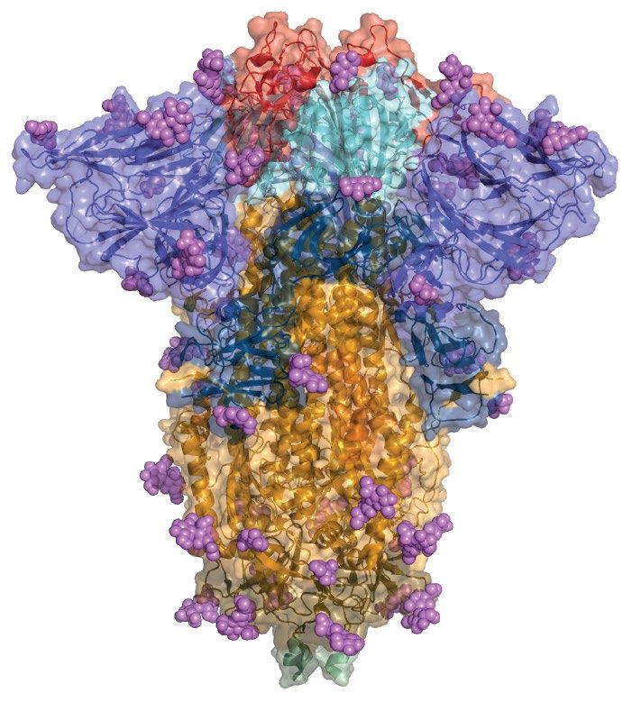
Reviews
Mutations
and transmissibility10. Several other spike mutations of from infection18. Another RBM amino acid change,
The substitutions, insertions note have now arisen and are discussed in this Review, Y453F — associated with increased ACE2-binding
or deletions of one or more with particular focus on mutations affecting antigenicity. affinity19 — received considerable attention following
nucleotides in the virus RNA The extent to which mutations affecting the anti- its identification in sequences associated with infec-
genome. Non-synonymous
nucleotide substitutions in
genic phenotype of SARS-CoV-2 will enable variants tions in humans and mink; most notably one lineage
protein-coding sequence to circumvent immunity conferred by natural infection identified in Denmark and initially named ‘cluster 5’
result in a change in amino acid or vaccination remains to be determined. However, (now B.1.1.298)20. As of 5 November 2020, 214 humans
(referred to as a substitution there is growing evidence that mutations that change infected with SARS-CoV-2 related to mink were all car-
or replacement), whereas
the antigenic phenotype of SARS-C oV-2 are circu- rying the mutation Y453F21. The B.1.1.298 lineage also
synonymous nucleotide
substitutions do not change
lating and affect immune recognition to a degree that has Δ69–70, an amino-terminal domain (NTD) dele-
the amino acid. requires immediate attention. The spike protein medi- tion that has emerged several times across the global
ates attachment of the virus to host cell-surface recep- SARS-C oV-2 population, including in the second
Lineages tors and fusion between virus and cell membranes11 N439K lineage, B.1.258. Δ69–70 is predicted to alter the
Monophyletic clusters of
(Box 1). It is also the principal target of neutralizing anti- conformation of an exposed NTD loop and has been
viruses assigned on the basis
of the severe acute reparatory bodies generated following infection by SARS-CoV-2 reported to be associated with increased infectivity22.
syndrome coronavirus 2 (refs12,13), and is the SARS-CoV-2 component of both Genomic analyses indicate a change in host environ
(SARS-CoV-2) global mRNA and adenovirus-based vaccines licensed for use ment and signatures of increased selective pressures
phylogenetic tree.
and others awaiting regulatory approval14. Consequently, acting upon immunologically important SARS-CoV-2
dN/dS ratio
mutations that affect the antigenicity of the spike pro- genes sampled from around November 2020 (ref.23). This
The ratio of non-synonymous tein are of particular importance. In this Review, we coincided with the emergence of variants with higher
mutations per non-synonymous explore the literature on these mutations and their numbers of mutations relative to previous circulating
site (dN) to synonymous antigenic consequences, focusing on the spike protein variants. These lineages because of their association
mutations per synonymous site
(dS), which is used to estimate
and antibody-mediated immunity, and discuss them in with increased transmissibility were named ‘variants of
the balance between neutral the context of observed mutation frequencies in global concern’. They are defined by multiple convergent muta-
mutations, purifying selection sequence datasets. tions that are hypothesized to have arisen either in the
and positive selection acting context of chronic infections or in previously infected
on gene or a specific codon.
Spike mutations receiving early attention individuals24–29. In addition to understanding the trans-
Amino acid substitution
The rate of evolution of SARS-CoV-2 from December missibility and pathogenicity of these emerging variants,
A change in a specific amino 2019 to October 2020 was consistent with the virus it is crucially important to characterize their antigenicity
acid of a protein. This is caused acquiring approximately two mutations per month in and the level of cross-protection provided by infection by
by non-synonymous mutations. the global population15,16. Although our understanding earlier viruses that are genetically and antigenically sim-
By convention, an amino acid
substitution is written in the
of the functional consequences of spike mutations is ilar to the virus that first emerged in December 2019 and
form N501Y to denote rapidly expanding, much of this knowledge involves the which is used in all of the current vaccine preparations.
the wild-type amino acid reactive investigation of amino acid changes identified as Information on how spike mutations affect antigenic
(N (asparagine)) and the rapidly increasing in frequency or being associated with profiles can be derived from structural studies, mutations
substituted amino acid
unusual epidemiological characteristics. Following the identified in viruses exposed to mAbs or plasma con-
(Y (tyrosine)) at site 501 in
the amino acid sequence.
emergence of D614G, an amino acid substitution within taining polyclonal antibodies, targeted investigations of
the receptor-binding motif (RBM), N439K, was noted variants using site-directed mutagenesis and deep muta-
Monoclonal antibodies as increasing in frequency in Scotland in March 2020. tional scanning (DMS) experiments that systematically
(mAbs). Antibodies made Whereas this first lineage with N439K (designated investigate the possibility of mutations arising.
by cloning a unique white
blood cell, which usually has
B.1.141 with the Pango nomenclature system17) quickly
monovalent binding affinity for became extinct, another lineage that independently Immunogenic regions of spike
a specific epitope. Virus particles acquired N439K (B.1.258) emerged and circulated Several studies have probed the antigenicity of
can be saturated with mAbs, widely in many European countries18. N439K is note- the SARS-C oV-2 spike protein by epitope mapping
and the structure can be solved
worthy as it enhances the binding affinity for the ACE2 approaches, including solving the structure of the spike
to determine the antibody
footprint or mAbs can be used
receptor and reduces the neutralizing activity of some protein in complex with the antigen-binding fragment
to select for mutations that monoclonal antibodies (mAbs) and polyclonal antibod- of particular antibodies13,30–32. Serological analyses of
escape recognition. ies present in sera from people who have recovered almost 650 individuals infected with SARS-C oV-2
indicated that ~90% of the plasma or serum neutraliz-
ing antibody activity targets the spike receptor-binding
Author addresses
domain (RBD)12. A relative lack of glycan shielding may
1
institute of Biodiversity, animal Health and Comparative Medicine, College of Medical, contribute to the immunodominance of the RBD33. One
veterinary and Life sciences, university of Glasgow, Glasgow, uK. study reported structural, biophysical and bioinformat-
2
MrC–university of Glasgow Centre for virus research, Glasgow, uK. ics analyses of 15 SARS-CoV-2 RBD-binding neutral-
3
Department of Medicine, university of Cambridge, Cambridge, uK. izing antibodies31. Antibody footprints were generated
4
institute of evolutionary Biology, university of edinburgh, edinburgh, uK. by structural analyses of the spike residues considering
5
Cambridge institute of therapeutic immunology and infectious Disease, university of
potential hydrogen bonds and van der Waals interactions
Cambridge, Cambridge, uK.
6
Department of Clinical research, London school of Hygiene and tropical Medicine, with a mAb atom that were less than 4.0 Å. Structural
London, uK. analyses allowed the categorization of RBD-binding
7
wellcome sanger institute, Hinxton, uK. neutralizing antibodies into four classes (Fig. 1a,b): ACE2-
8
these authors contributed equally: william t. Harvey, alessandro M. Carabelli. blocking antibodies that bind the spike protein in the
*a list of members and their affiliations appears in supplementary information. open conformation (class 1); ACE2-blocking antibodies
www.nature.com/nrmicro
0123456789();: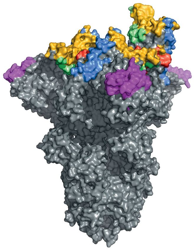
Reviews
Box 1 | Spike protein structure and function mAbs39 or convalescent plasma containing polyclonal
antibodies40,41; targeted characterization of particular
as with other coronaviruses, the entry of severe acute respiratory syndrome mutations18,42; and wider investigations of either large
coronavirus 2 (sars-Cov-2) into host cells is mediated by the transmembrane spike numbers of circulating variants43 or all possible amino
glycoprotein, which forms homotrimers on the surface of the virion. the sars-Cov-2
acid substitutions in the RBD39,44–46. For spike residues
spike protein is highly glycosylated, with 66 potential N-glycosylation sites per
where mutations have been shown to influence poly-
trimer98,99 (Fig. 4a).the sars-Cov-2 spike protein is post-translationally cleaved
by mammalian furin into two subunits: s1 and s2 (Fig. 4a). the s1 subunit largely clonal antibody recognition, the observation of an effect
consists of the amino-terminal domain and the receptor-binding domain (rBD), and on either mAbs or plasma is indicated in Fig. 1b. For a
is responsible for binding to the host cell-surface receptor, aCe2, whereas the s2 smaller number of residues, escape mutations emerg-
subunit includes the trimeric core of the protein and is responsible for membrane fusion ing in virus exposed to mAbs or polyclonal plasma have
(Fig. 4b). the presence of a polybasic furin cleavage site at the s1–s2 boundary, which been described (‘mAb emerge’ and ‘plasma emerge’ in
is unique within the subgenus Sarbecovirus, is important for infectivity and virulence100, Fig. 1b).
with furin cleavage facilitating the conformational change required for receptor In a DMS study, researchers assessed all possible
binding50. the spike protein transiently undergoes conformational changes between a single amino acid variants using a yeast-display system
closed conformation and an open conformation in which a hinge-like movement raises and detected variants that escape either nine neutraliz-
the rBD50. the residues comprising the receptor-binding motif are revealed on the
ing SARS-CoV-2 mAbs45 or convalescent plasma from
upright rBD, enabling binding to aCe2, which induces a progressively more open
structure until a fully open, three-aCe2-bound structure is formed, initiating s2 11 individuals taken at two time points after infection39
unsheathing and membrane fusion101. (shades of green in Fig. 1b). The resulting heat maps
provide rich data on the antigenic consequence of RBD
mutations, with the plasma escape mutations being of
that bind the RBD in both the open conformation and particular interest given that they impact neutraliza-
the closed conformation (class 2); antibodies that do not tion by polyclonal antibodies of the kind SARS-CoV-2
block ACE2 and bind the RBD in both the open con- encounters in infections, with significant levels of immu-
Epitope mapping
Experimental determination
formation and the closed conformation (class 3); and nity acquired through prior exposure or vaccination.
of the binding site, or epitope, neutralizing antibodies that bind outside the ACE2 site Although significant interperson and intraperson hetero
of an antibody. Approaches and only in the open conformation (class 4)31. Within geneity in the impact of mutations on neutralization by
include X-ray co-crystallography the RBD, RBM epitopes overlapping the ACE2 site are polyclonal serum has been described, the mutations that
or cryogenic electron
immunodominant, whereas other RBD sites generate reduce antibody binding the most occur at a relatively
microscopy of an antigen–
antibody complex and the lower and variable responses in different individuals12. small number of RBD residues, indicating substantial
mapping of systematic Although the RBD is immunodominant, there is immunodominance within the RBD39.
mutations introduced by evidence for a substantial role of other spike regions in Of all RBD residues for which substitutions affected
site-directed mutagenesis. antigenicity, most notably the NTD13,30,34. Early struc- recognition by convalescent sera, DMS identified E484
Glycan shielding
tural characterization of NTD-specific antibodies 4A8 as being of principal importance, with amino acid
The process by which a virus (ref.32) and 4–8 (ref.13) revealed similar epitope locations changes to K, Q or P reducing neutralization titres by
can cloak underlying protein, towards the upper side of the most prominently protrud- more than an order of magnitude39. E484K has also been
impeding antibody binding. ing area of the NTD. Cryogenic electron microscopy was identified as an escape mutation that emerges during
This is mediated by glycans,
used to determine the antibody footprint of the neutral- exposure to mAbs C121 and C144 (ref.40) and convales-
bulky sugar molecules that
are covalently attached to izing antibody 4A8, and showed key interactions involv- cent plasma41, and was the only mutation described in
amino acid side chains of ing spike residues Y145, H146, K147, K150, W152, R246 one study as able to reduce the neutralizing ability of a
the viral protein. and W258 (ref.32). Epitope binning of 41 NTD-specific combination of mAbs (REGN10989 and REGN10934)
mAbs led to the identification of six antigenic sites, to an unmeasurable level47. In an escape mutation study
Immunodominance
The phenomenon by
one of which is recognized by all known NTD-specific using 19 mAbs, substitutions at E484 emerged more
which the host immune neutralizing antibodies and has been termed the ‘NTD frequently than at any other residue (in response to
response against a viral supersite’, consisting of residues 14–20, 140–158 and four mAbs), and each of the four 484 mutants identi-
particle is mostly focused on 245–264 (ref.30) (Fig. 1a,b). The mechanism of neutraliza- fied (E484A, E484D, E484G and E484K) subsequently
a few antigens and mediated
tion by which NTD-specific antibodies act remains to be conferred resistance to each of four convalescent sera
by potently neutralizing
antibodies. fully determined, although it may involve the inhibition tested48. No other mAb-selected escape mutants escaped
of conformational changes or proposed interactions with each of the four sera, although the mutations K444E,
Antibody footprints auxiliary receptors such as DC-SIGN or L-SIGN32,35. G446V, L452R and F490S escaped three of the four sera
Amino acid residues of a Relatively little is known of antigenicity in the S2 sub- tested48.
3D folded protein that are
targeted and contacted by
unit, with immunogenicity thought to be impeded by Mutations at position 477 of the spike protein
a binding antibody. extensive glycan shielding36, and although both linear (S477G, S477N and S477R) rank prominently among
and cross-reactive conformational S2 epitopes have been mAb escape mutations identified by one study, and the
Glycoprotein described37,38, the biological significance of these is not mutation S477G conferred resistance to two of the four
A protein with oligosaccharide
yet known. sera tested48. However, substitutions at 477 were not
chains (glycans) covalently
attached to amino acid identified as being important in DMS with convales-
side chains. Virus surface Spike RBD mutations and immune escape cent plasma39. The mutation N439K increases affinity
glycoproteins embedded in the Several studies have contributed to the current under- for ACE2 (ref.19), is predicted to result in an additional
membrane often have a role standing of how mutations in the SARS-CoV-2 spike salt bridge at the RBM–ACE2 interface and is thought
in interactions with host cells,
including receptor binding,
protein affect neutralization. These studies include to preferentially reduce the neutralization potential
and are also commonly traditional escape mutation work that identifies muta- of plasma that already has low neutralizing activity18.
targeted by host antibodies. tions that emerge in virus populations exposed to either However, a DMS study39 did not find that the mutation
Nature Reviews | Microbiology
0123456789();: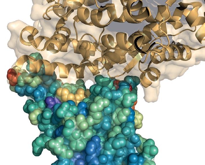
Reviews
a b 140 x xx 140
148 148
150 150
151 151
345 345
346 346
352 352
378 378
406 406 Antibody class
408 408 NTD
417 417 RBD 1
439 439 RBD 2
441 441 RBD 3
443 443 RBD 4
444 444
445 445 RBS distance (Å)
446 446 150
447 447 100
448 448 50
449 449 0
450 450
452 452 Described
453 453 No
455 455 Yes
180º
456 456 x Yes (deletion)
458 458
472 472 Plasma escape
fraction (DMS)
473 473
1
474 474
0.8
476 476
0.6
477 477
0.4
478 478
479 479 0.2
483 483 0
484 484
ACE2 binding (DMS)
485 485
1
486 486
0
487 487
–1
489 489
490 490 –2
493 493 –3
494 494
496 496
499 499
501 501
503 503
504 504
519 519
Amino acid position
RBS distance (Å)
mAb effect
mAb emerge
Plasma effect
Plasma emerge
Plasma average
Plasma max
Binding average
Binding max
DMS
N439K significantly alters neutralization by polyclonal Spike NTD mutations and immune escape
antibodies in plasma, in contrast to previous studies In the NTD, most of the evidence for immune evasion
that found that N439K reduced neutralization by mAbs focuses on a region centred at a conformational epitope
and convalescent plasma18. One explanation for this consisting of residues 140–156 (N3 loop) and 246–260
inconsistency is that the mechanism of immune escape (N5 loop), which includes the epitope of the antibody
conferred by N439K is through increased ACE2 affinity 4A832 (Fig. 1, magenta). In studies that identified the
Epitopes rather than by directly affecting antibody epitope recog- emergence of antibody escape mutations in virus popu-
The specific parts of an antigen
recognized by the immune
nition and that perhaps the experimental design of the lations exposed to convalescent plasma, mutations were
system: antibodies, B cells DMS study is less sensitive to detecting immune evasion roughly evenly distributed between the RBD and the NTD
or T cells. mutations of this type. (Fig. 1b). One study described the emergence of escape
www.nature.com/nrmicro
0123456789();: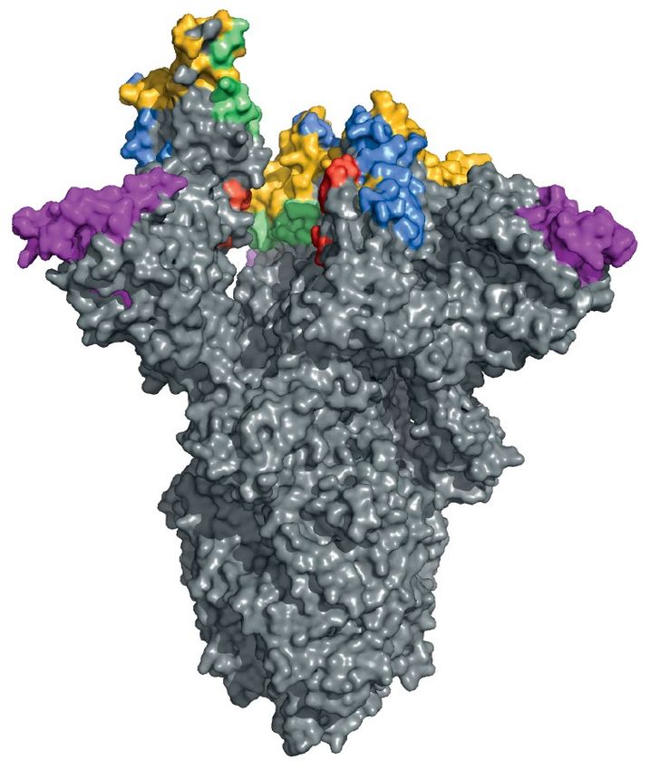
Reviews
◀ Fig. 1 | Neutralizing antibody classes defined by structural analyses and properties change across the NTD and the RBD can drastically evade
of spike protein residues. a | Amino acid residues of the severe acute respiratory the polyclonal antibody response. The Δ140+E484K
syndrome coronavirus 2 (SARS-CoV-2) spike protein are coloured according to the double mutant next acquired an 11-residue insertion in
class of the antibody that binds to an epitope. Receptor-binding domain (RBD) antibody the NTD N5 loop between Y248 and L249, completely
classes 1–4 (ref.31) are shown: green for class 1 (ACE2-blocking antibodies that bind the
abolishing neutralization. This insertion, which also
spike protein in the open conformation), yellow for class 2 (ACE2-blocking antibodies
that bind the RBD in both the open conformation and the closed conformations),
introduced a new glycosylation motif in the vicinity of
blue for class 3 (antibodies that do not block ACE2 and bind the RBD in both the open RDR4, is predicted to alter the structure of the antigenic
conformation and the closed conformations) and red for class 4 (neutralizing antibodies N3 and N5 NTD loops41. This finding further demon-
that bind outside the ACE2 site and only in the open conformation). When residues strates the structural plasticity of the NTD and indicates
belong to epitopes of multiple classes, priority colouring is given to antibodies that block that insertions and the acquisition of additional glyco-
ACE2 and bind the closed spike protein. The amino-terminal domain (NTD) supersite30 sylation motifs in the NTD are further mechanisms in
is coloured in magenta. b | Aligned heat maps showing properties of amino acid residues addition to deletion that lead to immune evasion. Other
where substitutions affect binding by antibodies in polyclonal human blood plasma or examples of mutations that impact the epitope–paratope
emerge as antibody escape mutations. The distance in angstroms to the ACE2-contacting interface indirectly include mutations in the signal pep-
residues that form the receptor-binding site (RBS) is shown in shades of orange; each
tide region and at cysteine residues 15 and 136, which
residue is classified as having evidence for mutations affecting neutralization by either
monoclonal antibodies (mAbs)40,43,47,48 or polyclonal antibodies in plasma from previously form a disulfide bond that ‘staples’ the NTD amino termi-
infected individuals (convalescent)39–41,43,48 or vaccinated individuals59 (‘mAb effect’ and nus against the galectin-like β-sandwich30. Mutations at
‘plasma effect’, respectively). A subset of these residues has mutations described as those sites (for example, C136Y and S12P, which alter the
emerging upon exposure (co-incubation) to mAbs40,47,48 or plasma40,41 in laboratory cleavage occurring between residues C15 and V16) have
experiments (‘mAb emerge’ and ‘plasma emerge’, respectively). When an observation been shown to affect the neutralizing activity of several
includes a deletion, this is indicated by a red cross. Shades of green depict the results of mAbs, likely disrupting the disulfide bond and therefore
deep mutational scanning (DMS) experiments where yeast cells expressing RBD mutants dislodging the supersite targeted by several antibodies30.
are incubated with multiple samples of human convalescent plasma39. The escape Across the spike protein, some mutations that con-
fraction (that is, a quantitative measure of the extent to which a mutation reduced fer escape to neutralizing mAbs have little impact on
polyclonal antibody binding) averaged across all amino acid substitutions at a residue
serum antibody binding39,40,44, possibly because those
(‘plasma average’) and the maximally resistant substitution (‘plasma max’) are indicated.
DMS data on ACE2-binding affinity19 are shown in shades of red or blue representing
mAbs are rare in polyclonal sera, targeting subdominant
higher or lower ACE2 affinity, respectively. The mean change in binding affinity averaged epitopes12,39,44. Escape mutations emerging in viruses
across all mutations at each site (‘binding average’) and alternatively the maximally exposed to convalescent plasma have been identified in
binding mutant (‘binding max’) is shown. Scores represent binding constants (Δlog10 KD) both the NTD (ΔF140, N148S, K150R, K150E, K150T,
relative to the wild-type reference amino acid. K150Q and S151P) and the RBD (K444R, K444N,
K444Q, V445E and E484K)40,41 (Fig. 1b). Notably, muta-
tions emerging under selective pressure from convales-
mutations in viruses exposed to convalescent plasma cent plasma may be different from those selected by the
from two individuals, one of which selected for NTD most frequent mAb isolated from the same plasma40.
mutations only (N148S, K150R, K150E, K150T, K150Q Potentially, observed differences arise because muta-
and S151P)40. This was despite the plasma being a source tions selected by convalescent plasma facilitate escape
of the highly potent RBD-targeting mAb C144 (ref.40). from multiple mAbs. Fewer data on the antigenic
NTD antibody escape mutations were not observed for effects of S2 mutations exist, though D769H has been
the other samples of plasma investigated, and further- described as conferring decreased susceptibility to neu-
more, the 148–151 mutants exhibited only marginal tralizing antibodies24. Residue 769 is positioned in a
reductions in sensitivity to the plasma tested, indicat- surface-exposed S2 loop, and D769H was found to arise,
ing individual immune responses may be differentially in linkage with Δ69–70, in an immunocompromised
affected by mutations of RBD and NTD epitopes40. individual treated with convalescent plasma24.
Deletions in the NTD have been observed repeatedly
in the evolution of SARS-CoV-2 and have been described Conformational epitopes in spike
as changing NTD antigenicity30,41,42. One study identified To evaluate potential antigenicity across the spike pro-
four recurrently deleted regions (RDRs) within the NTD tein, we analysed the protein using BEpro, a program
Epitope binning and tested five frequently observed deletions within these: for the prediction of conformational epitopes based
An approach that uses a Δ69–70 (RDR1), Δ141–144 and Δ146 (RDR2), Δ210 on tertiary structure 49. This approach calculates a
competitive immunoassay to (RDR3) and Δ243–244 (RDR4)42. Of the four RDRs, structure-based epitope score, which approximates anti-
sort a library of monoclonal
antibodies into discrete groups
RDR1, RDR2 and RDR4 correspond to NTD loops N2, body accessibility for each amino acid position. For each
of antibodies that compete for N3 and N5, whereas RDR3 falls between N4 and N5 in residue, the calculated score accounts for the local pro-
access to overlapping epitopes. another accessible loop (Fig. 2a, asterisk). Both RDR2 tein structure: half-sphere exposure measures and pro-
deletions, Δ141–144 and Δ146, and Δ243–244 (RDR4) pensity scores each depend on all atoms within 8–16 Å of
Convalescent plasma
abolished binding of 4A8 (ref.42). Further evidence of the target residue, with weighting towards closer atoms.
Blood serum of a previously
infected individual that usually the role of RDR2 deletions in immune escape was pro- Due to this aggregation, calculated scores are relatively
contains a mixture of different vided by a study that describes the emergence of Δ140 insensitive to the effects of single amino acid substitu-
antibodies referred to as in SARS-CoV-2 co-incubated with potently neutralizing tions. Scores were calculated for the spike protein in both
polyclonal antibodies. convalescent plasma, causing a fourfold reduction in neu- the closed conformation and the open conformation
Similarly, postvaccination
serum includes polyclonal
tralization titre41. This Δ140 spike mutant subsequently (Fig. 2). It has been estimated that ~34% of spike pro-
antibodies generated by acquired the E484K mutation, resulting in a further four- teins are closed and 27% are open (with the remainder
vaccination. fold drop in neutralization titre, and thus a two-residue in an intermediate form) following furin cleavage50.
Nature Reviews | Microbiology
0123456789();: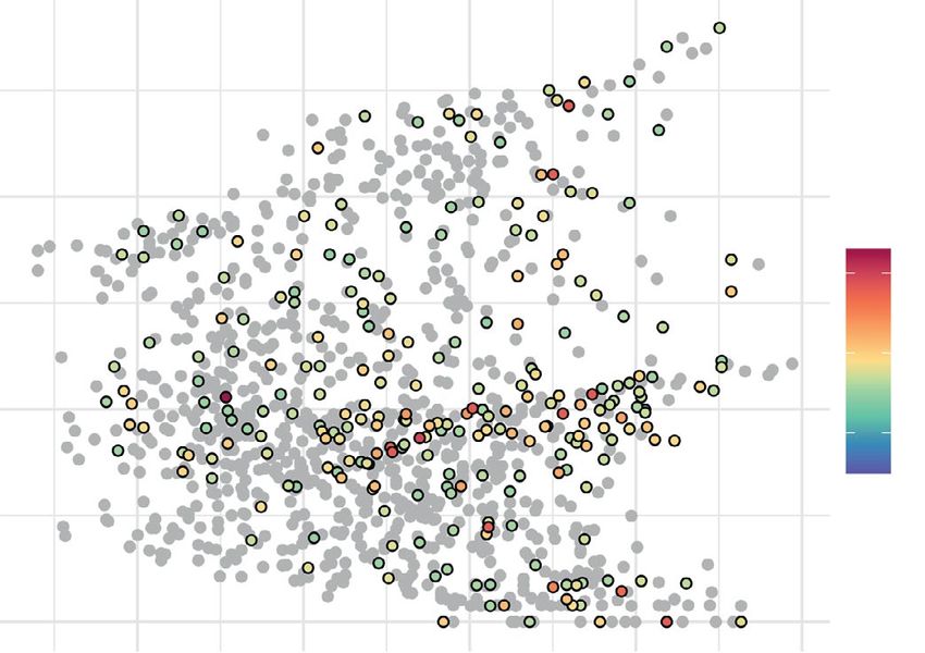
Reviews
Scores rescaled between 0 and 1 are plotted for the closed Fig, 1b). Notably, scores for residues with mutations
conformation in Fig. 2a and are represented on the struc- described as affecting plasma antibody recognition are
ture in Fig. 2b. A limitation of this approach is that it does also slightly higher on average compared with those with
not account for glycan shielding of residues and likely mutations described as affecting mAbs only. Epitope
overestimates scores at the base of the ectodomain for scores are particularly high for residues with mutations
residues closest to the carboxy terminus. described as emerging during exposure to convalescent
Comparisons with reporting of antibody footprints plasma40,41 (Supplementary Fig. 1b). Experimental data
and the impact of mutations on antigenicity indicate on the emergence of mutations under selective pressure
that residues with mutations described as affecting from polyclonal antibodies are relatively rare, although
recognition by mAbs or antibodies in convalescent these trends for higher scores associated with such muta-
plasma (Fig. 1b) tend to occur at residues with higher tions indicate that information from structural analysis
structure-based antibody accessibility scores compared approaches of this kind can contribute to the ranking of
with other residues belonging to epitope footprints and residues at which substitutions are likely to impact the
residues not implicated in antigenicity (Supplementary polyclonal antibody response.
a NTD RBD b
1.0 N3
N2 Closed
N1 N4 spike
* N5
0.8 477
501 Accessibility
Antibody accessibility score
score
70 439 1.0
0.6 0.8
69 0.6
453 0.4
0.4 0.2
222
0.0
0.2
614
0.0
d ACE2-binding site
0 100 200 300 400 500 600 700 800 900 1,000 1,100
Amino acid position Upright
RBD
c
More
0.4 accessible Open
when open spike
0.2
0.0
Adjustment to antibody accessibility
−0.2
More
0 100 200 300 400 500 600 700 800 900 1,000 1,100 accessible
when closed
0.4
0.2
0.0
−0.2
0 100 200 300 400 500 600 700 800 900 1,000 1,100
0.4
0.2
0.0
−0.2
0 100 200 300 400 500 600 700 800 900 1,000 1,100
Fig. 2 | Structure-based analysis of conformational epitopes on the spike view along this axis (right). c | The extent to which each spike residue becomes
protein. a | Structure-based antibody accessibility scores for each spike more or less accessible when the spike protein is in its open form is shown. For
protein ectodomain residue in the closed form were calculated with BEpro49. each spike monomer (upright receptor-binding domain (RBD) (yellow), closed
Black diamonds at the top and bottom of the plot indicate the positions of RBD clockwise adjacent (green) and closed RBD anticlockwise adjacent
ACE2-contacting residues. Accessible amino-terminal domain (NTD) loops (blue)), the difference relative to the score calculated for the closed form
N1–N5 are labelled, and a loop falling between these is indicated with an (shown in part a) is shown. d | Two surface colour representations of antibody
asterisk. b | Two surface colour representations of antibody accessibility scores accessibility scores for the spike protein in the open conformation with a
for the spike protein in the closed conformation according to the colour single monomer with an upright RBD are shown: a trimer axis vertical view
scheme in part a: a trimer axis vertical view (left) and an orthogonal top-down (left) and an orthogonal top-down view along this axis (right).
www.nature.com/nrmicro
0123456789();: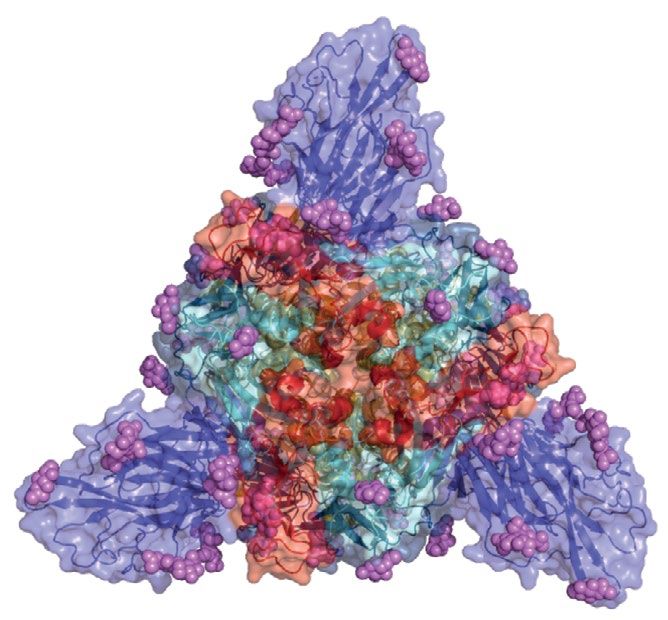
Reviews
Within the RBD, the two areas with high structure- frequency of mutations in circulating variants (Fig. 3).
based antibody accessibility scores for the closed spike Globally, the highest number of amino acid variants,
structure (Fig. 2a, peaks with consecutive residues with mapped against the Wuhan-Hu-1 reference sequence
scores greater than 0.8) are centred at residues 444–447 (MN908947), are recorded at amino acid positions
and residues 498–500. These areas are represented as 614, 222 and 18 (Fig. 4a) (among 426,623 high-quality
yellow patches near the centre of the top-down view of sequences retrieved from the GISAID database on
the spike structure in Fig. 2b. Figure 2c shows that, in 3 February 2021 and processed using CoV-GLUE).
general, residues become more accessible and are likelier Residues at positions 614 and 222 have relatively
to form epitopes when the spike protein is in the open low antibody access scores and are positioned ~50 Å
conformation, and this is especially true for the RBD, from the RBS residues when the spike protein is in
particularly for the upright RBD (Fig. 2c, yellow). In the the open conformation (Fig. 3a,b). As mentioned ear-
open form, residues close to the ACE2-binding site (405, lier, there is evidence indicating that D614G confers
415, 416, 417 and 468) become much more exposed on a moderate advantage for infectivity8,9 and increases
both the upright RBD and the clockwise adjacent closed transmissibility10. The spike amino acid substitution
RBD (Fig. 2c, green). The effect of mutations at these with the second highest frequency is A222V, which is
positions is likely to be greater for antibodies belonging present in the 20A.EU1 SARS-CoV-2 cluster (also des-
to RBD class 1. Residues centred at 444–447 and 498–500 ignated lineage B.1.177). This lineage has spread widely
maintain high scores on the upright RBD and are in Europe and is reported to have originated in Spain52.
joined by residues in areas 413–417 and 458–465. The There is no evidence for a notable impact of A222V on
only RBD residues that become notably less accessible virus phenotype (that is, infectivity and transmissibility),
in the open spike structure are residues 476, 477, 478, and so its increase in frequency is generally presumed to
586 and 487 of the closed RBD clockwise adjacent to have been fortuitous rather than a selective advantage.
the upright RBD, which become blocked by the upright The substitution L18F has occurred ~21 times in the
RBD (Fig. 2c, green). Several RBD-specific antibodies are global population53 and is associated with escape from
able to bind only the open spike protein (RBD classes 1 multiple NTD-binding mAbs30.
and 4 (ref.31)), and interestingly, it has been observed that Among the 5,106 independent substitutions observed
D614G makes the spike protein more vulnerable to neu- in the spike protein (Box 1), 161 are described as affecting
tralizing antibodies by increasing the tendency for the recognition by mAbs or polyclonal antibodies in sera, of
open conformation to occur51. which 22 are present in more than 100 sequences. On
Within the NTD, the highest-scoring spike residues in average, variant frequency is higher at amino acid posi-
the closed form belong to a loop centred at residues tions where mutations are described as affecting anti-
147–150, which each have scores greater than 0.9 (Fig. 2a, body recognition than at positions with no described
yellow patch to the extreme right of the structure viewed substitutions of antigenic importance (Supplementary
from the side in Fig. 2b). This loop, known as the N3 Fig. 1a), and high levels of amino acid substitutions are
loop, is described as forming key interactions with the observed at some amino acid positions where mutations
neutralizing antibody 4A8 (ref.32). One study described are described as affecting recognition by antibodies in
the structure of five previously unmodelled, protruding convalescent plasma, including positions 439 and 484.
NTD loops, denoting them N1–N5. In addition to N3, This indicates that, generally, the amino acid positions
high-scoring residues (greater than 0.7) are found at at which antibody escape mutations have been detected
positions 22–26 (N1), 70 (N2), 173–187 (N4), 207–213 in vitro tolerate mutations at least to some degree in vivo.
(Fig. 2a, asterisk) and 247–253 (N5). Structural analysis Within the RBD, the positions at which amino acid
indicates NTD-binding antibodies are likely able to bind substitutions are present at the highest frequency are
epitopes when the spike protein is in either the closed located close to the RBD–ACE2 interface (Fig. 3). Of the
conformation or open the conformation (Fig. 2c). Outside three RBD amino acid substitutions present in several
the NTD and the RBD, the highest-scoring residues are thousand sequences, N439K and N501Y were described
residues 676 and 689 (which lie on either side of the earlier, and N501Y is discussed in more detail in the next
loop containing the S1–S2 furin cleavage site, which section in the context of variants of concern. The other
is disordered in both the open conformation and the substitution, S477N, is estimated to have emerged at
closed conformation50), 793–794, 808–812, 1,099–1,100 least seven times in the global SARS-CoV-2 population
and 1,139–1,146 (Fig. 2a). When the spike protein is in and has persisted at a frequency of between 4% and 7%
the open conformation, increased accessibility results of sequences globally since mid-June 2020 (ref.53). One
in substantially higher potential epitope scores for S2 study described multiple mAbs that selected for the
residues centred at 850–854, which become more emergence of S477N and found this mutant to be resist-
accessible on all three spike monomers (Fig. 2c), and ant to neutralization by the entire panel of RBD-targeting
residues 978–984, which become more accessible on mAbs that were tested. By contrast, when tested with
the monomer anticlockwise adjacent to the upright RBD convalescent serum, neutralization of the S477N mutant
monomer (Fig. 2c, blue). was similar to that of the wild type48. In common with
N439K and N501Y, S477N results in increased affinity
Structural context of spike mutations for the ACE2 receptor, although to a lesser extent19,54. As
To assess the impact of spike mutations and their immuno described in Box 2, substitutions may facilitate immune
logical role in the global SARS-C oV-2 population, escape by increasing receptor-binding affinity inde-
we combined structural analyses with the observed pendently of any effect that they may have on antibody
Nature Reviews | Microbiology
0123456789();: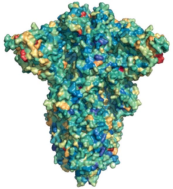
Reviews
a b
477
Distance from receptor-binding site (Å)
18
100 Variant
frequency 69–70
100,000
222 144
614
222 69 70 1,000
614
50
10
439
501
477
0
453 417 501 446
0.2 0.4 0.6 484 0.8 1.0
Antibody accessibility score
477
d
c
ACE2
477 501
417 501
453 439 439
453
Upright RBD
477
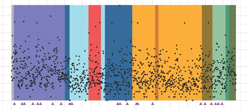
Fig. 3 | Structural context of spike amino acid mutations in the global b | Spike protein in closed form with all residues coloured according to the
virus population. Spike amino acid residues are coloured according to the frequency scale shown; a trimer axis vertical view (left) and an orthogonal
frequency of amino acid substitutions or deletions. Variants (retrieved from top-down view along this axis (right) are shown. c | A close-up view of the
CoV-GLUE) are based on 426,623 high-quality sequences downloaded receptor-binding domain (RBD) bound to ACE2 (RCSB Protein Data Bank ID
from the Global Initiative on Sharing All Influenza Data (GISAID) database 6M0J95), with RBD residues shown as spheres coloured by amino acid variant
on 3 February 2021. a | Points representing each spike amino acid residue frequency and ACE2 shown in gold. Amino acid variants are present at
are positioned according to the antibody accessibility score and the high frequency in positions at the RBD–ACE2 interface. d | Spike protein
distance to the nearest residue in the receptor-binding site. Residues with in open form with residues where at least 100 sequences possessing a
at least 100 sequences possessing a substitution or deletion are coloured substitution are highlighted; a trimer axis vertical view (left) and an
according to the frequency scale shown, with the remainder shaded grey. orthogonal top-down view along this axis (right) are shown.
recognition of epitopes; therefore, it is possible that such was detected in Denmark spreading among farmed mink
a mechanism contributes to the impact of S477N on and a small number of people20. This lineage is character-
neutralization. Variant frequency is also moderately high ized by four amino acid differences, ΔH69–V70, Y453F,
at RBD–ACE2 interface amino acid positions 417, 453 I692V and M1229I (Fig. 5). Of these, the Y453F substitu-
and 446. Of these positions, 446 occurs in a location in tion occurs at a residue within the ACE2 footprint and
the spike structure that is predicted to be highly anti- has been shown by DMS to increase ACE2 affinity19. In
genic, and substitutions at this site are described as addition, Y453F has been described as reducing neu-
affecting neutralization by both mAbs and antibodies tralization by mAbs47. In late 2020 and early 2021, the
present in polyclonal serum39,43,46,48. Substitutions at emergence and sustained transmission of lineages with
amino acid positions 417 and 453 are described in the mutations that affect the characteristics of the virus
next section in the context of variants of concern. received much attention, most notably lineages B.1.1.7,
B.1.351 and P.1 (also known as 501Y.V1, 501Y.V2 and
Variants of interest or concern 501Y.V3, respectively). The locations of the spike muta-
In addition to single mutations of note, more heavily tions in the B.1.1.298, B.1.1.7, B.1.351 and P.1 lineages
mutated SARS-CoV-2 lineages have emerged. Arguably are annotated in Fig. 5a, and information on the struc-
the first variant of interest defined by the presence of sev- tural context and consequences of mutations for antibody
eral spike mutations, and referred to as B.1.1.298 (cluster 5), recognition and ACE2 binding are shown in Fig. 5b.
www.nature.com/nrmicro
0123456789();: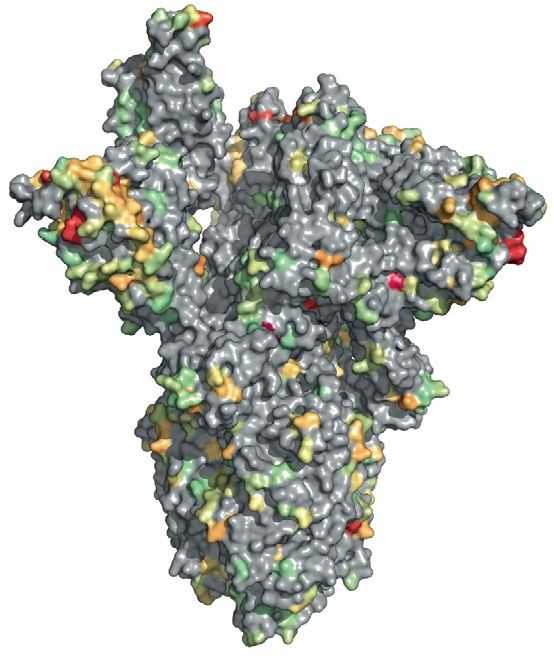
Reviews
Of the lineages summarized in Fig. 5, several amino in lineages B.1.1.298 and B.1.1.7. Additionally, lineages
acid substitutions are convergent, having arisen inde- B.1.351 and P.1 possess alternative amino acid substi-
pendently in different lineages: N501Y, which is present tutions K417N and K417T, respectively. Further line-
in lineages B.1.1.7, B.1.351 and P.1; E484K, which is pres- ages with these mutations have also been identified; for
ent in lineages B.1.351 and P.1 and has been detected as example, an independent emergence of N501Y in the
emerging within the B.1.1.7 lineage55; and ΔH69–V70 B.1.1.70 lineage, which is largely circulating in Wales.
a S1 S2
NTD RBD RBM FP CD TM CT
614
100,000 222
69–70
18 144 501 570 681 716 982
477 1,118
10,000 439
Variant frequency
1,000
100
10
0
0 100 200 300 400 500 600 700 800 900 1,000 1,100 1,200
Amino acid position
b RBM
c
RBD
NTD
S1 S2
FP
CD
Fig. 4 | Spike protein sequence variability and structure. a | The domain particularly within the amino-terminal domain (NTD). The most frequently
organization of the severe acute respiratory syndrome coronavirus 2 detected NTD deletion is the two-residue deletion at positions 69 and 70
(SARS-CoV-2) spike protein showing amino acid sequence variability. (Δ69–70), present in 45,898 sequences. The S1–S2 boundary is at amino
The spike protein is synthesized as a 1,273 amino acid polypeptide, and the acid position 685. b | Spike protein monomer displaying an upright
frequency of amino acid variants, including both substitutions and receptor-binding domain (RBD). c | Spike protein structure in the closed
deletions, at each of the positions is shown. These variants, relative to the conformation overlaid with surface representations shown with a trimer
Wuhan-Hu-1 reference sequence, were identified with use of CoV-GLUE96, axis vertical view (left) and an orthogonal top-down view along this axis
which filters out Global Initiative on Sharing All Influenza Data (GISAID) (right). Domains are coloured as in part a. The RCSB Protein Data Bank IDs
sequences97 identified as being of low quality or from non-human hosts for the SARS-CoV-2 spike protein structures are 6ZGG and 6ZGE50. The
(sequences retrieved from the GISAID database on 3 February 2021). magenta spheres represent glycans, and the magenta triangles represent
Among 426,623 genomes after filtering, 5,106 different amino acid potantial N-linked glycosylation sites. The scissors represent the S1–S2
replacements or substitutions across 1,267 spike positions were identified, boundary at amino acid position 685. CD, connecting domain;
of which 320 at 259 positions were observed in at least 100 sequences. CT cytoplasmic tail; FP, fusion peptide; RBM, receptor-binding motif;
In addition to substitutions, several deletions have been observed, TM, transmembrane domain.
Nature Reviews | Microbiology
0123456789();: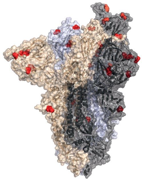
Reviews
Box 2 | Mechanisms of antigenic change antigenic effect, both K417N and K417T are expected
to moderately decrease ACE2-binding affinity19 (Fig. 5b).
in common with other virus surface glycoproteins responsible for attachment to The ΔH69–V70 deletion has been identified in variants
host cell-surface receptors, such as influenza virus haemagglutinin and the envelope associated with immune escape in immunocompro-
glycoprotein GP120 of Hiv, the severe acute respiratory syndrome coronavirus 2 mised individuals treated with convalescent plasma24.
(sars-Cov-2) spike glycoprotein is an important target for neutralizing antibodies.
Experiments have shown that ΔH69–V70 does not
there are various distinct mechanisms by which mutations can alter the antigenic
properties of a glycoprotein. reduce neutralization by a panel of convalescent sera;
however, it may compensate for infectivity deficits asso-
Amino acid substitutions that alter the epitope ciated with affinity-boosting RBM mutations that may
a change in the biophysical properties of an epitope residue directly diminishes
emerge due to immune-mediated selection22.
antibody binding. For example, the neutralizing antibody 4a8 forms salt bridges with
spike protein residues K147 and K150, and therefore substitutions at these residues
The first genomes belonging to the B.1.1.7 lineage
are likely to inhibit binding. the e484K amino acid substitution has received attention were sequenced in the south of England in September
for its effect on monoclonal antibodies and convalescent plasma neutralizing activity. 2020. Lineage B.1.1.7 is defined by the presence of
its position has been described as belonging to the footprint of several antibodies, 23 nucleotide mutations across the genome that map
and a change in charge caused by replacement of a glutamate residue with a lysine to a single branch of the phylogenetic tree3. Of these
residue has the potential to diminish antibody binding. 23 mutations, 14 encode amino acid changes and three
increasing receptor-binding avidity are deletions, including six amino acid substitutions in
substitutions that individually increase receptor-binding affinity can shift the binding the spike protein (N501Y, A570D, P681H, T716I, S982A
equilibrium between glycoprotein and neutralizing antibodies in favour of a higher- and D1118H) and two NTD deletions (ΔH69–V70 and
vidity interaction between glycoprotein and the cellular receptor102. the spike amino
a ΔY144)3. The lineage has been associated with a rapidly
acid substitution N501Y, which increases aCe2-binding affinity19, has been described increasing proportion of reported SARS-CoV-2 cases,
as emerging in individuals treated with convalescent plasma, potentially as a means and phylogenetic analyses indicate that this lineage was
of immune escape. associated with a growth rate estimated to be 40–70%
changes in glycosylation higher than that of other lineages60,61. There is also evi-
Glycans are bulky sugar molecules that may shield epitopes from antibody binding. dence that this lineage may be associated with a higher
N-linked glycans are typically prominent in glycan shielding of virus surface glycoprotein viral load62. In addition to N501Y, for which there is some
epitopes33, although O-linked glycans can also contribute103. a substitution can introduce evidence that it reduces neutralization by a small propor-
an additional N-linked glycosylation motif. the acquisition of epitope-masking glycans
tion of RBD antibodies63, there is evidence for an anti-
during the evolution of human influenza viruses is well described104.
genic effect of ΔY144 (Fig. 5b). This deletion is expected to
Deletions and insertions alter the conformation of the N3 NTD loop (amino acid
the deletion or insertion of residues has the potential to alter epitope conformation, positions 140–156) and has been demonstrated to abol-
diminishing antibody binding. several deletions in the spike amino-terminal domain (NtD) ish neutralization by a range of neutralizing antibodies30.
that affect recognition by neutralizing antibodies have been described41,42. in laboratory
The B.1.1.7 spike mutations have been shown to dimin-
experiments, a multiresidue insertion in the spike NtD has been described as emerging
and contributing to escape from polyclonal antibodies in convalescent plasma41. ish neutralization of a higher proportion of NTD-specific
neutralizing antibodies (9 of 10; 90%) than RBD-
Allosteric structural effects specific neutralizing antibodies (5 of 31; 16%) 63.
similarly to deletions or insertions, an amino acid substitution outside an epitope
Given the immunodominance of the RBD, this could
footprint may affect antibody binding by changing the protein conformation in such
a way that an epitope is altered or differently displayed. in the spike NtD, changes
explain the modest reductions in neutralizing activity
to disulfide bonds are thought to reduce binding by multiple monoclonal antibodies of convalescent sera against authentic B.1.1.7 or pseudo
through this mechanism30. viruses carrying the B.1.1.7 spike mutations64,65. The
co-occurrence of ΔY144 and E484K is concerning with
respect to the polyclonal antibody response as the N3
Residue 501 is at the RBD–ACE2 interface (Fig. 2c), and loop, which ΔY144 changes, is predicted to be among the
N501Y has been shown experimentally to result in one most immunogenic regions of the spike protein (Fig. 2a),
of the highest increases in ACE2 affinity conferred by a and amino acid substitutions at position 484 diminish
single RBD mutation19. E484 has been identified as an neutralization by a range of RBD-targeting antibodies.
immunodominant spike protein residue, with various Reports of lineages with N501Y circulating in the UK
substitutions, including E484K, facilitating escape from were followed by reports of another lineage possessing
several mAbs40,47,48 as well as antibodies in convales- N501Y circulating in South Africa (lineage B.1.351),
cent plasma39–41,48. E484K is estimated to have emerged which has been rapidly expanding in frequency since
repeatedly in the global SARS-C oV-2 population53 December 2020 (ref.66). In addition to N501Y, lineage
and has been described in two other lineages origi- B.1.351 is defined by the presence of five further spike
nating from lineage B.1.1.28 in addition to lineage P.1 amino acid substitutions (D80A, D215G, K417N, E484K
reported to be spreading in the state of Rio de Janeiro and A701V) and a deletion in the NTD, Δ242–244. High
in Brazil (lineage P.2)56 and in the Philippines (line- numbers of B.1.351 viruses also have the spike amino
Avidity age P.3)57. Whereas K417 is described in the epitopes acid substitutions L18F, R246I and D614G. A similar
Also referred to as functional of RBD class 1 and class 2 antibodies31, alterations to NTD deletion, Δ243–244, abolishes binding by the anti-
affinity, the accumulated K417 tend to affect class 1 antibody binding and are body 4A8 (ref.42), and L18F and R246I also occur within
binding strength of multiple therefore somewhat less important for the polyclonal the NTD supersite and likely affect antibody binding as
affinities of individual
interactions, such as between
antibody response to the RBD, which is dominated by well30. The co-occurrence of K417N, E484K and these
a virus receptor-binding site class 2 antibody responses, which are more susceptible NTD substitutions suggests that lineage B.1.351 may
and its cellular receptor. to substitutions such as E484K44,58,59. In addition to their overcome the polyclonal antibody response by reducing
www.nature.com/nrmicro
0123456789();:Reviews
IC50
neutralization by class 1 and class 2 RBD-specific anti- between December 2020 and February 2021, indicating
The half-maximal inhibitory bodies and NTD-specific antibodies (Fig. 5b) . Data that this amino acid substitution is probably the result
concentration, a quantitative reported in one study showed that nearly half of exam- of viral adaptation due to increasing immunity in the
measure that indicates ined convalescent plasma samples (21 of 44; 48%) had population75. L452R is also present in the A.27 lineage
how much of an inhibitory
no detectable neutralization activity against the B.1.351 associated with a cluster of cases identified on the island
substance (for example,
postvaccination serum) is variant58. Other experiments with pseudotyped viruses of Mayotte76. The lineage B.1.526 has been found to carry
required to inhibit a biological showed that the B.1.351 variant was also resistant to the either S477N or E484K, among other lineage-defining
process (for example, virus neutralizing activity of some mAbs (12 of 17; 70%)67. mutations77,78, both of which were described as antigen-
forming plaques or regions
Similarly, a study showed that the neutralizing effect of ically important above. A new variant carrying E484K
of infected cells in culture)
by 50%.
convalescent plasma collected from 14 individuals was currently designated A.VOI.V2 was recently identified
strongly reduced against a live (authentic) B.1.351 virus in Angola from cases involving travel from Tanzania79.
(with IC50 reduced by 3.2-fold to 41.9-fold relative to the This variant carries several amino acid substitutions in
first-wave virus)68. the spike protein and three deletions in the NTD, some
The P.1 lineage, a sublineage of B.1.1.28, was first of which are within the antigenic supersite79. Another
detected in Brazil69 and in travellers from Brazil to variant within the A lineage, the prevalence of which is
Japan70. Lineage P.1 is characterized by the presence rising in Uganda (A.23.1), shares with the B.1.1.7 lineage
of several amino acid substitutions in the spike pro- a substitution at position 681 within the furin cleavage
tein: L18F, T20N, P26S, D138Y, R190S, K417T, E484K, site (P681R has been found in the A lineage, whereas
N501Y, H655Y and T1027I69. In addition to substitutions P681H has been found in the B.1.1.7 lineage), and addi-
at positions 417, 484 and 501 discussed above, the P.1 lin- tionally has the amino acid substitutions R102I, F157L,
eage has a cluster of substitutions close to the described V367F and Q613H. Q613H is speculated to be impor-
antigenic regions of the NTD, including L18F, which is tant as it occurs at a position neighbouring D614G80.
known to reduce neutralization by some antibodies30. Amino acid position 157 has been identified as an
The substitutions T20N and P26S also occur in or epitope residue, with F157A reducing neutralization by
near the NTD supersite30 at residues with high anti- the mAb 2489 (ref.34).
body accessibility scores (Fig. 5b), and T20N introduces New variants will continue to emerge, and although
a potential glycosylation site that could result in glycan it is important to understand the phenotypes of emerg-
shielding (Box 2) of part of the supersite. The P.1 line- ing variants in terms of infectivity, transmissibility, vir-
age has also been associated with a reinfection case in ulence and antigenicity, it is also important to quantify
Manaus, Brazil27, indicating it is contributing to anti- the phenotypic impacts of specific mutations present
genic circumvention of what might have been an oth- in variants, both individually and in combination with
erwise effective immune response. Analyses integrating other mutations. As new variants with unforeseen com-
genomic and mortality data estimate that P.1 may be binations of mutations continue to emerge, such insights
1.7 to 2.4-fold more transmissible and that previous will allow predictions of virus phenotype. For example,
infection by non-P.1 SARS-C oV-2 provides 54–79% recently detected viruses of lineage B.1.617.1 were antici
of the protection against P.1 infection compared with pated to show altered antigenicity due to the presence of
non-P.1 lineages71. More details of the frequency and the substitutions L452R and E484Q, which have been
geographic distribution of the P1 lineage can be found described as affecting antibody recognition39,43,45,48,81.
at the Pango lineages website72. Moving forwards, the experimental characterization
Increasingly, lineages possessing independent of SARS-CoV-2 spike mutations to date will continue
occurrences of mutations in common with the variants to provide extremely useful information on individual
of concern B.1.1.7, B.1.351 and P.1 are being detected, mutations or combinations of mutations that may not
demonstrating convergent evolution. For example, yet have been seen in circulating viruses.
viruses of lineage B.1.525, which has been observed in
several countries, albeit at low frequency to date, have Vaccine efficacy against new variants
NTD deletions ΔH69–V70 and ΔY144 in common with To date, vaccines have been licensed and rolled out very
viruses of the B.1.1.7 lineage; E484K in common with the successfully in several countries, but the number of indi-
B.1.351 and P.1 lineages; and spike amino acid substitu- viduals vaccinated still represents a small fraction of the
tions Q52R, Q677H and F888L73. Repeated amino acid global population (Supplementary Table 1). To assess
substitutions at position 677 and the independent emer- the impacts of mutations on vaccine efficacy, authentic
gence of Q677H in several lineages in the USA provides viruses and pseudoviruses possessing particular spike
strong evidence of adaptation, potentially through an mutations (either individually or in combination) and
effect of this mutation on the proximal polybasic furin larger sets of mutations representing variants of concern
cleavage site, although further experiments are required and other circulating spike mutations have been assessed
to determine its impact74. Other novel variants have by neutralization assays with postvaccination sera
been identified spreading in California and New York, (Supplementary Table 1). Typically, studies report a fold
USA (B.1.427 and B.1.429, and B.1.526, respectively). change in variant virus, or pseudovirus, neutralization rel-
The B.1.427 and B.1.429 variants carry an antigeni- ative to wild-type virus (the serum concentration at which
cally noteworthy substitution, L452R75, which has been 50% neutralization (IC50) is achieved with the variant
shown to reduce neutralization by several mAbs43,45,48,59 divided by the average IC50 for the wild-type virus).
and convalescent plasma 43. L452R independently Postvaccination sera from a cohort of 20 volun-
appeared in several other lineages around the globe teers immunized with the mRNA vaccine mRNA-1273
Nature Reviews | Microbiology
0123456789();:You can also read