STRUCTURAL BASIS OF RIBOSOMAL FRAMESHIFTING DURING TRANSLATION OF THE SARS-COV-2 RNA GENOME - SCIENCE
←
→
Page content transcription
If your browser does not render page correctly, please read the page content below
RESEARCH ARTICLES

                                                                                                                                   Cite as: P. R. Bhatt et al., Science
                                                                                                                                   10.1126/science.abf3546 (2021).

Structural basis of ribosomal frameshifting during
translation of the SARS-CoV-2 RNA genome
Pramod R. Bhatt1,2,3†, Alain Scaiola1†, Gary Loughran2, Marc Leibundgut1, Annika Kratzel4,5,6, Romane Meurs7,
René Dreos7, Kate M. O’Connor2, Angus McMillan8, Jeffrey W. Bode8, Volker Thiel4,5, David Gatfield7,
John F. Atkins2,3,9*, Nenad Ban1*
1Department of Biology, Institute of Molecular Biology and Biophysics, ETH Zurich, Zurich, Switzerland. 2School of Biochemistry and Cell Biology, University College Cork,

Cork T12 XF62, Ireland. 3School of Microbiology, University College Cork, Cork T12 K8AF, Ireland. 4Institute of Virology and Immunology, University of Bern, Bern,
Switzerland. 5Department of Infectious Diseases and Pathobiology, Vetsuisse Faculty, University of Bern, Bern, Switzerland. 6Graduate School for Cellular and Biomedical
Sciences, University of Bern, Bern, Switzerland. 7Center for Integrative Genomics, Génopode, University of Lausanne, 1015 Lausanne, Switzerland. 8Laboratorium für
Organische Chemie, Department of Chemistry and Applied Biosciences, ETH Zurich, Zurich, Switzerland. 9MRC Laboratory of Molecular Biology, Cambridge CB2 0QH, UK.
†These authors contributed equally to this work. *Corresponding author. Email: j.atkins@ucc.ie (J.F.A.); ban@mol.biol.ethz.ch (N.B.)

Programmed ribosomal frameshifting is a key event during translation of the SARS-CoV-2 RNA genome

                                                                                                                                                                             Downloaded from http://science.sciencemag.org/ on July 6, 2021
allowing synthesis of the viral RNA-dependent RNA polymerase and downstream proteins. Here we present
the cryo-electron microscopy structure of a translating mammalian ribosome primed for frameshifting on
the viral RNA. The viral RNA adopts a pseudoknot structure that lodges at the entry to the ribosomal mRNA
channel to generate tension in the mRNA and promote frameshifting, whereas the nascent viral polyprotein
forms distinct interactions with the ribosomal tunnel. Biochemical experiments validate the structural
observations and reveal mechanistic and regulatory features that influence frameshifting efficiency.
Finally, we compare compounds previously shown to reduce frameshifting with respect to their ability to
inhibit SARS-CoV-2 replication, establishing coronavirus frameshifting as a target for antiviral intervention.

Ribosomal frameshifting, a process during which the reading                             produces polyprotein 1a ending with Nsp10 followed by the
frame of translation is changed at the junction between open                            short Nsp11. On the other hand, when the frameshift occurs,
reading frames 1a and 1b, is one of the key events during                               the polyprotein 1ab is generated, which contains almost 2700
translation of the severe acute respiratory syndrome corona-                            additional amino acids and in which the viral RdRp, Nsp12,
virus 2 (SARS-CoV-2) positive sense single-stranded RNA ge-                             is produced after Nsp10 as a consequence of translation in
nome. This programmed -1 translational frameshifting is                                 the -1 frame. A putative secondary structure element in the
conserved in all coronaviruses and is necessary for synthesis                           viral RNA that forms a loop upstream of the shift site has
of viral RNA-dependent RNA polymerase (RdRp or Nsp12)                                   been proposed to play an attenuating role in frameshifting
and downstream viral non-structural proteins encoding core                              and is referred to as the 5′ attenuator loop (8). Maintaining
enzymatic functions involved in capping of viral RNA, RNA                               the precise level of coronavirus frameshifting efficiency is
modification and processing, and RNA proof-reading (1). Alt-                            crucial for viral infectivity, evidenced by the remarkable fact
hough the translational machinery typically prevents                                    that mutation of a single nucleotide in the frameshifting re-
frameshifting as a potential source of one of the most disrup-                          gion of the SARS-CoV-1 RNA results in a concomitant abro-
tive errors in translation (2, 3), many viruses rely on pro-                            gation of viral replication (13). Therefore, the importance of
grammed ribosomal frameshifting to expand and fine-tune                                 3-stemmed pseudoknot-dependent -1 ribosomal frameshift-
the repertoire and stoichiometry of expressed proteins (4).                             ing for the propagation of SARS-related coronaviruses, a pro-
    Programmed -1 frameshifting in SARS-related corona-                                 cess that has not been seen to occur on any endogenous
viruses occurs at the slippery sequence U_UUA_AAC in the                                human transcript in human cells, presents itself as an oppor-
context of a 3′ stimulatory RNA sequence that was predicted                             tune drug-target with minimal tolerance for drug-resistant
to form a 3-stemmed pseudoknot structure (5), and in parallel                           mutations.
was independently tested by our lab and others (6–8). The                                   Due to its importance in the life cycle of many important
frameshifting occurs with high efficiency (25-75%) depending                            viruses and coronaviruses in particular, programmed
on the system used (6, 7, 9–11) and changes the reading frame                           frameshifting has been extensively studied using a range of
to UUU_AAA_C (12) (Fig. 1A). Consequently, two viral poly-                              structural and functional approaches (4). The structure of a
proteins are synthesized, one encoded by the ORF1a when                                 3′ stimulatory pseudoknot in isolation or in context of the vi-
frameshifting does not take place, whereas ORF1ab is ex-                                ral genome has been proposed recently by various groups us-
pressed as a result of frameshifting. Translation of ORF1a                              ing techniques that include molecular dynamics, nuclease

First release: 13 May 2021                                               www.sciencemag.org                 (Page numbers not final at time of first release) 1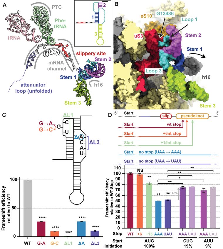
mapping, in vivo selective 2′-hydroxyl acylation analyzed by       channel suggestive of a structured RNA, which after focused
primer extension (SHAPE), nuclear magnetic resonance               classification revealed a prominent density for a complete 3′
(NMR) and cryo-electron microscopy (cryo-EM) (7, 14–17).           frameshifting stimulatory pseudoknot at the entry of the
Furthermore, a ribosomal complex with a frameshift stimu-          mRNA channel on the 40S subunit (Fig. 1, C and D). The res-
latory pseudoknot from the avian infectious bronchitis virus       olution of this reconstruction ranged from 2.4 Å at the core
was reported at low resolution (18). Here, to provide a struc-     of the ribosome to ~7 Å at the periphery, where the most flex-
tural and mechanistic description of the events during ribo-       ible regions of the pseudoknot are located (figs. S2 and S6).
somal frameshifting, we investigated mammalian ribosomes           Based on the high-resolution maps that allowed visualization
captured in distinct functional states during translation of a     of the codon-anticodon interactions and modifications in the
region of SARS-CoV-2 genomic RNA where -1 programmed               tRNA (Fig. 1E and fig. S6, A and B), we could unequivocally
frameshifting occurs.                                              determine that a Phe-tRNA(Phe) was bound at the P-site (22).
                                                                   The mRNA does not adopt any unusual structure in the A-
Structure determination of a frameshifting-primed                  site of the ribosome as was observed for the HIV-1 frameshift-
ribosomal complex                                                  ing sequence visualized on the bacterial ribosome (23). This
We captured a 0 frame, pre-frameshift ribosomal complex by         implied that the ribosome is paused by the downstream pseu-

                                                                                                                                        Downloaded from http://science.sciencemag.org/ on July 6, 2021
introducing a stop codon in place of the second codon of the       doknot located at the entrance to the mRNA channel such
slippery site (U_UUA_AAC to U_UUA_UAA) (Fig. 1A) and               that the P-site tRNA interacts with the UUU codon just prior
adding mutant eukaryotic Release Factor 1 [eRF1 (AAQ)] that        to the first codon, UUA, of the slippery site (Fig. 2A).
is unable to release the nascent polypeptide. Translating com-
plexes were prepared in an in vitro translation reaction using     The pseudoknot causes ribosomal pausing prior to
an in-house generated rabbit reticulocyte lysate (RRL) system      -1 frameshifting
that supported efficient frameshifting in the previously re-       The observation that the pseudoknot acts as an obstacle to
ported range of around 50% (19) according to dual luciferase       slow down translation as the ribosome approaches the slip-
experiments (see methods). The ribosomes were pro-                 pery site is mechanistically reasonable. Since the pseudoknot
grammed with mRNA encoding an affinity tag and harboring           is a stable structural element in the mRNA, it will resist un-
a region of the SARS-CoV-2 genome that encodes proteins            folding and consequently generate a back-pull on the viral
Nsp10 (C terminus), Nsp11 and the majority of Nsp12. West-         RNA, resulting in an increased chance of -1 frameshifting as
ern blotting showed that when using the WT RNA template,           the tRNAs are translocated. A pause in translocation at a co-
frameshifting was efficient, while the stop codon mutation         don that precedes the slippery site, characterized by a >10
prevented frameshifting and led to ribosome pausing. This          times longer occupancy prior to the slippage event, was ob-
effect was further enhanced when eRF1 (AAQ) was present in         served in an analogous case of heptanucleotide -1 frameshift-
excess over endogenous wild type eRF1 (Fig. 1B).                   ing on the bacterial dnaX gene using single molecule
    The cryo-EM 3D reconstruction of ribosome-nascent              experiments (24). According to this model, it would be antic-
chain complexes (RNCs) affinity purified from the reactions        ipated that a further round of translocation results in un-
supplemented with eRF1 (AAQ) revealed two distinct riboso-         winding of Stem 1 of the downstream stimulatory pseudoknot
mal complexes captured in the process of translating the slip-     structure. Consistently, in our structure of the eRF1 (AAQ)-
pery sequence (figs. S1 and S2). One represented a                 bound ribosome that advanced one codon further along the
termination complex that contained the ATP-binding cas-            mRNA, no clear secondary structure is visible at the entrance
sette transporter 1 (ABCE1) known to be involved in termina-       to the mRNA channel as the mRNA now becomes disordered
tion and recycling together with mutant eRF1 interacting           at this position (figs. S1 and S3, A and B).
with the stop codon (fig. S3). The second reconstruction re-           In order to investigate the slowdown of translation on the
solved translating 80S ribosomes containing P- and E-site          wild type slippery sequence, we performed disome footprint
tRNAs bound (fig. S2). This reconstruction at 2.2 Å resolution     profiling, a method to identify translational pause sites
allowed us to build the most accurate structure of a mamma-        through the analysis of transitory ribosome collisions (25–27)
lian 80S ribosome so far and directly visualize many protein       (see methods). Notably, recent studies using conventional ri-
and virtually all rRNA modifications identified for the human      bosome profiling methodology reported a lack in monosome
ribosome based on quantitative mass spectrometry and as in-        footprint coverage across the frameshifting region on the
terpreted in a recent human ribosome structure (20, 21), con-      SARS-CoV-2 RNA (11, 28) – possibly because ribosomes in this
sistent with the complete conservation of all modified             area became trapped in temporary collisions. Moreover, the
residues between rabbit and human rRNAs (figs. S4 and S5;          highly structured pseudoknot at the entry to the mRNA chan-
and tables S1 to S3). Importantly, this reconstruction also fea-   nel would likely preclude efficient trimming by RNase I, the
tured additional density at the entrance to the mRNA               enzyme used for footprint generation, further reducing

First release: 13 May 2021                             www.sciencemag.org       (Page numbers not final at time of first release)   2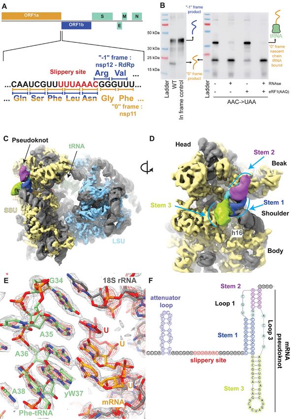
efficient monosome footprint capture. Using a modified nu-         simultaneous increase in the tension of the mRNA and un-
clease treatment protocol (see methods) that recovered mon-        winding of the GC-rich base of Stem 1 upon entering into the
osome footprints from the frameshift region (Fig. 3, A and C),     mRNA entry channel, comparable to the situation when the
our experiments revealed that ribosome collisions occur as a       ribosome proceeds to the engineered stop codon as observed
result of ribosomal pausing at the same position that is ob-       in our eRF1 (AAQ)-stalled structure (fig. S3).
served in the structure of the pseudoknot-engaged ribosome             The pseudoknot structure also reveals a hitherto unob-
(Fig. 3, B and D). Apparently, although the base substitutions     served and possibly unappreciated role for the distal site of
creating a stop codon in the 3′ adjacent slippery site did not     the mRNA entrance channel in helicase activity. While
change the features of pausing, it increased the dwell time of     mRNA unwinding studies outside the mRNA entrance chan-
the ribosomes at the pause site sufficiently to allow visualiza-   nel have so far implicated only a helix in the C-terminal do-
tion in the cryo-EM experiment.                                    main of uS3 (32), we notice that Loop 1 of the pseudoknot
    The results of our disome profiling experiments prompted       contacts the N-terminal domain of uS3 as well as the C-ter-
us to structurally investigate disomes by cryo-EM. We were         minal tail of eS10 (Fig. 2B and fig. S6D), whereas the flipped-
able to visualize the pseudoknot-paused ribosome followed          out base G13486 in this loop forms specific interactions (Fig.
by a closely trailing ribosome. Upon focused refinement, we        2B). Furthermore, as the pseudoknot is located at the entry

                                                                                                                                        Downloaded from http://science.sciencemag.org/ on July 6, 2021
obtained a high-resolution (3.1 Å) structure of the trailing ri-   to the mRNA channel, helix h16 of the 18S rRNA is noticeably
bosome in a rotated state (fig. S1). In congruence with our        pushed outwards due to a direct contact with the minor
estimated positioning of the ribosomes in disome profiling         groove of Stem 1 (Fig. 2B and fig. S7A). Since the pseudoknot
(Fig. 3D), the purine-pyrimidine pattern of codon-anticodon        wedges between the head and the body of the small riboso-
pairs in the structure of the colliding ribosome revealed that     mal subunit, it would restrict their relative motions that need
the pause occurs with CCC and AUG triplets in the P- and A-        to take place during translocation. This is consistent with the
sites, respectively (Fig. 3C).                                     studies on dynamics of coronavirus frameshifting, which re-
                                                                   vealed that the mechanism of -1 frameshifting involves re-
The SARS-CoV-2 RNA pseudoknot specifically interacts               striction of small subunit head motion (33).
with ribosomal proteins and 18S rRNA                                   The structure also reveals another key aspect of the archi-
The intermediate local resolution (5-7 Å) of the cryo-EM map       tecture of the pseudoknot as the ribosome encounters it. The
in the area of the pseudoknot allowed us to visualize the over-    start of the pseudoknot is shifted relative to the predicted sec-
all fold of the RNA and readjust its previously predicted sec-     ondary structure (14–17, 19) by two nucleotides. The two op-
ondary structure (14–17, 19) (Fig. 1, C, D, and F). The            posed nucleotides, which were assumed to base pair with
stimulatory pseudoknot forms an H-type pseudoknot with             Stem 1, are actually forming the start of Stem 3 by pairing
Stem 1 and Stem 2 coaxially stacked on top of each other to        with bases predicted to be in the single-stranded linker 2 (Fig.
form a quasi-continuous helix, while Stem 3 stands out al-         1F and fig. S7, B and C). Our cryo-EM density reveals that
most perpendicular to this plane (Figs. 1D and 2B). This cork-     Loop 3 accommodates a total of 4 nucleotides, three of which
screw-like formation provides a bulky and well-structured          were originally attributed to Stem 2. Thus, we observe that
obstacle wedged at the mRNA entry channel, having the po-          Loop 3 is shifted and expanded relative to the initially pre-
tential to resist unwinding by the helicase activity of the ri-    dicted secondary structures (14–17, 19).
bosome and generating tension on the upstream mRNA up                  To functionally support our structural findings and con-
to the decoding center. Stem 1 of the pseudoknot forms a 9         firm the nature and specificity of the pseudoknot interac-
bp helix which is GC rich at the bottom (Fig. 1F). The penul-      tions, we performed structure-guided mutagenesis
timate nucleotides of the ‘spacer region’ prior to Stem 1 are      experiments using dual luciferase reporter assays in
located at the mRNA entry tunnel, where they interact with         HEK293T cells (see methods) and monitored the frameshift-
several basic residues in the C-terminal domain of uS3 on one      ing efficiency relative to the WT (Fig. 2C). Mutation of G13486
side and are supported by uS5 from the other, with an addi-        of Loop 1 to another purine reduced the frameshifting effi-
tional weak contact contributed by the C-terminal end of           ciency to 30% of the WT level, and mutation of this base to a
eS30. uS3 and eS30 are primary components of the ribosome          pyrimidine further reduced frameshifting to 15%. As expected
helicase and uS5 has been proposed to be a component of the        from our structural data, deletions of the nucleotides of the
ribosomal helicase processivity clamp at the mRNA entry site       spacer regions also had a deteriorating effect on frameshift-
(29, 30). The observed distance between the P-site UUU co-         ing. Loss of Loop 1 entirely abolished frameshifting. Deletion
don and Stem 1 of the pseudoknot underscores the critical          of a single nucleotide of Loop 3 in agreement with its pro-
dependence of the frameshifting efficiency on the length of        posed role in forming the base pairing interactions dimin-
the spacer region (31). Translocation to the next codon would      ished the frameshifting rate to 25% of the WT level. Loss of
place the frameshifting codon UUA into the P-site, with a          the entire Loop 3 reduced frameshifting to 10% of WT levels.

First release: 13 May 2021                             www.sciencemag.org       (Page numbers not final at time of first release)   3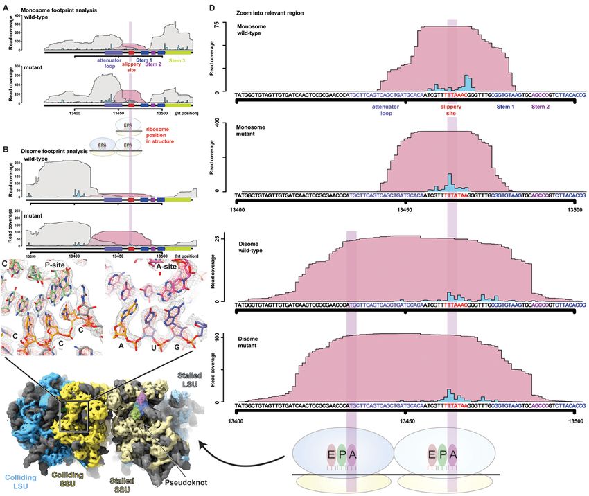
Frameshifting efficiency depends on the position of the            ribosome, the nascent chain that corresponds to the viral pol-
“0” frame stop codon                                               yprotein was visible along the entire length of the ribosomal
In SARS-CoV-2, the 0 frame stop codon is located 5 codons          exit tunnel (Fig. 4A). The density corresponded to the C-ter-
downstream of the frameshift site and is a constituent of          minal region of Nsp10, which is the activator of the viral
Stem 1. The placement of the stop codon in such proximity to       proofreading exonuclease and N7-methyltransferase Nsp14
the frameshift site is a common feature in coronaviruses, and      (34, 35), and then (depending on the frameshifting event)
its presence in a critical region of the stimulatory pseudoknot    continues as either the viral RNA-dependent RNA polymer-
prompted us to probe the effect of the distance of the 0 frame     ase Nsp12 (6) or as protein Nsp11, whose function is yet un-
stop codon on frameshifting. To this end, knowledge of the         known (Figs. 1A and 4B). The nascent chain makes several
3D structure of the pseudoknot helped us to confidently ma-        specific interactions with the ribosomal tunnel, one of which
nipulate the stop codon without hampering pseudoknot for-          is at the constriction site where arginine 4387 of Nsp10 inter-
mation. We introduced mutations to incrementally extend            acts with A1555 of the 28S rRNA [corresponding to A1600 in
the stop codon from the WT position and to completely re-          humans, numbering according to PDB 6EK0 (36)] and is sta-
move the occurrence of a stop codon in the 0 frame (Fig. 2D        bilized by the preceding leucine 4386 (Fig. 4C). Notably, these
and fig. S8). While introducing a stop codon 6 nucleotides         two amino acids are highly conserved across multiple coro-

                                                                                                                                        Downloaded from http://science.sciencemag.org/ on July 6, 2021
downstream of the WT position only marginally decreased            naviruses (Fig. 4G), although they are located in the unstruc-
the frameshifting rate (98% of WT), a stronger attenuation         tured C-terminal region of Nsp10 and therefore considered
was observed when the distance of the stop codon was in-           not to be important for the fold of the protein (37).
creased to 15 nucleotides from the WT stop (80% of WT). Fi-            Further down the tunnel, the C-terminal end of Nsp10
nally, removal of the stop codon by two different point            adopts a partially folded zinc finger motif (Fig. 4, D and E),
mutations led to a reduction of frameshifting efficiency to        which upon superposition reveals similarity with the corre-
50% of WT levels. To test whether reduced ribosomal loading        sponding fully folded C-terminal domain previously observed
rescues the effect of stop codon removal, we analyzed the          in the crystal structure of SARS-CoV-1 Nsp10 (37). Trypto-
frameshifting efficiency in the context of weaker initiation       phan 4376 located between the two pairs of cysteines that
codons such as CUG and AUU (Fig. 2D). These constructs led         form the zinc finger stacks with A2261 (A2418), an interaction
to a 45% rescue of the reduction in frameshifting compared         that might serve to promote the change of nascent chain di-
to stop codon mutants initiating at an AUG start.                  rection and facilitate folding of the zinc finger at the end of
    Taken together, these observations suggest that the stop       the exit tunnel. Co-translational events, such as insertion of a
codon position plays an important role in maintaining opti-        transmembrane domain at the exit of the ribosomal tunnel,
mum frameshift efficiency. We propose that the stop codon          was shown to promote -1 ribosomal frameshifting in alpha-
serves to prevent the closely trailing ribosome from encoun-       viruses (38).
tering a viral RNA that was unfolded by the leading ribosome.          To investigate whether the observed contacts between the
In this case, upon encountering a stop codon, termination          nascent chain and the ribosomal tunnel are specific, and
and subunit disassembly will occur, which will provide an op-      whether these interactions and co-translational folding of
portunity for the pseudoknot to refold without the con-            Nsp10 might play a role in modulating the frameshifting pro-
straints of the mRNA channel (see Conclusions). According          cess, we employed our dual luciferase reporter assay to meas-
to this model, although the wild type stop codon will make         ure the frameshifting efficiency of WT and mutant nascent
the frameshifting efficiency less sensitive to ribosome loading    chain sequence constructs. As our measurements in
in the “no-frameshifting” scenario, the frameshifting events       HEK293T cells did not reveal an appreciable change of
that occur following a -1 frameshift will nevertheless be more     frameshift efficiency, we carried out the same experiments in
likely when the ribosomes are spaced further apart. Our            vitro using RRL to monitor the effects in a single mRNA
measurements of the efficiency of frameshifting for the wild       setup. Replacement of the entire nascent chain with an unre-
type sequence in the context of different rates of translation     lated sequence leads to a 35% increase in frameshifting (Fig.
initiation are in agreement with this hypothesis (fig. S9). This   4F). Importantly, this effect was provoked by the change in
mechanism, consistent with our biochemical data, increases         peptide sequence and not simply by the loss of the 5′ attenu-
the efficiency of frameshifting to the levels required by SARS-    ator loop, given that a reporter containing silent attenuator
CoV-2 and may be used by viruses in general when high-effi-        loop mutations resulted in only a slight increase in
ciency frameshifting is required.                                  frameshifting (Fig. 4F). Mutation of the leucine 4386 and ar-
                                                                   ginine 4387 to alanine led to a considerable (30%) increase in
Nascent chain forms specific interactions with the                 frameshifting (Fig. 4, F and G), implying that these nascent
ribosomal exit tunnel                                              chain interactions with the ribosomal exit tunnel play an im-
Strikingly, in the reconstruction of the paused translating        portant role in regulating frameshifting levels, possibly

First release: 13 May 2021                             www.sciencemag.org       (Page numbers not final at time of first release)   4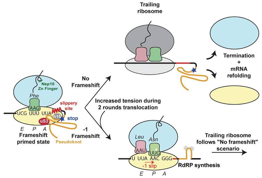
mechanistically akin to the well-studied SecM stalling system        ribosomal tunnel, at the level of RNA folding that leads to the
in bacteria (39), where it was shown that co-translational           formation of the frameshift stimulatory pseudoknot, or to
folding and the translocon-induced mechanical force can res-         perturb the interactions between the pseudoknot and the
cue the stall induced by interactions between the nascent            mRNA channel, represent a viable strategy in our search for
chain and the ribosomal tunnel (40). These observations also         new drugs against SARS-CoV-2, the virus that is currently
suggest that any cellular nascent-chain factors (41, 42) might       causing the global COVID-19 pandemic. Our results will also
influence the rate of frameshifting.                                 be useful for understanding the mechanism of programmed
                                                                     ribosomal “-1” frameshifting (4) including that employed by
Inhibition of viral replication by a compound that                   many other medically important viruses.
targets the SARS-CoV-2 pseudoknot                                    REFERENCES AND NOTES
The sensitivity of the coronavirus to the finely controlled          1. P. V’kovski, A. Kratzel, S. Steiner, H. Stalder, V. Thiel, Coronavirus biology and
frameshifting levels (13) may present an opportunity to de-               replication: Implications for SARS-CoV-2. Nat. Rev. Microbiol. 19, 155–170 (2021).
velop compounds that interfere with the frameshifting pro-                doi:10.1038/s41579-020-00468-6 Medline
                                                                     2. J. Parker, Errors and alternatives in reading the universal genetic code. Microbiol.
cess and thus inhibit replication of the virus. Using                     Rev. 53, 273–298 (1989). doi:10.1128/MR.53.3.273-298.1989 Medline
computational modeling and reporter assays, compounds                3. J. M. Ogle, A. P. Carter, V. Ramakrishnan, Insights into the decoding mechanism

                                                                                                                                                                    Downloaded from http://science.sciencemag.org/ on July 6, 2021
that have been predicted to bind the pseudoknot and inhibit               from recent ribosome structures. Trends Biochem. Sci. 28, 259–266 (2003).
SARS-CoV-2 frameshifting were described (19, 43), but never               doi:10.1016/S0968-0004(03)00066-5 Medline
                                                                     4. J. F. Atkins, G. Loughran, P. R. Bhatt, A. E. Firth, P. V. Baranov, Ribosomal
tested with respect to their ability to inhibit viral replication.        frameshifting and transcriptional slippage: From genetic steganography and
Furthermore, the fluoroquinolone compound merafloxacin                    cryptography to adventitious use. Nucleic Acids Res. 44, 7007–7078 (2016).
was recently reported to also inhibit -1 frameshifting effi-              doi:10.1093/nar/gkw530 Medline
ciency of SARS-CoV-2 and other betacoronaviruses (44). To            5. F. Dos Ramos, M. Carrasco, T. Doyle, I. Brierley, Programmed -1 ribosomal
                                                                          frameshifting in the SARS coronavirus. Biochem. Soc. Trans. 32, 1081–1083
demonstrate that the inhibition of frameshifting is a plausi-             (2004). doi:10.1042/BST0321081 Medline
ble strategy for drug development, we compared two of the            6. P. V. Baranov, C. M. Henderson, C. B. Anderson, R. F. Gesteland, J. F. Atkins, M. T.
previously described compounds with respect to their ability              Howard, Programmed ribosomal frameshifting in decoding the SARS-CoV
to reduce viral levels in infected African green monkey                   genome. Virology 332, 498–510 (2005). doi:10.1016/j.virol.2004.11.038 Medline
                                                                     7. E. P. Plant, G. C. Pérez-Alvarado, J. L. Jacobs, B. Mukhopadhyay, M. Hennig, J. D.
VeroE6 cells (fig. S10 and methods). Our experiments show                 Dinman, A three-stemmed mRNA pseudoknot in the SARS coronavirus frameshift
that merafloxacin is a better candidate compound as it                    signal. PLOS Biol. 3, e172 (2005). doi:10.1371/journal.pbio.0030172 Medline
showed a concentration dependent inhibition of frameshift-           8. M. C. Su, C. T. Chang, C. H. Chu, C. H. Tsai, K. Y. Chang, An atypical RNA pseudoknot
ing, whereas, contrary to earlier reports (19, 43) MTDB under             stimulator and an upstream attenuation signal for -1 ribosomal frameshifting of
                                                                          SARS coronavirus. Nucleic Acids Res. 33, 4265–4275 (2005).
our experimental conditions did not specifically inhibit                  doi:10.1093/nar/gki731 Medline
frameshifting (fig. S10). The two compounds showed no cel-           9. I. Brierley, P. Digard, S. C. Inglis, Characterization of an efficient coronavirus
lular toxicity and resulted in a 3 to 4 orders of magnitude               ribosomal frameshifting signal: Requirement for an RNA pseudoknot. Cell 57,
reduction of SARS-CoV-2 titer, with the half maximal inhibi-              537–547 (1989). doi:10.1016/0092-8674(89)90124-4 Medline
                                                                     10. N. Irigoyen, A. E. Firth, J. D. Jones, B. Y. W. Chung, S. G. Siddell, I. Brierley, High-
tory concentration (IC50) of 48 μΜ for MTDB and an order                  resolution analysis of coronavirus gene expression by RNA sequencing and
of magnitude higher efficacy of merafloxacin with an IC50 of              ribosome         profiling.    PLOS      Pathog.       12,     e1005473        (2016).
4.3 μΜ (fig. S10). Since MTDB did not appear to affect                    doi:10.1371/journal.ppat.1005473 Medline
frameshifting in our reporter construct experiments in vitro         11. Y. Finkel, O. Mizrahi, A. Nachshon, S. Weingarten-Gabbay, D. Morgenstern, Y.
                                                                          Yahalom-Ronen, H. Tamir, H. Achdout, D. Stein, O. Israeli, A. Beth-Din, S.
and in vivo, it is possible that it inhibits SARS-CoV-2 replica-          Melamed, S. Weiss, T. Israely, N. Paran, M. Schwartz, N. Stern-Ginossar, The
tion by a different mechanism. Although the potency range                 coding capacity of SARS-CoV-2. Nature 589, 125–130 (2021).
for these compounds is not what would be expected from po-                doi:10.1038/s41586-020-2739-1 Medline
tential drug candidates, it nevertheless provides a starting         12. V. Thiel, K. A. Ivanov, Á. Putics, T. Hertzig, B. Schelle, S. Bayer, B. Weißbrich, E. J.
                                                                          Snijder, H. Rabenau, H. W. Doerr, A. E. Gorbalenya, J. Ziebuhr, Mechanisms and
point for high-throughput screening and establishes that                  enzymes involved in SARS coronavirus genome expression. J. Gen. Virol. 84,
frameshifting is a viable target for therapeutic intervention             2305–2315 (2003). doi:10.1099/vir.0.19424-0 Medline
against SARS-CoV-2.                                                  13. E. P. Plant, R. Rakauskaite, D. R. Taylor, J. D. Dinman, Achieving a golden mean:
                                                                          Mechanisms by which coronaviruses ensure synthesis of the correct
                                                                          stoichiometric ratios of viral proteins. J. Virol. 84, 4330–4340 (2010).
Conclusions                                                               doi:10.1128/JVI.02480-09 Medline
Our results provide a mechanistic description of frameshift-         14. R. Rangan, I. N. Zheludev, R. J. Hagey, E. A. Pham, H. K. Wayment-Steele, J. S.
ing that occurs during translation of the SARS-CoV-2 genome               Glenn, R. Das, RNA genome conservation and secondary structure in SARS-CoV-
and reveal the features that may be exploited by the virus to             2 and SARS-related viruses: A first look. RNA 26, 937–959 (2020).
                                                                          doi:10.1261/rna.076141.120 Medline
finely control the stoichiometry of viral proteins at different      15. K. Zhang, I. N. Zheludev, R. J. Hagey, M. T. Wu, R. Haslecker, Y. J. Hou, R. Kretsch,
stages of infection (Fig. 5). Interfering with the frameshifting          G. D. Pintilie, R. Rangan, W. Kladwang, S. Li, E. A. Pham, C. Bernardin-Souibgui, R.
process at the level of nascent chain interactions with the               S. Baric, T. P. Sheahan, V. D Souza, J. S. Glenn, W. Chiu, R. Das, Cryo-electron

First release: 13 May 2021                               www.sciencemag.org             (Page numbers not final at time of first release)                      5
microscopy and exploratory antisense targeting of the 28-kDa frameshift 34. M. Bouvet, A. Lugari, C. C. Posthuma, J. C. Zevenhoven, S. Bernard, S. Betzi, I.
stimulation element from the SARS-CoV-2 RNA genome. bioRxiv Imbert, B. Canard, J. C. Guillemot, P. Lécine, S. Pfefferle, C. Drosten, E. J. Snijder,
2020.07.18.209270 [Preprint]. 20 July 2020; E. Decroly, X. Morelli, Coronavirus Nsp10, a critical co-factor for activation of
https://doi.org/10.1101/2020.07.18.209270. multiple replicative enzymes. J. Biol. Chem. 289, 25783–25796 (2014).
16. T. C. T. Lan, M. F. Allan, L. E. Malsick, S. Khandwala, S. Y. Nyeo, Y. Sun, J. U. Guo, doi:10.1074/jbc.M114.577353 Medline
M. Bathe, A. Griffiths, S. Rouskin, Insights into the secondary structural 35. E. C. Smith, J. B. Case, H. Blanc, O. Isakov, N. Shomron, M. Vignuzzi, M. R. Denison,
ensembles of the full SARS-CoV-2 RNA genome in infected cells. bioRxiv Mutations in coronavirus nonstructural protein 10 decrease virus replication
2020.06.29.178343 [Preprint]. 19 February 2021; fidelity. J. Virol. 89, 6418–6426 (2015). doi:10.1128/JVI.00110-15 Medline
https://doi.org/10.1101/2020.06.29.178343. 36. S. K. Natchiar, A. G. Myasnikov, H. Kratzat, I. Hazemann, B. P. Klaholz,
17. N. C. Huston, H. Wan, M. S. Strine, R. de Cesaris Araujo Tavares, C. B. Wilen, A. M. Visualization of chemical modifications in the human 80S ribosome structure.
Pyle, Comprehensive in vivo secondary structure of the SARS-CoV-2 genome Nature 551, 472–477 (2017). doi:10.1038/nature24482 Medline
reveals novel regulatory motifs and mechanisms. Mol. Cell 81, 584–598.e5 37. J. S. Joseph, K. S. Saikatendu, V. Subramanian, B. W. Neuman, A. Brooun, M.
(2021). doi:10.1016/j.molcel.2020.12.041 Medline Griffith, K. Moy, M. K. Yadav, J. Velasquez, M. J. Buchmeier, R. C. Stevens, P. Kuhn,
18. O. Namy, S. J. Moran, D. I. Stuart, R. J. C. Gilbert, I. Brierley, A mechanical Crystal structure of nonstructural protein 10 from the severe acute respiratory
explanation of RNA pseudoknot function in programmed ribosomal frameshifting. syndrome coronavirus reveals a novel fold with two zinc-binding motifs. J. Virol.
Nature 441, 244–247 (2006). doi:10.1038/nature04735 Medline 80, 7894–7901 (2006). doi:10.1128/JVI.00467-06 Medline
19. J. A. Kelly, A. N. Olson, K. Neupane, S. Munshi, J. San Emeterio, L. Pollack, M. T. 38. H. R. Harrington, M. H. Zimmer, L. M. Chamness, V. Nash, W. D. Penn, T. F. Miller
Woodside, J. D. Dinman, Structural and functional conservation of the III, S. Mukhopadhyay, J. P. Schlebach, Cotranslational folding stimulates
programmed -1 ribosomal frameshift signal of SARS coronavirus 2 (SARS-CoV-2). programmed ribosomal frameshifting in the alphavirus structural polyprotein. J.
Downloaded from http://science.sciencemag.org/ on July 6, 2021
J. Biol. Chem. 295, 10741–10748 (2020). doi:10.1074/jbc.AC120.013449 Medline Biol. Chem. 295, 6798–6808 (2020). doi:10.1074/jbc.RA120.012706 Medline
20. M. Taoka, Y. Nobe, Y. Yamaki, K. Sato, H. Ishikawa, K. Izumikawa, Y. Yamauchi, K. 39. H. Nakatogawa, K. Ito, The ribosomal exit tunnel functions as a discriminating
Hirota, H. Nakayama, N. Takahashi, T. Isobe, Landscape of the complete RNA gate. Cell 108, 629–636 (2002). doi:10.1016/S0092-8674(02)00649-9 Medline
chemical modifications in the human 80S ribosome. Nucleic Acids Res. 46, 9289– 40. D. H. Goldman, C. M. Kaiser, A. Milin, M. Righini, I. Tinoco Jr., C. Bustamante,
9298 (2018). doi:10.1093/nar/gky811 Medline Ribosome. Mechanical force releases nascent chain-mediated ribosome arrest in
21. W. Li, S. T. L. Chang, F. R. Ward, J. H. D. Cate, Selective inhibition of human vitro and in vivo. Science 348, 457–460 (2015). doi:10.1126/science.1261909
translation termination by a drug-like compound. Nat. Commun. 11, 4941 (2020). Medline
doi:10.1038/s41467-020-18765-2 Medline 41. G. Kramer, D. Boehringer, N. Ban, B. Bukau, The ribosome as a platform for co-
22. G. Keith, G. Dirheimer, The primary structure of rabbit, calf and bovine liver translational processing, folding and targeting of newly synthesized proteins. Nat.
tRNAPhe. Biochim. Biophys. Acta 517, 133–149 (1978). doi:10.1016/0005- Struct. Mol. Biol. 16, 589–597 (2009). doi:10.1038/nsmb.1614 Medline
2787(78)90041-2 Medline 42. K. Döring, N. Ahmed, T. Riemer, H. G. Suresh, Y. Vainshtein, M. Habich, J. Riemer,
23. C. Bao, S. Loerch, C. Ling, A. A. Korostelev, N. Grigorieff, D. N. Ermolenko, mRNA M. P. Mayer, E. P. O’Brien, G. Kramer, B. Bukau, Profiling Ssb-nascent chain
stem-loops can pause the ribosome by hindering A-site tRNA binding. eLife 9, interactions reveals principles of Hsp70-assisted folding. Cell 170, 298–311.e20
e55799 (2020). doi:10.7554/eLife.55799 Medline (2017). doi:10.1016/j.cell.2017.06.038 Medline
24. J. Choi, S. O’Loughlin, J. F. Atkins, J. D. Puglisi, The energy landscape of -1 43. S. J. Park, Y. G. Kim, H. J. Park, Identification of RNA pseudoknot-binding ligand
ribosomal frameshifting. Sci. Adv. 6, eaax6969 (2020). that inhibits the -1 ribosomal frameshifting of SARS-coronavirus by structure-
doi:10.1126/sciadv.aax6969 Medline based virtual screening. J. Am. Chem. Soc. 133, 10094–10100 (2011).
25. P. Han, Y. Shichino, T. Schneider-Poetsch, M. Mito, S. Hashimoto, T. Udagawa, K. doi:10.1021/ja1098325 Medline
Kohno, M. Yoshida, Y. Mishima, T. Inada, S. Iwasaki, Genome-wide survey of 44. Y. Sun, L. Abriola, Y. V. Surovtseva, B. D. Lindenbach, J. U. Guo, Restriction of
ribosome collision. Cell Rep. 31, 107610 (2020). SARS-CoV-2 replication by targeting programmed −1 ribosomal frameshifting in
doi:10.1016/j.celrep.2020.107610 Medline vitro. bioRxiv 2020.10.21.349225 (2020). 10.1101/2020.10.21.349225 Medline
26. A. B. Arpat, A. Liechti, M. De Matos, R. Dreos, P. Janich, D. Gatfield, Transcriptome- 45. A. Sharma, M. Mariappan, S. Appathurai, R. S. Hegde, In vitro dissection of protein
wide sites of collided ribosomes reveal principles of translational pausing. translocation into the mammalian endoplasmic reticulum. Methods Mol. Biol. 619,
Genome Res. 30, 985–999 (2020). doi:10.1101/gr.257741.119 Medline 339–363 (2010). doi:10.1007/978-1-60327-412-8_20 Medline
27. S. Meydan, N. R. Guydosh, Disome and trisome profiling reveal genome-wide 46. S. Q. Zheng, E. Palovcak, J. P. Armache, K. A. Verba, Y. Cheng, D. A. Agard,
targets of ribosome quality control. Mol. Cell 79, 588–602.e6 (2020). MotionCor2: Anisotropic correction of beam-induced motion for improved cryo-
doi:10.1016/j.molcel.2020.06.010 Medline electron microscopy. Nat. Methods 14, 331–332 (2017). doi:10.1038/nmeth.4193
28. M. Puray-Chavez, K. Tenneti, H. R. Vuong, N. Lee, Y. Liu, A. Horani, T. Huang, J. B. Medline
Case, W. Yang, M. S. Diamond, S. L. Brody, J. Dougherty, S. B. Kutluay, S. B. 47. K. Zhang, Gctf: Real-time CTF determination and correction. J. Struct. Biol. 193, 1–
Kutluay, The translational landscape of SARS-CoV-2 and infected cells. bioRxiv 12 (2016). doi:10.1016/j.jsb.2015.11.003 Medline
2020.11.03.367516 [Preprint]. 16 November 2020; 48. J. Zivanov, T. Nakane, B. O. Forsberg, D. Kimanius, W. J. H. Hagen, E. Lindahl, S. H.
https://doi.org/10.1101/2020.11.03.367516. W. Scheres, New tools for automated high-resolution cryo-EM structure
29. J. Rabl, M. Leibundgut, S. F. Ataide, A. Haag, N. Ban, Crystal structure of the determination in RELION-3. eLife 7, e42166 (2018). doi:10.7554/eLife.42166
eukaryotic 40S ribosomal subunit in complex with initiation factor 1. Science 331, Medline
730–736 (2011). doi:10.1126/science.1198308 Medline 49. A. Punjani, J. L. Rubinstein, D. J. Fleet, M. A. Brubaker, cryoSPARC: Algorithms for
30. S. Takyar, R. P. Hickerson, H. F. Noller, mRNA helicase activity of the ribosome. rapid unsupervised cryo-EM structure determination. Nat. Methods 14, 290–296
Cell 120, 49–58 (2005). doi:10.1016/j.cell.2004.11.042 Medline (2017). doi:10.1038/nmeth.4169 Medline
31. Z. Lin, R. J. C. Gilbert, I. Brierley, Spacer-length dependence of programmed -1 or 50. A. Punjani, D. J. Fleet, 3D variability analysis: Resolving continuous flexibility and
-2 ribosomal frameshifting on a U6A heptamer supports a role for messenger RNA discrete heterogeneity from single particle cryo-EM. J. Struct. Biol. 213, 107702
(mRNA) tension in frameshifting. Nucleic Acids Res. 40, 8674–8689 (2012). (2021). doi:10.1016/j.jsb.2021.107702 Medline
doi:10.1093/nar/gks629 Medline 51. T. C. Terwilliger, O. V. Sobolev, P. V. Afonine, P. D. Adams, Automated map
32. H. Amiri, H. F. Noller, Structural evidence for product stabilization by the ribosomal sharpening by maximization of detail and connectivity. Acta Crystallogr. D Struct.
mRNA helicase. RNA 25, 364–375 (2019). doi:10.1261/rna.068965.118 Medline Biol. 74, 545–559 (2018). doi:10.1107/S2059798318004655 Medline
33. N. Caliskan, V. I. Katunin, R. Belardinelli, F. Peske, M. V. Rodnina, Programmed -1 52. S. Shao, J. Murray, A. Brown, J. Taunton, V. Ramakrishnan, R. S. Hegde, Decoding
frameshifting by kinetic partitioning during impeded translocation. Cell 157, 1619– mammalian ribosome-mRNA states by translational GTPase complexes. Cell 167,
1631 (2014). doi:10.1016/j.cell.2014.04.041 Medline 1229–1240.e15 (2016). doi:10.1016/j.cell.2016.10.046 Medline
First release: 13 May 2021 www.sciencemag.org (Page numbers not final at time of first release) 653. E. F. Pettersen, T. D. Goddard, C. C. Huang, G. S. Couch, D. M. Greenblatt, E. C. 72. V. G. George, J. C. Hierholzer, E. W. Ades, “Cell culture” in Virology Methods
Meng, T. E. Ferrin, UCSF Chimera—A visualization system for exploratory Manual, B. W. J. Mahy, H. O. Kangro, Eds. (Academic Press, 1996), pp. 3–24.
research and analysis. J. Comput. Chem. 25, 1605–1612 (2004). 73. J. Schindelin, I. Arganda-Carreras, E. Frise, V. Kaynig, M. Longair, T. Pietzsch, S.
doi:10.1002/jcc.20084 Medline Preibisch, C. Rueden, S. Saalfeld, B. Schmid, J. Y. Tinevez, D. J. White, V.
54. P. Emsley, B. Lohkamp, W. G. Scott, K. Cowtan, Features and development of Coot. Hartenstein, K. Eliceiri, P. Tomancak, A. Cardona, Fiji: An open-source platform for
Acta Crystallogr. D Biol. Crystallogr. 66, 486–501 (2010). biological-image analysis. Nat. Methods 9, 676–682 (2012).
doi:10.1107/S0907444910007493 Medline doi:10.1038/nmeth.2019 Medline
55. D. Liebschner, P. V. Afonine, M. L. Baker, G. Bunkóczi, V. B. Chen, T. I. Croll, B. 74. J. Mutterer, E. Zinck, Quick-and-clean article figures with FigureJ. J. Microsc. 252,
Hintze, L. W. Hung, S. Jain, A. J. McCoy, N. W. Moriarty, R. D. Oeffner, B. K. Poon, 89–91 (2013). doi:10.1111/jmi.12069 Medline
M. G. Prisant, R. J. Read, J. S. Richardson, D. C. Richardson, M. D. Sammito, O. V. 75. T. D. Goddard, C. C. Huang, E. C. Meng, E. F. Pettersen, G. S. Couch, J. H. Morris,
Sobolev, D. H. Stockwell, T. C. Terwilliger, A. G. Urzhumtsev, L. L. Videau, C. J. T. E. Ferrin, UCSF ChimeraX: Meeting modern challenges in visualization and
Williams, P. D. Adams, Macromolecular structure determination using x-rays, analysis. Protein Sci. 27, 14–25 (2018). doi:10.1002/pro.3235 Medline
neutrons and electrons: Recent developments in Phenix. Acta Crystallogr. D 76. G. Cardone, J. B. Heymann, A. C. Steven, One number does not fit all: Mapping
Struct. Biol. 75, 861–877 (2019). doi:10.1107/S2059798319011471 Medline local variations in resolution in cryo-EM reconstructions. J. Struct. Biol. 184, 226–
56. K. J. Webb, C. I. Zurita-Lopez, Q. Al-Hadid, A. Laganowsky, B. D. Young, R. S. 236 (2013). doi:10.1016/j.jsb.2013.08.002 Medline
Lipson, P. Souda, K. F. Faull, J. P. Whitelegge, S. G. Clarke, A novel 3- 77. V. Chandrasekaran, S. Juszkiewicz, J. Choi, J. D. Puglisi, A. Brown, S. Shao, V.
methylhistidine modification of yeast ribosomal protein Rpl3 is dependent upon Ramakrishnan, R. S. Hegde, Mechanism of ribosome stalling during translation of
the YIL110W methyltransferase. J. Biol. Chem. 285, 37598–37606 (2010). a poly(A) tail. Nat. Struct. Mol. Biol. 26, 1132–1140 (2019). doi:10.1038/s41594-
doi:10.1074/jbc.M110.170787 Medline 019-0331-x Medline
Downloaded from http://science.sciencemag.org/ on July 6, 2021
57. A. Brown, S. Shao, J. Murray, R. S. Hegde, V. Ramakrishnan, Structural basis for 78. W. A. Cantara, P. F. Crain, J. Rozenski, J. A. McCloskey, K. A. Harris, X. Zhang, F. A.
stop codon recognition in eukaryotes. Nature 524, 493–496 (2015). P. Vendeix, D. Fabris, P. F. Agris, The RNA modification database, RNAMDB: 2011
doi:10.1038/nature14896 Medline update. Nucleic Acids Res. 39, D195–D201 (2011). doi:10.1093/nar/gkq1028
58. N. W. Moriarty, P. D. Adams, Iron-sulfur clusters have no right angles. Acta Medline
Crystallogr. D Struct. Biol. 75, 16–20 (2019). doi:10.1107/S205979831801519X
Medline ACKNOWLEDGMENTS
59. P. P. Chan, T. M. Lowe, GtRNAdb 2.0: An expanded database of transfer RNA genes We thank A. Jomaa for advice on cryo-EM data processing, Noreen Casey for support
identified in complete and draft genomes. Nucleic Acids Res. 44, D184–D189 with technical resources and A. Picenoni for help with the grid preparation. We
(2016). doi:10.1093/nar/gkv1309 Medline are indebted to the ETH scientific center for optical and electron microscopy
60. N. K. Sinha, A. Ordureau, K. Best, J. A. Saba, B. Zinshteyn, E. Sundaramoorthy, A. (ScopeM) for access to electron microscopes, and in particular to M. Peterek
Fulzele, D. M. Garshott, T. Denk, M. Thoms, J. A. Paulo, J. W. Harper, E. J. Bennett, and D. Boehringer. We thank Junjie Guo for helpful discussions and for providing
R. Beckmann, R. Green, EDF1 coordinates cellular responses to ribosome merafloxacin compound. Funding: This work was supported by the Swiss
collisions. eLife 9, e58828 (2020). doi:10.7554/eLife.58828 Medline National Science Foundation (SNSF) to NB and VT (grant 31003A_182341 and
61. A. W. Schüttelkopf, D. M. F. van Aalten, PRODRG: A tool for high-throughput 310030_173085) via the National Center of Excellence in RNA and Disease
crystallography of protein-ligand complexes. Acta Crystallogr. D Biol. Crystallogr. (project funding 182880) to N. Ban, V. Thiel and D. Gatfield; The Federal Ministry
60, 1355–1363 (2004). doi:10.1107/S0907444904011679 Medline of Education and Research, Germany (BMBF; grant RAPID, #01KI1723A) to AK
62. V. B. Chen, W. B. Arendall III, J. J. Headd, D. A. Keedy, R. M. Immormino, G. J. and VT; The Irish Research Council Advanced Laureate (IRCLA/2019/74) to
Kapral, L. W. Murray, J. S. Richardson, D. C. Richardson, MolProbity: All-atom J.F.A., who thanks Prof. G.F. Fitzgerald for funds from Carbery Group Ltd. Author
structure validation for macromolecular crystallography. Acta Crystallogr. D Biol. contributions: PRB and NB initiated the project, and together with JFA designed
Crystallogr. 66, 12–21 (2010). doi:10.1107/S0907444909042073 Medline the experiments. PRB carried out biochemical experiments and sample
63. F. Sievers, A. Wilm, D. Dineen, T. J. Gibson, K. Karplus, W. Li, R. Lopez, H. preparation. AS and PRB prepared grids. AS carried out data collection and
McWilliam, M. Remmert, J. Söding, J. D. Thompson, D. G. Higgins, Fast, scalable calculated EM maps. AS and ML performed molecular model building and
generation of high-quality protein multiple sequence alignments using Clustal refinement. AS, ML, PRB and NB interpreted the structure. PRB, AS, ML and NB
Omega. Mol. Syst. Biol. 7, 539 (2011). doi:10.1038/msb.2011.75 Medline drafted the manuscript. AS prepared the figures. GL and KMOC performed dual-
64. X. Robert, P. Gouet, Deciphering key features in protein structures with the new luciferase reporter assays. AM prepared and purified the drug-like ligand MTDB.
ENDscript server. Nucleic Acids Res. 42, W320–W324 (2014). AK and VT formulated experiments to test viral infected cells. AK carried out
doi:10.1093/nar/gku316 Medline testing of ligand on viral-infected cells. RM, RD and DG designed, carried out,
65. G. Loughran, M. T. Howard, A. E. Firth, J. F. Atkins, Avoidance of reporter assay analyzed and interpreted ribosome profiling experiments. All authors
distortions from fused dual reporters. RNA 23, 1285–1289 (2017). contributed to the final version of the manuscript. Competing interests: The
doi:10.1261/rna.061051.117 Medline authors declare no competing interest. Data and materials availability: The
66. B. W. Dyer, F. A. Ferrer, D. K. Klinedinst, R. Rodriguez, A noncommercial dual structure and cryo-EM map for the high-resolution reconstruction is available in
luciferase enzyme assay system for reporter gene analysis. Anal. Biochem. 282, the Protein Data Bank (PDB) as PDB ID 7O7Y and in the Electron Microscopy
158–161 (2000). doi:10.1006/abio.2000.4605 Medline Data Bank (EMDB) as EMD-12756, respectively. The structure refined into the
67. N. J. McGlincy, N. T. Ingolia, Transcriptome-wide measurement of translation by further classified particle set reconstruction and the corresponding maps are
ribosome profiling. Methods 126, 112–129 (2017). available as PDB-7O7Z and EMD-12757, respectively. The structure of the
doi:10.1016/j.ymeth.2017.05.028 Medline ribosome bound with eRF1 (AAQ) and ABCE1 reconstruction and the
68. M. Martin, Cutadapt removes adapter sequences from high-throughput corresponding maps are available as PDB-7O80 and EMD-12758, respectively.
sequencing reads. EMBnet.journal 17, 10 (2011). doi:10.14806/ej.17.1.200 The structure of the colliding ribosome reconstruction and the corresponding
69. T. Smith, A. Heger, I. Sudbery, UMI-tools: Modeling sequencing errors in Unique maps are available as PDB-7O81 and EMD-12759, respectively. Structure of the
Molecular Identifiers to improve quantification accuracy. Genome Res. 27, 491– composite map of the disome is available in the EMDB as EMD-12760. Ribosome
499 (2017). doi:10.1101/gr.209601.116 Medline profiling data have been deposited at GSE167421. This work is licensed under a
70. B. Langmead, S. L. Salzberg, Fast gapped-read alignment with Bowtie 2. Nat. Creative Commons Attribution 4.0 International (CC BY 4.0) license, which
Methods 9, 357–359 (2012). doi:10.1038/nmeth.1923 Medline permits unrestricted use, distribution, and reproduction in any medium,
71. R. Edgar, M. Domrachev, A. E. Lash, Gene Expression Omnibus: NCBI gene provided the original work is properly cited. To view a copy of this license, visit
expression and hybridization array data repository. Nucleic Acids Res. 30, 207– https://creativecommons.org/licenses/by/4.0/. This license does not apply to
210 (2002). doi:10.1093/nar/30.1.207 Medline figures/photos/artwork or other content included in the article that is credited
First release: 13 May 2021 www.sciencemag.org (Page numbers not final at time of first release) 7to a third party; obtain authorization from the rights holder before using such
material.
SUPPLEMENTARY MATERIALS
science.sciencemag.org/cgi/content/full/science.abf3546/DC1
Materials and Methods
Figs. S1 to S11
Tables S1 to S4
References (44–78)
MDAR Reproducibility Checklist
20 October 2020; resubmitted 24 February 2021
Accepted 7 May 2021
Published online 13 May 2021
10.1126/science.abf3546
Downloaded from http://science.sciencemag.org/ on July 6, 2021
First release: 13 May 2021 www.sciencemag.org (Page numbers not final at time of first release) 8Downloaded from http://science.sciencemag.org/ on July 6, 2021 First release: 13 May 2021 www.sciencemag.org (Page numbers not final at time of first release) 9
Fig. 1. The SARS-CoV-2 pseudoknot interacts with the ribosome and pauses translation
          upstream of the slippery site. (A) Schematic of the SARS-CoV-2 main ORF. In the close up view of
          the frameshift event, codons and corresponding amino acids are shown. During -1 frameshifting, the
          ‘slippery site’ codons UUA (Leu) and AAC (Asn) are the last codons decoded in the 0 frame. Upon -1
          frameshifting of the AAC codon to AAA, translation resumes at the CGG (Arg) triplet, where
          elongation proceeds uninterrupted to produce full-length Nsp12. (B) In vitro translation reaction
          depicting pausing at the frameshift site. Efficient frameshifting is observed for the WT template,
          consistent with our dual luciferase assays (see methods). Samples for cryo-EM originally intended
          to be trapped by dominant negative eRF1 (AAQ) show a tRNA-bound pause in proximity of the
          frameshift site. The tRNA-associated band is lost upon RNase treatment. Reactions without added
          eRF1 (AAQ) produce a similarly paused product. (C) Overview of the density low pass filtered to 6Å
          with the pseudoknot found close to the entry of the mRNA channel on the small subunit (SSU). The
          SSU proteins are colored in yellow, the large subunit (LSU) proteins in blue and the rRNA in grey. The
          pseudoknot is colored according to its secondary structure as in (F), and the P-site tRNA is colored
          in green. (D) Close-up view of the pseudoknot from the solvent-exposed side of the SSU. Helix h16
          of the 18S rRNA interacts with the base of Stem 1. Unpaired loop-forming nucleotides are colored in
          cyan. (E) P-site codon-anticodon interactions reveal a Phe (UUU) codon interacting with tRNA(Phe).
          (F) Schematic of the revised secondary structure elements in the pseudoknot necessary for -1 PRF

                                                                                                                                 Downloaded from http://science.sciencemag.org/ on July 6, 2021
          with different functional regions labeled and colored accordingly.

First release: 13 May 2021                         www.sciencemag.org     (Page numbers not final at time of first release) 10
Downloaded from http://science.sciencemag.org/ on July 6, 2021 First release: 13 May 2021 www.sciencemag.org (Page numbers not final at time of first release) 11
Fig. 2. Critical features of the ribosome-bound pseudoknot. (A) Overview of the frameshift-primed
          state. The stimulatory pseudoknot pauses the ribosome at the penultimate codon (UUU) of the
          slippery site (red), with P- (green) and E- (pink) sites occupied by tRNAs, and an empty A-site
          awaiting decoding in the non-rotated state. The length of the spacer region (grey) is critical for exact
          positioning of the pseudoknot as the spacer exerts tension at the entry of the mRNA channel
          (fig. S6C). (B) The backbone of Loop 1 (UGC) (cyan) of the pseudoknot interacts with the N-terminal
          domain of uS3 (red) and the C-terminal tail of eS10 (orange). mRNA residue G13486 is flipped out
          and interacts with uS3 (fig. S6D). (C) Mutagenesis experiments using dual luciferase assays in
          HEK293T cells indicate that the G13486 interaction is specific. Mutation of G13486 to other residues
          leads to a marked reduction in frameshifting efficiency, and deletion of Loop 1 (Δ L1) completely
          abolishes frameshifting. Similarly, deletion of a single nucleotide (A13537) in Loop 2 reduces
          frameshifting, while deletion of the entire loop (ΔL2) abolishes frameshifting. Normalized
          (Firefly/Renilla) luciferase activities were calculated for each construct as a percentage of their
          individual normalized in-frame controls. Data are presented as mean values ± standard deviations of
          three biological replicates (sets of translation reactions) averaged after three measurements, with
          error bars representing standard deviations. (D) Mutagenesis experiments using dual luciferase
          reporter assays in HEK293T cells show that the position of the 0 frame stop codon influences
          frameshifting. Leaving the pseudoknot unaltered, incremental increase in the distance of the 0 frame

                                                                                                                                   Downloaded from http://science.sciencemag.org/ on July 6, 2021
          stop codon from the frameshift site leads to a concomitant decrease in frameshifting levels. Loss of
          the stop codon in 0 frame leads to a sharp decline in frameshifting levels. This reduction is rescued
          by ~45% upon decreasing ribosome loading levels by implementing weaker initiation codons. The
          graph is normalized relative to the WT frameshifting of 25%. Mutations and complementary
          mutations are shown in fig. S8.

First release: 13 May 2021                          www.sciencemag.org      (Page numbers not final at time of first release) 12
Downloaded from http://science.sciencemag.org/ on July 6, 2021
Fig. 3. Pseudoknot-mediated pause occurs prior to the -1 frameshifting event. (A and B) Footprint
coverage for WT and mutant constructs determined by monosome- and disome-selective ribosome
profiling. Pileup of reads from the indicated areas are plotted separately for reads that overlap (pink)
vs. do not overlap (grey) the frameshift site (indicated by red bar below x-axis). The predicted A-sites
of the ribosomes giving rise to the footprints are depicted as blue peaks. A-site predictions were
carried out as described in Supplementary Material. (C) Zoom into the frameshift region from (A)
and (B) reveals that monosome profiles show transient occupancy in the vicinity of the frameshift
site, while disome profiles – indicative of strong pause sites – show a similarly enhanced occupancy
at the first codon (UUA) of the frameshift site in both WT and mutant constructs. A-site codons of
the leading and trailing ribosome are highlighted with a translucent bar and correspond to those seen
in the disome structure in (D). (D) In high resolution cryo-EM reconstructions of disomes at the
frameshift site, the P- and A-sites of the trailing ribosome show occupancy of CCC and AUG codons,
respectively, corresponding to the positions estimated by disome profiling. Disome maps were
calculated by separately refining the orientational parameters for each ribosome.
First release: 13 May 2021 www.sciencemag.org (Page numbers not final at time of first release) 13Downloaded from http://science.sciencemag.org/ on July 6, 2021 First release: 13 May 2021 www.sciencemag.org (Page numbers not final at time of first release) 14
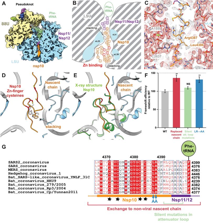
Fig. 4. The nascent viral polypeptide co-translationally folds and specifically interacts with the
          ribosomal tunnel. (A) Cross-section of the pseudoknot-paused ribosome structure showing the exit
          tunnel. The nascent C terminus of Nsp10 (orange) and the N terminus of Nsp11/12 (purple) are
          visible from the PTC to the periphery of the ribosome exit tunnel (LSU in blue). (B) Schematic
          representation of the path of nascent peptide along the exit tunnel. Arg 4387 stacks with 28S rRNA
          residue A1555 at the constriction site. Further down, where the tunnel widens, the C-terminal zinc
          finger domain of Nsp10 folds co-translationally, with Trp 4376 stacking on A2261 of 28S rRNA.
          (C) Well-ordered density is visible for Arg 4387 of Nsp10 as it stacks onto A1555 of 28S rRNA at the
          constriction site and is stabilized by Leu 4386. The structure is shown within the cryo-EM map
          contoured at two different levels (grey and red). (D and E) The overlay of the co-translationally folded
          zinc finger domain with the crystal structure of Nsp10 [green, PDB 2FYG (37)] reveals the structural
          similarity. (F) Probing the role of nascent chain interactions with the ribosome exit tunnel using an
          RRL in vitro system. Mutations of the interacting residues were tested for their effect on
          frameshifting shown in comparison to the wild type frameshifting (41% frameshifting was
          normalized to 100%). Replacement of the entire nascent chain with an unrelated sequence leads to
          a 35% relative increase in frameshifting, which is only in part due to loss of the 5′ attenuator loop.
          Interactions around the constriction site likely serve to attenuate frameshifting, as replacement of
          the interacting Arg 4387 and stabilizing Leu 4386 with Ala increases frameshifting by 30%.

                                                                                                                                   Downloaded from http://science.sciencemag.org/ on July 6, 2021
          (G) Alignment of SARS2 with closely related sequences of other coronaviruses highlighting the
          conservation of the mutated residues [colored as in (F)]. The shown sequence stretch encompasses
          the C-terminal zinc finger domain of Nsp10 (orange) and parts of Nsp11/Nsp12 (purple) visible in our
          reconstruction. Nascent chain residues Leu 4386 and Arg 4387 interacting with the ribosomal exit
          tunnel are strictly conserved, while the conservation of neighboring residues is lower. Stars
          represent the four cysteines of the Nsp10 zinc finger.

First release: 13 May 2021                          www.sciencemag.org      (Page numbers not final at time of first release) 15
You can also read