Danggui-Shaoyao-San Improves Gut Microbia Dysbiosis and Hepatic Lipid Homeostasis in Fructose-Fed Rats - Frontiers
←
→
Page content transcription
If your browser does not render page correctly, please read the page content below
ORIGINAL RESEARCH
                                                                                                                                                  published: 13 July 2021
                                                                                                                                         doi: 10.3389/fphar.2021.671708

                                           Danggui-Shaoyao-San Improves Gut
                                           Microbia Dysbiosis and Hepatic Lipid
                                           Homeostasis in Fructose-Fed Rats
                                           Jing Yin 1, Jiaxi Lu 1, Peng Lei 1, Mingshuai He 1, Shengjie Huang 1, Jialin Lv 1, Yan Zhu 1,
                                           Zhidong Liu 1 and Miaomiao Jiang 1,2*
                                           1
                                            State Key Laboratory of Component-Based Chinese Medicine, Tianjin University of Traditional Chinese Medicine, Tianjin, China,
                                           2
                                            Department of Pharmacy, Institute of Traditional Chinese Medicine, Tianjin University of Traditional Chinese Medicine, Tianjin,
                                           China

                                           Metabolic syndrome (MetS) is a pathological state of many abnormal metabolic sections.
                                           These abnormalities are closely related to diabetes, heart pathologies and other vascular
                                           diseases. Danggui-Shaoyao-San (DSS) is a traditional Chinese medicine formula that has
                         Edited by:        been used as a therapy for Alzheimer’s disease. DSS has rarely been reported in the
                 Juei-Tang Cheng,          application of MetS and its mechanism of how it improves gut microbia dysbiosis and
     Chang Jung Christian University,
                                           hepatic lipid homeostasis. In this study, three extracts of DSS were obtained using water,
                             Taiwan
                                           50% methanol in water and methanol as extracting solvents. Their chemical substances
                      Reviewed by:
                      Chao-Zhan Lin,       were analyzed by ultra-performance liquid chromatography coupled with quadrupole time-
     Guangzhou University of Chinese       of-flight mass (UPLC-Q/TOF-MS). Pharmacodynamic effect of the extracts were evaluated
                     Medicine, China
                     Guanglin Zhang,
                                           by comparison of biochemical factors, 16S rRNA sequencing test for gut microbiota
      UCLA Department of Integrative       analysis, as well as metabonomic and transcriptomic assessments on liver tissues from
Biology and Physiology, United States
                                           fructose-fed rats. This study aimed at investigating DSS’s mechanism of regulating blood
                 *Correspondence:
                                           lipid, anti-inflammation and reducing blood glucose. The results showed that the 50%
                      Miaomiao Jiang
          miaomiaojiang@tjtcm.edu.cn       methanol extract (HME) was more effective. It was worth noting that hydroxysteroid 17β-
                                           dehydrogenase 13 (HSD17β13) as a critical element of increasing blood lipid biomarker-
                    Specialty section:     triglyceride (TG), was decreased markedly by DSS. The influence from upgraded
         This article was submitted to
                  Ethnopharmacology,       hydroxysteroid 17β-dehydrogenase 7 (HSD17β7) may be stronger than that from
               a section of the journal    downgraded Lactobacillus in the aspect of regulating back blood lipid biomarker-total
            Frontiers in Pharmacology
                                           cholesterol (TC). The differential down-regulation of tumornecrosis factor alpha (TNF-α)
        Received: 24 February 2021
                                           and the significant up-regulation of Akkermansia showed the effective effect of anti-
          Accepted: 28 June 2021
           Published: 13 July 2021         inflammation by DSS. The declining glycine and alanine induced the lowering glucose
                              Citation:    and lactate. It demonstrated that DSS slowed down the reaction of gluconeogenesis to
Yin J, Lu J, Lei P, He M, Huang S, Lv J,   reduce the blood glucose. The results demonstrated that DSS improved pathological
      Zhu Y, Liu Z and Jiang M (2021)
 Danggui-Shaoyao-San Improves Gut          symptoms of MetS and some special biochemical factors in three aspects by better
Microbia Dysbiosis and Hepatic Lipid       regulating intestinal floras and improving hepatic gene expressions and metabolites.
   Homeostasis in Fructose-Fed Rats.
         Front. Pharmacol. 12:671708.      Keywords: Danggui-Shaoyao-San, metabolic syndrome, gut microbiadysbiosis, anti-inflammation, hepatic lipid
      doi: 10.3389/fphar.2021.671708       homeostasis

Frontiers in Pharmacology | www.frontiersin.org                                     1                                             July 2021 | Volume 12 | Article 671708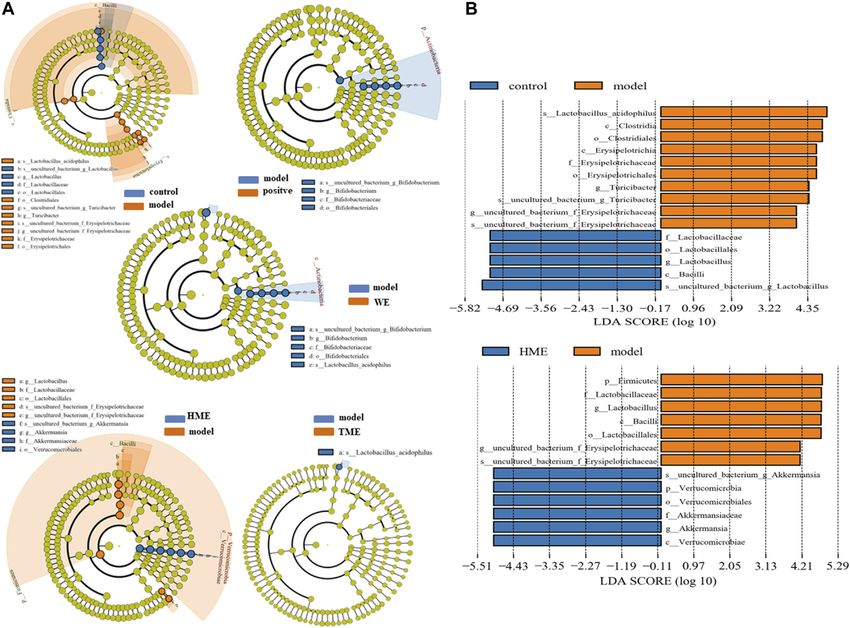
Yin et al.                                                                                                       Dss Improves MetS in Three Aspects

INTRODUCTION                                                                  et al., 2009; Niu and Wang, 2014). Monoterpenoid glycosides,
                                                                              phenolic acids, phthalides and lactones from DSS are active in
Metabolic syndrome (MetS) is a pathological state of many                     protecting the cardiovascular system, including regulating blood
abnormal metabolic sections (Boehm and Claudi-boehm,                          lipid, preventing atherosclerosis and improving hemodynamics
2005) such as overweight, dyslipidemia, hypertension,                         (Dong et al., 2014). Total glucosides of paeony including
hyperuricemia and insulin resistance (IR). These abnormalities                albiflorin and paeoniflorin from Baishao have significantly
are closely related to the emergency with diabetes, especially with           analgesic, anti-inflammatory and anticoagulant effects (Zhou
type 2 diabetes, heart pathologies and other vascular diseases.               et al., 2012; Shen et al., 2020). In this study, fructose-induced
    Intestinal microorganisms refer to bacteria existing in the gut of        MetS rats were employed to evaluate the action of DSS extracts.
the host body. They are involved in the pathogenesis of obesity or            We found that 50% methanol extract (HME) showed greater
metabolic diseases. Intestinal flora is an important correlation               advantages in adjusting the biomarker parameters, the structure
between the gut and liver. Normally, only a small amount of                   of intestinal flora. Subsequently, we studied the improvement of
bacteria and endotoxin enter into the liver through the portal vein,          HME in hepatic metabolites and transcriptomic changes and found
and later most of them are cleared by hepatic macrophages.                    the internal connections. All the results revealed that HME could
Imbalance of intestinal flora results in the destruction of the                alleviate the MetS symptoms through a variety of interactions of
intestinal barrier and the increase of bacteria. When bacterial               hepatic metabolites, genes and intestinal microorganisms.
metabolites exceed the scavenging capacity of the liver, through
pattern recognition receptor, it can activate inflammatory reaction
and accelerate hepatic disease progression (Wieland et al., 2015).            MATERIALS AND METHODS
    Danggui-Shaoyao-San (DSS) has been recorded by Zhongjing
Zhang in Jin Gui Yao Lue (Synopsis of Prescriptions of the Golden             Materials
Chamber) of the Eastern Han dynasty. It is used for bolstering the            Danggui (Tongrentang, Beijing, China), Baishao (Tongrentang,
blood cycle and alleviating stasis due to blood stasis and deficiency of       Beijing, China), Chuanxiong (Tongrentang, Beijing, China),
spleen Qi (Song, 2009). It consists of six herbals: smoke-dried root of       Fuling (Tongrentang, Beijing, China), Baizhu (Tongrentang,
Angelica sinensis (Oliv.) Diels (Danggui, Angelicae Sinensis Radix),          Beijing, China) and Zexie (Tongrentang, Beijing, China). All
sun-dried root of Paeonia lactiflora Pall. (Baishao, Paeoniae Radix            the traditional Chinese medicine decoction pieces were
Alba), oven-dried rhizome of Ligusticum chuanxiong Hort.                      authenticated by Dr. Honghua Wu from Tianjin University of
(Chuanxiong, Chuanxiong Rhizoma), dried sclerotia of Poria                    Traditional Chinese Medicine.
cocos (Schw.) Wolf (Fuling, Poria), oven or sun dried rhizome of                 Adenosine (Meilun, Dalian, China), gallic acid (Meilun,
Atractylodes macrocephala Koidz. (Baizhu, Atractylodis                        Dalian, China), ferulic acid (Meilun, Dalian, China), albiflorin
macrocephalae rhizoma), and dried stem tuber of Alisma                        (Meilun, Dalian, China), paeoniflorin (Meilun, Dalian, China),
plantago-aquatica Linn. (Zexie, Alismatis Rhizoma). DSS is also               atractylenolide I (Meilun, Dalian, China), alisol B-23-acetate
known as Tokishakuyakusan in Japanese, which is one of Kampo                  (Meilun, Dalian, China), Z-ligustilide (Meilun, Dalian, China),
formulas traditionally used for patients with irregular menstruation,         Pioglitazone (Yuanye, Shanghai, China) (HPLC > 98%),
fatigue, and anaemia (Kampo.ca Japanese Traditional Medicine &                D-fructose (Meilun, Dalian, China).
Therapeutics, 2014). In the 1980s, DSS was used in the treatment of
dementia firstly reported by Japanese scientists (Mizushima and
Ikeshita, 1989). Several clinical studies have reported that DSS              Preparation for Fructose Solution and Test
provides reductions in symptoms of Alzheimer’s disease (AD)                   Drugs
and vascular dementia (VD) making it as a promising anti-                     Seven hundred grams of D-fructose was added into 3950 ml of
dementia drug candidate (Matsuoka et al., 2012; Fu et al., 2015;              water and the mixed solution was stirred until fructose was
Kim and Cho, 2020). Moreover, the syndrome of spleen deficiency                dissolved to yield 15% fructose water (w/w).
and fluid retention has been demonstrated as the important                        Danggui, Baishao, Chuanxiong, Fuling, Baizhu, and Zexie
pathogenesis for the incidence and development of AD (Yu et al.,              were finely grounded and mixed with a dose proportion of
2014). Comorbidities of dementia include obesity, diabetes,                   450 g:1500 g:750 g:600 g:600 g:750 g. DSS was divided into
hypertension, and cardiovascular diseases. The risk factors for               several parts and every part was refluxed for 2 h by water,
these comorbidities are collectively referred to as MetS                      50% methanol in water (v/v) or methanol, respectively. The
(Pugazhenthi, 2017). IR and visceral fat during MetS have been                extracting solutions were concentrated and freeze-dried to
suggested to be important links between MetS and cognitive                    yield three extracts, water extract (WE), 50% methanol extract
dysfunction (Ryu et al., 2019; Ivanova et al., 2020).                         (HME) and methanol extract (TME). The yields of these dried
Pharmacological intervention in MetS represents a risk reduction              extracts were all about 20%.
for mild cognitive impairment (MCI) and dementia including AD
and VD later in life. Although there are numerous reviews about the
application of DSS, this study is the first report of DSS on MetS.             Liquid Chromatography–Mass
Herbal powders of DSS are inconvenient to take orally making the              Spectrometry Analysis for Test Drugs
studies of the oral extracts more prevail. Numerous identified                 The qualitative analysis was carried out on Waters Xevo G2-S
compounds have been reported from extracts of DSS (Chen                       UPLC-Q-TOF/MS (Waters Milford, United States). The

Frontiers in Pharmacology | www.frontiersin.org                           2                                    July 2021 | Volume 12 | Article 671708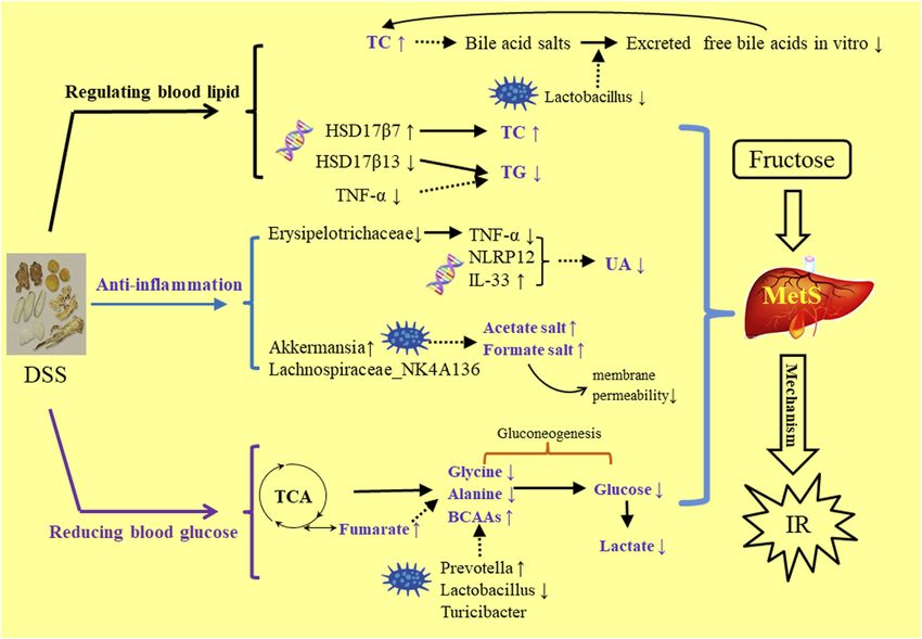
Yin et al.                                                                                                   Dss Improves MetS in Three Aspects

separation was carried out on a Waters ACQUITY UPLC BEH                  (ALT). Hepatic tumor necrosis factor-α (TNF-α) and
C18 (100 × 2.1mm, 1.7 μm) at 30°C. The mobile phase was 0.1%             intestinal lipopolysaccharide (LPS) were quantified using
formic acid water (A) and methanol (B). The gradient program of          enzyme-linked immunosorbent assay (Elisa) kits following
mobile phase was as follows: 0–3 min, 5–14% B; 3–16 min,                 the manufacturer’s instructions.
14–56% B; 16–23 min, 56–80% B; 23–25 min, 80–95% B;
25–30 min, 95–5% B. The injection volume was 2 μl. The                   16S rRNA Gene Sequencing and Analysis
quasi-molecular ions including [M-H]−, [M-H+HCOOH]−,                     DNA for gut microbiota analysis was extracted from
[M-H-CO2]−, [M-H-H2O]−, [M+H-H2O]+, [M+H]+ and                           approximately 100 mg of faeces. DNA was amplified using a
[M+Na]+ were selected as precursor ions and subjected to                 primer set targeting the V3+V4 region of 16S rDNA by carrying
target-MS/MS analyses. The acquisition parameters of Q/TOF               Barcode specific primer. The V3-V4 region of 16S rRNA was
were as follows: drying gas (N2) flow rate, 50 L/h; drying gas            amplified using the primers 341F (CCTACGGGNGGCWGCAG)
temperature, 400°C; the collision energy in high channels (MSE           and 806R (GGACTACHVGGGTATCTAAT) by PCR. Raw reads
pattern) was set at 20–60V; the mass range from m/z 50–100 Da            were first confirmed using basic statistics. The processed pair-end
             ™
Lock spray flow rate was 5 μl/min, and data acquisition was
based on the Centroid Mode.
                                                                         reads were then merged using FLASH software (Version 1.2.11,
                                                                         United States) to generate representative complete sequences.
                                                                         The reads were qualitatively filtered in Trimmomatic0.33 and
Animals and Sample Collection                                            then clustered into 97% identity Operational Taxonomic Units
A total of 60 male Wistar rats (120–150 g per rat) were                  (OTUs) using UCHIME4.2 to get effective tags. The effective tags
purchased from National Institutes for Food and Drug                     were determined quantitatively and analyzed by Quanti-
Control, License number: SCXK (Beijing) 2016-0006. The                   Fluorimeter and the Hiseq2500 system (Illumina, Inc., San
animal ethics approval number was TJAB-TJU20180041. Rats                 Diego, CA, United States). LEfSe software (http://huttenhower.
were maintained in SPF grade animal house in a 12-h dark-light           sph.harvard.edu/lefse/) is one way of statistical Beta diversity
cycle and had free access to food and water. Care and husbandry          analysis. LEfSe was used for differential analysis inter groups. In
followed standard guidelines. After one week, rats were                  this paper, LEfSe was shown to differential results. The selected
randomly and equally divided into six groups, including                  differences were sorted by linear discriminant analysis
control group (fed with purified water; i.g. homologous saline            (LDA) > 4.0.
per day), model group (fed with high-fructose drink; i.g.
                                                                         1
homologous saline per day), positive group (fed with high-                H-NMR Analysis of Hepatic Metobolites
fructose drink; i.g. 4 mg/kg dosage of pioglitazone), WE group           The 100 mg liver tissue was homogenized by 600 μl of pre-cold
(fed with high-fructose drink; i.g. 1.8 g/kg dosage of water             CH3OH/H2O (2:1) on the ice. The extractive solution was
extract), HME group (fed with high-fructose drink; i.g.                  vortexed for 30 s and centrifuged at 1600 rpm and 4°C for
1.8 g/kg dosage of 50% methanol extract), TME (fed with                  10 min. The supernatant of 600 μl in a new tube was dried
high-fructose drink; i.g. 1.8 g/kg dosage of methanol extract).          under nitrogen. Added with 600 μl of phosphate buffered
Treatments were continued for 8 weeks.                                   saline (pH 7.4) containing 0.01% Sodium 3-trimethysilyl
    The body weight and the level of fasted blood glucose were           [2,2,3,3-d4] propionate (TSP-d4). The solution was vortexed
measured every week. At the end of treatment, animals were kept          and centrifuged again. The amount of 550 μl supernatant was
in an empty cage without bedding to gather fresh stool samples           pipetted and transferred into 5 mm NMR tube. The NMR tube
into tubes. The rats were subjected to 12 h of fasting before they       was stored at 4°C for test.
                                                                             1
were sacrificed. Anaesthesia with 5% chloral hydrate was                        H-NMR spectrum was recorded on Bruker AVIII 600 MHz
administered, blood was collected from the abdominal aorta               NMR (Bruker, Germany) spectrometer (proton NMR frequency
and centrifuged to yield serum samples. The liver and                    at 600.13 MHz) equipped with ultra-low temperature probe by
intestine were precisely dissected from the thoracic and                 using a CPMG (CARR Purcell Meiboom Gill) pulse sequence.
abdominal cavity. All the samples were immersed immediately              Key parameters of data acquisition were set as follows: the
in liquid nitrogen and stored at −80°C for further analysis.             temperature at 300 K, spectrum width of 12,019.2 Hz,
                                                                         relaxation delay time of 4 s, 90° pulse width of 12 μs,
Histological Assay                                                       acquisition time of 2.7263 s, scan times of 64, receiving gain
Samples of liver were fixed with 10% formalin, embedded in                191 and sampling data point of 65,536.
paraffin, sectioned into 5 μm thickness, stained with                         Raw data were dealt with MestReNova software (Version 6.1.0,
hematoxylin/eosin (H&E) and finally analyzed by a NIKON                   Spain). The process was carried out by phase correction and
ECLIPSE CI microscope (Nikon, Japan).                                    baseline correction before the methyl resonance of TSP-d4 was
                                                                         referenced to δ 0.000 ppm. The 1H NMR spectra from δ
Determination of Biochemical Parameters                                  0.7–9.0 ppm was bucketed into bins with an integral step of
Levels of fasted blood glucose (FBG) and serum uric acid (UA)            0.001 ppm. The normalized integrity data were imported into
were measured by commercial kits on a biochemical                        Simca (Version 13.0, Sweden) to perform partial least squares
automatic analyzer. Hepatic triglyceride (TG), total                     discriminant analysis (PLS-DA). Differential metabolites were
cholesterol (TC), high-density lipoprotein (HDL-C), low-                 screened by Variable importance in the project (VIP) > 1 and
density lipoprotein (LDL-C), alanine aminotransferase                    t-test (p < 0.05).

Frontiers in Pharmacology | www.frontiersin.org                      3                                     July 2021 | Volume 12 | Article 671708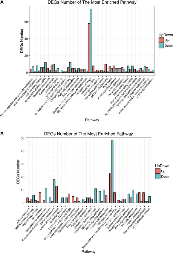
Yin et al. Dss Improves MetS in Three Aspects
Microarray Analysis of Hepatic Gene different substances between WE group and HME group,
Expression which were gallic acid, ferulic acid, albiflorin, alisol B-23-
Total RNA was extracted from liver according to the test kit’s acetate, senkyunolide I, lactiflorin, mudanpioside I,
instruction and then treated with DNase I. Interrupt reagent was Senkyunolide A, Z-ligustilide and cnidilide A. Ten different
used to break mRNA into short segments. Six base random substances were screened out between WE group and TME
hexamers were used to synthesize cDNA based on the short group, including gallic acid, albiflorin, isomaltopaeoniflorin,
segments of mRNA. The test kit was used to purify, repair, alisol B-23-acetate, mudanpioside I, senkyunolide A,
connect the test sequence and filter cDNA. The filtering Z-ligustilide, cnidilideA, alisol C-23-acetate, and tokinolide B.
conditions included: Removing the sequence that contains the Six different substances were screened out between HME group
connector, Sequencing with more than 5% N base removed and and TME group, including atractylenolideI, alisol B-23-acetate,
removing more than 50% of the sequences with base mass less senkyunolide I, lactiflorin, senkyunolide A, and Z-ligustilide. The
than 10. The cDNA was amplified and enriched by PCR and QC PLS-DA analysis indicated the obvious classification and clustar
tested by Agilent 2100 Bioanalyzer&ABI StepOnePlus Real-Time among three extracts (Supplementary Figure 3). Seven chemical
PCR System (Agilent Technologies Inc., California, compounds were screened out between WE and HME/TME.
United States). The data was sequenced by Illumina platform They were separately gallic acid, paeoniflorin, alisol B-23-acetate,
and the reads were compared by HISAT2 software (Version mudanpioside I, senkyunolide A, Z-ligustilide and cnidilide A.
2.0.5). Main parameter: no-mixed -I 100 -X 500—no-unal. In Results indicated that the mixture of methanol and water or
RNA SEQ analysis, we can use HTseq (Version 0.6.1) to further methanol upregulated the content of seven compounds as shown
map the reads of the alignment genome to the gene exon region, in Supplementary Figure 4. It has been reported recently that
and then count the number of reads on each gene alignment to phthalides (cnidilide, ligustilide, senkyunolide) from Chuanxiong
estimate the gene expression level. Main parameters of HTseq: -s have anti-inflammatory,vascellum protection, anti-thrombotic,
no -a 0 -t exon -m intersection-nonempty. Differential analysis anti-oxidant, anti-hypertensive properties (Yue et al., 2018;
was performed by DEGseq&DESeq platform among control, Ningsih et al., 2020). Cnidilide is found to lower LPS,
model and HME group. Differential genes were shown in inflammatory factors such as TNF-α, interleukin-1beta (IL-1β)
volcano diagram and selected by absolute log2fold change and interleukin-6 (IL-6) (Lee et al., 2016). Gallic acid is well
(absolute FC) ≥ 0.5 and Q-value (or FDR) ≤ 0.01. The known not only as antioxidant capacity but also for
biological functions of differential genes were analyzed by neuroprotective elements (Daglia et al., 2014). We concluded
KEGG (http://www.kegg.jp/). Differential pathway enrichments that different solvents can yield different extracts with different
were screened by Q-value (or FDR) ≤ 0.01 and t-test (p < 0.05). compounds, which may influence the MetS symptoms by these
differential compounds.
Statistical Analysis
The data were analyzed by GraphPad Prism software (Version Danggui-Shaoyao-San Effects on Liver
6.0.4, United States) and expressed as mean ± SD. One-way Histopathology and Biochemical
analysis of variance (ANOVA) with Dunnett test was employed Parameters
to evaluate the significance of differences among animal groups. Pathological study and biomarkers can be used to preliminarily
Differences were considered statistically significant at p < 0.05. evaluate which extracts can influence the symptoms of MetS. The
Partial least squares discriminant analysis (PLS-DA) was liver pathological sections of rats in each group were observed
performed on Umetrics SIMCA (Version 13.0, Sweden). A under 100x high power microscope (Figure 1A). There were
value of *p or #p < 0.05, **p or ##p < 0.01, and ***p or tensile vacuoles with different sizes and density in the cytoplasm
###p < 0.001 were considered statistically significant difference of hepatocytes in the model group, compared with the control
for all analyses. group. According to the scale bar, the diameters of the vacuoles
were about 2–15 μm. These vacuoles were supposed to generate
due to lipid homeostasis in the liver. In different treatment
RESULTS groups, there was less vacuole in quantity and size. The results
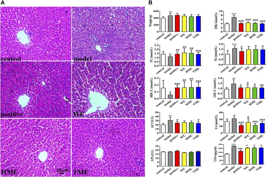
of H&E staining indicated that DSS protected liver tissue.
Chemical Profiling of Compared with the control group, FBG, body weight, UA and
Danggui-Shaoyao-San Extracts hepatic TG, LDL-C, ALT statistically up-regulated in the model
Twenty-three kinds of chemical compounds from WE, HME and group (p < 0.05), while hepatic TC and HDL-C statistically down-
TME were identified by UPLC-Q/TOF-MS (Supplementary regulated (p < 0.05, Figure 1B). It indicated that metabolic
Figure 1), mainly including seven monoterpene glycosides, syndrome and hepatic injury of rats in the model group were
nine phthalides, three prototerpane triterpene glycosides, two obvious. Enteral LPS and hepatic TNF-α were also detected, and
cyclic peptides, two organic acids and adenosine. There were the results showed a significant increase of TNF-α (p < 0.001) but
three kinds of differential MS fingerprints as shown in no obvious change of LPS in the model group (Figure 1B). It was
Supplementary Figure 2. The results about difference analysis found that FBG, TNF-α, LDL-C and UA significantly decreased
(VIP > 1, p < 0.05) showed that there were some differences in the (all p < 0.05) and TC and HDL-C increased (all p < 0.05) after the
chemical composition of the three extracts. There were ten use of pioglitazone or three extracts. ALT had got regulation (p <
Frontiers in Pharmacology | www.frontiersin.org 4 July 2021 | Volume 12 | Article 671708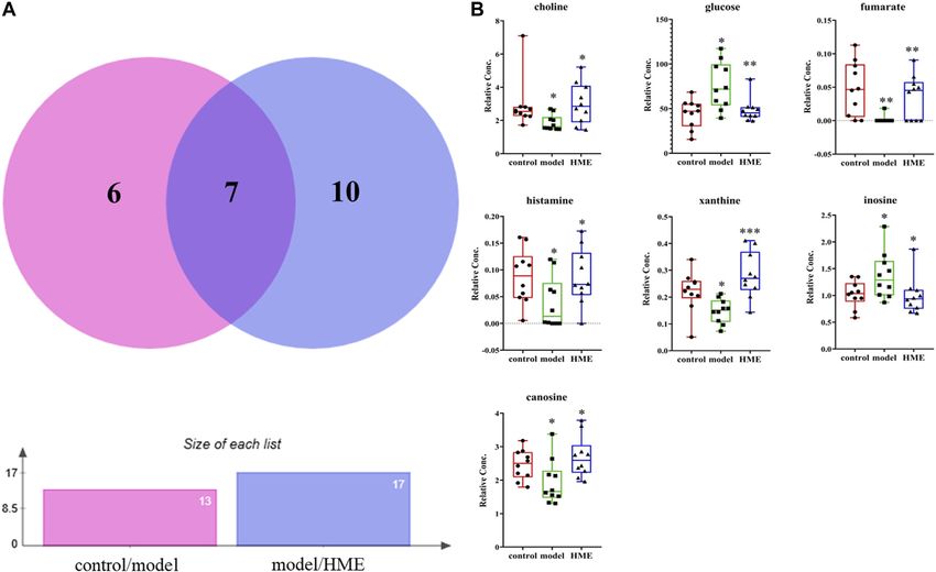
Yin et al.                                                                                                                                  Dss Improves MetS in Three Aspects

  FIGURE 1 | Histopathological assessment in liver (H&E stain) and biochemical assessments in serum or liver. (A) pathological paraffin section (100X) (B) Levels of
  fasted blood glucose (FBG), serum uric acid (UA), hepatic triglyceride (TG), total cholesterol (TC), high density lipoprotein (HDL-C), low density lipoprotein (LDL-C), alanine
  aminotransferase (ALT), tumor necrosis factor-α (TNF-α) and intestinal levels of lipopolysaccharide (LPS). All values are mean ± SD (n  10). *p < 0.05, **p < 0.01, ***p <
  0.001 between model group and control group, #p < 0.05, ##p < 0.01, ###p < 0.001 between model group and WE/HME/TME group.

0.05) after the intervention of pioglitazone or TME. Although                                relative abundance of Firmicutes is positively related to the occurrence
pioglitazone or different extracts had no obvious effect on the                              of metabolic diseases such as obesity (Koliada et al., 2017). It has also
improvement of body weight, body weight had the down                                         been reported that Akkermansia, belonging to Verrucomicrobia, is
tendency in HME or TME group, especial HME group. The                                        negatively related to obesity, and the higher the relative abundance in
statistical biomarkers, including TC, HDL-C, LDL-C, ALT and                                  the intestine, the less obesity the host appears (Brahe et al., 2015; Dao
UA, revealed that HME or TME had stronger protective actions                                 et al., 2016; Derrien et al., 2017).
for liver functions and regulated the MetS better than WE.                                       LEfSe was used to analyze the difference of gut microbes. The
                                                                                             result at the genus level was expressed by Cladogram pattern or LDA
Changes in Gut Microbiota Structure                                                          histogram (LDA > 4.0). From the results, we concluded that there was
Gut microbiota plays an important role in evaluating all kinds of                            more differential gut microbiota from HME group (Figure 3A) as
organism, balancing the unhealthy environment in our bodies and                              compared with the control group. The differences from the model
keeping us healthy. We calculated the OUT expression of each                                 group was mainly reflected in the up-regulation of the relative
sample at all kinds of classification level, especially at the level of                       abundance of Turicibacter and Erysipelotrichaceae, and the down-
phylum and genus to find which extract can influence the kinds and                             regulation of Lactobacillus (LDA > 4.0, Figure 3B) by LDA histogram
enrichment in intestinal microbiota to alleviate abnormal symptoms                           analysis. The HME group contained the richest gut microbes
about MetS. As shown in Figure 2 dominant bacteria were                                      including downregulated Lactobacillus and Erysipelotrichaceae, and
Firmicutes, Bacteroides, Verrucomicrobia in sequence at the                                  the notably upregulated Akkermansia (LDA > 4.0). It is reported that
phylum level. The relative abundance of Firmicutes had its                                   Akkermansia is beneficial bacteria to reduce obesity (Everard et al.,
downward trend in the WE, HME, TME group, while Bacteroides                                  2013; Shin et al., 2014) and Erysipelotrichaceae is harmful by
and its genus almost kept constant. In addition, Verrucomicrobia in                          increasing intestinal inflammation (Wu et al., 2018). Based on the
the different treatment groups had a growing tendency, while it was                          changes in gut microbes, it can be concluded that HME can play a
almost absent in the model group. It is reported that the change of                          more diversifying and beneficial effect on regulating gut microbiota

Frontiers in Pharmacology | www.frontiersin.org                                          5                                                 July 2021 | Volume 12 | Article 671708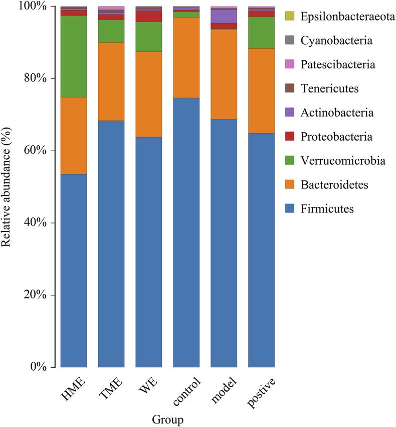
Yin et al.                                                                                                                Dss Improves MetS in Three Aspects

  FIGURE 2 | Histogram of mean value of intestinal microflora enrichment in rats at phylum level in each group.

structure than WE and TME. Based on all the results, further studies                  fumarate, histamine, xanthine, inosine (Figure 4). From thirteen
on metabolomic and transcriptomic changes in the liver were carried                   differential metabolites obtained between the control and model
only by the HME treatment.                                                            group, we concluded that there were certain metabolic disorders in
                                                                                      the model group. By the intervention of DSS, seven differential
Metabolomic Changes in Liver                                                          metabolites can be regulated back in the same changed metabolic
The changes of metabolites in the liver can show whether the                          routes by fructose. The other ten differential metabolites between
hepatic functions or metabolic pathways have been influenced by                        the model and HME group indicated that DSS could influence the
fructose. According to NMR data, a variety of metabolites in the                      new metabolic pathways. The results showed that DSS played an
liver were identified, mainly including amino acids, organic acids,                    effective role in improving the MetS.
alkaloids, sugars and nucleotides. Choose VIP > 1, p < 0.05 as
screening criteria to screen the differential metabolites between                     Transcriptomic Changes in Liver
different groups. Thirteen differential metabolites were identified                    The differential transcripts between different groups were screened
between the control group and model group, which were 3-                              at absolute FC ≥ 0.5, Q-value and FDR ≤ 0.01. By analyzing the
hydroxybutyrate, alanine, carnosine, choline, glycine, glucose,                       metabolic pathway by differential gene expressions, we determined
fumarate, anserine, tyramine, histamine, xanthine, inosine,                           which pathways had been disorganized by fructose in the model
formate, respectively. Seventeen differential metabolites were                        group and which were regulated back after DSS. Compared with
identified between the model group and HME group, which                                the control group, 422 transcripts were up-regulated, 377
were respectively isoleucine, leucine, valine, lactate, acetate,                      transcripts were down-regulated and 18,867 transcripts were the
succinate, carnosine, cadaverine, creatine, choline, glutamine,                       same in the model group. Compared with the model group, there
glucose, ADP + ATP, fumarate, histamine, xanthine and                                 were 132 up-regulated transcripts, 218 down-regulated transcripts
inosine. There were seven shared metabolites by Venny analysis                        and 19,336 transcripts were the same in HME group. The results
(http://jvenn.toulouse.inra.fr/app/index.html) between control/                       showed that high fructose-fed did change the hepatic gene
model and model/HME, including carnosine, choline, glucose,                           expressions of the liver gene in normal rats, while to a certain

Frontiers in Pharmacology | www.frontiersin.org                                   6                                     July 2021 | Volume 12 | Article 671708
Yin et al. Dss Improves MetS in Three Aspects
FIGURE 3 | LEfSe results of species differences in intestinal microflora enrichment in rats between different groups. (A) Comparative analysis by cladogram pattern
between control and model group or model and WE/HME/TME group. (B) Comparative analysis by LDA histogram only in control/model or model/HME contrast
(LDA > 4.0).
extent, DSS did alleviate the abnormal situation of hepatic as glucose and fumarate also showed the correlation with
transcripts in the model group. carbohydrate metabolism. The genes of HSD family, such as
Pathway analysis helps understand the biological function of hydroxysteroid 17β-dehydrogenase 7 (HSD17β7) and
genes. In this study, KEGG analyzed how the gene expressions hydroxysteroid 17β-dehydrogenase 13 (HSD17β13), existed in
were enriched between different groups. The top 30 differential the pathway of steroid hormone biosynthesis. They regulated
pathways at the Level 3 were analyzed between the control and blood lipid and were related to TC and TG metabolites. These
model group (Figure 5A). Another result was got between the results showed that fructose could induce differential gene changes
model and HME group as shown in Figure 5B. The pathway with and pathway discordances and DSS could regulate back these
the most enriched differential genes was focused on the metabolic abnormalities of carbohydrate, lipid and amino acids at the
pathway. Compared with the control group, fifty eight differential Level 2 of the KEGG pathway.
genes in the model group were upregulated and seventy five genes
were downregulated in the metabolic pathway. Compared with the
model group, twenty three differential genes in the HME group
were upregulated and forty eight were downregulated. Other ten
DISCUSSION
shared pathways were arginine biosynthesis, steroid hormone
biosynthesis, biosynthesis of amino acids, carbon metabolism,
The Relationship Between Metabolic
circadian rhythm, fatty acid metabolism, metabolic pathways, Syndrome in Fructose-Fed Rat Modeling
nitrogen metabolism, PPAR signalling pathway and pyruvate and Insulin Resistance
metabolism. In these pathways, some were related to the A large body of recent literature has shown that excess fructose
carbohydrate metabolism. Hepatic differential metabolites such comsumption can induce MetS (Li et al., 2018). 15% of fructose is
Frontiers in Pharmacology | www.frontiersin.org 7 July 2021 | Volume 12 | Article 671708Yin et al. Dss Improves MetS in Three Aspects
FIGURE 4 | Differential metabolites in common in rat liver between different groups. (A) Differential metabolites changes in common by Wayne analysis in pie chart
between control/model or model/HME contrast. (B) Individual relative content changes of differential metabolites in common by histogram pattern (differential screening
criterion: VIP > 1 and *p < 0.05, **p < 0.01, ***p < 0.001).
similar to American human soft drinks (Jürgens et al., ATP by KHK (Asipu et al., 2003; Softic et al., 2016) for fructose
2005).Many researchers have reported that 10–20% fructose phosphoration. This process converts fructose into fructose-1
for 4–8 weeks could induce MetS modeling or one of MetS phosphate (F-1-P) and finally produces plenty of UA (Nakagawa
symptoms in rat or mice (Jürgens et al., 2005; De Moura et al., 2006). Excessive UA directly inhibits insulin signaling not
et al., 2008; Zhang et al., 2011). The modeling conditions are only in hepatocytes (Zhu et al., 2014), but also in endothelial cells
similar with our research. IR is reported as the mechanism of (Choi et al., 2014) and cardiomy-ocytes in vitro (Zhi et al., 2016).
MetS and chronic fructose ingestion, but not acute fructose The inhibition leads to insulin resistance in potassiumoxonate-
feedings can lead to hyperinsulinemia and insulin resistance induced hyperuricemia mice (Zhu et al., 2014). The second
(Haas et al., 2012; Pan and Kong, 2018). Thus, we reviewed catalytic enzyme is ALDOB, which can help part of F-1-P
some literature on mechanism how IR develops high fructose- directly converted into dihydroxyacetone phosphate (DHAP)
induced MetS. and indirectly into glycerol-3-phosphate (GA3P) (Nomura and
Fructose directly reaches the hepatocytes after being absorbed Yamanouchi, 2012). DHAP and GA3P are catabolized for DNL
from the intestine via portal vein. The metabolic pathway of most via glycolytic pathway by intermediate acetyl-CoA, to be used in
of fructose is the firs-pass metabolism in the liver (Softic et al., tricarboxylic acid (TCA) cycle (also known as citric acid cycle),
2016). Fructolysis is much faster than glycolysis in the for energy production (Softic et al., 2016). High level of DNL leads
hepatocytes for bypassing the step of phosphofructokinase to alteration of TG, TC and free fatty acid (FFA) and other lipid
(Pan and Kong, 2018), which usually limits the metabolic metabolites and induces insulin resistance under high fructose
speed. Limitless fructolysis can produce high level of de novo condition (Huang et al., 2017).
lipogenesis (DNL) and UA metabolites (Pan and Kong, 2018). Oxidative stress and inflammation play causal roles in insulin
They are unique features of fructolysis and high level of them can resistance (Pan and Kong, 2018). More and more evidence
induce insulin resistance. After entering the hetatocytes, fructolsis demonstrates that they impair insulin signaling to induce
is catalyzed by ketohexokinase (KHK) and aldolase B (ALDOB), insulin resistance in high fructose-induced MetS (Bettaieb
which are two specific enzymes and highly expressed in the et al., 2015; Porto et al., 2015). High level of Reactive oxygen
hepatocytes (Pan and Kong, 2018). In the first-pass species (ROS) or disability of anti-oxidation stimulated by FFAs,
metabolism, fructolysis happens quickly with comsuming more especially saturated FFAs, induces oxidative stress (Inoguchi
Frontiers in Pharmacology | www.frontiersin.org 8 July 2021 | Volume 12 | Article 671708Yin et al. Dss Improves MetS in Three Aspects FIGURE 5 | Up or down about differential gene experessions in top 30 enrichment pathways in rat liver between control/model or model/HME. (A) Comparative analysis by histogram pattern between control group and model group. (B) Comparative analysis by histogram pattern between model group and HME group. (differential screening criterion: absolute FC ≥ 0.5, Q-value or FDR ≤ 0.01). Frontiers in Pharmacology | www.frontiersin.org 9 July 2021 | Volume 12 | Article 671708
Yin et al.                                                                                                            Dss Improves MetS in Three Aspects

et al., 2000). Lipid metabolites, such as FFAs, TG, diacylglycerol               transformation from TC to bile acid. The excretion pathway of bile
(DAG), and ceramides, can activate pro-inflammatory kinases,                      acids can be the regulated targeting to lower blood lipid.
thus impair insulin signaling in hepatic or extrahepatic tissues                 Lactobacillus (Supplementary Table 1) decreased (LDA > 4.0) in
(Xu et al., 2003; Choi et al., 2008). Pro-inflammatory kinases                    the model group compared with the control group. Compared with
relate to protein kinase C (PKC), c-jun N-terminal kinase (JNK)                  the model group, Lactobacillus was continuously decreased in the
and inhibitor of nuclear factor kappa B (NF-kB) (IkB) kinase                     HME group. TC finally increased (p < 0.01) and was even higher
(IKK) (Bergman and Ader, 2000). On the other hand, fructose-                     than the control group in the HME group. Based on the facts, we
induced UA metabolite also causes ROS production in hepatic                      concluded that the significant up-regulation of TC might be possibly
(Lanaspa et al., 2012) and extrahepatic tissues (Sautin et al., 2007;            due to the double effect from the upgrade of HSD17β7 and the
Hu et al., 2009; Lanaspa et al., 2012; Cirillo et al., 2015; Madlala             downgrade of Lactobacillus.
et al., 2016) and activates inflammatory response by induction of
IL-1β, TNF-α, transforming growth factor (TGF)-β1 and
monocyte chemotactic protein1 (Umekawa et al., 2003;
                                                                                 The Mechanism of Anti-Inflammation by
Palanisamy et al., 2011; Chen et al., 2013) Finally these                        Danggui-Shaoyao-San in Metabolic
inflammatory factors also induce IR. Therefore, DNL and UA                        Syndrome Rats
are dangerous factors induced by chronic fructose ingestion. IR,                 A high level of DNL and UA can result in insulin resistance, which is
whose underlying common mechanisms are oxidative stress and                      the key pathological event in developing MetS. It happens mostly
inflammation, is the key pathological mechnism in MetS.                           through oxidative stress and inflammation (Nakagawa et al., 2006;
                                                                                 Schwarz et al., 2017; Pan and Kong, 2018). Fructose-induced UA can
                                                                                 trigger an inflammatory response. ROS induction can induce
The Mechanism of Regulating Blood Lipid                                          inflammatory factors such as interleukin, tumour necrosis factor
by Danggui-Shaoyao-San in Metabolic                                              and transforming growth factor (Umekawa et al., 2003; Palanisamy
Syndrome Rats                                                                    et al., 2011; Chen et al., 2013; Pan and Kong, 2018). In comparison
Compared with the control group, part of biomarkers, body weights,               with the control group, UA (Figure 1B) in the model group
pathological sections in the model group have shown that there was a             significantly increased (p < 0.001 indicating an inflammatory
disorder in the hepatic functions and carbohydrate metabolism. This              reaction. After DSS treatment, UA decreased significantly (p <
indicated the occurrence of nonalcoholic fatty liver disease (NAFLD).            0.001), indicating an anti-inflammatory effect of DSS. There are
NAFLD is closely related to MetS. It is reported that inducing                   many kinds of inflammation genes to perform the anti-inflammation,
NAFLD is related to the rapid increase of hepatic TNF-α (Wang                    including the NOD-like receptor (NLR) family, TNF family and IL
et al., 2012). HSD17β13 is also identified as a pathogenic protein in             family (Huang et al., 2019). Some part of inflammation genes
the development of NAFLD (Kampf et al., 2014; Su et al., 2014). In               overexpresses when inflammation occurs. In this study, NOD-like
general, HSD family takes part in several kinds of metabolism, but the           receptor pyrin containing 12 (NLRP12) (Q ≤ 0.01) and TNF-α (p <
HSD17β13 mainly takes part in lipid metabolism (Fujimoto et al.,                 0.05) upgraded markedly in the model group, while IL-33 (Q ≤ 0.01)
2004; Moeller and Adamski, 2009; Marchais-Oberwinkler et al., 2011;              downgraded statistically (Supplementary Table 2). After the cure of
Su et al., 2014).The upgrade of TG is reported due to overexpression             DSS, although IL-33 upgraded markedly (Q ≤ 0.01), the level of IL-33
of HSD17β13. In our study, HSD17β13 (Q ≤ 0.01) (Supplementary                    was still lower than that in the control group. TNF-α was regulated
Table 2) and TNF-α (p < 0.05) were both found upregulated.TG                     back to a normal level statistically (p < 0.05). It revealed that the
(Figure 1B) had its marked upgraded trend (p < 0.001). On contrary,              inflammation was being alleviated. NLRP12 was gradually increased
all the results had their downgrade without statistics (p > 0.05) after          using DSS but had no significant difference. All the evidence
the cure with DSS. All the data conformed to the conclusion and the              suggested that there was a possible relationship between NLRP12,
literature that TG is positively and directly related to HSD17β13 and            TNF-α, IL-33 and DSS’s anti-inflammatory mechanism.
TNF-α (Su et al., 2014).                                                         Inflammation occurs due to the changes from genes, intestinal
    It is reported that HSD17β7 in a rat is beneficial to increase TC             flora and its metabolites. It is reported that Erysipelotrichaceae,
(Matsuoka et al., 2020). In this gene study, as compared with the                Turicibacter and others are related to the elevation of TNF levels,
control group, HSD17β7 (Supplementary Table 2) statistically                     IL levels in the Muc2 deficient mice with colitis (Wu et al., 2018).
decreased (Q ≤ 0.01) in the model group. TC (Figure 1B) in the                   Erysipelotrichaceae (Supplementary Table 1) markedly upgraded in
model group abnormally decreased (p < 0.01). Conversely, TC (p <                 model group (LDA > 4.0), accompanied with the increase of TNF-α.
0.01) and HSD17β7 (Q ≤ 0.01) increased markedly in the HME                       On the contrary Erysipelotrichaceae (LDA > 4.0) and TNF-α (p <
group. This showed that there was a positive relationship between                0.05) synchronously markedly downgraded after the cure of DSS.
TC and HSD17β7. It is reported that TC can produce bile acid after a             This result further demonstrated that Erysipelotrichaceae can be
series of metabolism. Bile acid is the main way to excrete cholesterol           directly related to inflammation and DSS worked as an anti-
(Iritani et al., 1982). Most bile acids exist as conjugated bile salts. A        inflammatory by reducing the enrichment of Erysipelotrichaceae
small part of bile salts are decomposed into insoluble free bile acids           and regulating back TNF-α level.
by the bile salt hydrolytic enzymes secreted by the Lactobacillus in                 One of the characteristics of MetS is IR, which is an
the intestine (Gilliland et al., 1985; Rasic et al., 1992; Klaver and van        inflammatory disease (Donath and Shoelson, 2011). It is also
der Meer, 1993). These free bile acids can be excreted along with                reported that intestinal flora ferments amino acids to produce
faeces in vitro. The lowered bile acid salts in vivo accelerated the             acetate, butyrate and formate which belong to short-chain fatty

Frontiers in Pharmacology | www.frontiersin.org                             10                                      July 2021 | Volume 12 | Article 671708
Yin et al. Dss Improves MetS in Three Aspects
acids salt with the anti-inflammation effect (Davila et al., 2013). Other studies indicated that the increase of BCAAs in the blood
For example, Lachnospiraceae family can produce butyrate of mice fed by Prevotella_9 resulted in IR, glucose intolerance and
(Costantini et al., 2017). Akkermansia is another new and Type 2 diabetes mellitus (T2DM). The increase of BCAAs in blood
strongly beneficial bacterium except for Lactobacillus which is has been used as a marker of IR and T2DM (Lynch and Adams, 2014;
resistant to human obesity and MetS (Brahe et al., 2015). It exists Pedersen et al., 2016). Other literature has reported that compared
in less quantity in the intestine of an obese human and plays an with the Normal group, Prevotella_9 in T2DM model group increases
important role in maintaining intestinal health and integrity. It significantly, accompanied by the increase of the metabolite-BCAAs
releases acetate through the degradation of mucin to regulate in the blood (Pedersen et al., 2016; Yue et al., 2018). Results obtained
membrane permeability (Dao et al., 2016). Results demonstrated demonstrated that increase of BCAAs is positively correlated with
that all kinds of intestinal flora can play a role in different ways Prevotella_9. By analyzing the data of this study, we found that
about anti-inflammation. In comparison with the control group, compared with the control group, the BCAAs of the model and HME
Lachnospiraceae_NK4A136_group (Supplementary Table 1) groups were gradually increasing with the increase of relative
increased in the model group without statistics (LDA < 4.0). abundance of Prevotella_9. It indicated that the trend of this
Akkermansia (Supplementary Table 1) almost absent experiment was consistent with the literature and there was a
statistically (LDA > 4.0). Hepatic metabolites-acetate strong positive correlation between BCAAs and Prevotella_9.
(Supplementary Table 3) increased without statistics (LDA < Lactate is the product of anaerobic fermentation of glucose under
4.0). After the treatment of DSS, the relative abundance of anaerobic conditions. The increase of lactate shows the growth of
Lachnospiraceae_NK4A136_group (Supplementary Table 1) Glycolysis and gluconeogenesis accompanied by more glucose in the
slightly downgraded with no statistics. But acetate and formate blood. In this study, compared with the control group, the increase of
(Supplementary Table 3) both increased markedly (VIP > 1, p < lactate in the blood in the model group indicated that
0.05.0) after the cure of DSS. The reason for the change could be gluconeogenesis generated more glucose. On the contrary,
due to the very rapid increase of Akkermansia (LDA > 4.0) and compared with the model group, HME statistically regulated the
there was a strong positive correlation. The efficient ingredient in lactate (Supplementary Table 3) and FBG (Figure 1B) (VIP > 1, p <
DSS had the function of anti-inflammation. DSS may contribute 0.05). There were a lot of changes in the intestinal flora. These
to the increase of Akkermansia and its metabolite-short-chain changes included Lactobacillus and Turicibacter (Supplementary
fatty acids salt to regulate membrane permeability and anti- Table 1) which produced lactate (Li et al., 2017). Compared with the
inflammation. control group, Lactobacillus producing lactate markedly came down
(LDA > 4.0) and Turicibacter markedly upgraded (LDA > 4.0) in the
model group. This suggested that Turicibacter may be the key factor
The Mechanism of Reducing the Blood to producing lactate. Compared with the model group, there was
Glucose by Danggui-Shaoyao-San in marked downgrade of lactate (VIP > 1, p < 0.05) and a marked
Metabolic Syndrome Rats downgrade of Lactobacillus (LDA > 4.0) after the cure with DSS.
The level of amino acid in the blood can predict the occurrence of Turicibacter had a slight upgrade without statistics after the cure with
diabetes as amino acid is an important material of gluconeogenesis DSS. In this aspect, we concluded that the change of lactate in blood
and it leads to the increase of blood glucose (Chen et al., 2016). was mainly due to the downgrade of Lactobacillus. All the results
Fumarate is reported to be a crucial enzyme in TCA cyclic reaction showed that the change of lactate in blood was not completely
and has a catalytic role in amino acid metabolism (Suzuki et al., 1989; induced by Lactobacillus, also by other bacteria which can produce
Arts et al., 2016). The higher the amino acid metabolic speed, the lactate, such as Turicibacter. In conclusion, the combined action of
lower is the concentration of amino acids in the blood. Compared host differential metabolites and intestinal flora can play a role in
with the control group, the differential down-regulation of fumarate regulating the abnormal blood glucose in MetS.
(Supplementary Table 3) in the model group (VIP > 1, p < 0.05)
slowed down the speed of amino acid metabolism, which led to the
significant up-regulation of glycine and alanine (Supplementary CONCLUSION
Table 3) in liver and glucose in the blood (VIP > 1, p < 0.05).
Compared with model group, HME group regulated back the DSS is a traditional prescription with bolstering blood cycle and
concentration of fumarate (VIP >1, p < 0.05). It led to decreasing alleviating stasis due to blood stasis and deficiency of spleen Qi.
the content of glycine and alanine, slowing down the gluconeogenesis, This study was the first to report about alleviating the MetS
and significantly reducing the glucose levels (VIP >1, p < 0.05) symptoms. In the comparison of the three extractive solvents, it
(Supplementary Table 3). Besides glycine and alanine, there were was found that HME or TME had seven different chemical
other branched amino acids (BCAAs) in the blood which were valine, components with higher content than WE. These compounds
leucine and isoleucine. In this study, BCAAs (Supplementary have been recorded in the aspects of vascellum protection, anti-
Table 3) in the different groups were relatively low. Glycine and inflammatory, anti-thrombotic, anti-oxidant effect. In the HE-
alanine contributed more to gluconeogenesis in the aspect of both staining pharmacological section, liver biochemical parameters,
content and changing amplitude and played major roles in the 16S rRNA intestinal flora research, it was preliminarily
decreasing of glucose level in the blood. Even though we should concluded that the effect of 50% methanol extract was more
discuss the relationship between BCAAs and Prevotella_9 prominent to be used to MetS. By the analysis of relationships in
(Supplementary Table 1), the relationship is also important. HME group between different liver metabolites, liver
Frontiers in Pharmacology | www.frontiersin.org 11 July 2021 | Volume 12 | Article 671708Yin et al. Dss Improves MetS in Three Aspects
FIGURE 6 | The mechanism study of DSS’s regulating MetS symptoms in three aspects of blood lipid, anti-inflammation, blood gluctose.
transcriptional genome and intestinal flora, we can understand AUTHOR CONTRIBUTIONS
how DSS played a role in reducing blood lipid, anti-inflammatory
and hypoglycemic (Figure 6). The results laid a foundation for the MJ conceived and designed the experiments. JY drafted the paper
further exploration of clinical application and effective mechanism and partially analyzed the data. JL conducted the main experiments
of DSS in MetS in the future. This study can give some guidance on and analyzed the partial data. PL, MH, SH and JL conducted part
more valid components of traditional Chinese medicine in the experiments. YZ and ZL reviewed and edited the paper. All authors
aspect of improving gut microbia dysbiosis and hepatic lipid revised the paper and approved the final manuscript.
homeostasis in fructose-fed rats.
FUNDING
DATA AVAILABILITY STATEMENT
This work was supported by the Tianjin Science and Technology
The datasets presented in this study can be found in article, Program (No. 20ZYJDJC00120), the Natural Science Foundation
Supplementary Material and online repository: Gene expression of Tianjin City (No. 18JCZDJC97700), the National Key Research
omnibus (GEO) from NCBI. The accession number can be found and Development Program of China (No. 2018YFC1707403) and
below: GEO, GSE176572. the National Natural Science Foundation of China (No. 81573547).
ETHICS STATEMENT SUPPLEMENTARY MATERIAL
The animal study was reviewed and approved by the Institutional The Supplementary Material for this article can be found online at:
Animal Ethical Committee of Xuhe Pharmaceutical Science and https://www.frontiersin.org/articles/10.3389/fphar.2021.671708/
Technology Co., Ltd. full#supplementary-material
Frontiers in Pharmacology | www.frontiersin.org 12 July 2021 | Volume 12 | Article 671708Yin et al. Dss Improves MetS in Three Aspects
REFERENCES Donath, M. Y., and Shoelson, S. E. (2011). Type 2 Diabetes as an Inflammatory
Disease. Nat. Rev. Immunol. 11, 98–107. doi:10.1038/nri2925
Dong, P., Zhang, T., Yin, X., Wen, H., Qu, N., and Han, H. (2014). Effect of
Arts, R. J., Novakovic, B., ter Horst, R., Carvalho, A., Bekkering, S., Lachmandas, E., DangguiShaoyao Powder on Experimental Hyperlipidemia Model Rats (II).
et al. (2016). Glutaminolysis and Fumarate Accumulation Integrate Acta Chin. Med. Pharmacol. 42, 103–105. doi:10.19664/j.cnki.1002-
Immunometabolic and Epigenetic Programs in Trained Immunity. Cell 2392.2014.04.036
Metab. 24, 807–819. doi:10.1016/j.cmet.2016.10.008 Everard, A., Belzer, C., Geurts, L., Ouwerkerk, J. P., Druart, C., Bindels, L. B., et al.
Asipu, A., Hayward, B. E., O’Reilly, J., and Bonthron, D. T. (2003). Properties of (2013). Cross-talk between Akkermansia Muciniphila and Intestinal
Normal and Mutant Recombinant Human Ketohexokinases and Implications Epithelium Controls Diet-Induced Obesity. Proc. Natl. Acad. Sci. U. S. A.
for the Pathogenesis of Essential Fructosuria. Diabetes 52, 2426–2432. 110, 9066–9071. doi:10.1073/pnas.1219451110
doi:10.2337/diabetes.52.9.2426 Fu, X., Wang, Q., Wang, Z., Kuang, H., and Jiang, P. (2015). Danggui-Shaoyao-San:
Bergman, R. N., and Ader, M. (2000). Free Fatty Acids and Pathogenesis of Type 2 New Hope for Alzheimer’s Disease. Aging Dis. 6, 342–348. doi:10.14336/
Diabetes Mellitus. Trends Endocrinol. Metab. 11, 351–356. doi:10.1016/S1043- AD.2015.1220
2760(00)00323-4 Fujimoto, Y., Itabe, H., Sakai, J., Makita, M., Noda, J., Mori, M., et al. (2004).
Bettaieb, A., Jiang, J. X., Sasaki, Y., Chao, T.-I., Kiss, Z., Chen, X., et al. (2015). Identification of Major Proteins in the Lipid Droplet-Enriched Fraction Isolated
Hepatocyte Nicotinamide Adenine Dinucleotide Phosphate Reduced Oxidase 4 from the Human Hepatocyte Cell Line HuH7. Biochim. Biophys. Acta. 1644(1):
Regulates Stress Signaling, Fibrosis, and Insulin Sensitivity during Development 47-59. doi:10.1016/j.bbamcr.2003.10.018
of Steatohepatitis in Mice. Gastroenterology 149, 468–480. e10. doi:10.1053/ Gilliland, S. E., Nelson, C. R., and Maxwell, C. (1985). Assimilation of Cholesterol
j.gastro.2015.04.009 by Lactobacillus Acidophilus. Appl. Environ. Microbiol. 49, 377–381.
Boehm, B. O., and Claudi-boehm, S. (2005). The Metabolic Syndrome. Scand. doi:10.1128/aem.49.2.377-381.1985
J. Clin. Lab. Invest. 65, 3–13. doi:10.1080/00365510500236044 Haas, J. T., Miao, J., Chanda, D., Wang, Y., Zhao, E., Haas, M. E., et al. (2012).
Brahe, L. K., Le Chatelier, E., Prifti, E., Pons, N., Kennedy, S., Hansen, T., et al. (2015). Hepatic Insulin Signaling Is Required for Obesity-dependent Expression of
Specific Gut Microbiota Features and Metabolic Markers in Postmenopausal SREBP-1c mRNA but Not for Feeding-dependent Expression. Cel Metab. 15,
Women with Obesity. Nutr. Diabetes 5. e159, doi:10.1038/nutd.2015.9 873–884. doi:10.1016/j.cmet.2012.05.002
Chen, L., Lan, Z., Lin, Q., Mi, X., He, Y., Wei, L., et al. (2013). Polydatin Ameliorates Hu, Q.-H., Wang, C., Li, J.-M., Zhang, D.-M., and Kong, L.-D. (2009). Allopurinol,
Renal Injury by Attenuating Oxidative Stress-Related Inflammatory Responses Rutin, and Quercetin Attenuate Hyperuricemia and Renal Dysfunction in Rats
in Fructose-Induced Urate Nephropathic Mice. Food Chem. Toxicol. 52, 28–35. Induced by Fructose Intake: Renal Organic Ion Transporter Involvement. Am.
doi:10.1016/j.fct.2012.10.037 J. Physiol. Physiol. 297, F1080–F1091. doi:10.1152/ajprenal.90767.2008
Chen, L., Qi, J., Chang, Y. xu., Zhu, D., and Yu, B. (2009). Identification and Huang, J.-P., Cheng, M.-L., Hung, C.-Y., Wang, C.-H., Hsieh, P.-S., Shiao, M.-S.,
Determination of the Major Constituents in Traditional Chinese Medicinal et al. (2017). Docosapentaenoic Acid and Docosahexaenoic Acid Are Positively
Formula Danggui-Shaoyao-San by HPLC-DAD-ESI-MS/MS. J. Pharm. Associated with Insulin Sensitivity in Rats Fed High-Fat and High-Fructose
Biomed. Anal. 50, 127–137. doi:10.1016/j.jpba.2009.03.039 Diets. J. Diabetes 9, 936–946. doi:10.1111/1753-0407.12505
Chen, T., Ni, Y., Ma, X., Bao, Y., Liu, J., Huang, F., et al. (2016). Branched-chain and Huang, J., Yang, D., Li, X., and Yang, S. (2019). Inflammasome and Intestinal
Aromatic Amino Acid Profiles and Diabetes Risk in Chinese Populations. Sci. Mucosal Immunity. J. Microbes Infect. 14, 113–123. doi:10.3969/j.issn.1673-
Rep. 6. 20594. doi:10.1038/srep20594 6184.2019.02.007
Choi, C. S., Befroy, D. E., Codella, R., Kim, S., Reznick, R. M., Hwang, Y.-J., et al. Inoguchi, T., Li, P., Umeda, F., Yu, H. Y., Kakimoto, M., Imamura, M., et al. (2000). High
(2008). Paradoxical Effects of Increased Expression of PGC-1α on Muscle Glucose Level and Free Fatty Acid Stimulate Reactive Oxygen Species Production
Mitochondrial Function and Insulin-Stimulated Muscle Glucose Metabolism. through Protein Kinase C--dependent Activation of NAD(P)H Oxidase in Cultured
Proc. Natl. Acad. Sci. 105. 19926–1993. doi:10.1073/pnas.0810339105 Vascular Cells. Diabetes 49, 1939–1945. doi:10.2337/diabetes.49.11.1939
Choi, Y.-J., Yoon, Y., Lee, K.-Y., Hien, T. T., Kang, K. W., Kim, K.-C., et al. (2014). Iritani, N., Nara, Y., and Yamori, Y. (1982). Cholesterol and Bile Acid Metabolism
Uric Acid Induces Endothelial Dysfunction by Vascular Insulin Resistance in Hypertensive Arteriolipidosis-Prone Rats (ALR). Jpn. Circ. J. 46, 151–155.
Associated with the Impairment of Nitric Oxide Synthesis. FASEB J. 28, doi:10.1253/jcj.46.151
3197–3204. doi:10.1096/fj.13-247148 Ivanova, N., Liu, Q., Agca, C., Agca, Y., Noble, E. G., Whitehead, S. N., et al. (2020).
Cirillo, P., Pellegrino, G., Conte, S., Maresca, F., Pacifico, F., Leonardi, A., et al. White Matter Inflammation and Cognitive Function in a Co-morbid Metabolic
(2015). Fructose Induces Prothrombotic Phenotype in Human Endothelial Syndrome and Prodromal Alzheimer’s Disease Rat Model.
Cells. J. Thromb. Thrombolysis 40, 444–451. doi:10.1007/s11239-015-1243-1 J. Neuroinflammation 17. 29. doi:10.1186/s12974-020-1698-7
Costantini, L., Molinari, R., Farinon, B., and Merendino, N. (2017). Impact of Jürgens, H., Haass, W., Castañeda, T. R., Schürmann, A., Koebnick, C.,
omega-3 Fatty Acids on the Gut Microbiota. Int. J. Mol. Sci. 18. 2645, Dombrowski, F., et al. (2005). Consuming Fructose-Sweetened Beverages
doi:10.3390/ijms18122645 Increases Body Adiposity in Mice. Obes. Res. 13, 1146–1156. doi:10.1038/
Daglia, M., Lorenzo, A. D., Nabavi, S. F., Nabavi, Z. S. T., and Nabavi, S. M. (2014). oby.2005.136
Polyphenols: Well beyond the Antioxidant Capacity: Gallic Acid and Related Kampf, C., Mardinoglu, A., Fagerberg, L., Hallström, B. M., Edlund, K., Lundberg,
Compounds as Neuroprotective Agents: You Are what You Eat! Curr. Pharm. E., et al. (2014). The Human Liver-specific Proteome Defined by
Biotechnol. 15, 362–372. doi:10.2174/138920101504140825120737 Transcriptomics and Antibody-Based Profiling. FASEB J. 28, 2901–2914.
Dao, M. C., Everard, A., Aron-Wisnewsky, J., Sokolovska, N., Prifti, E., Verger, E. doi:10.1096/fj.14-250555
O., et al. (2016). Akkermansia Muciniphila and Improved Metabolic Health Kampo.ca Japanese Traditional Medicine & Therapeutics (2014).
during a Dietary Intervention in Obesity: Relationship with Gut Microbiome Tokishakuyakusan. Available at: https://kampo.ca/herbs-formulas/formulas/
Richness and Ecology. Gut 65, 426–436. doi:10.1136/gutjnl-2014-308778 tokishakuyakusan/.
Davila, A. M., Blachier, F., Gotteland, M., Andriamihaja, M., Benetti, P. H., Sanz, Kim, Y., and Cho, S.-H. (2020). Danggui-Shaoyao-San for Dementia: A PRISMA-
Y., et al. (2013). Intestinal Luminal Nitrogen Metabolism: Role of the Gut Compliant Systematic Review and Meta-Analysis. Medicine (Baltimore). 99(4):
Microbiota and Consequences for the Host. Pharmacol. Res. 68, 95–107. e18507. doi:10.1097/md.0000000000018507
doi:10.1016/j.phrs.2012.11.005 Klaver, F. A., and van der Meer, R. (1993). The Assumed Assimilation of
De Moura, R. F., Ribeiro, C., de Oliveira, J. A., Stevanato, E., and de Mello, M. A. R. Cholesterol by Lactobacilli and Bifidobacterium Bifidum Is Due to Their
(2008). Metabolic Syndrome Signs in Wistar Rats Submitted to Different High- Bile Salt-Deconjugating Activity. Appl. Environ. Microbiol. 59, 1120–1124.
Fructose Ingestion Protocols. Br. J. Nutr. 101, 1178–1184. doi:10.1017/ doi:10.1128/aem.59.4.1120-1124.1993
S0007114508066774 Koliada, A., Syzenko, G., Moseiko, V., Budovska, L., Puchkov, K., Perederiy, V.,
Derrien, M., Belzer, C., and de Vos, W. M. (2017). Akkermansia Muciniphila and et al. (2017). Association between Body Mass index and Firmicutes/
its Role in Regulating Host Functions. Microb. Pathog. 106, 171–181. Bacteroidetes Ratio in an Adult Ukrainian Population. BMC Microbiol. 17.
doi:10.1016/j.micpath.2016.02.005 120, doi:10.1186/s12866-017-1027-1
Frontiers in Pharmacology | www.frontiersin.org 13 July 2021 | Volume 12 | Article 671708You can also read