MODULATION OF HIV-1 GAG/GAG-POL FRAMESHIFTING BY TRNA ABUNDANCE - MPG.PURE
←
→
Page content transcription
If your browser does not render page correctly, please read the page content below
5210–5222 Nucleic Acids Research, 2019, Vol. 47, No. 10                                                                 Published online 10 April 2019
doi: 10.1093/nar/gkz202

Modulation of HIV-1 Gag/Gag-Pol frameshifting by
tRNA abundance
Natalia Korniy1 , Akanksha Goyal1 , Markus Hoffmann2 , Ekaterina Samatova1 , Frank Peske1 ,
Stefan Pöhlmann2,3 and Marina V. Rodnina1,*

                                                                                                                                                                  Downloaded from https://academic.oup.com/nar/article-abstract/47/10/5210/5436774 by MPI Biophysical Chemistry user on 17 June 2019
1
  Department of Physical Biochemistry, Max Planck Institute for Biophysical Chemistry, Am Fassberg 11, 37077
Göttingen, Germany, 2 Infection Biology Unit, German Primate Center, Kellnerweg 4, 37077 Göttingen, Germany and
3
  Faculty of Biology and Psychology, University of Göttingen, Wilhelm-Weber-Str. 2, 37073 Göttingen, Germany

Received February 11, 2019; Revised March 12, 2019; Editorial Decision March 14, 2019; Accepted April 08, 2019

ABSTRACT                                                                          translation extracts (5,11,14–22), suggesting that the virus
                                                                                  exploits evolutionary conserved features of the translational
A hallmark of translation in human immunodefi-                                    apparatus.
ciency virus type 1 (HIV-1) is a –1 programmed ri-                                   –1FS is governed by two cis-acting elements in the
bosome frameshifting event that produces the Gag-                                 mRNA, the slippery site (SS1) U1 UUU4 UUA7 that en-
Pol fusion polyprotein. The constant Gag to Gag-Pol                               codes Phe (UUU) and Leu (UUA) in the 0-frame (5), and
ratio is essential for the virion structure and infectiv-                         a stem-loop (SL) structure downstream of the slippery site
ity. Here we show that the frameshifting efficiency is                            (SL1; Figure 1A). SS1 gives rise to two frameshifting prod-
modulated by Leu-tRNALeu that reads the UUA codon                                 ucts, one that contains the 0-frame peptide Phe-Leu fol-
at the mRNA slippery site. This tRNALeu isoacceptor                               lowed by the –1-frame sequence (FLR product; Figure 1A
is particularly rare in human cell lines derived from                             and B), and another with a second Phe incorporated in-
T-lymphocytes, the cells that are targeted by HIV-1.                              stead of Leu (FFR product). In mammalian cells ∼30% of
                                                                                  frameshifting ribosomes do not insert Leu, but are likely
When UUA decoding is delayed, the frameshifting fol-
                                                                                  to insert Phe at the same position (5). Also in E. coli, both
lows an alternative route, which maintains the Gag to                             products are produced, with the FLR constituting about
Gag-Pol ratio constant. A second potential slippery                               80% of the product (5,15,23,24). Further HIV frameshift-
site downstream of the first one is normally ineffi-                              ing product heterogeneity may result from –2 frameshift-
cient but can also support –1-frameshifting when al-                              ing (25). In addition, the gag-pol gene has a second, puta-
tered by a compensatory resistance mutation in re-                                tive slippery site (pSS2) 38 nt downstream of the canoni-
sponse to current antiviral drug therapy. Together                                cal SS1 (26–30). This slippery site is also conserved albeit
these different regimes allow the virus to maintain a                             to a lesser degree than the first slippery site (31). The se-
constant –1-frameshifting efficiency to ensure suc-                               quence of pSS2 (U1 UUU4 CUU7 ) is not particularly slip-
cessful virus propagation.                                                        pery, but a substitution of C5 with U (C5 U), which ap-
                                                                                  pears as a compensatory resistance mutation during anti-
                                                                                  HIV therapy, may facilitate additional FS at this otherwise
INTRODUCTION                                                                      silent site (26–28,30).
                                                                                     The mechanism of frameshifting on the gag-pol mRNA
Many viruses use programmed ribosome frameshifting to                             and the factors that define the ratio between the two –1FS
increase the coding capacity of their genome and to regu-                         products are unclear. The variety of proposed mechanisms
late stoichiometric ratio between viral proteins (1–4). The                       of –1FS (23), the uncertain significance of the second slip-
two HIV-1 genes, gag and pol, encode viral structural pro-                        pery site, and the lack of mechanistic information about al-
teins and enzymes, respectively, and overlap by 205 nt.                           ternative slippages (e.g., –2FS or +1FS) have prompted us
Synthesis of the Gag-Pol polyprotein requires –1 ribosome                         to study gag-pol frameshifting in real time in a fully recon-
frameshifting (–1FS) (5). The ratio between Gag and Gag-                          stituted in vitro translation system. We show that FS path-
Pol is crucial for virus propagation and its dysregulation is                     way and efficiency are determined by the availability of Leu-
detrimental for replication, virion formation and infectivity                     tRNALeu reading the UUA codon. The potential alternative
of HIV-1 (6–9). The efficiency of gag-pol –1FS in human                           +1 and –2 slippages can also operate when other aminoacyl-
cells is about 10%, ranging from 2% to 11% with different re-                     tRNAs (aa-tRNAs) are in limited supply. We show that the
porters (10–13). This –1FS efficiency has been recapitulated                      UUA-specific tRNALeu is particularly rare in human cell
in vivo or in vitro in mammalian, yeast or Escherichia coli

* To   whom correspondence should be addressed. Tel: +49 551 201 2900; Fax: +49 551 201 2905; Email: rodnina@mpibpc.mpg.de

C The Author(s) 2019. Published by Oxford University Press on behalf of Nucleic Acids Research.
This is an Open Access article distributed under the terms of the Creative Commons Attribution Non-Commercial License
(http://creativecommons.org/licenses/by-nc/4.0/), which permits non-commercial re-use, distribution, and reproduction in any medium, provided the original work
is properly cited. For commercial re-use, please contact journals.permissions@oup.com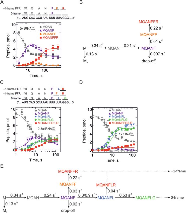
Nucleic Acids Research, 2019, Vol. 47, No. 10 5211
Downloaded from https://academic.oup.com/nar/article-abstract/47/10/5210/5436774 by MPI Biophysical Chemistry user on 17 June 2019
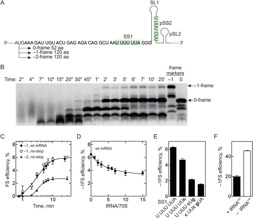
Figure 1. –1FS on HIV-1 gag-pol mRNA. (A) Scheme of the gag-pol frameshifting site. Slippery site (SS1) and the putative second slippery site (pSS2) are
highlighted in green; the stimulatory mRNA structure element downstream of the SS1 is indicated as a stem-loop (SL1). Amino acids incorporated into
0-frame and –1-frame peptides as well as the potential –1FS routes and in vivo efficiencies are shown below the frameshifting sites. (B) Top panel: Amino
acids incorporated into 0- and –1-frames are shown above the mRNA sequence. Bottom panel: –1FS efficiency with the wild-type (wt) mRNA and U4 C
derivative with disrupted SS1 measured at limiting amounts of Leu-tRNANAA Leu (molar ratio 0.3 tRNA to 70S ribosome) at the end of translation (2
min). The 0-frame is the sum of MQANF and MQANFLG peptides, –1-frame corresponds to MQANFFR/FLR peptides. MQANF was identified based
on its position on the chromatogram while MQANFFR/FLR and MQANFLG products were quantified using [14 C]Arg and [3 H]Gly, respectively. (C)
Concentration dependence of –1FS efficiency on the Leu-tRNANAA Leu (tRNANAA Leu , closed circles) or a mixture of tRNALeu isoacceptors reading CUN
codons (tRNANAG Leu , open circles). –1FS product was detected using [14 C]Arg. (D) Change in the FS regime with the Leu-tRNANAA Leu concentration.
The ratio of FFR route (open circles) versus FLR (closed circles) route was calculated from peptides with different radioactive labels as follows. The sum
of FFR and FLR frameshifting products was calculated using [14 C]Arg. To determine the amount of FLR, the mRNA was translated to the 0-frame
peptide fMet-Gln-Asn-Phe-Leu-Gly-Lys-Ile (MQANFLGKI). The presence of Ile allows for separation between 0-frame MQANFLGKI and –1-frame
MQANFLR peptides. The FFR peptide was then determined by subtracting the FLR from the total Arg-containing product. (E) –1FS efficiency in the
presence of varying concentrations of Gly-tRNAGly in the presence of excess Arg-tRNAArg (2 M) (green squares) or with varying concentrations of
Arg-tRNAArg in the presence of 3 or 6 M Gly-tRNAGly (red and light red squares, respectively).
lines derived from T-lymphocytes, the cells that are targeted and subsequent affinity chromatography of the EF-Tu–
by HIV-1. Furthermore, we have characterized the role of GTP–aa-tRNA ternary complexes on Protino Ni-IDA 2000
the second slippery site in supporting –1FS. Multiple ways Packed Columns (Macherey-Nagel) followed by phenoliza-
to modulate the frameshifting efficiency could help the virus tion and ethanol precipitation of aa-tRNA. tRNANAA Leu ,
to maintain the Gag to Gag-Pol ratio, which is crucial for elongator tRNAMet , tRNATyr and a mixture of isoacceptors
its viability. tRNALeu collectively reading all four CUN codons for Leu
were prepared by consecutive column chromatographies
on Sepharose 4B (GE Healthcare), Phenyl Sepharose (GE
MATERIALS AND METHODS Healthcare), and DEAE Toyopearl (Tosoh Bioscience).
Buffer tRNATrp was prepared by T7 RNA-polymerase transcrip-
tion from pUC18 plasmid carrying the E. coli trp gene (40).
All experiments with prokaryotic translation components, tRNAs were charged with 14 C-, or 3 H-labeled or unlabeled
including kinetic measurements summarized in Supplemen- amino acids (41). Leu-tRNANAA Leu , mixture of isoaccep-
tary Table S1, were carried out in HiFi buffer (50 mM tors Leu-tRNA for the CUN codons, Met-tRNAe Met , Ile-
Tris–HCl, pH 7.5, 70 mM NH4 Cl, 30 mM KCl, 3.5 mM tRNAIle and Tyr-tRNATyr were purified by reversed-phase
MgCl2 , 8 mM putrescine, 0.5 mM spermidine, 1 mM 1,4- HPLC on a WP-300 RP-18 column (250 mm × 10.5 mm,
dithiothreitol (DTT)) at 37◦ C if not stated otherwise. Merck) equilibrated with 20 mM ammonium acetate, pH
5.0, 10 mM magnesium acetate, 400 mM NaCl using a
gradient of 0 to 15% ethanol. Concentrations were deter-
tRNA preparation mined spectrophotometrically at 260 nm and by liquid–
70S ribosomes from E. coli MRE600, initiation fac- liquid scintillation counting where applicable (Ultima Gold
tors (IF1, IF2, IF3), elongation factors (EF-Tu, EF-G), XR, Perkin Elmer). Total human aa-tRNA was prepared
RF1, fMet-tRNAfMet , BODIPY-Met-tRNAfMet and Phe- from HeLa S10 cell extracts. The cytoplasmic fraction of
tRNAPhe were prepared from E. coli as described (32,33– the cell lysate was phenolized and aa-tRNA was purified
39). Gln-, Ala- and Asn-tRNA mixture, Arg-tRNAArg , Gly- by anion-exchange chromatography on a HiTrap Q HP
tRNAGly , Val-tRNAVal were prepared from E. coli total column (5 ml, GE Healthcare) equilibrated with 50 mM
tRNA by aminoacylation with the respective amino acid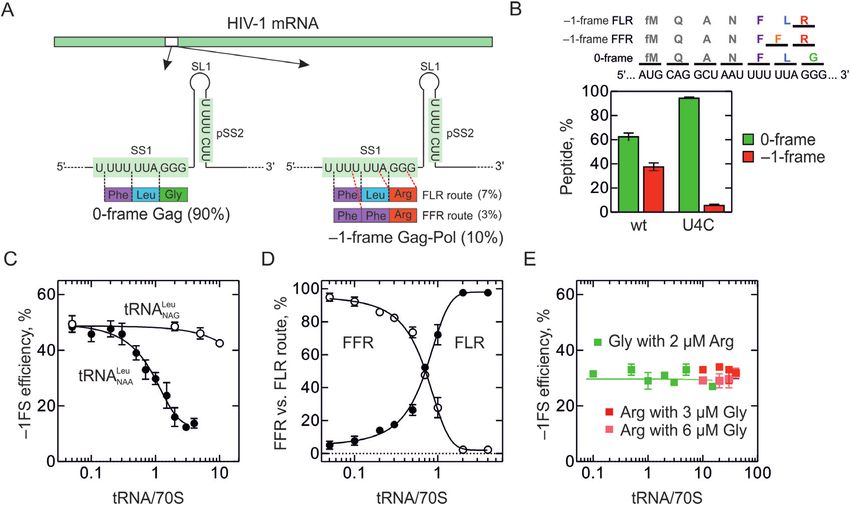
5212 Nucleic Acids Research, 2019, Vol. 47, No. 10
sodium acetate, pH 4.5 and 10 mM MgCl2 using a gradi- Codon-walk assay
ent of 0 to 1.1 M KCl.
To form EF-Tu–GTP–aa-tRNA ternary complexes (TCs),
EF-Tu (25–30 M or 3-fold excess over aa-tRNA) was in-
mRNA constructs cubated with GTP (1 mM), phosphoenolpyruvate (3 mM)
and pyruvate kinase (0.1 mg/ml) in buffer A with DTT
We used a native sequence of the gag-pol HIV-1 frameshift-
(1 mM) for 15 min at 37◦ C. Then aa-tRNAs were added
ing motif (nt 1601–1961 in the HIV-1 complete genome,
and incubated for 1 min at 37◦ C. The concentrations of aa-
NCBI Reference Sequence NC 001802.1) cloned into pEX-
tRNAs were optimized to ensure the maximum binding at
Downloaded from https://academic.oup.com/nar/article-abstract/47/10/5210/5436774 by MPI Biophysical Chemistry user on 17 June 2019
A2 vector. Mutations were introduced by site-directed mu-
their respective codon: 1.6 M each for Gln-tRNAGln , Ala-
tagenesis using Q5 DNA-polymerase (NEB) (42) (Supple-
tRNAAla , Asn-tRNAAsn , Phe-tRNAPhe and Arg-tRNAArg ,
mentary Tables S2–S4). mRNAs were prepared by in vitro
1.2 M for Gly-tRNAGly , and different concentrations of
transcription with T7 RNA-polymerase (43,44) and puri-
Leu-tRNANAA Leu as indicated. IC (0.16 M) was mixed
fied using the RNeasy maxi kit (Qiagen) according to the
with TCs (about 20 M final concentration of EF-Tu),
manufacturer’s recommendations. Control mRNAs used
EF-G (1.6 M), GTP (1 mM) phosphoenolpyruvate (2.4
to determine the rate of Arg-tRNAArg incorporation in 0-
mM) and pyruvate kinase (0.08 mg/ml) in HiFi buffer at
frame (Supplementary Figure S3B) were made by chemical
37◦ C. Incubation times were 0–10 min for time courses or 2
synthesis (IBA, Göttingen) and contained an E. coli Shine-
min for end-point measurements. The stability of peptidyl-
Dalgarno (SD) sequence inserted 9 nt upstream of the start
tRNA binding to the ribosome was tested by nitrocellu-
codon AUG. For translation in bacterial translation sys-
lose filter binding assay (Supplementary Figure S3D). To
tem, a SD sequence was inserted into the HIV mRNA 6
prepare samples for the HPLC analysis, the reactions were
nt upstream of the start codon AUG. In the short mRNAs
quenched with KOH (0.5 M) and hydrolyzed for 30 min
used in the codon walk experiments, AUG was introduced
at 37◦ C; then the samples were neutralized by the addi-
8 nt upstream of the SS1 and the length of the sequence
tion of acetic acid. Translation products were separated
following the SS1 was 56 nt encompassing SL1 and pSS2.
by reversed-phase HPLC on an RP-8 column (LiChro-
In the mRNAs used to study +1 and –2 frameshifting, the
Spher100, Merck) applying an adapted gradient of acetoni-
GGG codon (Gly) following the slippery site was mutated
trile (0–65%) with 0.1% trifluoroacetic acid. Eluted frac-
to UGG (Trp) to distinguish between the –1-, –2- and +1-
tions were mixed with Ultima Gold XR scintillation liq-
frameshifting products (10). The nearest natural AUG in
uid (Perkin Elmer) and analyzed by scintillation counting.
gag mRNA was used as a start codon in the long mRNAs
The peptide products up to MQAN were not separated
to study gag-pol translation products by PAGE. The stop
from each other, while all other peptides could be identi-
codon UAG was introduced in the 0-frame 156 nt down-
fied by either position shift on a chromatogram or by the
stream of the AUG and 120 nt after the SS1 to allow the
radioactive label of the respective amino acid. The amount
separation between the 0-frame (52 aa) and –1-frame (120
of each product was determined as a ratio between 3 H-
aa) products. 0-, –1- and –2-frame control mRNAs contain
counts in the respective peak and total 3 H-counts in the elu-
respective sequence cloned in-frame with SS1 and pSS2 be-
ate. For samples with [3 H]Gly-tRNAGly , [14 C]Arg-tRNAArg
ing mutated to prevent slippages; these mRNAs contained
or [14 C]Leu tRNANAA Leu , the respective peaks were calcu-
324 nt after the SS1 harboring SL1, pSS2 and pSL2. HIV
lated in pmol. Where necessary, the amount of MQAN-
mRNAs for eukaryotic translation contained the native 5’
FLR peptide was calculated by subtracting MQANFLG
UTR from rabbit -globin mRNA and a Kozak sequence
from MQANFL in pmol. Likewise, the MQANFFR pep-
with an embedded AUGG site to allow for efficient initia-
tide was calculated by subtracting MQANFLR from the
tion. The start codon AUG was placed 8 nt upstream of the
MQANFLR/FFR mixture product in pmol. Time courses
slippery site, and the next codon CAG (Gln) was mutated
were evaluated by numerical integration in KinTek Ex-
to GAG (Val) to improve initiation and facilitate product
plorer software (45). Frameshifting efficiency was calcu-
separation by HPLC. Eukaryotic mRNAs contained 167
lated as a ratio between the –1-frame peptide (MQANFFR
nt downstream of SS1 covering SL1, pSS2 and pSL2. All
and MQANFLR) and the sum of –1- and all 0-frame pep-
mRNA sequences are listed in Supplementary Tables S2–
tides (MQANF, MQANFL, MQANFLG) multiplied by
S4 (AUG is in bold, slippery sites are underlined, UAG stop
100%.
codon in 0-frame is in italic and mutated nucleotides are in
small letters in bold).
End-point translation assay of –2 / +1 mRNA
Preparation of initiation complexes
Translation of –2 / +1 mRNA was carried out as de-
70S initiation complexes (IC) were prepared by incubat- scribed for the codon-walk assay, but with 0.4 M of
ing 70S ribosomes (1 M) with mRNA (3–10 M), ini- each Gln-tRNAGln , Ala-tRNAAla , Asn-tRNAAsn , 0.8 M
tiation factors IF1, IF2 and IF3 (1.5 M each), initiator of Phe-tRNAPhe , 0.08 M of Leu-tRNANAA Leu and 0.4 M
f[3 H]Met-tRNAfMet or BODIPY-Met-tRNAfMet (2 M), each of Trp-tRNATrp , Met-tRNAe Met and Tyr-tRNATyr . IC
DTT (1 mM) and GTP (1 mM) in HiFi buffer for 30 min at (0.08 M) was mixed with TCs (about 10 M final concen-
37◦ C. ICs used in the codon-walk experiments were purified tration of EF-Tu), EF-G (1.6 M), GTP (1 mM) phospho-
by ultracentrifugation through a 1.1 M sucrose cushion in enolpyruvate (2.4 mM) and pyruvate kinase (0.08 mg/ml) in
buffer A (50 mM Tris-HCl pH 7.5, 70 mM NH4 Cl, 30 mM HiFi buffer at 37◦ C. The efficiency of frameshifting peptide
KCl, 7 mM MgCl2 ) and dissolved in HiFi buffer. synthesis was calculated by dividing the amount of the re-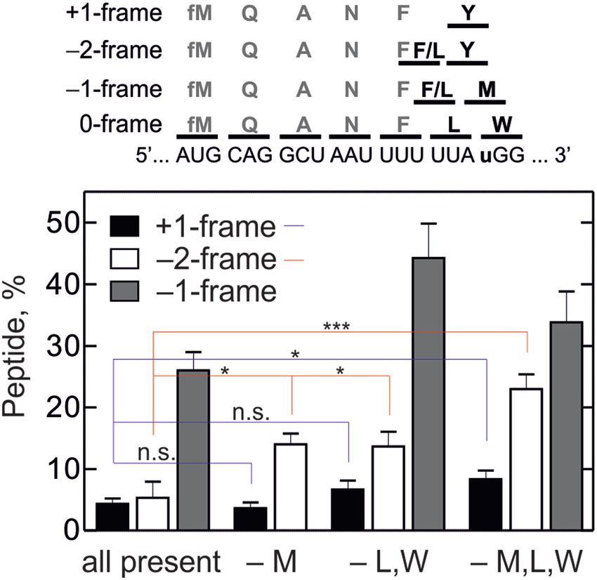
Nucleic Acids Research, 2019, Vol. 47, No. 10 5213
spective peptide in pmol by the sum of all peptides in trans- An elongation reaction mix was prepared by incubating
lation excluding MQAN multiplied by 100%. eEF1A (10.5 M), eEF2 (5 M), elongator tRNA mix (100
M total human tRNA aminoacylated with Val, Ala, Asn,
Arg-tRNAArg incorporation assay Phe, Leu, Arg, and Gly), phosphoenol pyruvate (3 mM), 1
mM MgCl2 and pyruvate kinase (0.1 g/l) in MT buffer
To form post-translocation complexes, purified ICs (0.16 for 8 min at 37◦ C. In experiments shown in Supplemen-
M final) were mixed with Phe-tRNAPhe (1.6 M) or Leu- tary Figure 4F [14 C]Leu and [3 H]Arg were used; in Figure
tRNANAA Leu (0.16 M) in the presence of EF-G (0.008 5F only [3 H]Arg was radioactive labeled. To start transla-
M, 1/20 of the IC concentration) in HiFi buffer and in-
Downloaded from https://academic.oup.com/nar/article-abstract/47/10/5210/5436774 by MPI Biophysical Chemistry user on 17 June 2019
tion, an equal volume of the elongation reaction mix was
cubated for 1 min at 37◦ C. Post-translocation complexes added to the initiation reaction mix and incubated for 4
were then mixed with Arg-tRNAArg (1.6 M) and EF-G min at 37◦ C. Sample preparation for HPLC, HPLC anal-
(1.6 M) and reacted for 1 to 100 s at 37◦ C. The position of ysis and quantification of radioactive peptides were done
MFR and MLR peptides was identified based on [14 C]Arg as described above for the bacterial translation system.
counts and their amounts were calculated in pmol. The rate –1-frame peptides were identified based on [3 H]Arg. In the
of Arg incorporation was estimated by exponential fitting presence of Leu, total translation efficiency was quantified
in GraphPad Prism software. using [14 C]Leu and the frameshifting efficiency was calcu-
lated as a ratio between Arg-containing peptides and to-
Translation assay tal translation products multiplied by 100%. In the ab-
IC prepared with BODIPY-Met-tRNAfMet (0.08 M) was sence of Leu, total translation efficiency was calculated us-
incubated with EF-Tu (80 M), total aa-tRNA from HeLa ing –1-frame control, which shows the maximum level of
cells (3–10 M), EF-G (1.6 M) and RF1 (0.8 M), GTP (1 frameshifting peptide produced under the given conditions.
mM), phosphoenolpyruvate (2.4 mM) and pyruvate kinase Here the frameshifting efficiency was calculated as the ratio
(0.08 mg/ml) in HiFi buffer at 37◦ C as indicated in the time between Arg-containing wt peptides and –1-frame control
course of translation or for 30 min for single-point measure- peptides multiplied by 100%.
ments. In case of ␥ B-crystallin, translation was carried out
using IC (0.02 M), EF-Tu (45 M), total aa-tRNA from
HeLa (10 M), EF-G (1 M), GTP (0.8 mM), phospho- Quantification of tRNA levels using qRT-PCR
enolpyruvate (1.4 mM) and pyruvate kinase (0.05 mg/ml)
for 30 min in HiFi buffer at 37◦ C. To prepare the samples Sup-T1 cells were derived from non-Hodgkin’s T-cell lym-
for PAGE, the reactions were stopped with NaOH (0.4 M) phoma isolated from a pleural effusion of an 8-year-old
and hydrolyzed as described for the HPLC sample prepara- male and subcloned on soft agar. Jurkat cells were derived
tion. HEPES (0.2 M, pH 5) was added to neutralize the re- from human T-cell lymphoblast. PM1 cells are a deriva-
actions. The samples were separated by Tris-Tricine gel elec- tive of HUT78, a human cutaneous T-cell lymphoma cell
trophoresis (46). Fluorescent peptides were visualized us- line derived from peripheral blood of a patient with Sezary
ing an Typhoon™ FLA-9000 scanner (GE Healthcare Life syndrome. 174xCEM cells are a fusion product of human
Sciences) and the band intensities were evaluated using the B-cell line 721.174 and a human T-cell line CEM. 293T
MultiGauge software. Frameshifting efficiency was calcu- is an epithelial cell line derived from human embryonic
lated from the band intensities of the –1-frame product to kidney cells and expressing large T antigen. Information
the sum of –1- and 0-frames products as well as of trans- about cell lines is taken from https://aidsreagent.org/ and
lation intermediates appearing at 20 s of translation. The http://www.lgcstandards-atcc.org/.
correct length of the peptides was confirmed using control 293T (DSMZ-German Collection of Microorganisms
0-frame, –1-frame and –2-frame mRNAs. and Cell Cultures, ACC 635) cells were maintained in Dul-
becco’s modified Eagle medium (DMEM, Pan Biotech)
supplemented with 10% fetal calf serum (Biochrom, FCS)
Translation of HIV mRNAs in a fully reconstituted homolo-
and 1% of 100× concentrated penicillin/streptomycin
gous mammalian in vitro translation system
(pen/strep) mix (Pan Biotech). For subculturing, cells were
40S and 60S ribosomal subunits from HeLa, translation detached by resuspension in phosphate-buffered saline
initiation factors eIF1, eIF1A, eIF2, eIF3, eIF4A, eIF4B, (PBS [293T]) or incubation with trypsin/EDTA solution
eIF5, eIF5B, translation elongation factors eEF1A and (Pan Biotech). The human T-cell lines 174xCEM (NIH
eEF2 as well as Met-tRNAMet were prepared according to AIDS Reagent Program, NIH272, (49)), PM1 (50), Sup-T1
published protocols (47,48). (NIH AIDS Reagent Program, NIH100, (51)) and Jurkat
48S IC (0.3 M) was formed by incubating 40S subunits (NIH AIDS Reagent Program, NIH177, (52)) were main-
with a 2-fold molar excess of eIF2, eIF3 and eIF4B, a 3- tained in Roswell Park Memorial Institute 1640 medium
fold excess of eIF1, eIF1A and eIF4A, a 3-fold excess of (RPMI, Pan Biotech) supplemented with 10% FCS and
mRNA and a 5-fold excess of [3 H]Met-tRNAMet in mam- 1% of 100× concentrated pen/strep mix. For subculturing,
malian translation (MT) buffer (20 mM Tris–HCl, pH 7.5, culture medium containing the suspension cells was cen-
100 mM KCl, 2.5 mM MgCl2 , 0.25 mM spermidine, 1 mM trifuged (600 × g, 10 min, room temperature). Then, the su-
ATP and 0.5 mM GTP) for 15 min at 37◦ C. To form 80S IC, pernatant was discarded and pelleted cells were resuspended
an equal volume of MT buffer containing eIF5 (0.4 M), in 10 ml RPMI medium. Further, 1 ml of this suspension
eIF5B (0.5 M) and 60S subunits (0.6 M) was added. The was added to a new culture flask and filled up with 19 ml of
initiation reaction mix was incubated for 8 min at 37◦ C. RPMI medium.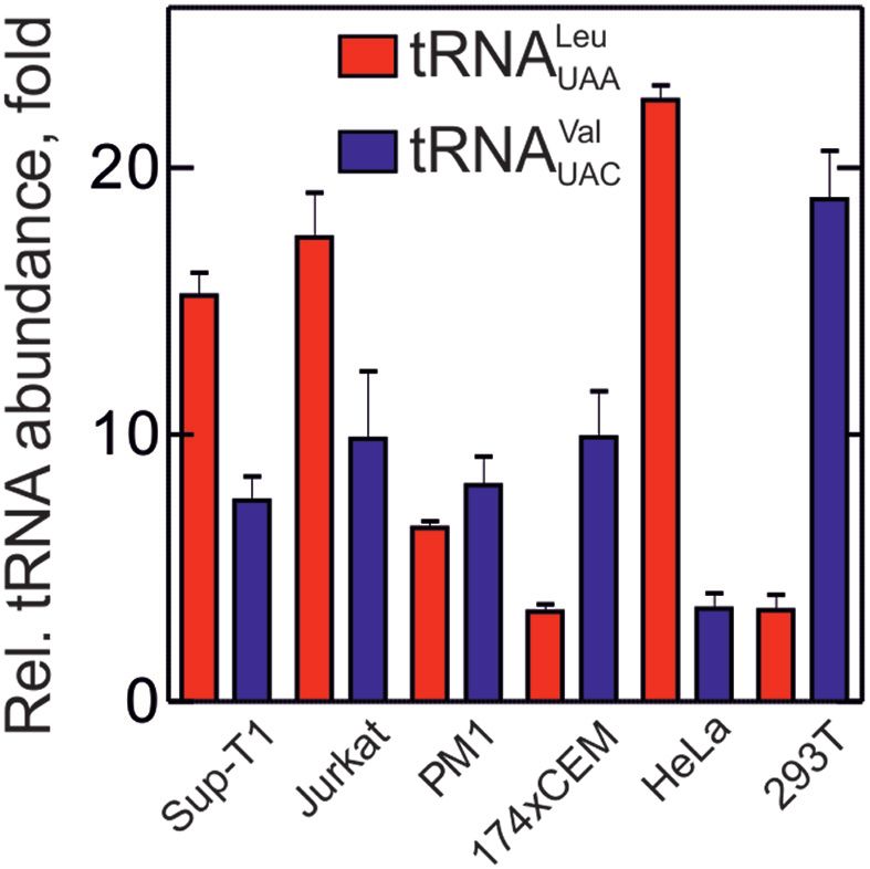
5214 Nucleic Acids Research, 2019, Vol. 47, No. 10
Cellular tRNA levels for tRNAUAA Leu , tRNACAG Leu , the mRNA and fMet-tRNAfMet and started translation
tRNAUAC Val and tRNACAC Val were quantified using a strat- by the addition of ternary complexes of elongation fac-
egy published by (53). Total cellular RNA was extracted us- tor (EF)-Tu–GTP with the desired combination of puri-
ing the RNeasy Mini Kit (Qiagen) according to the manu- fied aa-tRNAs and EF-G–GTP. Consecutive amino acid
facturer’s protocol. After elution, the RNA content was de- incorporation results in a 0-frame peptide fMet-Gln-Ala-
termined spectrophotometrically. To eliminate potentially Asn-Phe-Leu-Gly (MQANFLG) and the –1-frame peptides
co-isolated DNA from the samples, 0.5 g RNA was incu- fMet-Gln-Ala-Asn-Phe-Leu-Arg (MQANFLR) or fMet-
bated with DNase I (New England Biolabs) in a final vol- Gln-Ala-Asn-Phe-Phe-Arg (MQANFFR). Peptides were
Downloaded from https://academic.oup.com/nar/article-abstract/47/10/5210/5436774 by MPI Biophysical Chemistry user on 17 June 2019
ume of 10 l for 30 min at 37◦ C and finally heated to 65◦ C analyzed by reversed-phase HPLC (RP-HPLC) (Supple-
for 5 min to inactivate the enzyme. The primer for the re- mentary Figure S1). The overall frameshifting efficiency
verse transcription entailed a tRNA-specific sequence and was determined as a yield of –1-frame Arg incorporation
a stem-loop sequence (53). The tRNA-specific primer was relative to the sum of –1- and 0-frame peptides. With the
the same for tRNAUAA Leu and tRNACAG Leu (complemen- mRNA containing a native slippery sequence, a large frac-
tary to positions 47–55) and different for tRNACAC Val and tion of peptides contains Arg (Figure 1B), indicating effi-
tRNAUAC Val (complementary to positions 62–70) (Supple- cient –1FS. As expected, the U4 C mutation, which changes
mentary Table S5). cDNA synthesis was performed as de- the SS1 sequence to U UUC UUA (5), abolishes FS (Figure
scribed in the manufacturer’s instructions (for gene-specific 1B and Supplementary Figure S3A).
primers) using the SuperScript III First-Strand Synthesis In principle, –1FS can occur at the decoding step of the
System (ThermoFisher Scientific) and 5 l of DNase I- translation elongation cycle, when only peptidyl-tRNA is
digested RNA (0.25 g). Input RNA was removed by incu- bound in the P site of the ribosome while the A site is va-
bation with RNase H. To determine the levels of the tRNAs, cant (15,55), or during translocation, when two tRNAs to-
tRNAUAA Leu , tRNACAG Leu , tRNAUAC Val and tRNACAC Val gether with the mRNA move through the ribosome (56–59).
as well as 18S rRNA (housekeeping gene control), quan- If slippage occurred during decoding, the –1FS efficiency
titative PCR (qPCR) was performed employing the Quan- must depend on the competition between the 0-frame and
tiTect SYBR Green Kit (Qiagen) according to the manu- –1-frame aa-tRNAs, Leu-tRNA and Phe-tRNA, for bind-
facturer’s protocol on a Rotorgene Q device (Qiagen). The ing at the UUUA sequence in SS1. In fact, the addition of
primers used for qPCR were taken from (53); the forward Leu-tRNANAA Leu (E. coli tRNA that reads the UUA and
primer for the qPCR is highly specific to each tRNA and UUG codons; the anticodon is indicated as a subscript; N is
the reverse primer is universal (Supplementary Table S5). a post-transcriptionally modified nucleotide) dramatically
For each run, technical triplicates were analyzed for each lowers the –1FS efficiency from about 50% in the absence
sample and reactions containing water instead of template to 12% in the presence of Leu-tRNANAA Leu (Figure 1C). In
cDNA were used as negative control. Cycle conditions were contrast, a mixture of near-cognate Leu-tRNA isoacceptors
chosen as follows: one cycle at 95◦ C for 15 min, followed by that collectively read the CUN family of Leu codons has
45 cycles consisting of 15 s at 94◦ C, 30 s at 60◦ C and 30 s no effect. The high –1FS efficiency observed in the absence
at 72◦ C. Finally, a linear temperature increase from 60 to of Leu-tRNANAA Leu indicates that ribosomes can slip into
90◦ C with a ramp of 1◦ C per step and each step lasting 5 the –1-frame prior to, and independent of, Leu incorpora-
s was performed to obtain a melting curve for each reac- tion. By estimating the ratio of Leu, Phe and Arg incorpo-
tion. In order to compare tRNAUAA Leu /tRNACAG Leu and ration into the –1-frame product, we can determine how the
RNAUAC Val / tRNACAC Val tRNA ratio between cell lines, –1FS pathway changes with the Leu-tRNANAA Leu concen-
cycle threshold (ct ) values for each tRNA were normalized tration (Figure 1D). In the absence of Leu-tRNANAA Leu ,
against the respective ct values for 18S rRNA. only the FFR product is formed. Upon addition of Leu-
tRNANAA Leu , the FLR product becomes prevalent. Thus,
frameshifting at the gag-pol slippery site can switch between
RESULTS two regimes and their prevalence depends on the concentra-
Two regimes for –1FS on the gag-pol slippery site tion of the critical tRNA.
After Leu incorporation, the –1FS can follow different
We first aimed to solve the mechanism of –1FS on the routes: it can take place either during tRNALeu transloca-
HIV-1 mRNA. For the mechanistic work, we used the tion or upon decoding of the following Gly codon. Again,
fully reconstituted translation system from E. coli, be- if frameshifting took place during decoding, the 0-frame
cause the overall efficiency and the ratio of the FLR Gly-tRNAGly and –1-frame Arg-tRNAArg should compete
and FFR products is similar in mammalian cells and in for binding to the ribosome. This is, however, not observed,
E. coli (5,15,23,24), the functional centers of the ribo- as the –1FS efficiency is independent of Gly-tRNAGly and
somes where frameshifting takes place are highly con- Arg-tRNAArg concentrations (Figure 1E). This finding sug-
served between eukaryotes and prokaryotes (54), and it gests that slippage and commitment to the new reading
is not feasible to prepare materials for the reconstituted frame occur after Leu incorporation, but prior to decod-
eukaryotic translation system in amounts needed for this ing of the next codon by Gly- or Arg-tRNA. This is similar
type of analysis. We first tested the potential routes for to the well-studied cases of –1FS on IBV 1a/1b and dnaX
frameshifting. The model HIV-1 gag-pol mRNA encom- mRNAs, where slippage occurs at a late stage of transloca-
passes the natural frameshifting site with the translation tion of the slippery-site tRNAs, and suggests a similar two-
start codon AUG three codons upstream of the slippery tRNA frameshifting mechanism (55–60).
site (Figure 1B). We formed a 70S initiation complex withNucleic Acids Research, 2019, Vol. 47, No. 10 5215
and Met-tRNA, the products of all three alternative frames
are found. These data suggest that –2 and +1FS can occur
when one or more of the aa-tRNAs are lacking; however,
when all aa-tRNAs are available the –1FS pathway is preva-
lent.
Kinetics of FFR and FLR –1FS pathways
Downloaded from https://academic.oup.com/nar/article-abstract/47/10/5210/5436774 by MPI Biophysical Chemistry user on 17 June 2019
To understand the kinetic switch between the two differ-
ent –1FS regimes, we monitored translation and –1FS ef-
ficiency using the codon-walk approach (56) in the absence
and presence of Leu-tRNANAA Leu . Rate constants were cal-
culated by global fitting of the time courses using numeri-
cal integration according to the models shown in Figure 3.
As additional information, we estimated the rate constants
of Arg and Gly incorporation in independent experiments
Figure 2. tRNA limitation results in –1, +1 and –2FS. Top panel shows the using model mRNAs without frameshifting elements (Sup-
model mRNA and peptides synthesized in all frames. tRNAs for QANF plementary Figure S3B and C).
were added to all translation reactions; in addition, Met (M), Leu (L), For the –1FS model in the absence of Leu-tRNANAA Leu ,
Trp (W) and Tyr (Y) were added in the ‘all present’ sample; –M, –L,W
and –M,L,W indicate the aa-tRNAs that were omitted from the respec-
we introduced the steps that result in the formation
tive translation reaction. Positions of peaks were determined using [14 C]- of MQANF and the –1FS products MQANFF and
labeled Tyr, Met, Leu or Trp. Two-tailed two-sample equal variance t-test MQANFFR. In addition, we introduced two reaction
was performed between marked samples; blue lines for +1 peptides and red branches that account for the incomplete conversion of the
lines for –2 peptides. n.s., not significant, * indicates P ≤ 0.05, *** is P ≤ 70S IC into products as follows. Because a fraction of initi-
0.001.
ation complexes does not enter translation, we introduced a
step that accounts for this unproductive population (M →
The UUA codon is particularly rare in human cells Mn , non-reactive). We also noticed that MQANF-tRNAPhe
and the respective Leu-tRNANAA Leu is underrepresented in the absence of the A-site ligand tends to slowly dissoci-
is some cell types (see below and (53)). Because forma- ate from the ribosome (Supplementary Figure S3D); to ac-
tion of the –1-frame FFR product depends on the slip- count for this loss of peptidyl-tRNA we introduced the re-
page at the ‘hungry’ UUA codon, we further tested whether spective drop-off reaction. Global fitting of the time courses
this allows also –2 and +1FS. We note that normally such using numerical integration yielded a unique solution for
slippage events would lead to premature termination due all rate constants (Figure 3A and B; Supplementary Table
to stop codons appearing in the –2 or +1 frames down- S1). The step leading to the incorporation of the second
stream of the frameshifting site and that such peptides are Phe is slow, ∼0.01 s−1 , compared to all translation steps,
difficult to detect in vivo, but alternative slippage events which are at least 10 times faster. MQANFF peptides do
could change the ratio between the Gag and Gag-Pol not accumulate and are converted to the –1-frame peptide,
polyproteins. To distinguish between the products of the 0-, MQANFFR. Thus, the incorporation of the second Phe
–1-, –2- and +1-FS, we changed the GGG (Gly) codon residue is the rate-limiting step of frameshifting that com-
following the slippery site into UGG (Trp) (Figure 2 and mits the ribosome to –1-frame translation.
Supplementary Figure S2), a mutation that does not af- In the presence of Leu-tRNANAA Leu , the ribosome syn-
fect the –1FS efficiency in vivo in human cells (10). In thesizes the 0-frame MQANF peptide and then either con-
addition to the aa-tRNAs needed for translation of the tinues translation with Leu incorporation in the 0-frame
MQANFL sequence, we added purified Trp-tRNATrp (W), or shifts into the –1-frame before Leu-tRNANAA Leu can
elongator Met-tRNAMet (M) and Tyr-tRNATyr (Y). The bind. If Leu is incorporated, the 0-frame MQANFL prod-
expected 0-frame peptide is now MQANFLW and the –1- uct can partition between the 0-frame MQANFLG and the
frame peptides are MQANFFM and MQANFLM. Shift- –1-frame MQANFLR. Global fitting of the time courses
ing into the +1-frame should yield MQANFY and into the gives well-defined rate constants for most of the steps (Fig-
–2-frame MQANFFY and MQANFLY. When all required ure 3C–E). The rate-limiting step for the –1-frame FFR
aa-tRNAs are present, the –1-frame peptides account for pathway has a rate constant of ∼0.03 s−1 , similar to that
about 25% of product, consistent with the –1FS efficiency for the isolated FFR pathway. The efficiency of the FFR
on the native gag-pol sequence in the presence of equimo- pathway depends on the ratio of the rates of –1-slippage
lar amounts of Leu-tRNANAA Leu and ribosomes (Figure and Leu-tRNANAA Leu binding. While the rate of slippage
2), whereas the amounts of the +1 and –2 peptides are is constant, the rate of Leu-tRNANAA Leu binding increases
small. When Met-tRNAMet is omitted, –2FS increases more with concentration. This explains why the addition of excess
than 2-fold, whereas the +1FS is not changed. –2FS is en- Leu-tRNANAA Leu inhibits the FFR route. At high concen-
hanced because –1FS exposes a ‘hungry’ Met codon in the trations of Leu-tRNANAA Leu , the probability to bind Leu-
A site, which favors the slippage. In the absence of Leu- and tRNANAA Leu to the A site is higher than to slip into the –
Trp-tRNA, –1FS efficiency increases to 45%, as expected; 1 -frame. At this condition, the FFR pathway is suppressed
–2FS is unchanged; and again a small amount of the +1- and only the FLR pathway remains operational. After Leu
frame product is formed. Without addition of Leu-, Trp- incorporation, the –1FS efficiency of the FLR route is de-5216 Nucleic Acids Research, 2019, Vol. 47, No. 10
Downloaded from https://academic.oup.com/nar/article-abstract/47/10/5210/5436774 by MPI Biophysical Chemistry user on 17 June 2019
Figure 3. Kinetic mechanism of –1FS. (A) Time courses of translation in the absence of tRNANAA Leu reading the UUA codon. Peptides are MQAN
(gray circles), MQANF (purple triangles), MQANFF (orange circles) and MQANFFR (red squares). Global fits are shown as continuous lines. The top
panel shows amino acids in 0-frame and FFR –1-frame and respective codons on the mRNA. (B) Kinetic model of the FFR pathway in the absence
of tRNANAA Leu . Rates of all steps are calculated by global fitting. (C and D) Time courses of translation in the presence of limiting concentrations of
tRNANAA Leu (C, 0.3-fold per ribosome) and near-saturating concentrations of tRNANAA Leu (D, 1-fold per ribosome). Peptides are MQAN (gray circles),
MQANF (purple triangles), MQANFL (blue downward triangles), MQANFLG (green diamonds) and MQANFFR/MQANFLR (red squares). Global
fits are shown as continuous lines. The top panel shows amino acids in 0-frame and –1-frame and respective codons on the mRNA. (E) Kinetic model of
the FFR/FFL pathways in the presence of tRNANAA Leu . Rates of all steps are calculated by global fitting. 0- and –1-frames are indicated by dotted arrows.
*Incorporation of Leu-tRNANAA Leu is a bimolecular reaction and its rate depends on the concentration of tRNANAA Leu . The two rates correspond to
0.3- and 1.0-fold excess of tRNANAA Leu over ribosomes, respectively.Nucleic Acids Research, 2019, Vol. 47, No. 10 5217
the human aa-tRNA to the codon usage of mammalian
mRNA was validated by translation of an mRNA coding
for bovine ␥ B-crystallin. The codon usage of ␥ B-crystallin
matches the tRNA abundance of its eukaryotic host, but
not of E. coli (62). With native human aa-tRNA, ␥ B-
crystallin mRNA is translated efficiently (Supplementary
Figure S4D). Introducing synonymous mutations in the
mRNA to match the codon usage in E. coli (62) reduces the
Downloaded from https://academic.oup.com/nar/article-abstract/47/10/5210/5436774 by MPI Biophysical Chemistry user on 17 June 2019
yield of full-length product (Supplementary Figure S4D).
The average translation rate of the gag-pol or the native ␥ B-
crystallin mRNA in our system is 0.5–0.7 aa/s (Supplemen-
Figure 4. Relative abundance of tRNA isoacceptors in different cell types.
tary Figure S4C).
Plotted is the ratio of tRNACAG Leu to tRNAUAA Leu (red bars) and We then translated a gag-pol mRNA fragment encom-
tRNACAC Val to tRNAUAC Val (blue bars), which read frequent and rare passing the region from the nearest native (elongator) AUG
codons, respectively, in human cell lines. Error bars represent S.E.M of codon of the gag mRNA upstream of the SS1, the SS1
three biological replicates with three technical replicates each. Experimen- with its downstream SL1 and the second putative FS site,
tal design of qRT-PCR was according to (53). Human cell lines are in-
dicated below the graph. Sup-T1, Jurkat and PM1 are derived from hu- pSS2, with a 32-nt downstream sequence, which is predicted
man T-lymphocytes; 174xCEM is B-T-lymphocyte fusion; HeLa are de- to form a SL (pSL2) (Figure 5A). To distinguish 0- and
rived from cervical epithelial carcinoma; 293T is a kidney epithelial cell –1-frame translation products, we introduced a stop codon
line. UAG in the 0-frame to obtain a peptide 52 amino acids
(aa) in length; –1FS results in a 120-aa peptide product.
fined at the translocation step, because the partitioning be- To identify potential –2FS products, we mutated all na-
tween 0- and –1-frames takes place before decoding by Gly- tive stop codons in the –2-frame downstream of the pSS2
and Arg-tRNAs (Figure 1E). The ratio of the rate constants (mRNA denoted as no-stop). Translation of the no-stop
of Gly and Arg incorporation (0.53 and 0.04 s−1 , respec- mRNA leaves the product lengths in the 0- and –1-frames
tively; Supplementary Table S1) gives the –1FS efficiency unchanged, but additionally yields a 120-aa –2-frame
after Leu incorporation. product. Despite their identical length, the –1-frame and
–2-frame products have different electrophoretic mobility
due to their different amino acid composition (Figure 5B
–1FS with native human aa-tRNA
and Supplementary Figure S4E).
Our finding that the Leu-tRNALeu isoacceptor that reads The –1FS efficiency of the native gag-pol frameshifting
the UUA codon affects the mechanism and efficiency sequence is 6–7% (Figure 5C–E), consistent with earlier in-
of –1FS has prompted us to validate the key results vivo reports (10–13). Formation of 0-frame and –1-frame
in the eukaryotic system. First, we analyzed the rela- products starts after a 30-s delay that may be caused by
tive abundance of human Leu-tRNALeu that reads the an early translational pausing event (appearing as a promi-
UUA codon (tRNAUAA Leu ) in total tRNA from differ- nent peptide band between 7 and 30 s of translation (Fig-
ent human cell types using qRT-PCR (Figure 4). HIV- ure 5B and C; Supplementary Figure S4E). In contrast,
1 mainly infects CD4+ T-lymphocytes and macrophages the –2-frame product appears after a much longer delay of
(61). We determined the ratio of Leu-tRNAUAA Leu to Leu- 120 s. At this time, the synthesis of the 0-frame product
tRNACAG Leu reading the most abundant Leu codon CUG. is already completed on most ribosomes, suggesting that
Leu-tRNAUAA Leu is 7–17-fold less abundant than Leu- the –2FS may arise on a fraction of ribosomes that un-
tRNACAG Leu in cell lines derived from T lymphocytes, and dergo long translation pausing. Addition of exogenous Leu-
about 20-fold in HeLa cells, whereas in other types of tRNANAA Leu decreases the –1FS efficiency to 4% (Figure
human cells the ratio is about 1:3 (Figure 4). The Leu- 5D and Supplementary Figure S4F). A similarly reduced
tRNAUAA Leu concentration varies by as much as 10-fold, FS efficiency is observed when the UUA codon is mutated
whereas for Leu-tRNACAG Leu the differences are smaller, to UUC, which does not interrupt the slippery run of six
except for HeLa cells, where the Leu-tRNACAG Leu concen- Us, but changes the identity of the tRNA reading the sec-
tration is increased (Supplementary Figure S4A). As a con- ond slippery codon to the abundant tRNAPhe (Figure 5E).
trol, we quantified the relative abundance of Val-tRNAVal Thus, reassigning the second codon of the slippery site to
isoacceptors reading a rare GUA codon and an abundant an abundant tRNA has the same effect as adding excess of
GUG codon. The tRNAUAC Val isoacceptor reading the rare tRNANAA Leu to the native sequence. Shortening the slip-
codon is about 7–8 times less abundant than common pery site to four U residues decreased FS to 2%. Disrupt-
tRNACAC Val , except for the 293T epithelial cells, where the ing SS1 alone or together with pSS2 diminishes the FS effi-
amount of tRNAUAC Val is even lower (Figure 4). Because ciency to about 1%, which is the background level of these
the relative abundance of tRNAUAA Leu in HeLa cells is sim- experiments (Figure 5E and Supplementary Figure S5B).
ilar to that in cells used as a model for the HIV infection, we Finally, we tested whether addition of Leu-tRNAUAA Leu
used total human tRNA purified from HeLa cells for the in alters the frameshifting efficiency in a homologous reconsti-
vitro translation experiments described below. tuted mammalian translation system. Ribosomal subunits
We first tested how the HeLa aa-tRNA performs in trans- (40S and 60S), initiation factors (eIF1A, eIF1, eIF2, eIF3,
lation when combined with EF-Tu and ribosomes from eIF4A, eIF4B, eIF5, and eIF5B) and Met-tRNAi were used
E. coli (Supplementary Figure S4B,C). The conformity of to form the 80S initiation complex on an mRNA with an un-5218 Nucleic Acids Research, 2019, Vol. 47, No. 10
Downloaded from https://academic.oup.com/nar/article-abstract/47/10/5210/5436774 by MPI Biophysical Chemistry user on 17 June 2019
Figure 5. Translation and frameshifting with native human aa-tRNAs. (A) The mRNA used for translation experiments. SS1 and pSS2 are in highlighted
light green, SL1 and the potential stem-loop element downstream of the pSS2 (pSL2) are shown. Sizes of 0-, –1- and –2-frame peptides formed upon
translation of the mRNA are indicated. (B) Time course of 0-frame and –1-frame translation on wt mRNA. (C) Time courses of –1FS on wt mRNA
(closed circles) as well as –1FS (open circles) and –2FS (closed triangles) on mRNA where all stop codons in –2-frame were mutated to sense codons
(no-stop). (D) Concentration dependence of –1FS efficiency on exogenous tRNANAA Leu from E. coli measured on wt mRNA. (E) Effect of mutations in
SS1 on –1FS. The background of the measurements is ±1%. The S.E.M was calculated from 3 to 5 independent experiments. (F) –1FS efficiency measured
using a fully reconstituted homologous mammalian in vitro translation system with a gag-pol mRNA construct optimized for translation in eukaryotes.
Translation was carried out in the presence (+ tRNALeu ) and in the absence (– tRNALeu ) of total Leu-tRNALeu containing all isoacceptors in native ratios.
The –1-frame peptide was identified based on [3 H]Arg incorporation.
structured 5’UTR coding for the SS1 and the SL1 fragment the presence of native amounts of tRNAUAA Leu (Figure 5F
of HIV-1 (Figure 1A). To synthesize the 0-frame MVAN- and Supplementary Figure S4G). When leucine was omit-
FLG and the –1-frame MVANFLR peptides, we used the ted from the aminoacylation mixture, the –1FS efficiency
tRNA from HeLa cells aminoacylated with a mixture of the increases to 40% (Figure 5F). Thus, Leu-tRNAUAA Leu mod-
required amino acids with or without Leu; incorporation ulates –1FS efficiency on HIV mRNA also in the homolo-
of the –1-frame Arg was monitored by the HPLC analysis gous mammalian translation system.
(Supplementary Figure S4G). As controls, we included the
mRNA coding for MVANFLR in 0-frame (–1-frame con- The putative second slippery sequence
trol), which provides the estimate for the maximum Arg in-
corporation, and the mRNA without the SS1 but with SL1 To test the effect of mutations in the putative slippery se-
(0-frame control). The –1FS efficiency in this fully recon- quence (pSS2), we introduced mutations that should make
stituted eukaryotic translation system is about 20–25% in pSS2 either more or less slippery (Figure 6 and Supple-
mentary Figure S5). As long as the SS1 sequence is un-Nucleic Acids Research, 2019, Vol. 47, No. 10 5219
translation systems, which underscores the notion that this
frameshifting mechanism is universally conserved.
The ribosome can also slip into the –1-, –2-, or +1-
frames when some aa-tRNAs are lacking, but when all aa-
tRNAs are supplied the –1-product is predominant. Sim-
ilarly, translation of the E. coli dnaX mRNA can lead to
slippages into the –2-, +2- or –4-frames when aa-tRNAs are
in limiting supply (55,58). In the case of dnaX, the switch
Downloaded from https://academic.oup.com/nar/article-abstract/47/10/5210/5436774 by MPI Biophysical Chemistry user on 17 June 2019
to ‘hungry’ frameshifting may be caused by unfavorable
conditions, e.g. amino acid starvation (55,65–67). In con-
trast, HIV-1 can use both pathways constitutively due to
the inherently low concentration of the key tRNAUAA Leu
isoacceptor in the relevant human cells. The fraction of –
2PRF (25), which normally does not result in functional
Figure 6. Interplay between SS1 and pSS2. SS1 sequences are shown above
HIV products, may play a role in conjunction with the tran-
the bars, pSS2 sequences are indicated below the graph. –1FS is determined scription slippage of the reverse transcriptase (68).
with wt mRNA (black bars) or with no-stop mRNA (gray bars); –2FS is
measured with no-stop mRNA (white bars); the absence of pSL2 is indi-
cated by textured pattern. Two-tailed two-sample equal variance t-test was
performed between marked samples. n.s. means not significant, ** indi- Role of the tRNAUUA Leu
cates P ≤ 0.01, *** indicates P ≤ 0.001.
As the ratio of the Gag and Gag-Pol products is crucial
for virus propagation (6–9), HIV-1 must have evolved to
changed, mutations in pSS2 have little effect on overall – achieve the desired –1FS efficiency at the concentrations of
1FS, but change the –2FS efficiency, which, in turn, leads to tRNAUAA Leu prevalent in human cells. The UUA codon is
slight variations in –1FS. The –2FS efficiency is higher when rare in the human genome, as are all other A-ending codons.
both SS1 and pSS2 are slippery, suggesting that –2FS results The respective cognate tRNAUAA Leu is significantly under-
from dual –1-slippage on both sites rather than from –2- represented in the tRNA pool as compared to tRNACAG Leu
slippage on pSS2 alone. Replacing the rare CUU codon in that reads the abundant CUG codon (Figure 4) (53). While
pSS2 with the abundant CUG reduces –2FS indicating that in eukaryotes the tRNA expression is tissue-specific, the
the second slippage is due to ‘hungry’ FS on pSS2. When relative expression of tRNA isoacceptors in some tissues
the U-string in SS1 is disrupted, the frameshifting efficiency shows statistically significant correlation to the codon us-
is higher in the construct where pSS2 is slippery compared age of tissue-specific genes (69). The low relative abundance
to the native sequence. The presumed secondary structure of tRNAUAA Leu in the lymphocyte-derived cell types may
of the mRNA downstream of pSS2 (Supplementary Figure be a result of adaptation to the codon usage in these cells.
S5) has no effect. Disruption of both slippery sites decreases On the other hand, the rare UUA codon accounts for 45%
frameshifting to background levels. Thus, pSS2 supports a of all Leu codons in late-expressing HIV-1 genes including
low-level FS event that can rescue HIV-1 when SS1 is mu- gag and pol (70–72). Expression of the late HIV genes would
tated, but also causes –2FS, which in the native sequence increase the demand for a tRNAUAA Leu that is rare in the
leads to premature termination of translation. respective cells, which may delay the decoding at any UUA
codon, including the key codon in the slippery sequence.
DISCUSSION When the delivery to the UUA codon at the slippery site is
too slow, the ribosome switches to the FFR route leading to
Kinetic mechanism of frameshifting on the gag-pol mRNA
robust –1FS. The codon usage disparity between HIV and
We show that –1FS on HIV-1 mRNA operates in two its human host can be utilized to disrupt the late stage of
regimes, one that is caused by a limitation of the UUA- virus production (73). For example, the interferon-induced
specific Leu-tRNA isoacceptor and leads to the FFR – antiviral protein Schlafen 11 (SLFN11) selectively abro-
1-frame product, and another where ribosomes slip dur- gates the expression of late viral proteins in a codon-usage-
ing tRNAPhe –tRNALeu translocation over the slippery site dependent manner (73). SLFN11 induces specific cleavage
codons, yielding the FLR –1-frame product. The switch be- of tRNAs with a long variable loop, which include all ser-
tween the two regimes is modulated by the availability of ine and leucine tRNAs. If the already rare tRNA, such as
the UUA-specific tRNALeu , which we show to be rare in cell tRNAUAA Leu , is further reduced, tRNA availability might
lines derived from human immune cells. This notion is sup- manifest as the rate-limiting step in the synthesis of proteins
ported by in vivo experiments indicating that limitation of involving those tRNAs (73). A tRNA-dependent modula-
Leu in the culture media leads to increased –1FS in E. coli tion of frameshifting was also reported within the expanded
(15). The exact –1FS efficiency on the gag-pol mRNA dif- CAG stretch in the huntingtin gene (74): the translation of
fers depending on the type of model system, a phenomenon expanded CAG repeats in mutant huntingtin exon 1 leads
that has been noted before and attributed to different trans- to a depletion of charged Gln-tRNACUG Gln that pairs to the
lation rates in vivo and in vitro (18,19,63,64); presumably, the CAG codon, which results in translational frameshifting.
presence of the bulk aa-tRNA also plays a role (compare The frameshifting frequency varies strongly among differ-
Figures 1C and 5C). The UAA-specific Leu-tRNA modu- ent cell lines and is higher in cells with intrinsically lower
lates the –1FS efficiency in E. coli, mammalian or hybrid concentrations of tRNACUG Gln (74), which emphasizes the5220 Nucleic Acids Research, 2019, Vol. 47, No. 10
biological significance of tRNA concentrations in modulat- Author Contributions: N.K. prepared materials and per-
ing frameshifting efficiencies. formed most of experiments; A.G. performed experiments
with the mammalian reconstituted translation system;
The second slippery site M.H. performed qRT-PCR experiments; all authors con-
ceived the research, designed experiments and analyzed the
Anti-HIV therapy with protease inhibitors leads to accu- data; N.K. and M.V.R. wrote the paper with contributions
mulation of mutations in the HIV-1 protease that impair of all authors.
the recognition of its specific cleavage sites. To allow for
Downloaded from https://academic.oup.com/nar/article-abstract/47/10/5210/5436774 by MPI Biophysical Chemistry user on 17 June 2019
polyprotein maturation by the mutated protease, secondary
mutations arise at the pSS2, which harbors the p1/p6 cleav- FUNDING
age site of Gag polyprotein (75–79). The C5 U mutation in Deutsche Forschungsgemeinschaft [SFB860, Leibniz Prize
pSS2 substitutes Leu with Phe, which enhances van der to M.V.R.]; Boehringer Ingelheim Fonds, PhD Fellowship
Waals interactions between the substrate and the mutant [to N.K.]. Funding for open charge: MPG.
protease, thereby increasing the protease activity by about Conflict of interest statement. None declared.
10-fold (80). The same mutation produces a slippery se-
quence that can support –1FS, but the role of pSS2 depends
on the sequence of SS1. With native SS1, the joint activity REFERENCES
of SS1 and pSS2 is not different from SS1 alone, but when 1. Plant,E.P. (2012) In: Garcia,PM (ed). Viral Genomes - Molecular
SS1 is mutated to a non-shifty sequence, the C5 U mutation Structure, Diversity, Gene Expression Mechanisms and Host-Virus
Interactions. Intech.
in the pSS2 is sufficient to support a level of –1FS that may 2. Atkins,J.F., Loughran,G., Bhatt,P.R., Firth,A.E. and Baranov,P.V.
be sufficient for virus propagation. The finding that pSS2 (2016) Ribosomal frameshifting and transcriptional slippage: From
can alleviate the detrimental effects of SS1 mutations is con- genetic steganography and cryptography to adventitious use. Nucleic
sistent with previous in vitro and in vivo reports (26–30). Acids Res., 44, 7007–7078.
Interestingly, during antiviral therapy, drug-resistant her- 3. Brierley,I., Gilbert,R. and Pennell,S. (2010) In: Atkins,JF and
Gesteland,RF (eds). Recoding: Expansion of decoding rules enriches
pes simplex viruses also develop an unusual slippery site gene expression. Springer-Verlag, NY, pp. 149–174.
that supports both –1 and +1FS at levels sufficient for virus 4. Advani,V.M. and Dinman,J.D. (2016) Reprogramming the genetic
replication and pathogenicity despite the treatment (81,82). code: the emerging role of ribosomal frameshifting in regulating
Similarly, the C5 U mutation in the pSS2 of gag-pol HIV- cellular gene expression. Bioessays, 38, 21–26.
5. Jacks,T., Power,M.D., Masiarz,F.R., Luciw,P.A., Barr,P.J. and
1 mRNA does not only modulate the FS efficiency, but Varmus,H.E. (1988) Characterization of ribosomal frameshifting in
also improves the activity of mutant proteases that emerge HIV-1 gag-pol expression. Nature, 331, 280–283.
upon protease inhibitor treatment. The evolutionary pres- 6. Park,J. and Morrow,C.D. (1991) Overexpression of the gag-pol
sure that has led to the conservation of pSS2 in HIV-1 is not precursor from human immunodeficiency virus type 1 proviral
known, but this sequence seems to allow rapid adaptation genomes results in efficient proteolytic processing in the absence of
virion production. J. Virol., 65, 5111–5117.
to anti-viral therapy (31). Thus, HIV-1 constitutively uses 7. Karacostas,V., Wolffe,E.J., Nagashima,K., Gonda,M.A. and Moss,B.
different FS regimes that have evolved to ensure a low but (1993) Overexpression of the HIV-1 gag-pol polyprotein results in
crucial level of FS required for its proliferation. intracellular activation of HIV-1 protease and inhibition of assembly
and budding of virus-like particles. Virology, 193, 661–671.
8. Shehu-Xhilaga,M., Crowe,S.M. and Mak,J. (2001) Maintenance of
SUPPLEMENTARY DATA the Gag/Gag-Pol ratio is important for human immunodeficiency
virus type 1 RNA dimerization and viral infectivity. J. Virol., 75,
Supplementary Data are available at NAR Online. 1834–1841.
9. Biswas,P., Jiang,X., Pacchia,A., Dougherty,J. and Peltz,S. (2004) The
ACKNOWLEDGEMENTS human immunodeficiency virus type 1 ribosomal frameshifting site is
an invariant sequence determinant and an important target for
We thank Prof. Wolfgang Wintermeyer for critical read- antiviral therapy. J. Virol., 78, 2082–2089.
ing of the manuscript, Prof. Tatyana Pestova for intro- 10. Mathew,S.F., Crowe-McAuliffe,C., Graves,R., Cardno,T.S.,
McKinney,C., Poole,E.S. and Tate,W.P. (2015) The highly conserved
ducing us to eukaryotic translation and providing the ex- codon following the slippery sequence supports -1 frameshift
pression constructs, Prajwal Karki for providing RF1, Dr. efficiency at the HIV-1 frameshift site. PLoS One, 10, e0122176.
Dmitry Burakovskiy for providing eEF1A and eEF2, Dr. 11. Plant,E.P. and Dinman,J.D. (2006) Comparative study of the effects
Neva Caliskan for participation in the initial phase of the of heptameric slippery site composition on -1 frameshifting among
project, and Anna Pfeifer, Olaf Geintzer, Sandra Kappler, different eukaryotic systems. RNA, 12, 666–673.
12. Grentzmann,G., Ingram,J., Kelly,P., Gesteland,R. and Atkins,J.
Christina Kothe, Inga Nehlmeier, Theresia Niese, Tanja (1998) A dual-luciferase reporter system for studying recoding
Wiles, Franziska Hummel, Tessa Hübner, Vanessa Herold signals. RNA, 4, 479–565.
and Michael Zimmermann for expert technical assistance. 13. Cassan,M., Delaunay,N., Vaquero,C. and Rousset,J.P. (1994)
The following reagents were obtained through the National Translational frameshifting at the gag-pol junction of human
immunodeficiency virus type 1 is not increased in infected
Institutes of Health AIDS Reagent Program, Division of T-lymphoid cells. J. Virol., 68, 1501–1508.
AIDS, National Institute of Allergy and Infectious Dis- 14. Wilson,W., Braddock,M., Adams,S.E., Rathjen,P.D., Kingsman,S.M.
eases, National Institutes of Health: 174xCEM cells from and Kingsman,A.J. (1988) HIV expression strategies: ribosomal
Dr. Peter Cresswell, Sup-T1 cells from Dr. Dharam Ablashi, frameshifting is directed by a short sequence in both mammalian and
and Jurkat Clone E6–1 cells from Dr. Arthur Weiss. HeLa yeast systems. Cell, 55, 1159–1169.
15. Yelverton,E., Lindsley,D., Yamauchi,P. and Gallant,J.A. (1994) The
S10 cytoplasmic cell lysates were provided by the fermenta- function of a ribosomal frameshifting signal from human
tion facility of Max Planck Institute for Biophysical Chem- immunodeficiency virus-1 in Escherichia coli. Mol. Microbiol., 11,
istry, Göttingen, Germany. 303–313.You can also read