COQ6 mutations in human patients produce nephrotic syndrome with sensorineural deafness - JCI
←
→
Page content transcription
If your browser does not render page correctly, please read the page content below
COQ6 mutations in human patients produce nephrotic
syndrome with sensorineural deafness
Saskia F. Heeringa, … , Christian Faul, Friedhelm Hildebrandt
J Clin Invest. 2011;121(5):2013-2024. https://doi.org/10.1172/JCI45693.

 Research Article   Nephrology

Steroid-resistant nephrotic syndrome (SRNS) is a frequent cause of end-stage renal failure. Identification of single-gene
causes of SRNS has generated some insights into its pathogenesis; however, additional genes and disease mechanisms
remain obscure, and SRNS continues to be treatment refractory. Here we have identified 6 different mutations in
coenzyme Q10 biosynthesis monooxygenase 6 (COQ6) in 13 individuals from 7 families by homozygosity mapping. Each
mutation was linked to early-onset SRNS with sensorineural deafness. The deleterious effects of these human COQ6
mutations were validated by their lack of complementation in coq6-deficient yeast. Furthermore, knockdown of Coq6 in
podocyte cell lines and coq6 in zebrafish embryos caused apoptosis that was partially reversed by coenzyme Q10
treatment. In rats, COQ6 was located within cell processes and the Golgi apparatus of renal glomerular podocytes and in
stria vascularis cells of the inner ear, consistent with an oto-renal disease phenotype. These data suggest that coenzyme
Q10–related forms of SRNS and hearing loss can be molecularly identified and potentially treated.

Find the latest version:
https://jci.me/45693/pdf
Research article

                     COQ6 mutations in human patients
                       produce nephrotic syndrome
                       with sensorineural deafness
     Saskia F. Heeringa,1 Gil Chernin,1 Moumita Chaki,1 Weibin Zhou,1 Alexis J. Sloan,2 Ziming Ji,3
       Letian X. Xie,3 Leonardo Salviati,4 Toby W. Hurd,1 Virginia Vega-Warner,1 Paul D. Killen,5
  Yehoash Raphael,5 Shazia Ashraf,1 Bugsu Ovunc,1 Dominik S. Schoeb,1 Heather M. McLaughlin,1,6
         Rannar Airik,1 Christopher N. Vlangos,1 Rasheed Gbadegesin,1 Bernward Hinkes,1,7
      Pawaree Saisawat,1 Eva Trevisson,4 Mara Doimo,4 Alberto Casarin,4 Vanessa Pertegato,4
   Gianpietro Giorgi,4 Holger Prokisch,8,9 Agnès Rötig,10 Gudrun Nürnberg,11,12,13 Christian Becker,11
      Su Wang,5 Fatih Ozaltin,14 Rezan Topaloglu,14 Aysin Bakkaloglu,14 Sevcan A. Bakkaloglu,15
      Dominik Müller,16 Antje Beissert,17 Sevgi Mir,18 Afig Berdeli,18 Seza Özen,14 Martin Zenker,19
          Verena Matejas,7 Carlos Santos-Ocaña,20 Placido Navas,20 Takehiro Kusakabe,21
      Andreas Kispert,22 Sema Akman,23 Neveen A. Soliman,24 Stefanie Krick,2 Peter Mundel,2
           Jochen Reiser,2 Peter Nürnberg,11,12,13 Catherine F. Clarke,3 Roger C. Wiggins,5
                            Christian Faul,2 and Friedhelm Hildebrandt1,6,25
1Department     of Pediatrics, University of Michigan, Ann Arbor, Michigan, USA. 2Department of Medicine, University of Miami Miller School of Medicine, Miami,
   Florida, USA. 3Department of Chemistry and Biochemistry and Molecular Biology Institute, UCLA, Los Angeles, California, USA. 4Department of Pediatrics,
               University of Padua, Padua, Italy. 5Department of Internal Medicine, Department of Pathology, and Department of Otolaryngology and
       6Department of Human Genetics, University of Michigan, Ann Arbor, Michigan, USA. 7Institute of Human Genetics, University Hospital of Erlangen,

   Erlangen, Germany. 8Institute of Human Genetics, Helmholtz Zentrum Munich, German Research Center for Environmental Health, Neuherberg, Germany.
    9Institute of Human Genetics, Klinikum rechts der Isar, Technical University Munich, Munich, Germany. 10INSERM U781, Hôpital Necker-Enfants Malade,

                 Université René Descartes, Paris, France. 11Cologne Center for Genomics, 12Center for Molecular Medicine Cologne (CMMC), and
          13Cologne Excellence Cluster on Cellular Stress Responses in Aging-Associated Diseases (CECAD), University of Cologne, Cologne, Germany.
     14Department of Pediatric Nephrology, Hacettepe University, Ankara, Turkey. 15Department of Pediatric Nephrology, Gazi University Faculty of Medicine,

               Ankara, Turkey. 16Department of Pediatric Nephrology, Charité, Berlin, Germany. 17University Children’s Hospital, Würzburg, Germany.
        18Department of Pediatrics, Ege University, Izmir, Turkey. 19Institute of Human Genetics, University Hospital of Magdeburg, Magdeburg, Germany.
          20Centro Andaluz de Biología del Desarrollo, Universidad Pablo de Olavide-CSIC and CIBERER, ISCIII, Seville, Spain. 21Department of Biology,

             Faculty of Science and Engineering, Konan University, Kobe, Japan. 22Institut für Molekularbiologie, Medizinische Hochschule Hannover,
         Hannover, Germany. 23Department of Pediatric Nephrology, Akdeniz University, School of Medicine, Antalya, Turkey. 24Department of Pediatrics,
      Kasr Alainy School of Medicine, Cairo University, Cairo, Egypt. 25Howard Hughes Medical Institute, University of Michigan, Ann Arbor, Michigan, USA.

    Steroid-resistant nephrotic syndrome (SRNS) is a frequent cause of end-stage renal failure. Identification of
    single-gene causes of SRNS has generated some insights into its pathogenesis; however, additional genes and
    disease mechanisms remain obscure, and SRNS continues to be treatment refractory. Here we have identified
    6 different mutations in coenzyme Q10 biosynthesis monooxygenase 6 (COQ6) in 13 individuals from 7 fami-
    lies by homozygosity mapping. Each mutation was linked to early-onset SRNS with sensorineural deafness.
    The deleterious effects of these human COQ6 mutations were validated by their lack of complementation in
    coq6-deficient yeast. Furthermore, knockdown of Coq6 in podocyte cell lines and coq6 in zebrafish embryos
    caused apoptosis that was partially reversed by coenzyme Q10 treatment. In rats, COQ6 was located within cell
    processes and the Golgi apparatus of renal glomerular podocytes and in stria vascularis cells of the inner ear,
    consistent with an oto-renal disease phenotype. These data suggest that coenzyme Q10–related forms of SRNS
    and hearing loss can be molecularly identified and potentially treated.

Introduction                                                                                few exceptional cases that respond to treatment (3, 12); (b) that
Nephrotic syndrome (NS), a malfunction of the kidney glomer-                                SRNS-causing genes are expressed in a specialized cell type, the
ular filter, leads to proteinuria, hypoalbuminemia, and edema.                              glomerular podocyte (13); and (c) that the pathohistology ranges
Steroid-resistant NS (SRNS) represents a frequent cause of end-                             from the intrauterine-onset severe developmental phenotype of
stage renal failure (ESRF), which requires renal replacement ther-                          diffuse mesangial sclerosis (DMS) to the childhood-onset pheno-
apy for survival. Identification of single-gene causes of NS (1–7)                          type of focal segmental glomerulosclerosis (FSGS). Two-thirds of
has generated the first insights (8, 9) into its pathogenesis: (a)                          all SRNS cases with onset in the first year of life (14) and 10%–28%
that single-gene causes of NS result in SRNS (10, 11) with very                             of all childhood cases (15) are caused by single-gene mutations in
                                                                                            1 of only 4 genes, NPHS1 (1), NPHS2 (2), LAMB2 (16), and WT1
Authorship note: Saskia F. Heeringa and Gil Chernin contributed equally to                  (17). However, the molecular cause of more than 80% of all cases
this work.                                                                                  of SRNS is unknown, and treatment options have yet to be discov-
Conflict of interest: The authors have declared that no conflict of interest exists.        ered. We therefore performed total genome search for linkage to
Citation for this article: J Clin Invest. 2011;121(5):2013–2024. doi:10.1172/JCI45693.      identify further causative recessive genes.

                                       The Journal of Clinical Investigation     http://www.jci.org   Volume 121   Number 5   May 2011                     2013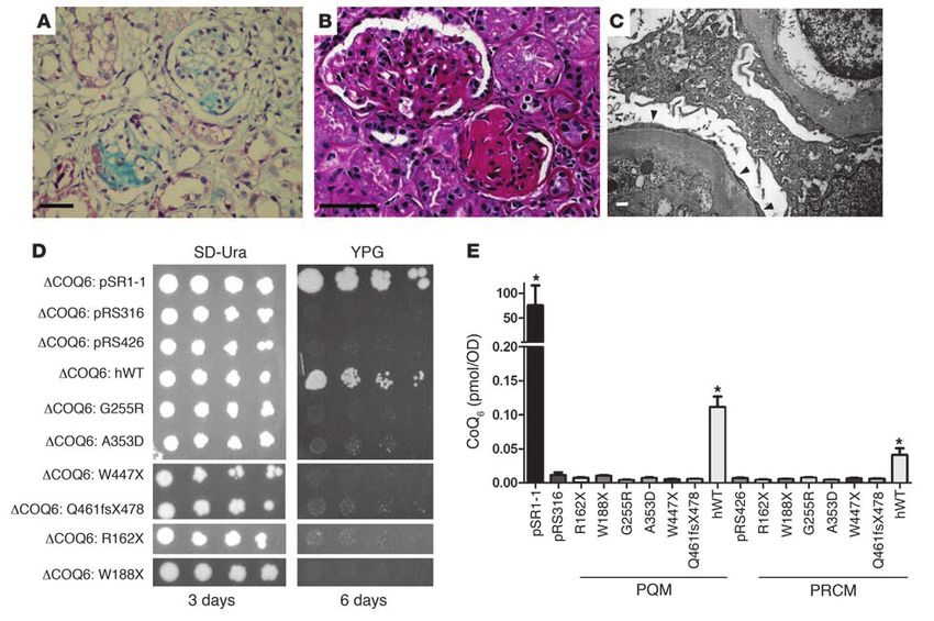
research article

Results                                                                           available online with this article; doi:10.1172/JCI45693DS1). Full-
COQ6 mutations cause SRNS with sensorineural deafness (SND). To iden-             length isoforms were expressed in multiple tissues, including the
tify further single-gene causes for SRNS, we performed a genome-                  kidney (Supplemental Figure 1, C–F). Using in situ hybridization
wide search for linkage in 14 different consanguineous families                   analysis, we demonstrated Coq6 mRNA expression in the meta-
with SRNS. Calculating lod under the hypothesis of locus heteroge-                nephric mesenchyme and forming nephrons of mouse kidney
neity yielded a significant maximum heterogeneity lod score of 4.9                (Supplemental Figure 1, G–L). Isoform a (encoding a 54-kDa pro-
(α = 52%) on human chromosome 14q24.3, covering a region of 6.4                   tein) is predicted to be expressed in mitochondria with a Predo-
Mb (Figure 1A). Of 7 families homozygous at this locus (SRNS2),                   tar score of 0.81 (http://urgi.versailles.inra.fr/predotar) because
families A1072 and F1082 from Turkey revealed haplotype sharing                   of a mitochondrial leader peptide, whereas isoform b (encoding a
(Figure 1B), restricting the critical genetic region to 2.1 Mb under              51-kDa protein), which uses an alternative exon 1, is considered
the hypothesis that an ancestor common to both families intro-                    only possibly mitochondrial, with a Predotar score of 0.45. Both
duced the disease allele (i.e., homozygosity by descent). This inter-             isoforms encode a flavin-dependent monooxygenase with 3 fla-
val contained 32 positional candidate genes (Figure 1C).                          vin adenine dinucleotide–binding motifs (18). The amino acid
   Exon sequencing of COQ6 yielded homozygous mutations in 3                      sequence is highly conserved throughout evolution, with identity
of the 7 families homozygous at the SRNS2 locus (Table 1). In both                to the human COQ6 protein sequence of 66% for Danio rerio and
affected individuals of family A1072, we detected a homozygous                    33% for the E. coli ortholog UbiH. The enzyme coenzyme Q10 mono-
A353D substitution (Figure 1G and Table 1) that was also found                    oxygenase 6 (COQ6) is required for biosynthesis of coenzyme Q10
in family F1082, as predicted from haplotype sharing at SRNS2                     (CoQ10; also referred to as ubiquinone) and is thought to catalyze
(Figure 1B). All 3 affected individuals that were examined in the                 one or more ring hydroxylation steps. Humans synthesize CoQ10,
northern Lebanese family F252 exhibited the homozygous change                     and yeast synthesizes CoQ6 (where CoQn designates a polyisoprene
G255R (Figure 1G and Table 1). This mutation was shared by 3                      residue with n isoprene units). CoQ10 operates as a redox carrier in
affected individuals of family A234 from southern Turkey (Table 1),               the mitochondrial respiratory chain shuttling electrons from respi-
most likely as a founder effect. Both missense mutations altered                  ratory chain complexes RCCI and RCCII to complex RCCIII (19).
amino acid residues that are uniformly conserved from E. coli to                  CoQ10 also serves as a lipid-soluble antioxidant and has previously
humans (Figure 1G). Another individual with SRNS from Turkey                      been implicated in protection from cell damage by ROS (20).
revealed 2 compound heterozygous truncating mutations, W447X                        Because rare recessive mutations have been described in syn-
and Q461fsX478 (Figure 1G and Table 1). All mutations were                        dromic and nonsyndromic forms of NS (21, 22) in other genes
absent from more than 90 healthy control subjects from central                    involved in CoQ10 biosynthesis (23–25), we performed sequencing
Europe and 60 healthy control subjects from Turkey. Segregation                   of all exons of the following 11 candidate genes: PDSS1, PDSS2,
of mutations was consistent with recessive inheritance (Table 1).                 COQ2, COQ3, COQ4, COQ5, COQ7, COQ8, COQ9, COQ10a, and
In all 9 individuals with SRNS that were examined for hearing loss,               COQ10b. We examined 42 individuals with SRNS who had extrare-
there was SND (Table 1). Exon sequencing of all other 31 genes                    nal symptoms suggestive of mitochondrial disease or in whom only
at the SRNS2 locus did not yield any mutations in the families                    1 heterozygous COQ6 mutation was found. However, we did not
F252, F1082, and A1072. We thus identified mutations in COQ6                      detect any further mutations. The 4 protein-truncating mutations
as a cause of recessive early-onset SRNS with SND (NPHS type 5),                  in COQ6 that we detected in subjects A988, A1904, and A3331-21
which we believe to be novel.                                                     (Figure 1G and Table 1) may be considered null alleles. Similarly,
   We then performed exon PCR of COQ6 in another 530 families:                    the fully conserved homozygous missense mutations G225R and
70 with SRNS, 35 that were homozygous for 3 markers at the COQ6                   A353D that we observed in 4 families with SRNS and SND may
locus, 55 with a neurologic phenotype, and 370 with a mitochon-                   also represent loss-of-function mutations (Figure 1G and Table 1).
driopathy. We identified a single heterozygous nonsense mutation,                 To test this hypothesis, we examined all 6 COQ6 mutations for
R162X, in A988-21, an individual with cyclosporine A–dependent                    potential deleterious effects by complementation in coq6-deficient
(CsA-dependent) NS, and a single heterozygous nonsense muta-                      yeast strains. Yeast cells harboring a deletion mutation in the coq6
tion, W188X, in A1904-21, an individual with DMS (Figure 1G).                     gene were unable to grow on yeast extract/peptone/glycerol media
As NPHS type 5 appears recessive, the second mutation most likely                 (YPG; in which glycerol acts as a nonfermentable carbon source)
escaped identification in these 2 families.                                       and were deficient in CoQ6 (Figure 2, D and E, and ref. 18). Expres-
   All 11 affected individuals of the 5 families with 2 recessive COQ6            sion of human WT COQ6 with an aminoterminal yeast mitochon-
mutations had SRNS. They manifested with proteinuria at a medi-                   drial leader sequence rescued both YPG growth and CoQ6 content
an age of 1.2 years (range, 0.2–6.4 years) and progressed to ESRF by              (Figure 2, D and E), although not as robustly as did yeast harboring
a median age of 1.7 years (range, 0.4–9.3 years); 5 individuals died in           the WT COQ6 gene (pSR1-1; Figure 2, D and E). Rescue of growth
early childhood (median age, 5.0 years). Renal biopsy revealed FSGS               on YPG was observed with either low–copy number (pQM; Figure
in 7 cases (Table 1 and Figure 2, A–C) and DMS in 1 case. Subject                 2D) or high–copy number (pRCM; data not shown) expression
A234-21 presented with seizures, and subject F1082-21 had white                   constructs. Both constructs also rescued the deficiency in CoQ6
matter abnormalities and seizures and died of multiorgan failure                  content (Figure 2E). In contrast, none of the Coq6 constructs har-
in sepsis; 2 other individuals had ataxia and facial dysmorphism                  boring the human mutations (Figure 1G and Table 1) were able to
(individuals F252-51 and A234-26, respectively; Table 1).                         rescue the growth deficiency phenotype or CoQ6 content pheno-
   COQ6 function. The COQ6 gene extends over 13.2 kb and con-                     types (Figure 2, D and E), thereby demonstrating the deleterious
tains 12 exons (Figure 1, D and E). There are 18 putative isoforms                effects of these mutations.
resulting from alternative splicing (www.aceview.org). Full-length                  Response to CoQ10 treatment. Monogenic variants of childhood NS
isoforms a and b contain 12 exons, but use alternative exons 1a                   are characterized by a lack of response to therapy (26). Because
and 1b (Supplemental Figure 1, A and B; supplemental material                     successful CoQ10 treatment had been described previously in an

2014                           The Journal of Clinical Investigation   http://www.jci.org   Volume 121   Number 5   May 2011
research article

Figure 1
Positional cloning of COQ6 mutations in individuals with NS and SND. (A) lod score profile across the human genome in affected children from 14
consanguineous kindred with SRNS. Parametric heterogeneity lod (HLOD) scores are plotted against human chromosomal mapping positions,
concatenated from p-ter (left) to q-ter (right). (B) Within the SRNS2 locus, haplotypes from 250k SNP analysis are shown for 3 of the 7 families
with homozygosity at the SRNS2 locus. Alleles are colored light green (AA), dark green (BB), and red (AB). (C) The 32 genes within the SRNS2
locus; 3 genes preferentially expressed in kidney podocytes are underlined. Mutations were found in COQ6. Transcriptional direction is indicated
by < or >. (D) COQ6 extends over 13.2 kb and contains 12 exons (boxes). (E) Exon structure of COQ6 cDNA. Arrows indicate relative positions of
mutations (see G). Positions of peptides for antibody generation are shown in yellow. (F) Domain structure of COQ6 protein. Extent of the mono-
oxygenase domain is shown in relation to encoding exon position (E). (G) 6 different COQ6 mutations in 7 families with SRNS. Family number
and amino acid change (see Table 1) are given above sequence traces. Arrows denote positions of mutations in relation to exons and protein
domains. For the 2 missense mutations, G255R and A363D, full conservation across evolution of altered amino acid residues is illustrated.

                               The Journal of Clinical Investigation   http://www.jci.org   Volume 121   Number 5   May 2011                2015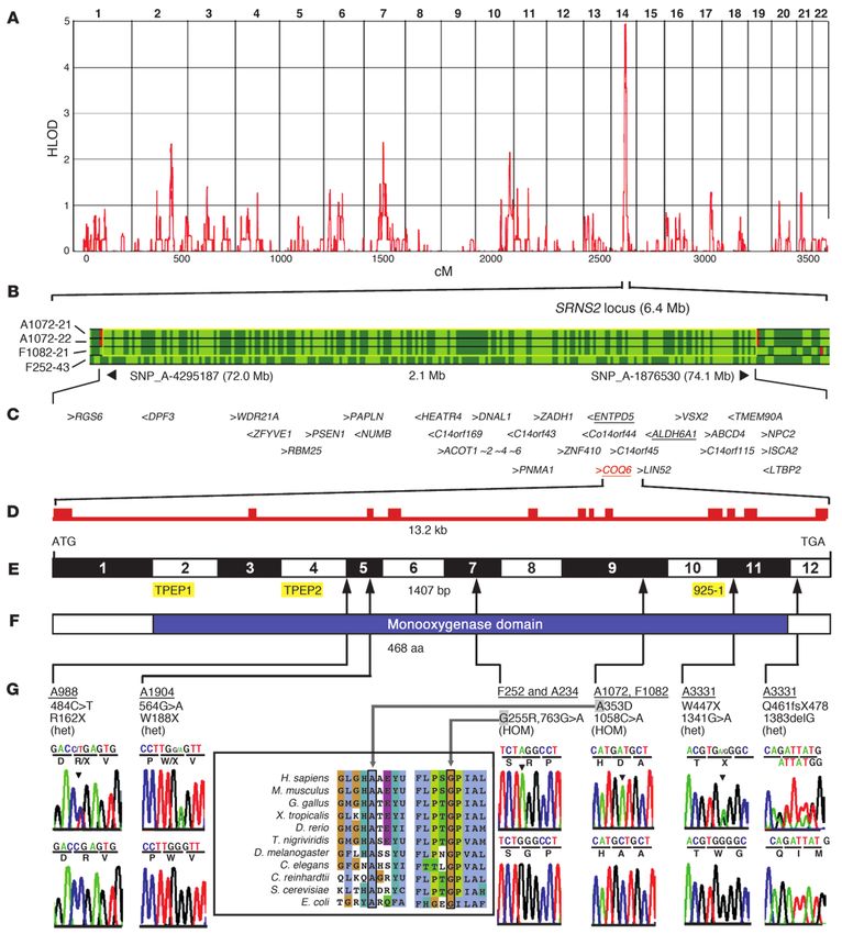
research article
individual with COQ2 mutations (12), we administered
CoQ10 in 2 children whose parents consented. Treatment
Seizures, white matter abnormalities (died, sepsis)
2 additional mutations (heterozygous only) were detected in families A998 (R162X) and A1904 (W188X) (see Figure 1G). ND, no data or DNA available. Consang, Consanguinity. AAll mutations were absent
SND (10 mo), bilateral nephrolithiasis (5 mo), GR
was administered orally, giving 1 Softgel CoQ10 capsule
individual; het, heterozygous in affected individual; M, heterozygous mutation identified in mother; P, heterozygous mutation identified in father. CACE-I, ACE inhibitor; CsA-S, CsA sensitive; CP-R, cyclo-
from more than 90 healthy control subjects from Central Europe and from 60 healthy control subjects from Turkey. All missense mutations were conserved down to E. coli. Bhom, homozygous in affected
(50 mg; GNC Preventive Nutrition) twice per day. Individ-
ual A234-27 presented with proteinuria without edema at
Congenital SND (died 17.5 yr)
Congenital SND (died 5.0 yr)
Extrarenal manifestations
SND, facial dysmorphism
SND, ataxia (died 6.5 yr)
2 months of age, when the COQ6 mutation was detected
SND (40% of normal)
SND (40% of normal)
in his sister (A234-26) and cousin (A234-21). Urine pro-
Congenital SND
Seizures (died)
SND (6 yr)
SND (4 yr)
tein/creatinine ratio was 40 mg/mg initially (normal, A
1058C>A
1058C>A
763G>A
763G>A
763G>A
763G>A
763G>A
763G>A
Cos-7 or HeLa cells or into murine podocyte cell lines,
ND
exogenous COQ6 isoform a colocalized quantitatively in
mitochondria with mitochondrial markers cytochrome c
and cytochrome c oxidase (COXIV; Figure 3, A and B).
Surprisingly, whereas α–COQ6-TPEP2 clearly detected
N
N
Y
Y
Y
Y
Y
Y
Y
Y
Y
Y
exogenously expressed COQ isoform a, thereby confirm-
ing specificity of the antibody, it detected an additional
F252-43 Lebanon
F252-46 Lebanon
F252-51 Lebanon
F252-42 Lebanon
endogenous signal that appeared to be localized in Golgi
Turkey
Turkey
Turkey
A1072-21 Turkey
A1072-22 Turkey
F1082-21 Turkey
A3331-21 Turkey
A3331-21 Turkey
Patient ID Origin
apparatus (Figure 3C). In fact, this endogenous signal
detected by α–COQ6-TPEP2 was located in Golgi, as
Table 1
confirmed by double labeling with the Golgi marker Gol-
A234-21
A234-26
A234-27
gin 97 (Figure 3D). Interestingly, α–COQ6-TPEP2 did
not reveal any endogenous COQ6 expression in mito-
chondria (Figure 3, D and E). Furthermore, exogenously
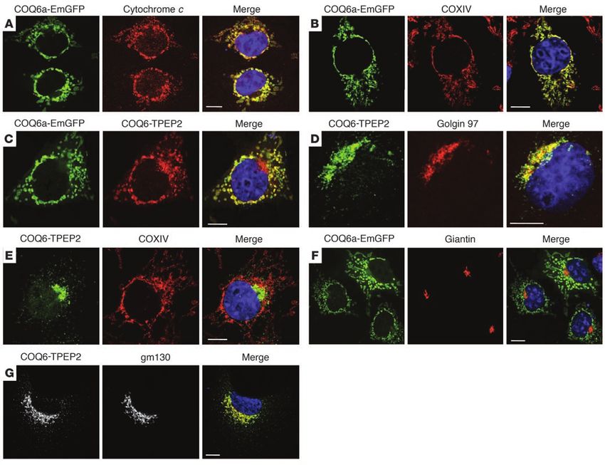
2016 The Journal of Clinical Investigation http://www.jci.org Volume 121 Number 5 May 2011research article
Figure 2
COQ6 loss-of-function causes glomerular damage in human kidney and growth defects in yeast. (A–C) Renal histology in individuals revealed
homozygous COQ6 mutation in A3331-21 FSGS, demonstrated by increased fibrosis (blue) in Trichrome-Masson staining (A), and in F1082-
21, demonstrated by excess PAS staining (red) (B). (C) Transmission electron microscopy for F1082 showed effacement of podocyte foot
processes, resulting in a continuous electron-dense layer (arrowheads). Scale bars: 50 μm (A and B); 250 nm (C). (D and E) Functional test of
human COQ6 mutations in yeast coq6-null mutants. (D) WT human COQ6 (hWT), but not mutations, rescued growth in yeast coq6-null mutants
plated on a nonfermentable carbon source. Yeast cells harboring the indicated low-copy plasmids were cultured in SD-Ura, seeded to both SD-
Ura and YPG plate media, and incubated at 30°C for the times indicated. pSR1-1 contains the yeast COQ6 gene (18) and served as positive
control. Empty vectors pRS316 and pRS426 served as negative controls. (E) WT human COQ6, but not mutations, rescued CoQ6 synthesis in
yeast coq6-null mutants. CoQ6 content of each yeast coq6-null mutant harboring 1 of the designated plasmids was determined as described
in Methods. Each CoQ6 measurement represents mean ± SD of 4 measurements from 2 independent samples. *P < 0.0005 versus negative
control (99% confidence level). PQM and PRCM indicate low– and high–copy number plasmids, respectively.
expressed full-length COQ6 isoform a spared Golgi expression, and C), which confirmed the results obtained in cell lines (Figure 3).
as demonstrated with Giantin as a Golgi marker (Figure 3F). Because nonmitochondrial localization and function has been
Likewise, in podocyte cell lines, the cell type central to the dis- previously described for CoQ6 (27), we hypothesized that non-
ease mechanism of SRNS, α–COQ6-TPEP2 detected endogenous mitochondrial expression would also apply to COQ7 and COQ9,
COQ6 in Golgi, but not in mitochondria (Figure 3G). The endog- which are known as mitochondrial proteins but share a multien-
enous COQ6 expression pattern was confirmed with the anti- zyme complex together with COQ6 for the biosynthesis of CoQ10
body α–COQ6–925-1 (Supplemental Figure 3, A and B), which and CoQ6 (21). Indeed, upon immunofluorescence, both COQ6
was derived from a different peptide (Figure 1E). and COQ7 fully colocalized to cellular processes and Golgi of rat
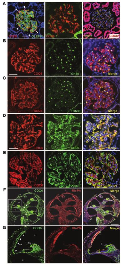
Endogenous COQ6, COQ7, and COQ9 colocalize to cell processes and glomerular podocytes (Figure 4, B and C), as did COQ7 and COQ9
Golgi in rat podocytes. Monogenic forms of SRNS are caused by dys- (Supplemental Figure 3C). We demonstrated specificity of this sig-
function of the glomerular podocyte, a terminally differentiated nal for α–COQ6-TPEP2 by showing the absence of the podocyte
cell critical for the filtering function of the kidney glomerulus. signal after preabsorption with the cognate peptide TPEP2, in
(13, 26). We therefore examined expression of the COQ6 protein contrast to its presence after preabsorption with the noncognate
in kidney. COQ6 was seen almost exclusively in glomeruli, rather peptide TPEP1 (Supplemental Figure 3D). In addition, COQ6
than in tubules (Figure 4A). As predicted, within glomeruli, COQ6 colocalized to podocyte cellular processes with podocin (Figure
was expressed specifically in podocytes, as marked by WT1 labeling 4E), another gene product that, if mutated, causes a Mendelian
(Figure 4A). Within podocytes, COQ6 was absent from mitochon- form of SRNS (2). These findings demonstrate that COQ6, COQ7,
dria (labeled with COXIV; Figure 4A); conversely, it was expressed and COQ9 are all 3 expressed in cellular processes and Golgi appa-
within cellular processes and within Golgi apparatus (Figure 4, B ratus of podocytes rather than in mitochondria.
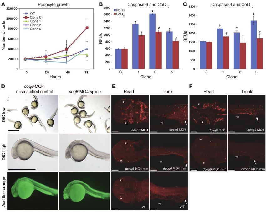
The Journal of Clinical Investigation http://www.jci.org Volume 121 Number 5 May 2011 2017research article Figure 3 Transient exogenous expression of full-length human COQ6 isoform a into Cos-7 cells, HeLa cells, or murine podocyte cell lines. (A and B) GFP-labeled full-length human COQ6 isoform a (COQ6a-EmGFP) exogenously expressed in Cos-7 cells colocalized quantitatively with mito- chondrial markers cytochrome c (A) and COXIV (B). (C–E) GFP-labeled full-length human COQ6 isoform a expressed exogenously in HeLa cells was detected by α–COQ6-TPEP2 upon immunofluorescence, thereby confirming specificity of the antibody (C). α–COQ6-TPEP2 detected an additional endogenous signal, which appeared to be localized in Golgi apparatus (C), as confirmed by double labeling with Golgi marker Golgin 97 (D). α–COQ6-TPEP2 did not reveal any endogenous COQ6 expression in mitochondria (D and E). (F) GFP-labeled full-length human COQ6 isoform a expressed exogenously in HeLa cells spared Golgi expression, as demonstrated with Giantin as a Golgi marker. (G) α–COQ6-TPEP2 detected endogenous COQ6 in Golgi of a podocyte cell line, as marked by gm130, but not in mitochondria. Scale bars: 5 μm. We then investigated expression of COQ6 in the inner ear, lyzed by RT-PCR for Coq6 expression levels. We compared clones because hearing loss is the other phenotype in this oto-renal transfected using a scrambled siRNA oligonucleotide with non- syndrome. Immunofluorescence studies using α–COQ6-TPEP2 transfected WT podocytes. The 3 clones that showed the highest revealed COQ6 expression in the spiral ganglion as well as in cells degree of Coq6 downregulation (clones 1, 2, and 5) were selected of stria vascularis and spiral ligament (Figure 4, F and G), which for functional studies (Supplemental Figure 1M). First, we ana- are involved in maintaining the high potassium concentration in lyzed the growth behavior of Coq6 knockdown clones when kept the cochlear duct necessary for sound transduction of the hair in an undifferentiated and proliferative state (Figure 5A). When cells of Corti organ. we seeded a defined number of cells and counted them after 24, Coq6 knockdown in cultured podocytes induces apoptosis that is reversed 48, and 72 hours of cultivation, we found that the growth rate of by CoQ10 treatment. In order to study COQ6 function in glomeru- the 3 Coq6 knockdown clones was strongly decreased, while the lar podocytes, the cell type involved in the disease phenotype, we scrambled siRNA clone C proliferated at the same rate as WT cells performed knockdown of Coq6 expression in vitro using a vector- (Figure 5A). This difference reached statistical significance after based siRNA approach (28). Murine podocytes (29) were stably 24 hours for clones 2 and 5 (P < 0.05) and after 72 hours for clone 1 transfected with COQ6 siRNA constructs, and clones were ana- (P < 0.005; Figure 5A). As the scrambled siRNA clone C did not 2018 The Journal of Clinical Investigation http://www.jci.org Volume 121 Number 5 May 2011
research article

                                                              Figure 4
                                                              COQ6 and COQ7 colocalize in rat podocytes to cell pro-
                                                              cesses and Golgi and in the inner ear to stria vascularis and
                                                              spiral ligament cells. (A) In rat renal glomeruli (marked with
                                                              an α-GLEPP1 antibody), α–COQ6-TPEP2 labeled podocyte
                                                              cytoplasm and cell processes (left). COQ6 was expressed
                                                              in podocytes, whose nuclei were marked with α-WT1 (mid-
                                                              dle), but not in mitochondria, marked with α-COXIV (right).
                                                              (B) COQ6 (red) was located in podocyte cellular processes
                                                              and in Golgi (marked with the trans-Golgi antibody TGN38),
                                                              demonstrating colocalization (yellow). (C) COQ7 exhibited
                                                              an expression pattern identical to that of COQ6 (see B).
                                                              (D) There was full colocalization of COQ6 and COQ7 in
                                                              podocyte cytoplasm, cell processes, and (by inference from
                                                              B and C) Golgi. (E) COQ6 colocalized in podocyte cellular
                                                              processes with podocin, another protein that, if mutated,
                                                              causes SRNS. COQ6 showed additional expression more
                                                              centrally in cell processes. (F) In cochlea (counterstained
                                                              with rhodamin-phalloidin [Rh-Ph] for F-actin), COQ6 was
                                                              expressed in spiral ganglion neurons in Rosenthal canal
                                                              (arrowheads). (G) Higher-magnification view of boxed
                                                              region in F showing COQ6 expression in stria vascularis
                                                              cells (arrowheads) and the spiral ligament (arrow). Signal in
                                                              tectorial membrane (tm) and adjacent structures most likely
                                                              represents background staining. Rm, Reissner membrane;
                                                              cd, cochlear duct; st, scala tympani; sv, scala vestibuli. In
                                                              merged images, nuclei are stained blue with DAPI. Scale
                                                              bars: 50 μm (A–E); 300 μm (F); 100 μm (G).

                                                              show any difference in growth behavior compared with
                                                              WT podocytes, the scrambled clone was used as negative
                                                              control for further experiments. To analyze whether the
                                                              diminished overall growth rate in podocytes after Coq6
                                                              knockdown is associated with activation of the intrinsic
                                                              apoptotic pathway, we next determined the inner mito-
                                                              chondrial membrane potential (ΔΨm) in these podocyte
                                                              clones by using the fluorescent dye tetramethylrhoda-
                                                              mine ethylester (TMRE). Compared with control cells, all
                                                              3 knockdown clones showed a substantial drop in TMRE
                                                              fluorescence (data not shown), indicative of an increased
                                                              percentage of podocytes with depolarized ΔΨm.
                                                                We then analyzed activity of caspase-9 (Figure 5B),
                                                              the major caspase downstream of mitochondria-medi-
                                                              ated apoptosis (30). Using fluorometric immunosorbent
                                                              enzyme assay (FIENA), we detected a more than 2-fold
                                                              increase of caspase-9 activity in all 3 knockdown clones
                                                              compared with control podocytes (P < 0.005; Figure 5B).
                                                              Moreover, caspase-3, the final downstream executioner cas-
                                                              pase of different apoptotic signaling pathways (31), showed
                                                              significant elevation of activity for all 3 siRNA clones
                                                              (P < 0.05; Figure 5C), consistent with the effect seen in
                                                              zebrafish (see below). These data suggest that the observed
                                                              decline in podocyte growth after Coq6 knockdown is caused,
                                                              at least in part, by increased apoptosis. Since mitochondria-
                                                              induced apoptosis is associated with depolarization of the
                                                              inner mitochondrial membrane (32) as well as caspase-9
                                                              activation (30), it is likely that lack of growth upon Coq6
                                                              knockdown is caused by activation of the intrinsic apop-
                                                              totic pathway. Because some of the SRNS patients in our
                                                              study that carry COQ6 mutations responded partially to
                                                              CoQ10 treatment, we then analyzed whether CoQ10 has a
                                                              beneficial effect on the phenotype caused by knockdown

The Journal of Clinical Investigation   http://www.jci.org   Volume 121   Number 5   May 2011                          2019
research article Figure 5 COQ6 knockdown causes apoptosis in podocytes and zebrafish embryos. (A–C) Coq6 downregulation in cultured mouse podocytes induced apoptosis that was diminished by CoQ10 treatment. Data are mean ± SEM. (A) Growth curve analysis of Coq6 knockdown clones. 20,000 podocytes were seeded per 24-well plate, plated, detached, and counted (n = 4). (B and C) Caspase-9 (B) and caspase-3 (C) activity in undif- ferentiated Coq6 knockdown clones by FIENA before and after CoQ10 treatment. RFUs derived from cleaved capase-9 or caspase-3 sub- strate peptide were measured in lysates from 1 × 10⁶ cells (n = 3). C, control clone; No Tx, no treatment. *P < 0.001, †P < 0.05 versus control; #P < 0.005, ‡P < 0.05 versus untreated. (D–F) coq6 knockdown in zebrafish embryos 28 hours after fertilization increases apoptosis. (D) coq6- MO4 directed against zebrafish coq6 intron 7 splice donor blocked proper splicing of coq6 mRNA (see Supplemental Figure 1N). Negative controls were injected with 0.1 mM coq6 mismatch MO. Note the gray appearance of zebrafish heads upon differential interference contrast (DIC) microscopy as a sign of increased cell death. (E and F) Zebrafish dorsal head and lateral trunk views. Embryos were injected as indicated with 0.1 mM coq6-MO4 splice targeting MO (E), MO targeting the AUG translation start site (dcoq6 MO1; F), coq6 mismatch (mm) MO nega- tive controls, or left uninjected (WT). Cells were stained by an antibody against cleaved caspase-3, a specific marker for apoptotic cells. Lens (asterisk), yolk sac (ys), and cloaca (arrow) are indicated. Scale bars: 1 mm (D); 100 μm (E and F). of Coq6 expression in podocytes in vitro. When culturing podocyte knockdown of coq6 in zebrafish. We demonstrate that coq6-MO4 clones 1, 2, and 5 in the presence of CoQ10 at a concentration of MOs directed against the zebrafish coq6 intron 7 splice donor 50 μM for 48 hours, we observed a decrease in caspase-9 and cas- site (Supplemental Figure 1N) and the AUG translation start site pase-3 activities (Figure 5, B and C). Compared with clones cultured induced apoptosis preferentially in heads and trunks of zebrafish in the absence of CoQ10, this decrease was statistically significant embryos 28 hours after fertilization (Figure 5, D–F). (P < 0.05), whereas we did not detect any changes in caspase activi- Because COQ6 mutations cause SND, it is interesting to note ties of negative control clones (Figure 5, B and C). that treatment with water-soluble CoQ10 decreased apoptosis To examine COQ6 loss of function in a vertebrate organism rather and activated caspase-3 and improved hearing in a guinea pig than cell lines, we performed morpholino oligonucleotide (MO) model of noise-induced hearing loss (33). The kidney phenotype 2020 The Journal of Clinical Investigation http://www.jci.org Volume 121 Number 5 May 2011
research article

of COQ6 mutations is remarkably similar to that of mutations in                  or the level of respiratory activity, explaining the wide spectrum
COQ2 (23) and PDSS2 in humans (34) and Pdss2 in mice (24), all of                of clinical features. Based on the surprising Golgi localization of
which respond to CoQ10 treatment (12, 35). Patients with defects                 COQ6, COQ7, and COQ9, we speculate that the antioxidant func-
in genes required for CoQ10 biosynthesis — PDSS2, COQ2, and                      tion of Q/QH2 in certain cells, such as podocytes, may depend on
COQ9 — exhibit renal disease, and in some cases there is a dramatic              targeted synthesis of CoQ in the Golgi for delivery to the plasma
response to CoQ10 therapy, but this is quite variable (36).                      membrane, which in the podocyte constitutes an enormous sur-
                                                                                 face area that must contain vulnerable lipid components. Whereas
Discussion                                                                       most other forms of monogenic childhood NS are characterized
In this study, we report what we believe to be a novel cause of SRNS             by a lack of response to therapy (10, 16, 17, 44), the identifica-
that appears to respond to oral CoQ10 supplementation. CoQ10,                    tion by mutation analysis of individuals with NS caused by CoQ10
an essential component of the mitochondrial electron transport                   biosynthesis defects, as described here, is important because those
chain and one of the most potent lipophilic antioxidants (37),                   individuals may respond to treatment with CoQ10.
is also required for pyrimidine nucleoside biosynthesis and has
been implicated in the inhibition of apoptosis by its prevention                 Methods
of inner mitochondrial membrane collapse (38). In multiple stud-
ies, the deleterious effect of CoQ10 deficiency to mitochondria                  Subjects
has been shown. CoQ10 deficiency can lead to the opening of the                  We obtained blood, tissue samples, and pedigrees following informed con-
mitochondrial permeability transition pore (MPTP) directly, but                  sent from individuals with NS and/or from their parents. Human subject
an increased amount of ROS caused by CoQ10 deficiency can also                   research was approved by the University of Michigan Institutional Review
induce the MPTP by opening of nonspecific high-conductance per-                  Board. The diagnosis of NS was made by a pediatric nephrologist based on
meability transition pores in the mitochondrial inner membrane                   either chronic or recurrent high-grade proteinuria (>40 mg/m2/h) or per-
(39). Experiments in HEK293 cells recently showed that CoQ10                     sistent low-grade proteinuria (>4 mg/m2/h) (45). Steroid-sensitive NS and
inhibits Bax-induced mitochondrial dysfunction and protects                      SRNS were defined according to standard criteria (45, 46). Renal biopsies
mitochondria from permeability transition pore opening (9). Here,                were evaluated by a reference renal pathologist. Age at onset of ESRF was
we confirmed that COQ6 mutations that cause CoQ10 deficiency                     defined as age at first renal replacement therapy, i.e., dialysis or renal trans-
led to the upregulation of proapoptotic factors. Interestingly,                  plantation. Clinical data were obtained using a standardized questionnaire
CsA inhibits the MPTP through interaction with cyclophilin D, an                 (www.renalgenes.org).
essential component of the MPTP (40). We showed that incuba-
tion of COQ6 knockdown podocytes with CsA had a mild rescue                      Genetic mapping and exon sequencing
effect and decreased activity of caspase-3. Patient A1072-22, who                We performed a genome-wide search for linkage by homozygosity mapping
had been treated with CsA in the past, also showed partial remis-                using the 250k Affymetrix SNP microarray (47). Data were evaluated by
sion of proteinuria after treatment with CsA.                                    calculating nonparametric lod scores and scoring for homozygosity (Zhom)
  Mutations in 5 other CoQ10 biosynthesis enzymes leading to                     across the whole genome in order to identify regions of homozygosity. The
CoQ10 deficiency have been recently implicated in other mono-                    GENEHUNTER-MODSCORE program (48) was used to calculate multi-
genic mitochondriopathies, generally characterized by central                    point lod scores assuming recessive inheritance with complete penetrance,
nervous system signs and myopathy (22, 34, 41–43); 1 patient                     a disease allele frequency of 0.001, and the marker allele frequencies for
with mutations in PDSS2 and 5 patients with mutations in COQ2                    individuals of mixed European descent specified by Affymetrix. Parametric
also presented with SRNS (22, 34, 43). So far, the exact patho-                  and nonparametric lod scores were plotted over genetic distance across the
genic mechanism has remained unclear. Interestingly, 1 patient                   entire human genome using GNUPLOT software (http://www.gnuplot.
with mutations in COQ2 was successfully treated with oral CoQ10                  info/). See Supplemental Table 2 for exon sequencing primers.
supplementation, showing progressive recovery of renal function
and a reduced level of proteinuria until 5 years after completion of             COQ6 mRNA expression
treatment (22). The podocyte-specific phenotype caused by PDSS2                  The probe for human Northern blot analysis was obtained by digesting
mutations leading to primary CoQ10 deficiency was demonstrated                   the COQ6 isoform a coding region cloned into pCRIITOPO with EcoRI
in the Pdss2 knockout mouse (24). Conditional knockouts targeted                 and was radiolabeled with α-32P dCTP (Amersham Biosciences) using the
to renal tubular epithelium, monocytes, or hepatocytes did not                   Random Primers DNA Labeling System kit (Invitrogen) and successively
show disease manifestation. It remains unclear why the podocyte                  purified with Quick-Spin columns (Roche) according to the manufac-
in particular is affected by PDSS2-dependent CoQ10 deficiency.                   turer’s protocol. Radiolabeled probes were hybridized to a commercial
  Interestingly, the presentation of primary CoQ10 deficiency                    preblotted membrane (FirstChoice Human Blot 1 membrane; Ambion)
caused by genetic mutations is very heterogeneous. It seems that                 containing 2 μg/lane poly(A)+ RNA from 10 human tissues. Prehybridiza-
the mutations in PDSS2 and COQ2 are partial loss-of-function                     tion and hybridization were both performed in a 50% formamide buffer at
mutations. Therefore, the phenotypes (ATP synthesis, ROS pro-                    42°C, with a working concentration of 1.5 × 10⁶ cpm and 0.1 mg salmon
duction) may depend on the content of Q in the cell, which is                    sperm DNA per milliliter. The excess probe was removed by washing at
determined by the severity of the mutation. Mutations in PDSS2,                  65°C for 15 minutes using a 0.1% SDS, 0.1× sodium chloride–sodium
for example, cause reduced ATP synthesis in cultured fibroblasts,                citrate buffer solution. Radioactivity was detected with a Storm Phos-
but no increase in ROS production, whereas mutations in COQ2                     phoImager (Molecular Dynamics) after overnight exposure. Selective
cause no difference in ATP synthesis in cultured fibroblasts, but                amplification of COQ6 isoform b was carried out using primers in exon 1b
increase ROS production (36). One could speculate that different                 and in exon 4. Simultaneous amplification of isoform a and isoform b
cell types and/or tissues react differently to ROS and ATP synthe-               was carried out using primers on exon 2 and on exon 4. PCR conditions
sis defects according to their antioxidant defense mechanisms                    were 94°C for 3 minutes followed by 35 cycles of 94°C for 30 seconds,

                              The Journal of Clinical Investigation   http://www.jci.org   Volume 121   Number 5     May 2011                                2021
research article
55°C for 30 seconds, and 72°C for 30 seconds and a final extension step of dextrose; YPGal, containing 1% yeast extract, 2% peptone, 2% galactose,
72°C for 7 minutes. Amplified fragments were also purified from agarose and 0.1% dextrose; YPG, containing 1% yeast extract, 2% peptone, and 3%
gel and sequenced. Primer sequences were as follows: exon 1b forward, glycerol; and SD-Ura, containing 0.18% nitrogen base without amino acids,
5′-TCTAGCTTTGGCGTCTGGTT-3′; exon 2 forward, 5′-CTTCTAAGCTT- 2% dextrose, 0.14% NaH2PO4, 0.5% (NH4)2SO4, and a complete amino acid
GATATGATATTCACTTTCATGACAAG-3′; exon 4 reverse, 5′-CTTCTG- supplement (56), minus uracil. Solid plate media contained 2% agar.
GATCCATGATGACATCATTCTCCAC-3′. Yeast transformation. Yeast was subjected to transformation as described pre-
viously (57), with either a low–copy number (pQM; ref. 58) or a high–copy
Generation of plasmids number (pRCM; ref. 59) yeast expression plasmid. These vectors were used to
Yeast COQ6 was amplified from genomic DNA extracted from a WT prepare plasmids containing the human COQ6 open reading frame with an
BY4741 strain and cloned in pCM189 vector. Site-specific mutagenesis in-frame aminoterminal yeast mitochondrial leader sequence (pQM_hCOQ6-
was performed using the QuickChange kit (Stratagene) according to the MLS and pRCM_hCOQ6-MLS). Each of the following mutations in human
manufacturer’s protocol. To generate constructs that expressed the gene COQ6 were introduced by site-directed mutagenesis (Stratagene): R162X,
under the control of the endogenous yeast coq6 promoter, the different W188X, G255R, A353D, W447X, and Q461fsX478. See Supplemental Table 1
pCM189–yeast COQ6 constructs were digested with SacI and MscI (to for a complete list of plasmids used in yeast transformation experiments.
eliminate the CYC1 promoter and control elements); religated with a frag- Lipid extraction and analysis. Yeast cells were grown in YPGal and harvest-
ment encompassing the yeast COQ6 promoter, the 5′ end of the gene, and ed during log phase growth (1.0 OD600nm). Lipid extracts were prepared
the MscI site; amplified from genomic DNA; and cut accordingly. Plasmid from yeast whole cells as described previously (60). Lipids were separated
maps were described previously (49). Yeast growth and transformation by reverse-phase high-performance liquid chromatography with a Phenyl-
were performed as described below. hexyl column (Phenomenex 5-μm, 100 × 4.6 mm). The column was equili-
brated with a mobile phase consisting of 95:5 methanol/2-propanol with
COQ6-GFP expression studies 2.5 mM ammonium formate as solvent A and a flow rate of 650 μl/min.
The complete coding region of full-length human COQ6 isoform a, lacking Upon sample injection, the percentage of solvent B (2-propanol with 2.5
termination codon but including the Kozak sequence, was PCR amplified mM ammonium formate) was increased linearly from 0% to 5% over 6 min-
from pCRIITOPO-COQ6 and subcloned into the HindIII and BamHI sites utes, and the flow rate was also increased linearly to 800 μl/min. The flow
of pEGFP-N1 (BD Biosciences — Clontech). The correctness of the construct rate and mobile phase were linearly changed back to initial conditions by
was verified by direct sequencing. HeLa cells stably expressing mitochondri- 8 minutes. Q was quantified with an Applied Biosystems-MDS Sciex 4000
ally targeted red fluorescent protein (50) were grown on coverslips in com- Q Trap (hybrid triple-quad linear ion trap analyzer with autosampler and
plete DMEM (Sigma-Aldrich) containing 10% fetal bovine serum until 70% a Turbo-V source equipped with ESI and APCI sources) as described pre-
confluent, then transfected with the purified plasmid using Effectene Trans- viously (35). The samples were analyzed in multiple reaction monitoring
fection Reagent (Qiagen) according to the manufacturer’s instructions. Cells (MRM) mode; MRM transitions were as follows: m/z 591.6/197.1 for Q6;
were visualized after 48 hours using a Nikon Video Confocal microscope. m/z 610.6/197.1 for Q6H2 with ammonium adduct; m/z 455.6/197.1 for Q4
(Sigma-Aldrich), used as internal standard.
In situ hybridization analysis in mice
Whole-mount in situ hybridization was performed following a standard pro- Podocyte expression studies
cedure with digoxigenin-labeled antisense riboprobes (51). Stained specimens Preparation of CoQ10 solution for use in cell culture. Pure CoQ10 in powder form
were transferred in 80% glycerol prior to documentation. In situ hybridization (Kaneka Corp.) was dissolved in 1 ml of molecular biology–grade isopro-
on 10-μm paraffin sections was done as described previously (52). panol at a concentration of 10 mM by heating it briefly in a 95°C water
bath. Thereafter, the CoQ10 solution was diluted 1:10 in podocyte culture
Immunofluorescence media to a final stock concentration of 1 mM and reheated for an addi-
Rat kidneys were perfusion-fixed with paraformaldehyde/lysine/periodate tional 5 minutes at 95°C with gentle shaking. This CoQ10 stock solution
and processed as previously described (3). For absorption experiments, was stored at 4°C, protected from light for up to 4 weeks, and diluted
the immunopurified α–COQ6-TPEP2, COQ7, and COQ9 antibodies were immediately before use at 1:20 in cell culture medium to a final working
preabsorbed with an equal weight of cognate or noncognate peptide over- concentration of 50 μM.
night at 4°C. The following antibodies were used: WT1 mouse monoclonal Podocyte culture. Immortalized murine podocytes were cultured as
antibody (catalog no. sc-7385; Santa Cruz Biotechnology Inc.); PLCe1 goat described previously (29). For treatment studies of undifferentiated podo-
polyclonal antibody (catalog no. sc-28404; Santa Cruz Biotechnology Inc.); cytes, cells were incubated in the presence of 50 μM CoQ10, 1 μM dexameth-
α–COQ6-TPEP2 immunopurified rabbit antipeptide antibody (against asone (Sigma-Aldrich), or 1 μM CsA (Sigma-Aldrich) for 48 hours. For anal-
peptide sequence N-DKDNLDDMGYIVEND-C; anti-rat Glepp1 mouse ysis of differentiated podocytes, undifferentiated podocytes were cultured
monoclonal antibody (1B4; ref. 53), CoQ7 goat polyclonal antibody (cata- under nonpermissive conditions in the presence of 1 μM dexamethasone for
log no. sc-66353; Santa Cruz Biotechnology Inc.); CoQ9 rabbit polyclonal 14 days. Cultivation was then continued in the absence of dexamethasone
antibody (catalog no. 14874-1-AP; Protein Tech); TGN38 monoclonal for an additional 48 hours. For determination of cell number and growth
mouse antibody (catalog no. 610898; BD Biosciences); and GM130 mouse curve analysis, cells were seeded in 24-well plates at 20,000 cells/well. After
monoclonal antibody (catalog no. 610823; BD Biosciences). 24, 48, or 72 hours, cells were trypsinized and counted using a hemocytom-
eter. Experiments were done 4 times with at least 3 internal replicates.
Yeast studies
Yeast growth. The S. cerevisiae coq6-null mutants W303ΔG63 (Mat α ade2-1 COQ6 RNAi
his 3-1,15 leu2-3,112 trp1-1 ura3-1 ΔCOQ6:HIS3; ref. 18) and W303ΔCOQ8 For the targeted downregulation of protein expression in podocytes, we
(Mat α ade2-1 his 3-1,15 leu2-3,112 trp1-1 ura3-1 ΔABC1/COQ8:HIS3; ref. used the pSuper RNAi System (OligoEngine) (28) following the manufac-
54) were used in this study. Yeast growth media were prepared as described turer’s instructions and as done previously (61). Briefly, we cloned a 60-nt
previously (55): YPD, containing 1% yeast extract, 2% peptone, and 2% oligo including a 19-nt COQ6 target sequence and a hairpin (5′-TTCAAGA-
2022 The Journal of Clinical Investigation http://www.jci.org Volume 121 Number 5 May 2011research article
GA-3′) into a modified pSuper vector, which also encoded for a zeocin selec- against D. rerio coq6 were 5′-GCTTAGCTTTAGCCAGAGACAGCAT-
tion marker (61). After transfection of undifferentiated podocytes using 3′ and 5′-ACACATAAGGTCAGCTCACGGGAAG-3′, respectively. The
FuGene 6 (Roche) followed by zeocin selection at 500 μg/ml for 7–10 days, 5-bp mismatch controls for the MOs were as follows: coq6-MO1mm,
appearing podocyte clones were covered by cloning chambers, trypsinized, 5′-GGTTACCTTTACCCAGACACACCAT-3′; coq6-MO4mm, 5′-ACA-
and expanded separately from each other in culture medium containing GATAACGTCACCTCAGGGCAAG-3′. Fertilized eggs were microin-
100 μg/ml zeocin. In total, 5 different COQ6 targeting sequences were used jected with the specified amount of MO dissolved in 0.1M KCl. For
(clone 1, AACAGAGTCAGCTCCATAT; clone 2, AATGACGTCATCATG- immunohistochemical labeling, embryos were fixed with 4% PFA in PBS
TATG; clone 3, CTGTGGCAGATCGAGTGAA; clone 4, AACTGTTGATTG- (pH 7.4) and stained with antibody against activated caspase-3 (1:200; BD
GTGCTGA; clone 5, AAGGACTTAGGCTCCATGA) as well as 1 scrambled Biosciences) following standard protocols (63).
control sequence (5′-CCGCGACTCGCCGTCTGCG-3′). Stable transfected
clones were expanded and used for RNA isolation and functional studies. Statistics
To evaluate caspase-3 and -9 activity in cell lines, Pearson χ2 test was used
RNA isolation and RT-PCR with a critical P value of 0.05 (Figure 5).
RNA was isolated from 2 × 10⁶ undifferentiated podocytes using the
RNeasy kit (Qiagen) following the manufacturer’s protocol. Prior to RT- Databases
PCR, total RNA samples were digested with DNase I (Roche) and RNA Genetic mapping was performed using http://genome.ucsc.edu (May
was transcribed into cDNA using Superscript II (Invitrogen). For RT-PCR, 2004 freeze).
100 ng cDNA, AmpliTaq Gold DNA polymerase (Applied Biosystems), and
sequence-specific, intron-spanning primers were used (COQ6 forward, Accession numbers
CAGACACCGTGTACGACGTG; COQ6 reverse, CCAACAGCTTTGCTCT- Accession numbers of COQ6 orthologs were NP_872282 for Homo sapiens,
CATAGAG; GAPDH forward, tatgtcgtggagtctactgg; GAPDH NP_001038869 for D. rerio, and ZP_00723222 E. coli.
reverse, agtgatggcatggactgtgg). 30 PCR cycles (5 minutes at
95°C, 30 seconds at 53°C, and 30 seconds at 72°C) were run on a Gene- Acknowledgments
Amp PCR System 9700 (Applied Biosystems). PCR reactions were analyzed We thank the affected individuals and their families for participa-
on a 2% agarose gel, and ethidium bromide signals were captured with a tion. We thank Corina Nailescu for contribution of clinical data.
Bio-Rad Gel Doc XR system. This research was supported by grants from the NIH to F. Hildeb-
randt (DK076683 and DK086542), C.F. Clarke (GM45952), and
Inner ΔΨm R.C. Wiggins (DK46073 and P30 DK081943) and by a grant from
To analyze changes in the ΔΨm of undifferentiated podocytes, we used the KMD Foundation and the Thrasher Research Fund to F. Hil-
TMRE (Invitrogen) as described previously (62). Briefly, 3 × 10⁵ cells were debrandt. F. Hildebrandt is an Investigator of the Howard Hughes
trypsinized and incubated in 25 nM TMRE for 30 minutes at 37°C. With this Medical Institute, a Doris Duke Distinguished Clinical Scientist,
concentration (nonquenching mode), depolarization causes a drop in TMRE and the Frederick G.L. Huetwell Professor. H. Prokisch was sup-
fluorescence. Cells were washed with PBS and analyzed by flow cytometry ported by the Impulse and Networking Fund of the Helmholtz
(FACSCalibur; BD), measuring TMRE fluorescence in FL-2. The relative fluo- Association in the framework of the Helmholtz Alliance for Mental
rescence of TMRE was compared with that of the respective untreated control Health in an Ageing Society (HA-215), by the German Network for
cells in each experiment. 3 replicates were analyzed per treatment group. Mitochondrial Disorders (mitoNET 01GM0862 and 01GM0867),
and by Systems Biology of Metabotypes (SysMBo 0315494A). The
FIENA work was further supported by a grant from the German Research
To determine caspase-3 and -9 activities in podocyte extracts, we used Foundation (DFG, SFB 423, TP B19) to M. Zenker, by the German
the Fluorometric Assay Kits from BioVision according to the manufac- Federal Ministry of Science and Education through the National
turer’s protocol. In brief, podocytes were cultured in 6- or 12-well plates, Genome Research Network to G. Nürnberg and P. Nürnberg, by
trypsinized, and counted using a hemocytometer. 1 × 10⁵ to 1 × 10⁶ cells the European Community FP7 program (EUNEFRON 201590) to
were lysed on ice for 10 minutes, and the derivatized peptide DEVD–7- D. Müller, by Fondazione CARIPARO Telethon Italy (GGP09207)
amino-4-trifluoromethyl coumarin (DEVD-AFC) caspase-3 or -9 cleav- to L. Salviati, by a Spanish FIS grant (PI080500) to P. Navas, and by
age substrate was added at a final concentration of 50 μM in 2× reaction the Young Investigator Career Development Grant from the Neph-
buffer containing 10 mM dithiothreitol. The reaction was incubated at Cure Foundation and the National Scientist Development Grant
37°C for 1–24 hours, and green fluorescence (λmax, 505 nm) of free AFC from the American Heart Association to C. Faul.
after caspase-3– or caspase-9–mediated cleavage was measured in relative
fluorescence units (RFU) using a fluorometer (SpectraMax M5; Molecular Received for publication November 6, 2010, and accepted in
Devices) at different time points. Values were normalized to cell lysates revised form February 9, 2011.
without substrate and to substrate in reaction buffer in the absence of cell
lysate. Experiments were done 3 times with at least 3 internal replicates. Address correspondence to: Friedhelm Hildebrandt, Howard Hughes
Medical Institute, Departments of Pediatrics and Human Genetics,
Zebrafish MO-mediated knockdown University of Michigan Health System, 8220C MSRB III, 1150 West
MOs were obtained from Gene Tools. The nucleotide sequences of the Medical Center Drive, Ann Arbor, Michigan 48109-5646, USA. Phone:
translation-blocking (coq6-MO1) and splice-blocking (coq6-MO4) MOs 734.615.7285; Fax: 734.615.1386; E-mail: fhilde@umich.edu.
1. Kestila M, et al. Positionally cloned gene for a novel protein podocin, is mutated in autosomal recessive tions in PLCE1 responsible for a nephrotic syndrome
glomerular protein--nephrin--is mutated in congeni- steroid-resistant nephrotic syndrome. Nat Genet. variant that may be reversible. Nat Genet. 2006;
tal nephrotic syndrome. Mol Cell. 1998;1(4):575–582. 2000;24(4):349–354. 38(12):1397–1405.
2. Boute N, et al. NPHS2, encoding the glomerular 3. Hinkes B, et al. Positional cloning uncovers muta- 4. Kaplan JM, et al. Mutations in ACTN4, encoding
The Journal of Clinical Investigation http://www.jci.org Volume 121 Number 5 May 2011 2023research article
alpha-actinin-4, cause familial focal segmental glo- 26. Tryggvason K, Patrakka J, Wartiovaara J. Hereditary 1988;1(8582):380–383.
merulosclerosis. Nat Genet. 2000;24(3):251–256. proteinuria syndromes and mechanisms of pro- 46. ISKDC. Primary nephrotic syndrome in children:
5. Reiser J, et al. TRPC6 is a glomerular slit dia- teinuria. N Engl J Med. 2006;354(13):1387–1401. Clinical significance of histopathologic variants of
phragm-associated channel required for normal 27. Padilla-Lopez S, Jimenez-Hidalgo M, Martin- minimal change and of diffuse mesangial hypercellu-
renal function. Nat Genet. 2005;37(7):739–744. Montalvo A, Clarke CF, Navas P, Santos-Ocana C. larity: A Report of the International Study of Kidney
6. Winn MP, et al. A mutation in the TRPC6 cation Genetic evidence for the requirement of the endo- Disease in Children. Kidney Int. 1981;20(6):765–771.
channel causes familial focal segmental glomeru- cytic pathway in the uptake of coenzyme Q6 in Sac- 47. Hildebrandt F, et al. A systematic approach to map-
losclerosis. Science. 2005;308(5729):1801–1804. charomyces cerevisiae. Biochim Biophys Acta. 2009; ping recessive disease genes in individuals from out-
7. Brown EJ, et al. Mutations in the formin gene INF2 1788(6):1238–1248. bred populations. PloS Genet. 2009;5(1):e1000353.
cause focal segmental glomerulosclerosis. Nat 28. Brummelkamp TR, Bernards R, Agami R. A system 48. Strauch K. Parametric linkage analysis with auto-
Genet. 2010;42(1):72–76. for stable expression of short interfering RNAs in matic optimization of the disease model param-
8. Somlo S, Mundel P. Getting a foothold in nephrot- mammalian cells. Science. 2002;296(5567):550–553. eters. Am J Hum Genet. 2003;73(suppl 1):A2624.
ic syndrome. Nat Genet. 2000;24(4):333–335. 29. Mundel P, et al. Rearrangements of the cytoskel- 49. Gari E, Piedrafita L, Aldea M, Herrero E. A set of vec-
9. Machuca E, Benoit G, Antignac C. Genetics of eton and cell contacts induce process formation tors with a tetracycline-regulatable promoter system
nephrotic syndrome: connecting molecular genet- during differentiation of conditionally immortal- for modulated gene expression in Saccharomyces cerevi-
ics to podocyte physiology. Hum Mol Genet. 2009; ized mouse podocyte cell lines. Exp Cell Res. 1997; siae. Yeast. 1997;13(9):837–848.
18(R2):R185–R194. 236(1):248–258. 50. Casarin A, et al. Functional characterization of human
10. Ruf RG, et al. Patients with mutations in NPHS2 30. Green DR. Apoptotic pathways: ten minutes to COQ4, a gene required for Coenzyme Q10 biosynthe-
(podocin) do not respond to standard steroid treat- dead. Cell. 2005;121(5):671–674. sis. Biochem Biophys Res Commun. 2008;372(1):35–39.
ment of nephrotic syndrome. J Am Soc Nephrol. 2004; 31. Wilson MR. Apoptosis: unmasking the executioner. 51. Wilkinson DG. Whole mount in situ hybridization
15(3):722–732. Cell Death Differ. 1998;5(8):646–652. of vertebrate embryos. In: Wilkinson DG, ed. In Situ
11. Weber S, et al. NPHS2 mutation analysis shows 32. Green DR, Kroemer G. The pathophysiol- Hybridization: A Practical Approach. Oxford, United
genetic heterogeneity of steroid-resistant nephrotic ogy of mitochondrial cell death. Science. 2004; Kingdom: IRL Press; 1992:75–83.
syndrome and low post-transplant recurrence. Kid- 305(5684):626–629. 52. Franco D, de Boer PA, de Gier-de Vries C, Lamers
ney Int. 2004;66(2):571–579. 33. Fetoni AR, Piacentini R, Fiorita A, Paludetti G, WH, Moorman AF. Methods on in situ hybridiza-
12. Montini G, Malaventura C, Salviati L. Early coenzyme Troiani D. Water-soluble Coenzyme Q10 formu- tion, immunohistochemistry and beta-galactosi-
Q10 supplementation in primary coenzyme Q10 lation (Q-ter) promotes outer hair cell survival in dase reporter gene detection. Eur J Morphol. 2001;
deficiency. N Engl J Med. 2008;358(26):2849–2850. a guinea pig model of noise induced hearing loss 39(3):169–191.
13. Wiggins RC. The spectrum of podocytopathies: a (NIHL). Brain Res. 2009;1257:108–116. 53. Yang DH, et al. Glomerular epithelial protein 1
unifying view of glomerular diseases. Kidney Int. 34. Lopez LC, et al. Leigh syndrome with nephropathy and podocalyxin-like protein 1 in inflammatory
2007;71(12):1205–1214. and CoQ10 deficiency due to decaprenyl diphos- glomerular disease (crescentic nephritis) in rabbit
14. Hinkes BG, et al. Nephrotic syndrome in the phate synthase subunit 2 (PDSS2) mutations. Am J and man. Lab Invest. 1996;74(3):571–584.
first year of life: two thirds of cases are caused by Hum Genet. 2006;79(6):1125–1129. 54. Do TQ, Hsu AY, Jonassen T, Lee PT, Clarke CF. A defect
mutations in 4 genes (NPHS1, NPHS2, WT1, and 35. Saiki R, et al. Coenzyme Q10 supplementation res- in coenzyme Q biosynthesis is responsible for the
LAMB2). Pediatrics. 2007;119(4):e907–e919. cues renal disease in Pdss2kd/kd mice with muta- respiratory deficiency in Saccharomyces cerevisiae abc1
15. Karle SM, Uetz B, Ronner V, Glaeser L, Hildebrandt F, tions in prenyl diphosphate synthase subunit 2. Am mutants. J Biol Chem. 2001;276(21):18161–18168.
Fuchshuber A. Novel mutations in NPHS2 detected in J Physiol Renal Physiol. 2008;295(5):F1535–F1544. 55. Burke D, Dawson D, Stearns T. Methods in Yeast
both familial and sporadic steroid-resistant nephrotic 36. Quinzii CM, et al. Respiratory chain dysfunction and Genetics. Plainview, New York, USA: Cold Spring
syndrome. J Am Soc Nephrol. 2002;13(2):388–393. oxidative stress correlate with severity of primary Harbor Laboratory Press; 2000.
16. Hasselbacher K, et al. Recessive missense mutations in CoQ10 deficiency. FASEB J. 2008;22(6):1874–1885. 56. Poon WW, et al. Yeast and rat Coq3 and Escherichia
LAMB2 expand the clinical spectrum of LAMB2-asso- 37. Bentinger M, Brismar K, Dallner G. The anti- coli UbiG polypeptides catalyze both O-methyl-
ciated disorders. Kidney Int. 2006;70(6):1008–1012. oxidant role of coenzyme Q. Mitochondrion. 2007; transferase steps in coenzyme Q biosynthesis. J Biol
17. Mucha B, et al. Mutations in the Wilms’ tumor 1 7(suppl):S41–S50. Chem. 1999;274(31):21665–21672.
gene cause isolated steroid resistant nephrotic syn- 38. Turunen M, Olsson J, Dallner G. Metabolism and 57. Elble R. A simple and efficient procedure for trans-
drome and occur in exons 8 and 9. Pediatr Res. 2006; function of coenzyme Q. Biochim Biophys Acta. 2004; formation of yeasts. Biotechniques. 1992;13(1):18–20.
59(2):325–331. 1660(1–2):171–199. 58. Hsu AY, Poon WW, Shepherd JA, Myles DC, Clarke
18. Gin P, et al. The Saccharomyces cerevisiae COQ6 39. Kim I, Rodriguez-Enriquez S, Lemasters JJ. Selec- CF. Complementation of coq3 mutant yeast by
gene encodes a mitochondrial flavin-dependent tive degradation of mitochondria by mitophagy. mitochondrial targeting of the Escherichia coli UbiG
monooxygenase required for coenzyme Q biosyn- Arch Biochem Biophys. 2007;462(2):245–253. polypeptide: evidence that UbiG catalyzes both
thesis. J Biol Chem. 2003;278(28):25308–25316. 40. Waldmeier PC, Zimmermann K, Qian T, Tintelnot- O-methylation steps in ubiquinone biosynthesis.
19. DiMauro S, Mancuso M. Mitochondrial diseases: ther- Blomley M, Lemasters JJ. Cyclophilin D as a drug Biochemistry. 1996;35(30):9797–9806.
apeutic approaches. Biosci Rep. 2007;27(1–3):125–137. target. Curr Med Chem. 2003;10(16):1485–1506. 59. Xie LX, et al. Expression of the human atypical
20. Hyun DH, Hernandez JO, Mattson MP, de Cabo R. 41. Lagier-Tourenne C, et al. ADCK3, an ancestral kinase, kinase ADCK3 rescues coenzyme Q biosynthesis
The plasma membrane redox system in aging. Age- is mutated in a form of recessive ataxia associated and phosphorylation of Coq polypeptide in yeast
ing Res Rev. 2006;5(2):209–220. with coenzyme Q10 deficiency. Am J Hum Genet. 2008; coq8 mutants [published online ahead of print
21. DiMauro S, Quinzii CM, Hirano M. Mutations in 82(3):661–672. February 3, 2011]. Biochim Biophys. doi:10.1016/
coenzyme Q10 biosynthetic genes. J Clin Invest. 2007; 42. Duncan AJ, et al. A nonsense mutation in COQ9 j.bbalip.2011.01.009.
117(3):587–589. causes autosomal-recessive neonatal-onset primary 60. Jonassen T, Clarke CF. Isolation and functional
22. Diomedi-Camassei F, et al. COQ2 nephropathy: a coenzyme Q10 deficiency: a potentially treatable expression of human COQ3, a gene encoding a
newly described inherited mitochondriopathy with form of mitochondrial disease. Am J Hum Genet. 2009; methyltransferase required for ubiquinone biosyn-
primary renal involvement. J Am Soc Nephrol. 2007; 84(5):558–566. thesis. J Biol Chem. 2000;275(17):12381–12387.
18(10):2773–2780. 43. Mollet J, et al. Prenyldiphosphate synthase, subunit 61. Asanuma K, et al. Synaptopodin regulates the actin-
23. Lopez-Martin JM, et al. Missense mutation of the 1 (PDSS1) and OH-benzoate polyprenyltransferase bundling activity of alpha-actinin in an isoform-spe-
COQ2 gene causes defects of bioenergetics and de (COQ2) mutations in ubiquinone deficiency and cific manner. J Clin Invest. 2005;115(5):1188–1198.
novo pyrimidine synthesis. Hum Mol Genet. 2007; oxidative phosphorylation disorders. J Clin Invest. 62. Krick S, et al. Mpv17l protects against mitochon-
16(9):1091–1097. 2007;117(3):765–772. drial oxidative stress and apoptosis by activation of
24. Peng M, et al. Primary coenzyme Q deficiency in 44. Antignac C. Molecular basis of steroid-resis- Omi/HtrA2 protease. Proc Natl Acad Sci U S A. 2008;
Pdss2 mutant mice causes isolated renal disease. tant nephrotic syndrome. Nefrologia. 2005; 105(37):14106–14111.
PLoS Genet. 2008;4(4):e1000061. 25(suppl 2):25–28. 63. Zhou W, Dai J, Attanasio M, Hildebrandt F.
25. Salviati L, et al. Infantile encephalomyopathy and 45. [No authors listed]. Short versus standard pred- Nephrocystin-3 is required for ciliary function in
nephropathy with CoQ10 deficiency: a CoQ10- nisone therapy for initial treatment of idio- zebrafish embryos. Am J Physiol Renal Physiol. 2010;
responsive condition. Neurology. 2005;65(4):606–608. pathic nephrotic syndrome in children. Lancet. 299(1):F55–F62.
2024 The Journal of Clinical Investigation http://www.jci.org Volume 121 Number 5 May 2011You can also read