The Amphibian (Xenopus laevis) Type I Interferon Response to Frog Virus 3: New Insight into Ranavirus Pathogenicity
←
→
Page content transcription
If your browser does not render page correctly, please read the page content below
The Amphibian (Xenopus laevis) Type I Interferon Response to Frog
Virus 3: New Insight into Ranavirus Pathogenicity
Leon Grayfer, Francisco De Jesús Andino, Jacques Robert
Department of Microbiology and Immunology, University of Rochester Medical Center, Rochester, New York, USA

ABSTRACT
The increasing prevalence of ranavirus (RV; Iridoviridae) infections of wild and commercially maintained aquatic species is rais-
ing considerable concerns. While Xenopus laevis is the leading model for studies of immunity to RV, amphibian antiviral inter-
feron (IFN) responses remain largely uncharacterized. Accordingly, an X. laevis type I interferon was identified, the expression
of the gene for this IFN was examined in RV (frog virus 3 [FV3])-infected tadpoles and adult frogs by quantitative PCR, and a
recombinant form of this molecule (recombinant X. laevis interferon [rXlIFN]) was produced for the purpose of functional
studies. This rXlIFN protected the kidney-derived A6 cell line and tadpoles against FV3 infection, decreasing the infectious viral

                                                                                                                                                                   Downloaded from http://jvi.asm.org/ on May 8, 2015 by guest
burdens in both cases. Adult frogs are naturally resistant to FV3 and clear the infection within a few weeks, whereas tadpoles
typically succumb to this virus. Hence, as predicted, virus-infected adult X. laevis frogs exhibited significantly more robust FV3-
elicited IFN gene expression than tadpoles; nevertheless, they also tolerated substantially greater viral burdens following infec-
tion. Although tadpole stimulation with rXlIFN prior to FV3 challenge markedly impaired viral replication and viral burdens, it
only transiently extended tadpole survival and did not prevent the eventual mortality of these animals. Furthermore, histological
analysis revealed that despite rXlIFN treatment, infected tadpoles had considerable organ damage, including disrupted tissue
architecture and extensive necrosis and apoptosis. Conjointly, these findings indicate a critical protective role for the amphibian
type I IFN response during ranaviral infections and suggest that these viruses are more pathogenic to tadpole hosts than was pre-
viously believed, causing extensive and fatal damage to multiple organs, even at very low titers.

IMPORTANCE
Ranavirus infections are threatening wild and commercially maintained aquatic species. The amphibian Xenopus laevis is exten-
sively utilized as an infection model for studying ranavirus-host immune interactions. However, little is known about amphib-
ian antiviral immunity and, specifically, type I interferons (IFNs), which are central to the antiviral defenses of other vertebrates.
Accordingly, we identified and characterized an X. laevis type I interferon in the context of infection with the ranavirus frog vi-
rus 3 (FV3). FV3-infected adult frogs displayed more robust IFN gene expression than tadpoles, possibly explaining why they
typically clear FV3 infections, whereas tadpoles succumb to them. Pretreatment with a recombinant X. laevis IFN (rXlIFN) sub-
stantially reduced viral replication and infectious viral burdens in a frog kidney cell line and in tadpoles. Despite reducing FV3
loads and extending the mean survival time, rXlIFN treatments failed to prevent tadpole tissue damage and mortality. Thus, FV3
is more pathogenic than was previously believed, even at very low titers.

A     mphibian populations are facing a serious threat of extinction
      (1), with a decline of approximately one-third (32%) of spe-
cies resulting from complex, poorly understood causes (2, 3). Due
                                                                                   tential to cross multiple species barriers and establish infections to
                                                                                   the detriment of new hosts (15–18).
                                                                                       The amphibian Xenopus laevis provides an ideal platform for
to the dramatic increases in the prevalence of ranavirus (RV; fam-                 studying RV-host immune interactions and elucidating the mech-
ily Iridoviridae) infections and the resulting mortalities of aquatic              anisms governing amphibian tadpole susceptibility and adult re-
species, these viruses are now believed to be a factor contributing                sistance to RV infections (19). In fact, X. laevis has successfully
to the amphibian decline and are recognized as ecologically and                    been employed as an FV3 infection model to underline several key
economically relevant agents (1–3). RVs are large, icosahedral,                    immune parameters that may contribute to these susceptibility
double-stranded DNA viruses that cause systemic diseases and                       differences (19–22). Unfortunately, studies of amphibian antiviral
mortalities resulting from hemorrhaging and necrotic death of                      immunity have been limited by the largely uncharacterized frog
multiple afflicted organs (1). These viruses infect amphibian spe-
cies across the world, including Asia (4–6), Australia (7, 8), the
United Kingdom (9, 10), and North America (11–14), and as such
are considered emerging infectious diseases (3). Of the three RV                      Received 26 January 2014 Accepted 4 March 2014
species presently plaguing amphibians, as recognized by the Inter-                    Published ahead of print 12 March 2014
national Committee on Taxonomy of Viruses, Bohle iridovirus                           Editor: G. McFadden
(BIV) and Ambystoma tiginum virus (ATV) have thus far re-                             Address correspondence to Jacques Robert,
mained confined to infecting their respective natural host species                    Jacques_Robert@urmc.rochester.edu.
(8, 14, 15). In contrast, frog virus 3 (FV3), initially isolated from                 Copyright © 2014, American Society for Microbiology. All Rights Reserved.
the leopard frog, Rana (Lithobates) pipiens (4), is now recognized                    doi:10.1128/JVI.00223-14
worldwide to be an amphibian pathogen with a threatening po-

5766   jvi.asm.org                                     Journal of Virology   p. 5766 –5777                                          May 2014 Volume 88 Number 10
Amphibian IFN Responses to the Ranavirus FV3
Downloaded from http://jvi.asm.org/ on May 8, 2015 by guest
FIG 1 Analysis of X. laevis IFN phylogeny (A), quantitative tissue gene expression (B), and capacity to elicit A6 kidney cell MX1 gene expression (C). (A) The
phylogenetic tree was constructed from a multiple-sequence alignment using the neighbor-joining method and bootstrapped 10,000 times; the bootstrap values
are expressed as percentages. (B) Tissues from outbred premetamorphic (stages 54 to 56) tadpoles and metamorphic (stage 64) and adult (2 years old) frogs were
assessed. Tissues from 3 individuals of each stage were examined. The individual letters above the bars correspond to tissues exhibiting significantly different
(P ⬍ 0.05) IFN gene expression: k, kidney; s, spleen; i, intestine; m, muscle; lu, lung; l, liver; t, thymus; g, gill; b, bone marrow. (C) A6 cells (1 ⫻ 106) were
incubated for the indicated times with 50 ng/ml rXlIFN or an equal volume of the vector control, harvested, and assessed by qRT-PCR for MX1 gene expression.
*, statistically significant difference (P ⬍ 0.05) in variance between the vector- and rXlIFN-treated A6 cell cultures over time. The level of expression of both genes
relative to that of the GAPDH endogenous control was examined (B and C). RQ, relative quantification values.
antiviral defenses, especially those orchestrated by type I inter- of cysteine patterns (28, 29) and further subdivided into four
feron (IFN) cytokines. groups (IFNa to IFNd) according to phylogeny (26, 29). Interest-
Notably, the cytokines belonging to the type I IFN family are ingly, whereas the mammalian multigene IFNs all utilize the same
central to vertebrate antiviral immunity. In mammals, these moi- receptor complex for cell signaling (30, 31), the fish group I and II
eties are encoded by intronless genes that comprise the multigene IFNs are thought to act through distinct receptor complexes (32).
alpha IFN (IFN-␣) family (13 in humans) and the single IFN- Functional studies have predominantly been performed on
(reviewed in reference 23). Akin to mammals, reptiles and birds group I, type I fish IFNs (28, 32–36), where these cytokines have a
also express single exon-encoded type I IFNs (24, 25). In contrast, range of antiviral activity, such as through induction of hallmark
lower vertebrates, such as cartilaginous and bony fish as well as antiviral genes (the genes for MX1, viperin, and PKR) and antivi-
amphibians, are now known to possess a distinct type I IFN sys- ral protection (32, 35, 37, 38). Interestingly, it has recently been
tem. These lower vertebrate IFN homologs also comprise multiple shown that salmonid IFNs a to d are under distinct transcriptional
members but are encoded on transcripts containing five exons regulation and possess different antiviral capacities (39). In fact,
and four introns (24–28). To date, only the teleost IFNs have been some of these IFN cytokines display potent antiviral effects, while
characterized in detail; they have now been subdivided into two at present, others are believed not to confer any antiviral proper-
groups (group I, 2 C residues; group II, 4 C residues) on the basis ties (39).
May 2014 Volume 88 Number 10 jvi.asm.org 5767Grayfer et al.
Amphibians such as Xenopus represent a key stage in the evo-
lution of vertebrate antiviral defenses. However, to date this arm
of the amphibian immune response has remained largely unchar-
acterized. Accordingly, we identified, cloned, and produced a re-
combinant form of an X. laevis type I interferon (rXlIFN), exam-
ined its gene expression in ranavirus (FV3)-infected tadpoles and
adult frogs, and assessed its capacity to protect Xenopus cells in
vitro and tadpoles in vivo against FV3 infection.
MATERIALS AND METHODS
Animals. Outbred premetamorphic (stages 54 to 56) tadpoles and meta-
morphic (stage 64) and adult (2 years old) frogs were obtained from our X.
laevis research resource for immunology at the University of Rochester
(http://www.urmc.rochester.edu/smd/mbi/xenopus/index.htm). Experi-
ments involving frogs and tadpoles were carried out according to the
Animal Welfare Act from the United States Department of Agriculture
(USDA), Public Health Service policy (A-3292-01), and the Public Health
Downloaded from http://jvi.asm.org/ on May 8, 2015 by guest
Act of New York State. Animal care and all the protocols were reviewed
and approved by the University of Rochester Committee on Animal Re-
sources (approval number 100577/2003-151).
Identification of X. laevis type I IFN. Partial X. laevis IFN cDNA was
identified using primers against X. tropicalis IFN5. Rapid amplification of
cDNA ends (RACE)-PCR was performed in accordance with the manu-
facturer’s directions (Clontech) to identify the 5= and 3= regions of the X.
laevis cDNA transcript.
In silico analyses. Protein sequence alignments were performed using
Clustal W software (http://www.ebi.ac.uk/clustalw/). Signal peptide re-
gions were identified using the SignalP (version 3.0) server (http://www
.cbs.dtu.dk/services/SignalP/). Phylogenetic analysis was performed with
Clustal X software using the neighbor-joining method and bootstrapping
10,000 times, with values expressed as percentages.
FV3 stocks and animal infections. Fathead minnow (FHM) cells
(ATCC CCL-42; American Type Culture Collection) were maintained in
Dulbecco modified Eagle medium (Invitrogen) supplemented with 10%
fetal bovine serum (Invitrogen), penicillin (100 U/ml), and streptomycin
(100 g/ml) at 30°C with 5% CO2. FV3 was grown by a single passage in
FMH cells and purified via ultracentrifugation on a 30% sucrose cushion.
Tadpole kidneys and A6 cells to be assessed for infectious FV3 burdens
were subjected to 3 rounds of sequential freeze-thaw lysis and intermittent
repeated passages through a 24-gauge needle; the resulting homogenates
were examined by plaque assays. All plaque assays were performed on
BHK cell monolayers under an overlay of 1% methylcellulose, as previ- FIG 2 Treatment of A6 cells with rXlIFN is protective against FV3 infection.
ously described (22). A6 cells were cultured for 8 h with medium alone (mock infection; not shown),
All tadpole infections were performed by intraperitoneal (i.p.) injec- 50 ng/ml rXlIFN, or an equal volume of the vector control, subsequently in-
tion of 1 ⫻ 104 PFU of FV3 in 10-l volumes. Adult frogs were infected i.p. fected with FV3 at an MOI of 0.3, and processed at 1 dpi (A) and 3 dpi (B). Cells
with 5 ⫻ 106 PFU of FV3 in 100-l volumes. At the desired times postin- were stained using rabbit anti-FV3 53R primary Ab and FITC-labeled goat
fection, frogs were euthanized by immersion in 0.5% tricaine methane antirabbit secondary Ab. Cellular nuclei were visualized using the Hoechst
sulfonate (MS-222), and tissues were removed and processed for RNA DNA stain. First column, phase-contrast images of A6 cells pretreated with the
and DNA isolation. vector control or rXlIFN prior to infection for 1 or 3 days with FV3; second
column, anti-FV3 53R Ab immunofluorescence images corresponding to
Semiquantitative (RT) and quantitative PCR gene expression anal-
those presented in the first column; third column, merged images of the re-
ysis. Total RNA and DNA were extracted from frog tissues using the spective Hoechst- and anti-FV3 53R Ab-stained cultures presented in the first
TRIzol reagent following the manufacturer’s directions (Invitrogen). All column. (C) Percentage of FV3-infected A6 cells. Digital images from the
cDNA syntheses were performed using an iScript cDNA synthesis kit ac- experiments represented in panels A and B were analyzed using Image-Pro
cording to the manufacturer’s directions (Bio-Rad, Hercules, CA) and Plus and ImageJ software. Results are presented as mean percentages ⫾ SEMs
500 ng of total DNase (Ambion)-treated RNA. Reverse transcription of infected cells (of total) per 10 fields. *, significant difference between vector
(RT)-PCR and quantitative RT-PCR (qRT-PCR) analyses were per- and rXlIFN treatment groups (P ⬍ 0.05).
formed using 2.5 l of cDNA templates and 50 ng of DNA templates.
RT-PCR products were resolved on 1.5% agarose gels, visualized with
ethidium bromide, and compared against a 1-kb-plus DNA marker (In- viral DNA (vDNA) polymerase II (Pol II) gene expression, absolute qRT-
vitrogen). PCR was performed on DNA and cDNA using a serially diluted standard
Relative qRT-PCR gene expression analyses of IFN and MX1 were curve. Briefly, an FV3 DNA Pol II PCR fragment was cloned into the
performed via the ⌬⌬CT threshold cycle (CT) method, with the level of pGEM-T Easy vector (Promega), and this construct was amplified in bac-
expression being examined relative to that of the GAPDH (glyceralde- teria, quantified, and serially diluted to yield 1010 to 101 plasmid copies of
hyde-3-phosphate dehydrogenase) endogenous control and normalized the vDNA Pol II. These dilutions were employed to create a standard
against the lowest observed level of expression. To measure FV3 loads and curve in subsequent absolute qRT-PCR experiments to assess viral ge-
5768 jvi.asm.org Journal of VirologyAmphibian IFN Responses to the Ranavirus FV3
Downloaded from http://jvi.asm.org/ on May 8, 2015 by guest
FIG 3 Administration of rXlIFN induces MX1 gene expression in tadpole spleens, kidneys, and peritoneal leukocytes. Tadpoles (stages 54 to 56) were injected
i.p. with 500 ng of rXlIFN or an equal volume of the vector control. Twenty-four hours later, tissues and cells were isolated for qRT-PCR analysis. Kidney and
spleen tissues from 4 individuals were examined. Peritoneal leukocytes were isolated from 3 individual tadpoles and pooled; 3 distinct pools were examined.
Results are means ⫾ SEMs, and the levels of MX1 gene expression relative to the level of expression of the GAPDH endogenous control were examined. *,
significant difference from vector-treated controls (P ⬍ 0.05).
nome and vDNA Pol II transcript copy numbers relative to those on this used for subsequent in vitro experiments, whereas 500 ng of total rXlIFN
standard curve. All experiments were performed using an ABI 7300 real- protein was used in tadpole protection studies.
time PCR system, PerfeCTa SYBR green FastMix, and carboxy-X-rhoda- Cell culture medium. The ASF culture medium used in these studies
mine (Quanta). Expression analysis was performed using ABI sequence has been previously described (19). All cell cultures were established using
detection system (SDS) software. All primers were validated prior to use. ASF supplemented with 10% fetal bovine serum, 20 g/ml kanamycin,
Primer sequences are available upon request. 100 U/ml penicillin, and 100 g/ml streptomycin (Gibco). Amphibian
Production of rXlIFN. The portion of the X. laevis type I IFN sequence phosphate-buffered saline (APBS) has been previously described (19).
corresponding to the signal peptide-cleaved fragment was ligated into the A6 cell maintenance, rXlIFN stimulation, and FV3 infection. A6 cell
pMIB/V5 His A insect expression vector (Invitrogen) and introduced into cultures were maintained in the medium described above with weekly
Sf9 insect cells (Cellfectin II reagent; Invitrogen). The rXlIFN-positive passages.
transfectants were selected using 10 g/ml blasticidin, scaled up into For A6 cell-MX1 gene expression studies, 1 ⫻ 106 A6 cells were seeded
500-ml liquid cultures, and grown for 5 days. The rXlIFN-containing into individual wells of 24-well plates and incubated for 1, 3, 6, or 12 h
supernatants were concentrated against polyethylene glycol flakes (8 with 50 ng/ml rXlIFN or an equal volume of the vector control, RNA was
kDa), dialyzed against 150 mM sodium phosphate, and passed through isolated, and cDNA was synthesized using the methods described above.
Ni-nitrilotriacetic acid agarose columns (Qiagen) to bind the recombi- On the day prior to FV3 infection, A6 cells were seeded onto sterile
nant cytokine. The column was washed twice with 10 volumes of high- microscope slides in 6-well plates at approximately 80% confluence. On
stringency wash buffer (0.5% Tween 20, 50 mM sodium phosphate, 500 the following day, cells were incubated for 8 h with medium alone (mock
mM sodium chloride, 100 mM imidazole) and five times with 10 volumes infection), 50 ng/ml rXlIFN, or an equal volume of the vector control and
of low-stringency wash buffer (like the high-stringency wash buffer de- subsequently infected with FV3 at an MOI of 0.3. At 1 and 3 days postin-
scribed above but with 40 mM imidazole). rXlIFN was eluted in fractions fection (dpi), cells were fixed and stained using rabbit anti-FV3 53R pri-
using 500 mM imidazole, and the purity of the recombinant protein was mary antibody (Ab; generously provided by Greg V. Chinchar) and fluo-
assessed by SDS-PAGE and Western blotting against the V5 epitope on rescein isothiocyanate (FITC)-labeled goat antirabbit secondary Ab.
rXlIFN. The rXlIFN-containing fractions were pooled, and a sample was Cellular nuclei were visualized using a Hoechst DNA stain. Slides were
taken to determine the protein concentration by the Bradford protein mounted onto microscope slides and examined using an Axiovert 200
assay (Bio-Rad). After addition of a protease inhibitor cocktail (Roche), inverted microscope and Infinity 2 digital camera (objective, ⫻40/0.6;
the rXlIFN preparation was aliquoted and stored at ⫺20°C until use. Zeiss). Digital images were analyzed using Image-Pro Plus and ImageJ
The vector control was derived by transfecting Sf9 cells with an empty software.
expression vector and following the methodology described for the gen- Tadpole rXlIFN stimulation and FV3 infection. For tadpole MX1
eration and isolation of rXlIFN. expression studies, tadpoles were injected i.p. with 500 ng of rXlIFN pro-
The antiviral activity of rXlIFN was assessed by pretreatment of A6 cell tein or an equal volume of the vector control. On the following day,
cultures at various doses (0.05, 5, 50, or 500 ng/ml rXlIFN) for 8 h, fol- tadpoles were euthanized in 0.5% tricaine methane sulfonate (MS-222),
lowed by FV3 infection at multiplicities of infection (MOIs) of 0.04, 0.2, and tissues were removed and processed for RNA.
and 1 for 24 h. A6 cells were harvested, lysed, and assessed by plaque assays For short-term rXlIFN protection studies, stage 54 tadpoles (n ⫽ 6
(as described above). For all MOIs, the virus loads were drastically (but tadpoles per treatment group) were injected i.p. with 500 ng of rXlIFN or
not completely) reduced at both the 50- and 500-ng/ml doses and still an equal volume of the vector control and 8 h later were injected with
significantly decreased at 5 ng/ml (data not shown). Thus, 50 ng/ml was either 104 PFU FV3 in APBS or APBS alone. At 6 days following treatment
May 2014 Volume 88 Number 10 jvi.asm.org 5769Grayfer et al.
Downloaded from http://jvi.asm.org/ on May 8, 2015 by guest
FIG 4 Administration of rXlIFN to tadpoles prior to FV3 infection reduces viral burdens and viral DNA Pol II expression. Stage 54 tadpoles (n ⫽ 6 per treatment
group) were injected i.p. with 500 ng of rXlIFN or an equal volume of the vector control. After 8 h, tadpoles were injected i.p. with 104 PFU FV3 in APBS or APBS
alone. At 6 dpi, peritoneal leukocytes and kidneys were removed for RNA and DNA isolation. Peritoneal leukocytes were isolated from 3 individual tadpoles and
pooled to obtain sufficient RNA and DNA for analysis, and 3 distinct pools were examined. The vDNA Pol II copy number in the derived cDNA and DNA samples
was measured by absolute qRT-PCR (using a vDNA Pol II standard curve) to determine FV3 gene expression and viral loads in kidneys (A and B) and PLs (C and
D). Results are means ⫾ SEMs. *, significant difference from vector-treated controls (P ⬍ 0.05).
and FV3 infection, peritoneal leukocytes (PLs) were isolated by i.p. flush- test was performed on the A6 cell MX1 gene expression data. A probability
ing with APBS using fine-tipped pulled-glass needles. Tissues were re- level of P equal to ⬍0.05 was considered significant. The Vassar Stat pro-
moved, and the RNA, DNA, and cDNA were derived as described above. gram was used for statistical computation (http://faculty.vassar.edu
PLs from 6 tadpoles were pooled to obtain sufficient RNA and DNA for /lowry//anova1u.html).
analysis. Nucleotide sequence accession number. The X. laevis cDNA se-
For tadpole survival studies, stage 50 tadpoles (n ⫽ 11 tadpole per quence determined in this study was deposited in GenBank under acces-
treatment group) were injected as described above and monitored over sion number KF597522.
the course of 50 days. Tadpoles were checked twice daily, and dead ani-
mals were immediately frozen and stored at ⫺20°C for DNA isolation.
RESULTS
Histology. Tadpoles were preinjected with the vector control or
rXlIFN (500 ng/tadpole), infected with FV3 or mock infected by injection Identification and in silico and phylogenetic analysis of an X.
of APBS as described above, and reared until they displayed characteristic laevis type I IFN. To investigate amphibian antiviral immunity
signs of terminal infection, including irregular swimming and trouble against FV3, we sought to identify an X. laevis type I IFN. Accord-
maintaining balance. At this point, the animals were euthanized in 0.5% ingly, we designed primers against the X. tropicalis type I IFN
tricaine methane sulfonate and fixed in buffered 10% formalin. Fixed sequences (GenBank accession no. BN001171), and via conven-
tadpoles were rinsed with phosphate-buffered saline (PBS), immersed in tional and RACE-PCR, we successfully identified the full-length
successively higher ethanol solutions (30, 50, 70%), embedded in paraffin,
cDNA transcript of an X. laevis IFN (data not shown). This X.
sectioned (5-m sections), and hematoxylin-eosin stained. The resulting
histology slides were examined using a Niko Eclipse E200 phase-contrast
laevis transcript encodes a 189-residue protein with a signal pep-
microscope, and images were taken using a Nikon SPOT Idea digital cam- tide, four structurally conserved cysteines, six mRNA instability
era and analyzed using SPOT Imaging software. motifs (ATTTA) (40, 41), and a conventional polyadenylation
Statistical analysis. Statistical analysis was performed using a one-way signal (AATAAA; data not shown).
analysis of variance (ANOVA) and Tukey’s post hoc test. A two-sample F To examine the evolutionary relationships among vertebrate
5770 jvi.asm.org Journal of VirologyAmphibian IFN Responses to the Ranavirus FV3
type I IFN proteins, we performed phylogenetic analyses (Fig. 1A).
Interestingly, X. tropicalis IFN1 and IFN2 branched as a separate
clade from X. tropicalis IFNs 3, 4, and 5 and the X. laevis IFN, albeit
with low bootstrap values (Fig. 1A). Furthermore, X. tropicalis
IFNs 1 and 2 branched ancestral to fish group I and II IFNs, X.
tropicalis IFNs 3, 4, and 5, and the X. laevis IFN (Fig. 1A), suggest-
ing that the former may have been retained from an ancestral
species, while the latter diverged with evolutionary time. The fish
and amphibian IFNs branched ancestral from the reptile, avian,
monotreme, marsupial, and mammalian type I IFNs (Fig. 1A).
Quantitative analysis of IFN gene expression in tissues of
tadpoles and metamorphic and adult X. laevis frogs. We per-
formed qRT-PCR gene expression analysis of X. laevis IFN in the
tissues of tadpoles (stage 54) and metamorphic (stage 64) and
adult frogs (Fig. 1B). Most tissues examined exhibited modest
levels of IFN transcripts, irrespective of developmental stage (Fig.
1B). However, IFN gene expression was strikingly high in tadpole
Downloaded from http://jvi.asm.org/ on May 8, 2015 by guest
thymi and declined through metamorphosis to much lower levels FIG 5 Pretreatment of A6 cell cultures and tadpoles with rXlIFN reduces
in adult frogs (Fig. 1B). Notably, considerably greater IFN tran- infectious viral yields. (A) A6 cells were treated for 8 h with 50 ng/ml rXlIFN or
an equal volume of the vector control, subsequently infected with FV3 at an
script levels were seen in the lungs of adults than in the lungs of MOI of 0.3, and processed at 1 and 3 dpi. The results are the means ⫾ SEMs of
tadpoles and metamorphs (Fig. 1B). Finally, relatively robust IFN the PFU/ml (103) from triplicate experiments. (B) Stage 54 tadpoles (n ⫽
gene expression was detected in the adult bone marrow (Fig. 1B). 3/treatment group) were injected i.p. with 500 ng of rXlIFN or an equal volume
rXlIFN enhances MX1 gene expression in A6 cells. To assess of the vector control. After 8 h, tadpoles were injected i.p. with 104 PFU of FV3
whether the identified X. laevis IFN possesses antiviral activity, we in APBS or APBS alone. At 6 dpi, kidneys were removed and plaque assays were
performed on their homogenates. Results are means ⫾ SEMs of the PFU/ml
produced a recombinant form of the signal sequence-cleaved X. (102). *, significant difference from vector-treated controls (P ⬍ 0.05).
laevis IFN protein (rXlIFN) using an insect protein expression
system. We incubated X. laevis kidney-derived A6 cell line cultures
with either this purified rXlIFN (50 ng/ml) or a vector control
(supernatants from Sf9 insect cells transfected with an empty vec- immune organ), kidney (the primary site of FV3 replication), and
tor and processed as described above for the rXlIFN isolation pro- PLs (the first cells to encounter the virus) 1 day after rXlIFN ad-
cedure). Cells were harvested at 1, 3, 6, 12, and 24 h following the ministration. Interestingly, MX1 transcript levels were signifi-
treatment, and the expression of the antiviral MX1 gene was ex- cantly elevated in the spleens and PLs of rXlIFN-injected animals,
amined (Fig. 1C). We observed time-dependent rXlIFN-induced whereas the kidney expression of MX1 was variable (Fig. 3).
MX1 gene expression in A6 cells, beginning at 6 h poststimulation Since rXlIFN conferred potent antiviral effects on A6 cells in
(Fig. 1C), indicating that rXlIFN possessed antiviral properties in vitro (Fig. 1C and 2) and elicited significant increases in MX1 gene
vitro. expression in tadpoles in vivo (Fig. 3), we next examined if this
rXlIFN confers antiviral protection against FV3 infection of cytokine could also protect the highly susceptible X. laevis tad-
A6 cells. To examine the antiviral protective capacity of rXlIFN in poles against FV3 infection. Accordingly, we pretreated tadpoles
vitro, we preincubated A6 cell cultures for 8 h with the recombi- by i.p. injection of rXlIFN (500 ng/tadpole) 8 h prior to infection
nant cytokine (50 ng/ml) or with the vector control and then with FV3 (104 PFU) and at 6 dpi assessed the viral loads and viral
infected the cells with FV3 at an MOI of 0.3 and examined the transcription by qRT-PCR (Fig. 4). Notably, both the kidneys and
efficacy of infection using anti-FV3 53R Ab (42) (Fig. 2). After 1 PLs from tadpoles that had been preadministered the rXlIFN
day of FV3 infection, a substantial proportion of the vector- showed significantly reduced FV3 loads compared to the vector-
treated A6 cells showed cytopathicity (Fig. 2A, first column, injected controls (Fig. 4A and C). Furthermore, rXlIFN treatment
phase-contrast images) and anti-FV3 53R Ab staining (Fig. 2A, also hampered FV3 gene expression, as shown by the significantly
middle column), indicative of infection. Conversely, rXlIFN-pre- diminished levels of the essential FV3 transcript vDNA Pol II (Fig.
treated cultures infected in parallel exhibited very low numbers of 4B and D).
FV3-infected cells and almost no cytopathicity (Fig. 2A, bottom). The observations presented above indicate that rXlIFN effi-
By 3 dpi, the majority of cells in vector-treated A6 cell cultures ciently triggers X. laevis tadpole antiviral immunity, effectively
were dead and lysed, with the remainder exhibiting extreme cyto- abrogating FV3 replication and transcriptional activity.
pathicity and irregular, clumped growth with active FV3 infection Pretreatment of tadpoles and A6 cells with rXlIFN reduces
being seen in over 70% of the remaining cells (Fig. 2B, top, and C). their infectious FV3 burdens. In order to examine the effects of
In stark contrast, at 3 dpi, rXlIFN-treated A6 cell cultures showed rXlIFN on the infectivity of FV3, we pretreated A6 cells with the
no cytopathicity and no detectable viral antigen (Fig. 2B, bottom, recombinant protein (or vector control) as described above, har-
and C). vested and homogenized cells at 1 and 3 dpi, and examined their
rXlIFN induces tadpole MX1 gene expression and ablates intracellular infectious viral burdens by plaque assays (Fig. 5A). In
FV3 replication and transcriptional activity. To examine corroboration of our prior findings, pretreatment of A6 cell cul-
whether rXlIFN could confer antiviral effects in vivo, we injected tures with rXlIFN significantly reduced the intracellular FV3 bur-
X. laevis tadpoles i.p. with the recombinant cytokine (500 ng/tad- dens (in numbers of PFU/ml) at 1 and 3 dpi compared to those for
pole) and examined MX1 gene expression in the spleen (a central vector-pretreated FV3-infected parallel A6 cell cultures (Fig. 5A).
May 2014 Volume 88 Number 10 jvi.asm.org 5771Grayfer et al.
In order to extend these in vitro findings to in vivo conditions,
tadpoles were pretreated with rXlIFN and infected with FV3 as
described above and sacrificed after 6 dpi, and their kidneys were
isolated, homogenized, and also assessed for intracellular viral
burdens by plaque assays (Fig. 5B). Consistent with the protective
properties conferred by rXlIFN in other assays, kidneys from an-
imals pretreated with the antiviral cytokine contained signifi-
cantly fewer PFU/ml of FV3 than vector control-pretreated in-
fected animals (Fig. 5B).
X. laevis tadpoles and adults exhibit distinct IFN gene ex-
pression patterns and viral burdens during FV3 infection. Since
rXlIFN pretreatment significantly abolished viral replication and
gene expression in tadpoles upon FV3 infection, we wanted to
determine whether larval susceptibility to FV3 (compared to adult
frog resistance) correlated with differences in IFN gene expres-
sion. Accordingly, we infected tadpoles and adults with FV3 and
examined IFN gene expression in their kidney, spleen, and liver
Downloaded from http://jvi.asm.org/ on May 8, 2015 by guest
tissues at 0, 1, 3, and 6 dpi (Fig. 6). In adult spleens and kidneys,
IFN gene expression was barely detectable at 1 dpi, was dramati-
cally elevated at 3 dpi, and diminished closer to the baseline level
by 6 dpi (Fig. 6A and B). The IFN gene expression kinetics were
relatively delayed in adult X. laevis frog livers, with IFN mRNA
levels being significantly increased at 3 dpi and further increased
by 6 dpi (Fig. 6C). In contrast, kidney IFN gene expression in
infected tadpoles was substantially delayed (the peak was at 6 dpi
rather than 3 dpi in the adults) and very modest (6 times lower
than that in the adults, on average) compared to that in the adults
(Fig. 6A). The changes to the tadpole spleen IFN gene expression
kinetics were even more modest following viral infection (Fig.
6B). This is particularly telling of the tadpole/adult FV3 suscep-
tibility paradigm, since the amphibian kidney is the primary
site of ranaviral replication, whereas the spleen represents the
central immune organ of these animals. Hence, the degree of
expression of an antiviral cytokine at these sites is critical for
the efficacy and outcomes of an antiranaviral response. Inter-
estingly, the baseline level of IFN expression in the liver was
significantly greater in tadpoles than adults, while the FV3-
elicited upregulation of this gene, albeit very modest, occurred
earlier in tadpoles (1 dpi) than in adult frogs (3 dpi) (Fig. 6C).
Despite this, at 3 days after FV3 infection, the IFN transcript
levels seen in adult liver tissues were significantly greater than
those seen in tadpole liver tissues (Fig. 6C). The adult liver IFN
mRNA levels seen at 6 dpi were even greater than those seen at
3 dpi, while the tadpole IFN transcript levels were comparable
to those detected at 3 dpi (Fig. 6C).
To elucidate the possible consequences of the differences in
IFN gene expression upon FV3 replication in tadpoles and
adult frogs, we assessed the FV3 loads in the kidney and liver
tissues of these animals by qRT-PCR (Fig. 7). Surprisingly and
contrary to the notion that susceptible animals are expected to
possess higher viral burdens, we observed that at all infection
FIG 6 FV3-infected X. laevis adults express higher levels of the type I IFN gene in
their tissue than virally infected tadpoles. Tadpoles were infected i.p. with 1 ⫻ 104
PFU of FV3 (21). Adult frogs were infected i.p. with 5 ⫻ 106 PFU of FV3 (22). At
the indicated times, animals were euthanized and tissues were collected. The levels
of IFN gene expression relative to the expression levels of the GAPDH endogenous
control in kidney (A), spleen (B), and liver (C) tissues from three individuals per
treatment group were determined. Results are means ⫾ SEMs. *, significant dif-
ference between the tadpole and adult stages (P ⬍ 0.05).
5772 jvi.asm.org Journal of VirologyAmphibian IFN Responses to the Ranavirus FV3
prior to FV3 infection (Fig. 4). We therefore anticipated that tad-
poles prestimulated with rXlIFN should not succumb to FV3. To
test this hypothesis, we injected tadpoles with rXlIFN (500 ng/
tadpole) or the vector control 8 h prior to FV3 infection (104 PFU)
and monitored tadpole survival daily for 60 days (Fig. 8A). Con-
trary to our prediction, although tadpoles prestimulated with
rXlIFN had significantly extended mean survival times, the major-
ity of these animals still died within 2 months of the time of infec-
tion (Fig. 8A).
When we examined the postmortem FV3 loads in the carcasses
of the tadpoles described above, we discovered that the tadpoles
that had been preinjected with rXlIFN prior to FV3 infection had
FV3 loads several log units lower than those of the vector control
tadpoles (Fig. 8B). This finding suggests that tadpole susceptibility
and mortalities are likely the result of variables unrelated to high
viral burdens.
rXlIFN does not protect X. laevis tadpoles against FV3-in-
Downloaded from http://jvi.asm.org/ on May 8, 2015 by guest
duced kidney and liver pathology. Since tadpoles that had been
preinjected with rXlIFN prior to FV3 inoculation still succumbed
to infection, even though IFN significantly lowered the viral bur-
dens (Fig. 8), we examined whether the rXlIFN-protected tadpoles
were dying from possible FV3-induced tissue damage. Accord-
ingly, tadpoles were rXlIFN pretreated and FV3 infected as de-
scribed above and then harvested once they displayed terminal
signs of infection (irregular swimming, balance problems) and
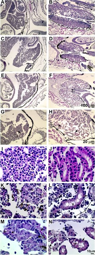
processed for histology analysis (Fig. 9). Compared to mock-in-
fected tadpoles that had been pretreated with the vector control,
tadpoles infected with FV3 after vector control treatment exhib-
ited significant damage to their livers (Fig. 9A and E, respectively)
and kidneys (Fig. 9B and F, respectively). These injuries included
the loss of tissue architecture, immune cell infiltration, and the
formation of intracytoplasmic inclusion bodies (Fig. 9G and H),
characteristic of RV pathology (43). Intriguingly, in corrobora-
tion of the rXlIFN-diminished FV3 loads (Fig. 8B), pretreatment
of animals with rXlIFN reduced the severity of the tissue damage
incurred from subsequent FV3 infections (Fig. 9). However, tad-
poles injected with this potent antiviral cytokine prior to viral
infection nonetheless incurred liver and kidney tissue damage,
including substantial cytopathology and a loss of tissue architec-
ture (Fig. 9C and D, respectively), which were not found in vector-
pretreated uninfected controls (Fig. 9A and B, respectively) or
rXlIFN-pretreated uninfected animals (data not shown). These
FIG 7 FV3-infected X. laevis adults exhibit greater kidney and liver viral bur- observations suggest that the extensive FV3-induced damage to
dens than virus-infected tadpoles. Tadpoles were infected i.p. with 1 ⫻ 104 multiple organs is a major contributor to the mortality of infected
PFU of FV3; adult frogs were infected i.p. with 5 ⫻ 106 PFU of FV3. At the animals, including those pretreated with rXlIFN.
indicated times, animals were euthanized, tissues were collected, and DNA was
isolated. Viral loads were measured by absolute qRT-PCR (vDNA Pol II) of 50
Histological examination at higher magnification revealed that
ng total DNA derived from kidney (A) and liver (B) tissues. Tissues from three rXlIFN treatment prior to FV3 infection resulted in fewer intracy-
individuals per treatment group were examined. Results are means ⫾ SEMs. *, toplasmic inclusion bodies (typical of FV3 infection) in liver and
significant difference between the tadpole and adult stages (P ⬍ 0.05). kidney tissues (Fig. 9K and L, respectively) than the number in
vector-treated FV3-infected animals (Fig. 9M and N, respec-
tively). This corroborates our earlier findings that rXlIFN stimu-
times examined, tadpoles had substantially lower (as much as lation of tadpoles significantly diminishes their viral loads. How-
10 times lower) FV3 loads in kidney and liver tissues than adult ever, despite the diminished viral burdens and irrespective of the
frogs (Fig. 7A and B). treatment (rXlIFN or vector), infection of tadpoles with FV3 re-
X. laevis tadpoles administered rXlIFN exhibit prolonged sulted in severe disruption of the cellular organization in both
mean survival times and lower viral burdens during FV3 infec- liver and kidney tissues as well as the presence of major necrotic
tion. Our initial rationale that tadpole viral susceptibility stems cell death (dark or black-stained cells and debris; indicated by the
from insufficient IFN responses during FV3 infection (compared letter n) and apoptotic cell death (vacuolated and blebbed cells;
to that in adults) was supported by the significant decreases in FV3 indicated by the letter a) in liver tissue (Fig. 9K and M, respec-
burdens observed following administration of rXlIFN to tadpoles tively) and kidney tissue (Fig. 9L and N, respectively). None of
May 2014 Volume 88 Number 10 jvi.asm.org 5773Grayfer et al.
Downloaded from http://jvi.asm.org/ on May 8, 2015 by guest
FIG 8 Pretreatment of tadpoles with rXlIFN prior to FV3 infection increases animal mean survival time and lowers postmortem viral burdens. Stage 50 tadpoles
(n ⫽ 11 per treatment group) were injected with rXlIFN (500 ng/tadpole) or an equal volume of the vector control and 8 h later were infected i.p. with 1 ⫻ 104
PFU of FV3 (in APBS) or an equal volume of APBS. (A) Animal survival was monitored over the course of 60 days. Tadpoles were checked twice daily, and dead
animals were immediately frozen and stored at ⫺20°C for DNA isolation. DNA was isolated from whole tadpoles postmortem, and FV3 loads were determined
by absolute qRT-PCR. (B) FV3 copy numbers from 11 vector-injected and FV3-infected tadpoles and 10 rXlIFN-injected and FV3-infected tadpoles are
represented as mean log numbers ⫾ SEMs. *, significant difference between the vector and rXlIFN treatment groups (P ⬍ 0.05).
these pathologies were observed in the liver and kidney tissues of signal through at least two distinct receptor complexes (32). It is
the tadpoles treated with the saline vector or tadpoles pretreated noteworthy that the recombinant form of the X. laevis IFN iden-
with rXlIFN and injected with the vehicle (Fig. 9I and J, respec- tified here displayed very potent antiviral properties. In addition,
tively, and data not shown). Finally, the degree of immune infil- our phylogenetic analysis suggests that X. tropicalis IFNs 1 and 2
tration seen within the tissues of afflicted animals (irrespective of may have been conserved from an ancestral IFN, whereas X. tropi-
treatment) was modest, considering the severity of the FV3-in- calis IFNs 3, 4, and 5, the X. laevis IFN, and the fish IFNs diverged
duced destruction (Fig. 9). more recently. Possibly, as in fish, the amphibian type I IFNs may
possess distinct functional properties and interact with different
DISCUSSION cognate receptors. With regard to the position of amphibians in
This article presents the first reported study of amphibian type I the context of the evolution of type I IFN defenses, it will be in-
IFN immunity. This is particularly relevant considering the key triguing to learn if, indeed, the individual Xenopus type I IFNs
place in vertebrate evolution represented by amphibian species display a disparity of antiviral functions akin to those seen in
such as X. laevis. Interestingly, a hallmark characteristic of fish and teleosts or act more like the higher vertebrate IFNs and signal
amphibian IFNs is the five-exon/four-intron genomic organiza- through a single receptor complex.
tion, which is very distinct from the intronless type I IFN tran- During our analysis of X. laevis type I IFN gene expression in
scripts encoded by reptiles, birds, and mammals (24, 29, 36). In the tissues of virus-infected tadpoles and adult frogs, we observed
fact, there has been considerable debate whether the fish type I that despite similar FV3 expansion kinetics across tissues, the liver
IFNs are ancestral homologues of the higher vertebrate type I or IFN gene expression was delayed in comparison to that seen in the
type III IFNs. Notably, the fish counterparts exhibit a five-exon/ kidneys of infected animals, particularly in adult frogs. It is diffi-
four-intron gene organization akin to that of the mammalian type cult to speculate on the precise explanation for this discrepancy.
III IFNs but possess prototypic sequence patterns, such as cysteine Presumably, these observations reflect the distinct immune cell
positioning and the C-terminal CAWE motifs conserved in higher compositions of these tissues, where the liver contains large quan-
vertebrate type I IFNs (24, 27, 28, 44). In fact, since fish are cur- tities of immunomodulatory Kupffer cells (reviewed in reference
rently believed not to possess type III IFNs, it is intriguing that 45), which would likely skew the immune outcomes within this
amphibians possess both type I IFNs akin to those of fish and bona site of infection.
fide type III IFNs (27). This suggests that the divergence of type I This work is the first to demonstrate the roles of the amphibian
and III IFNs occurred prior to the appearance of tetrapods (27) type I IFNs in immunity against the etiologically relevant ranavi-
and brings to question the relative biological roles of the amphib- rus FV3. Our findings underline both the efficacy with which the
ian type I IFNs compared to those of fish, which are currently X. laevis IFN is able to deter infections with a potent virus like FV3
thought to lack the type III antiviral cytokines. and the resilience of this pathogen, where, despite significant
The efficacy of function of the four fish IFN subgroups range rXlIFN-induced perturbation of viral replication and expansion,
from highly antiviral to nonfunctional (39) and are believed to FV3 was still ultimately lethal to the tadpole hosts. Indeed, we
5774 jvi.asm.org Journal of VirologyAmphibian IFN Responses to the Ranavirus FV3
observed that tadpoles mounted relatively delayed and meager
FV3-elicited IFN responses compared to those induced by the
virus in adult frogs. This difference is a likely factor contributing to
the FV3 susceptibility of tadpoles and the adult resistance to this
virus. Furthermore, as nothing is known regarding the receptor
system employed by the amphibian type I IFN, it is possible that
the tadpole susceptibility is further compounded at the receptor
level.
Considering the notion that X. laevis adults mount robust and
presumably effective IFN responses to FV3 that contribute to the
effective clearance of this pathogen within 2 weeks, it is intriguing
that (at least during the acute infection) adults exhibit signifi-
cantly greater FV3 loads than tadpoles, which are more susceptible
to and typically succumb to infection. This suggests that the in-
ability to contain and/or minimize the expansion of FV3 is not the
mechanism resulting in tadpole mortality. Conversely, our obser-
vations underline the inherent capacity of adult frogs to tolerate
Downloaded from http://jvi.asm.org/ on May 8, 2015 by guest
much greater FV3 burdens. This idea is substantiated by the find-
ings that while the rXlIFN-pretreated tadpoles possessed viral
loads several log units lower than those of the adults, the rXlIFN-
pretreated tadpoles still eventually succumbed to FV3 infection.
This mortality, despite the extended mean survival time and con-
siderably diminished viral burdens in tadpoles, suggests that even
at significantly reduced titers, FV3 confers irreversible damage to
multiple tadpole organs (as seen in our histology studies), includ-
ing an extensive loss of tissue architecture and cellular organiza-
tion through necrotic and apoptotic cell death. Furthermore,
the meager immune infiltration of these severely compromised
tissues suggests that this damage is likely incurred relatively
early in the infection and/or results primarily from virus-me-
diated cytopathy rather than inflammation.
It is noteworthy that FV3 infection of rats was employed as
a model for hepatitis over 30 years ago (46–48). In these exper-
iments, rat inoculation with FV3 resulted in necrotic Kupffer
cell death, cessation of hepatic clearance, and subsequent tox-
icity, culminating in severe hepatitis, inflammation, and ani-
mal deaths (46). Although the temperature of 37°C used for
these mammalian studies was not permissive to ranaviral rep-
lication (49), FV3 particles were detected in phagocytic vacu-
oles and endocytic compartments of mammalian Kupffer cells,
where approximately a quarter of these virions exhibited viral
FIG 9 Pretreatment of tadpoles with rXlIFN does not prevent FV3 infection-
induced liver and kidney damage. Tadpoles were preinjected with 500 ng of
total rXlIFN or an equal volume of the vector control and 8 h later were
infected with FV3 or mock infected by APBS injection. Animals were reared
until they displayed characteristic signs of terminal infection, sacrificed, and
prepared for histology analysis. The cellular compositions were as follows:
vector, APBS, liver (A); vector, APBS, kidney (B); rXlIFN, FV3, liver (C);
rXlIFN, FV3, kidney (D); vector, FV3, liver (E); vector, FV3, kidney (F); higher
magnification, vector, FV3, liver (G); and higher magnification, vector, FV3,
kidney (H). Arrows, intracytoplasmic inclusions characteristic of ranavirus-
induced pathology. (I to N) Pretreatment of tadpoles with rXlIFN does not
prevent FV3 infection-induced cellular damage, necrosis, and apoptosis. Tad-
poles were preinjected with rXlIFN (500 ng) or the vector control 8 h before
infection with FV3. Animals were reared until they displayed characteristic
signs of terminal infection, sacrificed, and prepared for histology analysis. The
cellular compositions were as follows: vector, APBS, liver (I); vector, APBS,
kidney (J); rXlIFN, FV3, liver (K); rXlIFN, FV3, kidney (L); vector, FV3, liver
(M); and vector, FV3, kidney (N). Arrows, FV3-induced intracytoplasmic in-
clusions; n, necrotic cells; a, apoptotic cells. Bars, 100 m (A to F), 20 m (G,
H), and 10 m (I to N).
May 2014 Volume 88 Number 10 jvi.asm.org 5775Grayfer et al.
core membrane-host membrane fusion and viral core material 11. Bollinger TK, Mao J, Schock D, Brigham RM, Chinchar VG. 1999.
release into cell cytoplasms (50). This nonspecific capacity to Pathology, isolation, and preliminary molecular characterization of a
novel iridovirus from tiger salamanders in Saskatchewan. J. Wildl. Dis.
infiltrate phagocytic cells of otherwise resistant hosts may ex- 35:413– 429. http://dx.doi.org/10.7589/0090-3558-35.3.413.
plain the recent increase in the prevalence and expansion of the 12. Donnelly TM, Davidson EW, Jancovich JK, Borland S, Newberry M,
host tropisms of ranaviruses. Furthermore, the inability of FV3 Gresens J. 2003. What’s your diagnosis? Ranavirus infection. Lab. Anim.
to replicate at nonpermissive mammalian body temperatures (NY) 32:23–25.
implies that the pathogenic effects described above were not 13. Greer AL, Berrill M, Wilson PJ. 2005. Five amphibian mortality events
associated with ranavirus infection in south central Ontario, Canada. Dis.
mediated through cell lytic mechanisms (at least in the animal Aquat. Organ. 67:9 –14. http://dx.doi.org/10.3354/dao067009.
models described above) but instead were mediated through 14. Jancovich JK, Davids EW, Seiler A, Jacobs BL, Collins JP. 2001. Trans-
prepackaged factors already present in the FV3 virion. In sup- mission of the Ambystoma tigrinum virus to alternative hosts. Dis. Aquat.
port of this, early research on FV3 in mammalian models also Organ. 46:159 –163. http://dx.doi.org/10.3354/dao046159.
15. Jancovich JK, Bremont M, Touchman JW, Jacobs BL. 2010. Evidence for
revealed that this virus induces rapid cellular RNA, DNA, and
multiple recent host species shifts among the ranaviruses (family Irido-
protein synthesis arrest (51) resulting from structural proteins viridae). J. Virol. 84:2636 –2647. http://dx.doi.org/10.1128/JVI.01991-09.
that can be solubilized from the viral particles (52) and that are 16. Becker JA, Tweedie A, Gilligan D, Asmus M, Whittington RJ. 2003.
sufficient for inhibiting host cell nucleic acid synthesis (49). Experimental infection of Australian freshwater fish with epizootic
Our findings indicate that the amphibian tadpole susceptibility haematopoietic necrosis virus (EHNV). J. Aquat. Anim. Health 25:66 –76.
http://dx.doi.org/10.1080/08997659.2012.747451.
to FV3 is most likely not the result of extensive viral burdens.
Downloaded from http://jvi.asm.org/ on May 8, 2015 by guest
17. Haislip NA, Gray MJ, Hoverman JT, Miller DL. 2011. Development and
Rather, we propose that prepackaged FV3 components and/or disease: how susceptibility to an emerging pathogen changes through
FV3 products generated during early phases of ranaviral infec- anuran development. PLoS One 6:e22307. http://dx.doi.org/10.1371
tion culminate in compounding long-term tissue damage and /journal.pone.0022307.
organ failure, prominently contributing to tadpole mortality. 18. Hyatt AD, Gould AR, Zupanovic Z, Cunningham AA, Hengstberger S,
Whittington RJ, Kattenbelt J, Coupar BE. 2000. Comparative studies of
piscine and amphibian iridoviruses. Arch. Virol. 145:301–331. http://dx
ACKNOWLEDGMENTS .doi.org/10.1007/s007050050025.
19. Robert J, Abramowitz L, Gantress J, Morales HD. 2007. Xenopus laevis:
We thank Daria Krenitsky for her assistance with histology analysis. We
a possible vector of Ranavirus infection? J. Wildl. Dis. 43:645– 652. http:
thank Greg V. Chinchar for generously providing the anti-FV3 53R Ab. //dx.doi.org/10.7589/0090-3558-43.4.645.
We thank Tina Martin and David Albright for animal husbandry. We 20. Chen G, Robert J. 2011. Antiviral immunity in amphibians. Viruses
thank Eva-Stina Eldhom for her critical review of the manuscript. 3:2065–2086. http://dx.doi.org/10.3390/v3112065.
This work was supported by grants R24-AI-059830 and IOB-074271 21. De Jesus Andino F, Chen G, Li Z, Grayfer L, Robert J. 2012. Suscepti-
from NIH and NSF, respectively. L.G. was supported by an NSERC post- bility of Xenopus laevis tadpoles to infection by the ranavirus frog-virus 3
doctoral fellowship program and an LSRF postdoctoral fellowship pro- correlates with a reduced and delayed innate immune response in com-
gram from the Howard Hughes Medical Institute. parison with adult frogs. Virology 432:435– 443. http://dx.doi.org/10
.1016/j.virol.2012.07.001.
22. Morales HD, Abramowitz L, Gertz J, Sowa J, Vogel A, Robert J. 2010.
REFERENCES Innate immune responses and permissiveness to ranavirus infection of
1. Chinchar VG. 2002. Ranaviruses (family Iridoviridae): emerging cold- peritoneal leukocytes in the frog Xenopus laevis. J. Virol. 84:4912– 4922.
blooded killers. Arch. Virol. 147:447– 470. http://dx.doi.org/10.1007 http://dx.doi.org/10.1128/JVI.02486-09.
/s007050200000. 23. Hervas-Stubbs S, Perez-Gracia JL, Rouzaut A, Sanmamed MF, Le Bon
2. Williams T, Barbosa-Solomieu V, Chinchar VG. 2005. A decade of A, Melero I. 2011. Direct effects of type I interferons on cells of the
advances in iridovirus research. Adv. Virus Res. 65:173–248. http://dx.doi immune system. Clin. Cancer Res. 17:2619 –2627. http://dx.doi.org/10
.org/10.1016/S0065-3527(05)65006-3. .1158/1078-0432.CCR-10-1114.
3. Chinchar VG, Hyatt A, Miyazaki T, Williams T. 2009. Family Iridoviridae: 24. Robertsen B. 2006. The interferon system of teleost fish. Fish Shellfish
poor viral relations no longer. Curr. Top. Microbiol. Immunol. 328:123–170. Immunol. 20:172–191. http://dx.doi.org/10.1016/j.fsi.2005.01.010.
4. Granoff A, Came PE, Breeze DC. 1966. Viruses and renal carcinoma of 25. Zou J, Secombes CJ. 2011. Teleost fish interferons and their role in
Rana pipiens. I. The isolation and properties of virus from normal and immunity. Dev. Comp. Immunol. 35:1376 –1387. http://dx.doi.org/10
tumor tissue. Virology 29:133–148. .1016/j.dci.2011.07.001.
5. Granoff A, Came PE, Rafferty KA, Jr. 1965. The isolation and properties 26. Chang M, Nie P, Collet B, Secombes CJ, Zou J. 2009. Identification
of viruses from Rana pipiens: their possible relationship to the renal ade- of an additional two-cysteine containing type I interferon in rainbow
nocarcinoma of the leopard frog. Ann. N. Y. Acad. Sci. 126:237–255. http: trout Oncorhynchus mykiss provides evidence of a major gene dupli-
//dx.doi.org/10.1111/j.1749-6632.1965.tb14278.x. cation event within this gene family in teleosts. Immunogenetics 61:
6. Zhan QY, Xiao F, Li ZQ, Gui JF, Mao J, Chinchar VG. 2001. Charac- 315–325. http://dx.doi.org/10.1007/s00251-009-0366-y.
terization of an iridovirus from the cultured pig frog Rana grylio with 27. Qi Z, Nie P, Secombes CJ, Zou J. 2010. Intron-containing type I and type
lethal syndrome. Dis. Aquat. Organ. 48:27–36. http://dx.doi.org/10.3354 III IFN coexist in amphibians: refuting the concept that a retroposition
/dao048027. event gave rise to type I IFNs. J. Immunol. 184:5038 –5046. http://dx.doi
7. Speare R, Freeland WJ, Bolton SJ. 1991. A possible iridovirus in eryth- .org/10.4049/jimmunol.0903374.
rocytes of Bufo marinus in Costa Rica. J. Wildl. Dis. 27:457– 462. http://dx 28. Zou J, Tafalla C, Truckle J, Secombes CJ. 2007. Identification of a second
.doi.org/10.7589/0090-3558-27.3.457. group of type I IFNs in fish sheds light on IFN evolution in vertebrates. J.
8. Speare R, Smith JR. 1992. An iridovirus-like agent isolated from the Immunol. 179:3859 –3871.
ornate burrowing frog Limnodynastes ornatus in northern Australia. Dis. 29. Sun B, Robertsen B, Wang Z, Liu B. 2009. Identification of an Atlantic
Aquat. Organ. 14:51–57. http://dx.doi.org/10.3354/dao014051. salmon IFN multigene cluster encoding three IFN subtypes with very dif-
9. Cunningham AA, Langton TE, Bennett PM, Lewin JF, Drury SE, Gough ferent expression properties. Dev. Comp. Immunol. 33:547–558. http:
RE, Macgregor SK. 1996. Pathological and microbiological findings from //dx.doi.org/10.1016/j.dci.2008.10.001.
incidents of unusual mortality of the common frog (Rana temporaria). 30. Li Z, Strunk JJ, Lamken P, Piehler J, Walz T. 2008. The EM structure of
Philos. Trans. R. Soc. Lond. B Biol. Sci. 351:1539 –1557. http://dx.doi.org a type I interferon-receptor complex reveals a novel mechanism for cyto-
/10.1098/rstb.1996.0140. kine signaling. J. Mol. Biol. 377:715–724. http://dx.doi.org/10.1016/j.jmb
10. Drury SE, Gough RE, Cunningham AA. 1995. Isolation of an iridovirus- .2007.12.005.
like agent from common frogs (Rana temporaria). Vet. Rec. 137:72–73. 31. Samuel CE. 2001. Antiviral actions of interferons. Clin. Microbiol. Rev.
http://dx.doi.org/10.1136/vr.137.3.72. 14:778 – 809. http://dx.doi.org/10.1128/CMR.14.4.778-809.2001.
5776 jvi.asm.org Journal of VirologyYou can also read