Serotonin involvement in okadaic acid induced diarrhoea in vivo - USC
←
→
Page content transcription
If your browser does not render page correctly, please read the page content below
Archives of Toxicology (2021) 95:2797–2813
https://doi.org/10.1007/s00204-021-03095-z

    ORGAN TOXICITY AND MECHANISMS

Serotonin involvement in okadaic acid‑induced diarrhoea in vivo
M. Carmen Louzao1 · Celia Costas1 · Paula Abal1 · Toshiyuki Suzuki2 · Ryuichi Watanabe2 ·
Natalia Vilariño1 · Cristina Carrera1 · Andrea Boente‑Juncal1 · Carmen Vale1 · Mercedes R. Vieytes3                               ·
Luis M. Botana1

Received: 22 April 2021 / Accepted: 15 June 2021 / Published online: 20 June 2021
© The Author(s) 2021

Abstract
The consumption of contaminated shellfish with okadaic acid (OA) group of toxins leads to diarrhoeic shellfish poisoning
(DSP) characterized by a set of symptoms including nausea, vomiting and diarrhoea. These phycotoxins are Ser/Thr phos-
phatase inhibitors, which produce hyperphosphorylation in cellular proteins. However, this inhibition does not fully explain
the symptomatology reported and other targets could be relevant to the toxicity. Previous studies have indicated a feasible
involvement of the nervous system. We performed a set of in vivo approaches to elucidate whether neuropeptide Y (NPY),
Peptide YY (PYY) or serotonin (5-HT) was implicated in the early OA-induced diarrhoea. Fasted Swiss female mice were
administered NPY, PYY(3–36) or cyproheptadine intraperitoneal prior to oral OA treatment (250 µg/kg). A non-significant
delay in diarrhoea onset was observed for NPY (107 µg/kg) and PYY(3–36) (1 mg/kg) pre-treatment. On the contrary, the
serotonin antagonist cyproheptadine was able to block (10 mg/kg) or delay (0.1 and 1 mg/kg) diarrhoea onset suggesting a
role of 5-HT. This is the first report of the possible involvement of serotonin in OA-induced poisoning.

Keywords Okadaic acid · Diarrhoeic shellfish poisoning (DSP) · 5-Hydroxytryptamine · Neuropeptide Y · Peptide YY

Introduction                                                                 recovery after 3 days (Yasumoto et al. 1978; EFSA 2008).
                                                                             Exposure to DSP has been frequently reported in various
Okadaic acid (OA) group of toxins comprise polyether fatty                   countries (Young et al. 2019; Vale 2020), representing the
acids synthetized by dinoflagellates of the genera Prorocen-                 primary cause of bans on the harvesting of aquaculture in
trum and Dinophysis. Bivalves may accumulate the toxins                      Japan and Europe (Reguera et al. 2014).
following the consumption of this toxic phytoplankton.                          Previous studies have revealed that OA inhibits serine/
Therefore, OA and related compounds enter the food chain                     threonine protein phosphatases (PPs) 1, 2A, 4, 5 and 6 activ-
reaching humans through toxin-containing seafood inges-                      ity (Bialojan and Takai 1988; Brewis et al. 1993; Chen et al.
tion causing diarrhoeic shellfish poisoning (DSP) (Yasumoto                  1994; Prickett and Brautigan 2006). PPs remove a phos-
et al. 1978). DSP can be developed fast, between 30 min                      phate group from the phosphorylated amino acid residue
and a few hours afterwards. Symptomatology includes nau-                     of a wide variety of proteins (Yadav et al. 2017), meaning
sea, vomiting, diarrhoea and abdominal pain, achieving full                  disturbance in their activity can modify downstream cel-
                                                                             lular pathways. OA in vitro has been described to induce
                                                                             cytoskeleton reorganization (Espina et al. 2010; Opsahl et al.
* M. Carmen Louzao                                                           2013; Louzao et al. 2015), cell death (Dietrich et al. 2020)
  mcarmen.louzao@usc.es                                                      and cell cycle alteration (Feng et al. 2018). However, during
1
                                                                             the last decade, it has been discussed whether OA-exerted
     Departamento de Farmacología, Facultad de Veterinaria,
     Universidade de Santiago de Compostela, 27002 Lugo,
                                                                             effects are fully explained by its PP inhibition (Espina et al.
     Spain                                                                   2010; Munday 2013).
2
     Fisheries Technology Institute, National Research
                                                                                Diarrhoea is defined as reduced stool consistency,
     and Development Agency, Japan Fisheries Research                        increased water content and number of evacuations. A
     and Education Agency, Yokohama 236‑8648, Japan                          wide array of causes and pathophysiological mechanisms
3
     Departamento de Fisiología, Facultad de Veterinaria,                    have been proposed for both infectious and non-infectious
     Universidade de Santiago de Compostela, 27002 Lugo, Spain

                                                                                                                                 13
                                                                                                                            Vol.:(0123456789)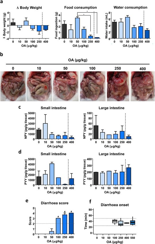
2798 Archives of Toxicology (2021) 95:2797–2813 diarrhoea (Thiagarajah et al. 2015; Anand et al. 2016; Materials and methods Camilleri et al. 2017). A considerable number of those mechanisms involve neuronal activation of the Enteric Animal model Nervous System (ENS). The ENS together with parasym- pathetic and sympathetic innervation throughout the gas- Mouse bioassay had been an accepted method for marine trointestinal tract coordinate and regulate essential func- biotoxins detection, though nowadays has been replaced by tions regarding pancreatic secretion, gut motility, fluid analytical methods on behalf of NC3R’s principles (Union secretion and nutrient absorption among others (Li et al. 2011). Based on the Organization for Economic Cooperation 2000; Hu and Spencer 2018). Within components of the and Development guidelines for acute oral toxicity studies, ENS some members of the Neuropeptide Y (NPY) family we decided to use female mice as an animal model (OECD/ have been closely related to functions such as fluid absorp- OCDE 2002). One-month-old Swiss female mice weighing tion and gastric emptying (Saria and Beubler 1985; Wang between 18 and 22 g from the colonies of the University of et al. 2010). These 36-aa peptides’ location include neu- Santiago de Compostela were employed for all the experi- ral and endocrine components (Ekblad and Sundler 2002; ments described. They were kept in controlled conditions of Mongardi Fantaguzzi et al. 2009). For instance, NPY is temperature (23 ± 2 °C), humidity (60–70%) and light/dark expressed in different regions of the brain, but also in cycles (12 h/ 12 h). Mice were placed individually on meta- sympathetic neurons and in the ENS (e.g., secretomotor bolic cages and fasted overnight with access to 5% glucose neurons) (Cox 2007; Mongardi Fantaguzzi et al. 2009). serum. Animals were randomly assigned to each treatment. On the contrary, enteroendocrine L cells are the mayor Mice received the toxin by oral gavage at 9 a.m. (10 mL/kg contributors of Peptide YY (PYY) in the body, though it body weight), moment at which food and drink were pro- has been likewise detected in myenteric neurons and in vided ad libitum. When any pre-treatment was studied, it was some brain areas (Ekblad and Sundler 2002; Morimoto given by intraperitoneal injection (1% body weight) prior to et al. 2008). In vitro, the DSP toxin OA downregulated the toxin. Note that the assays described hereafter were pre- NPY content and release of SH-SY5Y neuroblastoma cell ceded by these conditions. At the end of each experiment, line (Valdiglesias et al. 2012; Louzao et al. 2015). euthanasia by CO2 inhalation was conducted. All animal Another key signalling molecule in the gut is seroto- procedures were carried out in conformity to the European nin (5-HT), a bioamine mainly expressed along the diges- (EU directive 2010/63/EU), the Spanish legislation (Real tive tract (Erspamer and Testini 1959; Erspamer 1966; Decreto 53/2013, Decreto 296/2008) and to the principles Savelieva et al. 2008; Mawe and Hoffman 2013), whose approved by the Institutional Animal Care Committee of the physiological functions comprise intestine fluid secretion University of Santiago de Compostela under the procedure and motility (El-Salhy et al. 2013; Mawe and Hoffman Code: 01/17/LU-002 (approved on 22 September 2017). 2013; Coates et al. 2017; Camilleri et al. 2017; Hu and Spencer 2018). 5-HT is present in serotoninergic enteric neurons (Okamoto et al. 2014), though enterochromaffin Materials cells (ECCs) are its major producers which are scattered distributed along the epithelia (Sjolund et al. 1983). ECCs Okadaic acid employed in this study was kindly provided act as chemosensors (Braun et al. 2007; Lund et al. 2018) by the National Research Institute of Fisheries Science and mechanosensors (Fujimiya et al. 1997; Alcaino et al. (NRIFS) from the Fisheries Research and Education Agency 2018), triggering a response in the underlying nerve ter- (Yokohama, Japan). OA isolated from toxic dinoflagellate minals and the surrounding cells via neurotransmitters’ or Prorocentrum lima (Suzuki et al. 2014) was quantified by hormones’ signalling (Bertrand et al. 2000; Reynaud et al. PULCON method (Watanabe et al. 2016) on the quantita- 2016; Fazio Coles et al. 2020). tive NMR with an external standard. Purities (purity > 95%) Gathering the variety of pathophysiologic mechanisms were also confirmed by the NMR spectroscopy. Neuropep- resulting in diarrhoea and the important role of the ENS, tide Y and Peptide YY(3–36) were purchased from TOCRIS, we studied if OA-caused diarrhoea involves alteration of cyproheptadine hydrochloride sesquihydrate from Sigma- intestinal hormones (PYY) and/or neurotransmitters (5-HT Aldrich. All chemicals employed were analytical grade from and NPY). To elucidate this premise, we firstly performed Sigma-Aldrich Quimica S.A. (Madrid, Spain). an in vivo approach to determinate the dose–response of OA doses on diarrhoea outcome. Second, we assessed the effect of exogenous NPY or PYY(3–36) on OA-induced Dose–response of okadaic acid on diarrhoea diarrhoea. Furthermore, we evaluated 5-HT implication in DSP in mice using the 5-HT 1 and 5-HT 2 antagonist OA was previously reconstituted with ethanol. For adminis- cyproheptadine (CPH) prior to OA treatment. tration, OA doses were prepared by serial dilutions in 0.9% 13
Archives of Toxicology (2021) 95:2797–2813                                                                                 2799

saline solution. OA was given by oral gavage at 10, 50, 100,    Dose–response of cyproheptadine in vivo at 2 h
250 and 400 µg/kg doses. Control mice received the vehicle
alone. Diarrhoea onset time and diarrhoea score were regis-     Doses of 0.1, 1, 3 and 10 mg/kg CPH were tested as a pre-
tered along with the symptoms presented at 1, 3, 6, 9, 12 and   treatment to 250 µg/kg OA for 2 h. The experimental devel-
24 h. Anatomopathological examination took place when the       opment was as detailed above. Dosages were diluted in
necropsy was performed. Small and large intestines were         physiological solution. OA was given by oral gavage 30 min
removed and stored at − 20 °C.                                  after CPH dose was injected via intraperitoneal. Mice were
                                                                observed, stools were collected, diarrhoea score and time of
                                                                onset were measured 2 h of treatment. Then necropsy was
Pre‑treatment in vivo studies at 6 h                            performed and the gut was removed and stored at −20 °C.

NPY was previously reconstituted with milliQ water. Doses       Short‑time exposure CPH dose–response
of 550 µg/kg OA and 107 µg/kg NPY were prepared by
dilution of each compound in physiological solution. Four       CPH and OA doses were prepared as described in previous
treatment groups were set: (i) control, (ii) NPY, (iii) OA      sections. In this case, animals were split in six groups: (i)
and (iv) NPY plus OA. Each group was performed in dupli-        control, (ii) CPH, (iii) OA, (iv) 0.1 mg/kg CPH plus OA, (v)
cate. In this last case, intraperitoneal injection of NPY was   1 mg/kg CPH plus OA and (vi) 6 mg/kg CPH plus OA. CPH
performed 15 min prior to OA administration by oral gav-        was given via intraperitoneal 30 min before administering
age. The time of diarrhoea outbreak, diarrhoea score and        250 µg/kg OA by oral gavage. Mice receiving the toxin alone
symptomatology were registered and stools collected at 1, 3     were first treated, the time of diarrhoea onset was set as the
and 6 h of treatment. At the end of the experiment, animals     end of the experiment for the remaining treatments. Same
were subjected to necropsy. Small and large intestines were     performance and data as in the previous experiment were
sampled and kept at − 20 °C.                                    obtained, but for 30 min post-toxin administration.
   A similar approach was performed for PYY(3–36) pre-
treatment studies. PYY(3–36) was also reconstituted with        Diarrhoea score
milliQ water. The peptide was diluted in physiological solu-
tion to reach 1 mg/kg PYY(3–36). Mice were split into four      To assess the differences in terms of how severe the diar-
groups comprising control, PYY(3–36), OA and PYY(3–36)          rhoea was, we designed a scoring system (Table 1), meaning
plus OA. In this last group, PYY(3–36) was given by intra-      0 normal faeces; 1 soft faeces; 2 shapeless soft faeces; 3
peritoneal injection 15 min before oral administration of       watery diarrhoea; 4 having for more than once diarrhoea. To
550 µg/kg OA. Same performance and data as in the previ-        be considered a different time of diarrhoea, it was required
ous experiment were obtained, ending the experiment at 6 h      to be at least 20 min past the last defecation.
post-toxin administration.
   CPH pre-treatment approach was in line with the previous     Neuromodulators’ detection
ones. CPH was first reconstituted with ethanol. CPH was
diluted in physiological solution to prepare the dose 3 mg/     Small intestine (ileum) and large intestine (proximal colon)
kg. Different sets of animals were given vehicles, CPH or       were first extracted. Samples were cleansed in ice-cold PBS
OA each alone or CPH plus OA. Mice were first injected          and weighted immediately afterwards. PBS was added (1:9
CPH intraperitoneally 30 min prior to 250 µg/kg OA by oral      w/v) and tissues were homogenized and sonicated. Finally,
gavage. Same data as in the previous approaches were also       they were centrifuged for 5 min at 10,000×g at 4 °C and
collected.

                                                                Table 1  Diarrhoea scoring
                                                                system criteria                  0       Normal faeces
NPY or PYY(3–36) pre‑treatment of mice in 2 h
                                                                                                 1       Soft faeces
experiments
                                                                                                 2       Shapeless soft faeces
                                                                                                 3       Watery diarrhoea
Preparation of treatments and administration were performed
                                                                                                 4       Watery diarrhoea
as described in the preceding assessment. The peptides were                                                repeatedly
each diluted in physiological solution to obtain 107 µg/kg                                               (taking into account
NPY and 1 mg/kg PYY(3–36). NPY or PYY(3–36) was                                                            20 min between each
given intraperitoneal 15 min before oral administration of                                                 time)
250 µg/kg OA. During the following 2 h, the same data as                                         a
                                                                                                  A score equal or higher than 2
detailed in the above experiment were obtained.                                                  is considered diarrhoea

                                                                                                                    13
2800 Archives of Toxicology (2021) 95:2797–2813
stored at − 20 °C. When required, extracts were diluted for Results
the compounds’ concentration to fall within the linear range
of the standard solutions. In all cases, absorbance was meas- Dose–response of okadaic acid
ured in a Multi-mode Microplate Reader Synergy 4 (Biotek).
NPY was measured in samples from OA dose–response To determine at which concentration OA is able to trigger
at 24 h and from 6 h NPY pre-treatment studies. Enzyme- diarrhoea, several doses of the toxin (10, 50, 100, 250 and
linked Immunosorbent Assay (ELISA) Kit for Neuropep- 400 µg/kg) were administered to mice that were observed
tide Y from Cloud Clone Corp. was employed. The range of for 24 h. During the experiment, the symptomatology was
detection was 2.47–200 pg/mL and absorbance was meas- monitored in detail at 1, 3, 6, 9, 12 and 24 h of OA treat-
ured at 450 nm. ment. No symptoms were detected in 10 µg/kg treated mice
PYY was analysed in mice intestines from OA throughout the experiment. In the case of 50 µg/kg dose,
dose–response at 24 h and from 6 h PYY(3–36) pre-treat- these animals did not present any symptoms, but one mouse
ment experiments. ELISA Kit for Peptide YY (Cloud-Clone alone had soft faeces at 12 h, being normal at 24 h. Treat-
Corp.) was used. The detection range was 12.35–1000 pg/ ments of 100, 250 and 400 µg/kg induced squint-eyes, pilo-
mL and absorbance was read at 450 nm. erection, spasms, cyanosis and even death for the highest
5-HT was determined in samples stored from CPH pre- dose (Table 2). This approach allowed to register the time at
treatment (6 h), dose–response of CPH (2 h) and short time which mice recovered, i.e., had no symptoms. Clinical signs
exposure CPH dose–response experiments. The Serotonin were noted at 1 h for 100 µg/kg OA treatment, being absent
ELISA kit from Enzo Life Sciences was used for 5-HT at 3 h. None of the reported symptoms were identified at 9 h
detection. The range of detection was 0.49–500 ng/mL and for 250 µg/kg OA. Finally, no mice that received 400 µg/kg
absorbance was read at 405 nm. OA reached the 24 h of treatment.
Variations in body weight after 24 h of OA treatment
Statistical analysis were measured, as well as food and water consumption dur-
ing the experiment (Fig. 1a). Both body weight variation and
Graphpad Prism and RStudio were employed to perform food intake display a similar pattern.
the statistical analyses. First, the distribution and homosce- At the end of the experiment, mice were subjected to
dasticity of the data set were tested. If it followed a normal necroscopic analysis, focusing on anatomopathological eval-
distribution, t test was performed to compare two treatments uation of the gastrointestinal tract (Fig. 1b). While 10, 50
or one-way ANOVA plus Bonferroni multiple comparison and 100 µg/kg OA examination revealed no differences with
test in the case more groups were analysed. Conversely, control, 250 µg/kg dose was featured by a swollen stomach
under no normal distribution of data, Mann–Whitney test and fluid accumulation in the small intestine. In the case of
or Kruskal–Wallis test followed by Bonferroni multiple 400 µg/kg OA-treated mice, swollen stomach accompanied
comparison test was conducted. The significance threshold by moderate to strong fluid accumulation in the small intes-
was set at P < 0.05. tine were observed for all animals.
Neuropeptides like NPY and PYY are involved in the
regulation of nutrients absorption and exert a protective role
(Moriya et al. 2010; Tough et al. 2011). Thus, we aimed
to elucidate how OA affects NPY and PYY along the gut
in vivo after 24 h treatments. OA reduces NPY in the small
Table 2 Symptomatology of Symptoms Control OA (µg/kg)
24 h OA-treated animals (%)
10 50 100 250 400
Apathy 0 0 0 33.3 100 50
Piloerection 0 0 0 33.3 66.7 50
Cyanosis 0 0 0 0 0 50
Spasms 0 0 0 33.3 66.7 50
On hind legs 0 0 0 66.7 33.3 0
Squint-eyes 0 0 0 66.7 66.7 50
Diarrhoea 0 0 0 100 100 100
Mortality 0 0 0 0 0 100
13Archives of Toxicology (2021) 95:2797–2813 2801
intestine and in large intestine (Fig. 1c). Only 250 µg/kg NPY was measured in the intestine of these animals 6 h
OA treatment decreases PYY in small intestine while large after toxin administration (Fig. 2c, d). OA reduces NPY
intestine’s PYY is not affected by OA at any of the given concentration in the small intestine; meanwhile, in NPY-
doses at 24 h (Fig. 1d). OA-treated animals’ NPY resembles control concentration
The evaluation of clinical signs was focused on diar- (Fig. 2c). Conversely, the same treatment seems to induce a
rhoea (percent of mice with this symptom, diarrhoea onset modest rise of NPY in the large bowel (Fig. 2d).
time and diarrhoea score). To assess the severity of diar-
rhoea, faeces from each mouse were scored as described PYY(3–36) effect on OA poisoning in 6 h
above (Table 1). Based on diarrhoea score criteria, there is experiments
a dose-dependent increase up to the maximal punctuation
(Fig. 1e). Control, 10 µg/kg and 50 µg/kg OA had normal The enteric nervous system plays a vital role in the response
faeces, except for one mouse that received the latter dose. to various gastrointestinal stimuli. The peptides of this nerv-
Then the lowest dose tested for developing diarrhoea was ous system regulate gastrointestinal movement, secretion,
100 µg/kg, with several defecations. Since neither control, absorption and other complex functions through endocrine,
10 nor 50 µg/kg treated mice had diarrhoea, no onset is rep- paracrine and neuronal actions. Both NPY and Peptide YY
resented regarding these treatments (Fig. 1f). are important enteric peptides. The observation of a soft effect
To further assure diarrhoea in the following approaches, of NPY over OA intoxication led us to study PYY. Y 2 recep-
we also included the dose: 550 µg/kg OA (Fig. 1f). We found tors are not only in nerve terminals around myenteric neu-
no significant differences in diarrhoea onset between any of rons, but also in mucosa and muscle layers and its agonist
the OA doses that triggers the symptom, showing an all-or- PYY(3–36) has additionally been related to a clear anti-diar-
none response. rhetic effect (Moriya et al. 2010; Tough et al. 2011). Thus, we
aimed to check whether this agonist had the ability to relieve
NPY effect on OA poisoning in 6 h experiments OA-induced diarrhoea. PYY(3–36) (1 mg/kg) was adminis-
tered prior to OA (550 µg/kg) was given. Symptomatology
Based on how OA affected in vitro NPY expression in was monitored along 6 h (Table S2). Apathy, piloerection and
addition to the pro-absorptive role of the neuropeptide, we squint-eyes were developed following toxin treatment, alone or
designed a 6 h experiment in which mice were intraperito- in combination with PYY(3–36) [PYY(3–36)-OA]. Both OA
neally administered NPY prior to OA treatment. Due to the and PYY(3–36)-OA-treated mice still presented symptoms at
lack of differences between doses in diarrhoea onset (Fig. 1f) the end of the experiment.
along with the evaluation of a dose closer to the previously Subsequently, the balance of body weight (Fig. S2a), food
described oral LD50 for OA (760 µg/kg) (Abal et al. 2018), (Fig. S2b) and water consumption (Fig. S2c) were measured.
we considered 550 µg/kg to be suitable to perform this Anatomopathological evaluation revealed swollen stomachs
assessment. The time of the experiment, 6 h, was selected and small intestine fluid accumulation of the toxin-treated ani-
to assure not just the inhibition of diarrhoea detection, but mals, with or without PYY(3–36) (Fig. S2d).
any delay in diarrhoea onset. Symptomatology of the ani- Diarrhoea score shows no differences between OA and
mals was recorded during the experiment (Table S1). Most PYY(3–36)-OA (Fig. 3a). Diarrhoea outbreak of PYY(3–36)-
animals exhibited a variety of symptoms such as piloerec- OA-treated mice displays a slight time delay when compared
tion and squint-eyes. Symptoms were still observed in mice to OA alone [OA 43 ± 7.8 min; PYY(3–36)-OA 49 ± 4.2 min]
treated with OA alone or NPY-OA at the end of the experi- (Fig. 3b). Still, it is a remarkable fact that 100% of OA-treated
ment. It should be remarked the fact that all mice treated mice presented diarrhoea; meanwhile, 85.7% of PYY(3–36)-
with the toxin or with the combination of both, OA and NPY OA-treated mice showed this symptom (Table S2 and Fig. 3a).
(NPY-OA), developed diarrhoea. It was then of interest to analyse the effect of the toxin on
Body weight variations (Fig. S1a), food (Fig. S1b) and PYY concentration (Fig. 3c, d). The amount of PYY meas-
water intake (Fig. S1c) were measured. Necropsy of all ured in small intestine has a remarkable non-significant
animals revealed that the toxin induced fluid accumulation increase in PYY(3–36)-OA-treated mice (Fig. 3c). In the
along the small intestine, being modestly improved by NPY large intestine, PYY(3–36)-OA treatment induces a minor
(Fig. S1d). rise compared to control (Fig. 3d).
The type of diarrhoea was evaluated with the diarrhoea
score (Fig. 2a). Pre-treatment with NPY did not modify NPY and PYY(3–36) pre‑treatment effect on OA
OA-induced diarrhoea nor supressed it (Fig. 2a). Diarrhoea poisoning in 2 h experiments
onset time displays a short, non-significant, delay with NPY
pre-treatment (OA 33 ± 3.4 min; NPY-OA 43 ± 4.9 min) Neither NPY nor PYY(3–36) modified OA poisoning, still
(Fig. 2b). it was of interest to assure that this was independent of OA
132802 Archives of Toxicology (2021) 95:2797–2813 13
Archives of Toxicology (2021) 95:2797–2813 2803
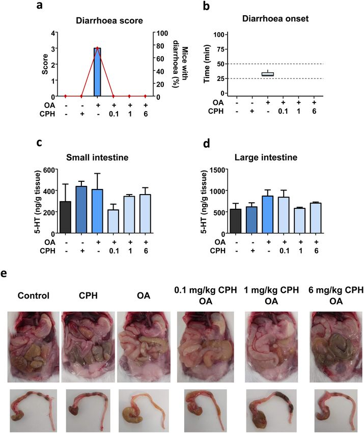
◂Fig. 1 Dose–response for diarrhoea induced by doses from 10 to induce diarrhoea, we administered 3 mg/kg CPH before OA
400 µg/kg okadaic acid in a 24 h study period. a Variation on mice treatment. The set of symptoms developed were observed
body weight, food and water intake. b Representative images of the
mice abdominal cavity at the end of the experiment. Image corre-
for 6 h and were similar in mice that received OA and OA
sponding to 400 µg/kg OA was taken 11:45 h after toxin administra- with CPH (CPH-OA) (Table S4). Diarrhoea was the repre-
tion, time at which the animal died. c Neuropeptide Y measured in sentative symptom and it should be highlighted that CPH
small and large intestines. d Peptide YY detected in small and large pre-treatment reduced the prevalence from 100% of the toxin
intestines. e Diarrhoea score. f Diarrhoea onset time. Inner box line
indicates median and dashed lines are set at 25 and 50 min. Graphs a,
alone to 61.5% (Table S4 and Fig. 5a).
c, d and e display mean ± SEM (n = 3). Since no animal treated with As in previous approaches, body weight variations (Fig.
400 µg/kg accomplished the experimental time, data for this dose S4a) as well as food (Fig. S4b) and water intake (Fig. S4c)
are separated by dotted line. Statistical analysis was conducted with were measured. Food intake was reduced in groups of mice
one-way ANOVA–Bonferroni multiple comparison test. In e, treat-
ments with ‘#’ over the bar are significantly different (P < 0.001) from
that received the toxin or CPH-OA (Fig. S4b). Macroscopic
those with a ‘ + ’. Significance is indicated with asterisks over the line evaluation of the abdominal cavity revealed mild fluid accu-
between treatments, so that *P < 0.05, otherwise non-significant mulation in the small intestine of animals that received OA,
resembling that of CPH-OA treated mice (Fig. S4d). Large
intestines were removed and examined (Fig. S4d). OA large
dose. Subsequently, 250 µg/kg OA was chosen to both assure intestines were featured by diarrhetic content; meanwhile,
diarrhoea (Fig. 1e–g) and avoid mortality (Table 2). Since CPH pre-treatment helped nearly restore normal intestinal
diarrhoea outbreak appears in less than 2 h, this was set as content.
the endpoint of the experiment. During this time, monitoring With regard to diarrhoea measured parameters, no sig-
of clinical signs identified symptoms such as piloerection or nificant difference in diarrhoea score was detected between
squint-eyes in all treatments involving the toxin (Table S3). OA and CPH-OA treated mice (Fig. 5a). Conversely, onset
Body weight variation (Fig. S3a) and food intake (Fig. of diarrhoea is significantly delayed by CPH administration
S3b) were measured showing no significant differences [OA 34 ± 3.7 min; CPH-OA 69.1 ± 3.2 min] (Fig. 5b). OA
between treatments. At the end of the experiment, mac- induced a stark increase in faeces wet weight that decreased
roscopic evaluation of the abdominal cavity was per- significantly with CPH pre-treatment (Fig. 5c).
formed (Fig. S3c). Animals treated with OA, NPY-OA and Intestine’s 5-HT was quantified in mice samples (Fig. 5d,
PYY(3–36)-OA revealed fluid accumulation mainly along e). OA and CPH-OA induced 5-HT increase in the small
the intestine even some mice that had no stools. Large intes- intestine (Fig. 5d). Although in large intestine, CPH-OA
tines were also removed and examined separately (Fig. S3c). treatment shows a non-significant increase over the other
Diarrhoeic content was observed in OA-treated mice alone treatments (Fig. 5e).
or in combination with NPY or PYY(3–36).
Diarrhoea was also evaluated (Fig. 4). The type of diar- Dose–response of CPH in OA poisoning
rhoea developed by animals pre-treated with either NPY or
PYY(3–36) was not different from that induced by OA alone The suppression of OA-triggered diarrhoea led us to perform
(Fig. 4a). Note that animals treated with control, NPY or a dose–response study. Since CPH effect occurs within 2 h
PYY(3–36) had no stools during the experimental time, so after toxin administration, experimental time was reduced
the score is 0 (Fig. 4a) and no diarrhoea onset is shown to 2 h. Here we assessed 0.1, 1, 3 and 10 mg/kg CPH as a
for these treatments (Fig. 4b). Regarding onset time of pre-treatment for 250 µg/kg OA. Clinical signs developed
diarrhoea, only a slight non-significant delay is observed by each group of treatment were monitored (Table S5). It is
in PYY(3–36) pre-treatment [OA 35 ± 2.1 min; NPY-OA noteworthy that diarrhoea was present in all mice adminis-
38 ± 8.1 min; PYY(3–36)-OA 45 ± 5.5 min] (Fig. 4b). tered with OA, but was absent in those pre-treated with 3 or
10 mg/kg CPH.
CPH effect on OA poisoning Body weight variation (Fig. S5a) as well as food inges-
tion (Fig. S5b) were measured. It was observed a tendency
The lack of a robust reaction to Y receptor ligands led us to in weight lost and reduced food intake. On the contrary,
study secretory pathways instead of pro-absorptive mecha- anatomopathological examination provides information
nisms. Serotonin is a key signalling molecule that medi- regarding the gastrointestinal tract at macroscopic level
ates physiological processes in the gut and its release is (Fig. S5c). Fluid accumulation in the stomach and intestine
stimulated by diarrhoeagenic compounds. Cyproheptadine was observed for OA-treated mice. In animals pre-treated
(CPH), an inverse agonist/antagonist of 5-HT receptors 1 with 0.1, 1 and 3 mg/kg CPH, OA still induced fluid accu-
and 2, has been described to elicit a response at the level mulation in the intestine and stomach. However, mice with
of other antisecretory drugs (Meddah et al. 2014). Conse- 10 mg/kg CPH pre-treatment displayed an ameliorated
quently, to assess the role of 5-HT on OA mechanism to fluid content in the intestine and a degree of solid content
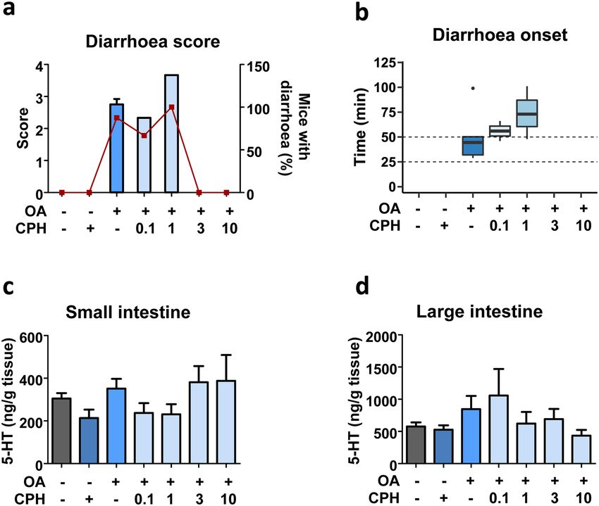
132804 Archives of Toxicology (2021) 95:2797–2813 Fig. 2 NPY effect on OA-induced diarrhoea and changes in NPY showing the median. c–d NPY concentration in small (c) and large in the gut at 6 h experiment. Animals were given 107 µg/kg NPY intestines (d). Data are mean ± SEM (n = 4 performed in duplicate). 15 min prior to 550 µg/kg OA administration. a Diarrhoea score Student t test resulted in no significant differences (b). For c and d, (bars) and percent of mice that developed diarrhoea (closed squares). statistical analyses were performed by one-way ANOVA and Bonfer- b Time of diarrhoea outbreak, values are expressed as boxplot roni multiple comparison test with no significant differences in the stomach. Complementary, large intestine state was Evaluation of CPH doses effect when OA induced evaluated (Fig. S5c), diarrhetic content is appreciated in diarrhoea mice administered the toxin alone or in combination with 0.1 mg/kg CPH. A mild improvement can be appreciated Based on the previous results, it was interesting to assess in the intestine of mice pre-treated with 1 mg/kg CPH, the effect of CPH when diarrhoea was triggered by OA. being back to normal with 3 and 10 mg/kg CPH. To elucidate this, first animals were pre-treated with dif- In a further evaluation of diarrhoea, score and onset ferent doses of CPH and 30 min later treated with 250 µg/ were studied (Fig. 6a, b). Diarrhoea score varies between kg OA and euthanised at the time when diarrhoea should 2.7 and 4 (Fig. 6a). There was a delay in diarrhoea onset appear. Average of OA-triggered diarrhoea outbreak was when mice were pre-treated with 0.1 or 1 mg/kg CPH 33 ± 2.3 min (Fig. 7b); therefore, this time was set as the (Fig. 6b). In opposite, 3 and 10 mg/kg CPH pre-treatment end of the experiment. Notice the fact that only OA-treated blocked OA-induced diarrhoea (Fig. 6a). It should be mice had diarrhoea as shown by diarrhoea score (Fig. 7a). taken into consideration the fact that control mice, or mice During the brief experimental time, symptoms developed treated with CPH or OA plus 3 or 10 mg/kg CPH had no were observed (Table S6). No mice treated with any dose of stools along the experiment (Fig. 6b). CPH plus OA developed diarrhoea. We examined if different doses of CPH had any effect Both mice body weight balance (Fig. S6a) and food con- on 5-HT concentration (Fig. 6c, d). Overall, no signifi- sumption (Fig. S6b) display subtle variations due to the cant variations were observed in small intestine’s 5-HT shortened experimental time. (Fig. 6c). In the large intestine, there was a dose-dependent Determination of 5-HT in the small (Fig. 7c) and large no significant decrease in 5-HT (Fig. 6d). intestine (Fig. 7d) was then conducted. OA increased 5-HT 13
Archives of Toxicology (2021) 95:2797–2813                                                                                             2805

Fig. 3  PYY(3–36) effect on OA-induced diarrhoea and changes            small (c) and large (d) intestine of mice 6 h after treatments. Data
in PYY in the gut at 6 h. PYY(3–36) (1 mg/kg) was given to mice         are presented as mean ± SEM (n = 3 performed in duplicate). Student
15 min previous to OA (550 µg/kg). a Diarrhoea score (bars) along       t test (b) or one-way ANOVA (a, c and d) were performed with no
with percent of mice with diarrhoea (closed squares). b Time of diar-   statistical significance detected in either case
rhoea onset; median is shown within each box. c–d PYY detected in

in the small intestine (non-significant) that decreased                 As described for the small intestine, 6 mg/kg CPH large
with CPH pre-treatments (Fig. 7c). Conversely, the toxin                intestines resembled those of control.
induced a modest 5-HT rise not observed in the presence
of 1 nor 6 mg/kg CPH in the large intestine (Fig. 7d).
   Necroscopy of mice allowed the evaluation of the OA-                 Discussion
induced effects along the gastrointestinal tract (Fig. 7e).
No clear differences between administration of OA alone                 Microalgae of the genera Dinophysis and Prorocentrum
or in combination with 0.1 or 1 mg/kg CPH were detected.                produce OA and form hazardous blooms leading to adverse
Yet, pre-treatment with 6 mg/kg CPH did improve the gas-                environmental consequences associated with the declines
trointestinal tract aspect bringing it closer to that of con-           of zooplankton populations (Gong et al. 2021). Besides, the
trol. In the large intestine, diarrhetic content was noticed            consumption of seafood contaminated by OA or their struc-
in mice treated with OA and OA plus 0.1 mg/kg CPH,                      tural derivatives, dinophysis toxins, causes DSP (Yasumoto
which was partially reversed by 1 mg/kg CPH (Fig. 7e).                  et al. 1978). Due to the human health concerns associated

                                                                                                                                13
2806 Archives of Toxicology (2021) 95:2797–2813
should not be ruled out. Thus, it is of great interest to eluci-
date the specific signalling pathway resulting in OA-induced
diarrhoea.
We designed an OA dose–response study to character-
ize, among others, effects caused by the toxin for 24 h and
particularly diarrhoea onset. In some mice, OA induced
various symptoms such as on hind legs, squint-eyes, apathy,
piloerection or spasms, but all animals that showed clinical
signs of intoxication developed diarrhoea. The toxic effects
of OA included fluid accumulation in the gastrointestinal
tract, and even death at high doses. To perform a risk assess-
ment, parameters as No-Observed-Adverse-Effect-Level
(NOAEL) derived from the estimated exposures have been
used to define Acute reference dose (ARfD) for humans.
In our hands, 50 µg/kg OA was the highest administered
dose at which no symptom or clinical sign was observed.
This NOAEL agrees with the one proposed by EFSA in
mice (2008). Besides our study revealed that 100 µg/kg OA
was the lowest dose developing symptoms according to the
LOAEL (Lowest-Observed-Adverse-Effect-Level) previ-
ously indicated in humans (Toyofuku 2006). Administra-
tion of doses equal or higher than 100 µg/kg OA triggers
diarrhoea reaching the score of 3 or 4, with no differences
in time onset indicating an all-or-none response. This could
suggest a neuronal pathway in OA pathophysiology as
was previously published in relation to the intestine water
absorption-secretion balance (Delbro and Lange 1997).
Recently, OA has also been involved in oxidative stress and
inflammation pathways activation in enteric glial cell culture
(Reale et al. 2019).
Fig. 4 NPY or PYY(3–36) pre-treatment effects on OA diarrhoea Diarrhoea represents an increase in water content of the
during 2 h experiments. NPY (107 µg/kg) or PYY(3–36) (1 mg/ stool and in the frequency of evacuation and mainly results
kg) were administered 15 min prior to receiving OA (250 µg/kg). a from dysregulation of either intestinal secretory function or
Diarrhoea score (bars) and percent of mice that developed diarrhoea
(closed circles). b Diarrhoea onset time, boxes show data distribution, colonic motor function (Moriya et al. 2010). These intestinal
indicating the median as the line within each box. Data are expressed activities are regulated by the enteric nervous system and
as mean ± SEM (n = 3 with duplicates). One-way ANOVA was con- implicate the Neuropeptide Y family as mediators (Vona-
ducted resulting in no significant differences in either case Davis and McFadden 2007). This family includes Neuropep-
tide Y (NPY) and Peptide YY (PYY) that act as hormone
with DSP, OA group of phycotoxins are tightly regulated and/or neurotransmitters/neuromodulators. They exert their
by European Union legislation (Union 2011). Even though functions through binding to Y-receptor subtypes of trans-
many in vitro and in vivo studies have been performed with membrane-domain G-protein-coupled receptors (El-Salhy
OA, there are still many gaps about the targets involved in its et al. 2020). PYY and NPY have similar biological effects
acute oral toxicity (Louzao et al. 2021; Huguet et al. 2020; and bind to and activate receptors Y1 and Y2 localized in
Dietrich et al. 2019; Reale et al. 2019; Tripuraneni et al. epithelial cells and submucosal and myenteric plexus neu-
1997; Ferron et al. 2014; Vilarino et al. 2018). It is stated in rons of the small intestine and colon (Mao et al. 1996; Cox
the literature that okadaic acid group of toxins are inhibitors et al. 2001; Wang et al. 2010). They delay gastric emptying
of Ser/Thr protein phosphatases 1 (PP1) and 2A (PP2A) and are mediators of the ileal break, also inhibit gastric and
which play many roles in the cell (Takai et al. 1992). How- pancreatic secretion and stimulate the absorption of water
ever, some challenging reports arise the possibility of differ- and electrolytes. In some ways, they provide an integrated
ent action mechanisms triggering gastrointestinal symptoms functional defence against luminal harmful factors includ-
(Vilarino et al. 2008; Espina et al. 2010; Munday 2013). ing toxins.
However, taking into account the rapid onset of this main Some diarrhetic agents have been proven to alter NPY
symptom, the involvement of the enteric nervous system and PYY expression (Moriya et al. 2010). Previous studies
13Archives of Toxicology (2021) 95:2797–2813 2807
Fig. 5 CPH pre-treatment effect
on OA-induced diarrhoea (6 h).
Mice were treated with CPH
(3 mg/kg) 30 min before OA
(250 µg/kg) administration. a
Diarrhoea score (bars) along
with the percent of animals
developing diarrhoea (closed
circles). b Time of diarrhoea
outbreak. Inside boxes median
is indicated. c Faeces wet
weight. d–e 5-HT concentra-
tion measured in small (d) and
large intestines (e) of mice.
Mean ± SEM (n = 7 of dupli-
cates) are presented. One-way
ANOVA (a, c) or Kruskal–Wal-
lis (d–e) followed by Bonfer-
roni multiple comparison test
was performed. Significance is
indicated by asterisks over the
line between groups, such as
*P < 0.05. Student t test com-
paring OA with CPH-OA was
conducted to study diarrhoea
onset (b), resulting in *P < 0.05
(Valdiglesias et al. 2012; Louzao et al. 2015) have also accumulation was only modestly improved. This was further
shown an impairment of NPY production on SH-SY5Y neu- supported by similar results obtained at a shorter time of
roblastoma cell line when treated with OA. In agreement exposure and with lower OA dose.
with these results, in vivo, we found a decrease in small PYY exerts multiple physiological effects on the gastro-
intestine NPY concentration mainly after 24 h of oral OA intestinal tract (El-Salhy et al. 2020). PYY inhibited Pros-
administration. This could be related to the increase in intes- taglandin E2 (PGE2) and Vasoactive Intestinal Peptide that
tinal fluid secretion observed in necropsy. Other diarrhoeic stimulated intestinal water secretion in the human small
compounds, for example, Cholera toxin, induce hyperexcita- intestine being a defence against diarrhoea (Moriya et al.
bility of secretomotor neurons in enteric pathways (Gwynne 2010; Roze et al. 1997). We detected a reduction in PYY
et al. 2009), while intraarterial infusion of the neuropeptide in the small intestine of mice 24 h after receiving OA but
notably reduced this enterotoxin-evoked fluid secretion in this decrease was not clear after 6 h of treatment. Besides,
cats (Sjoqvist et al. 1988). However, pre-treatment with NPY Y2 agonist PYY(3–36) administration had no effect on OA-
did not reduce OA-induced diarrhoea and intestinal fluid induced diarrhoea although it was previously reported that
132808 Archives of Toxicology (2021) 95:2797–2813 Fig. 6 Dose-dependent effect of CPH on OA-induced diarrhoea concentration measured in small (c) and large (d) intestines. Data are (2 h). Mice received 0.1, 1, 3 or 10 mg/kg CPH 30 min before the shown as mean ± SEM (n = 3 of duplicates). One-way ANOVA (a–b) administration of 250 µg/kg OA. a Diarrhoea score (bars) and per- or Kruskal–Wallis (c–d) and Bonferroni multiple comparison tests cent of mice developing diarrhoea (closed squares). b Time of diar- were performed as statistical analyses. No significant differences were rhoea onset; line within each box represents the median. c–d 5-HT detected PYY prevented faecal pellet output caused by dimethyl- several diarrhoeagenic agents have been strongly related PGE2 (Moriya et al. 2010) or inhibited propulsive colonic to this molecule (Ha et al. 2021; Westerberg et al. 2018; motor function through Y 2 receptor in conscious mice Singhal et al. 2017). Serotonin effects are achieved through (Wang et al. 2010). the action on epithelial 5-HT2 receptor and neuronal 5-HT1, Therefore, the addition of Y receptor agonist NPY ( Y1 5-HT3 and 5-HT4 receptors (Fidalgo et al. 2013). Cypro- and Y2 receptors) or PYY(3–36) (Y2 receptor) induced heptadine (CPH) a 5-HT1 and 5-HT2 receptor antagonist/ almost no improvement on intestinal and stomach fluid accu- inverse agonist has potent antiserotoninergic effects decreas- mulation even in mice that had no faeces. Besides, the lack ing contraction of longitudinal smooth muscles of small of a robust delay or prevention of OA-induced diarrhoea by intestine in mice (Fida et al. 2000). Our experiments are targeting pro-absorptive peptides suggests that other enteric the first to evaluate the effects of 5-HT receptor antagonist nervous pathways should be involved. during OA intoxication in vivo. In our CPH dose–response Serotonin is an endogenous signalling molecule involved study at 2 h, the highest dose (10 mg/kg CPH) prevented in the regulation of fluid and mucus secretion as well as the phycotoxin effects regarding diarrhoea, even lower regulation of ion transport in gastrointestinal tract (Ban- doses (0.1 and 1 mg/kg CPH) delayed OA-induced diar- skota et al. 2019) capable of altering intestinal motility rhoea onset. Average of OA-triggered diarrhoea outbreak and implicated in diarrhoea outcome (Thiagarajah et al. was 33 ± 2.3 min. At this time, serotonin measured in large 2015; Camilleri et al. 2017; Hu and Spencer 2018). In fact, intestine was slightly elevated in OA-treated mice and 13
Archives of Toxicology (2021) 95:2797–2813                                                                                           2809

Fig. 7  CPH pre-treatment effect on OA-induced diarrhoea onset.        diarrhoea onset presented as a boxplot indicating the median inside
Mice were pre-treated with 0.1, 1 or 6 mg/kg CPH and 30 min later      the box. c–d 5-HT concentration detected on small (c) and large
treated with 250 µg/kg OA. The end of the experiment was set at the    (d) intestines of mice. e Representative images of abdominal cavity
time OA triggered diarrhoea. a Diarrhoea score (bars) and percent of   and large intestine. Mean ± SEM (n = 3 of duplicates) is presented.
mice having diarrhoea (closed diamonds). All animals had normal        Kruskal–Wallis (c) or one-way ANOVA (d) resulted in no significant
or no stools (score of 0), but for those treated with OA. b Time of    differences

remains high in CPH pre-treated mice. Interestingly, our               increasing the secretion of water into intestinal lumen. It can
results showed no diarrhoea in mice pre-treated with CPH               modify gastrointestinal motility by stimulating secretomotor
at any dose; meanwhile, OA-treated mice have this symptom              neurons leading to the release of serotonin from enteroen-
30 min after receiving the toxin. Other potent diarrhoeagenic          docrine cells (Spencer and Hu 2020). This enterotoxin has
compounds, such as Cholera toxin, cause the symptom by                 been reported to prompt hypersecretion via 5-HT release in

                                                                                                                              13
2810 Archives of Toxicology (2021) 95:2797–2813
human (Bearcroft et al. 1996) and rat jejunum (Beubler et al. Author contributions Conceptualization: MCL; methodology: MCL,
1989) as well as in vitro primary enterochromaffin tumour CC, PA, TS and RW; investigation: CC, MCL, PA, NV and CCR;
formal analysis: CC, ABJ and CV; resources: LMB and MCL; writing-
cells (Hagbom et al. 2011). In accordance, 5-HT2 and 5-HT3 original draft: CC, ABJ and CV; writing-review and editing: MCL,
receptors have been described to mediate this toxin-induced CC and LMB; visualization: CC, ABJ and CV; supervision: MCL and
fluid secretion in rat jejunum (Beubler et al. 1989; Beubler MRV; funding acquisition: MCL and LMB.
and Horina 1990) while the antagonist of 5-HT2 receptor
ketanserin ameliorated fluid secretion evoked by the com- Funding Funding Open Access funding provided thanks to the CRUE-
CSIC agreement with Springer Nature. Funding sources are listed here
pound in rats (Harville and Dreyfus 1995). Our results as well as in Acknowledgements. This work was supported by Min-
showed that an increase in fluid secretion occurs within isterio de Economía, Industria y Competitividad AEI/FEDER, UE
30 min exposure of OA. This early secretion can be partially (AGL2016-78728-R), (IISCIII/PI19/001248); Conselleria de Cultura,
inhibited by CPH, making the contents of the large intestine Educacion e Ordenación Universitaria, Xunta de Galicia, 2017 GRC
GI-1682 (ED431C 2017/01); European Union Interreg AlertoxNet
normal (this was not achieved with pre-treatments with NPY (EAPA-317-2016), Interreg Agritox (EAPA-998-2018), and H2020
or PYY), suggesting a role for serotonin as a mediator during 778069-EMERTOX. Celia Costas and Andrea Boente-Juncal are
this stage. In relation to this, it was reported that CPH has a recipient of a scholarship from Ministerio de Ciencia, Innovación y
direct effect on the inhibition of electrogenic ion secretion Universidades Grant FPU18/05681 and FPU16/07129, respectively.
None of the funding sources was involved in the development of this
in the intestinal epithelium (Meddah et al. 2014). This effect study but for the financial support.
could also explain the clear improvement of clinical signs
and major gross findings of dilation of the large bowel appre- Data availability The datasets generated during and/or analysed dur-
ciated during necropsy in CPH pre-treated mice. Therefore, ing the current study are available from the corresponding author on
CPH inhibited the OA-induced diarrhoea by blocking sero- reasonable request.
tonin activity on 5-HT receptors. All these findings entail
Code availability Graphpad Prism and RStudio were the software used
an indication of neuronal signalling mediation in the patho- to perform statistical analysis.
physiology of DSP in mice, mainly involving 5-HT activity.
Declarations
Conflict of interest The authors have no conflicts of interest to declare
Conclusions that are relevant to the content of this article.
Ethics approval All animal procedures were carried out in compliance
The fast symptoms OA causes during shellfish poisoning with the European (EU directive 2010/63/EU), the Spanish legisla-
in humans (diarrhoea, nausea, vomiting and abdominal tion (Real Decreto 53/2013, Decreto 296/2008) and with the principles
pain) suggested a neurogenic component. We determined approved by the Institutional Animal Care Committee of the University
that diarrhoea onset is an all-or-none response independent of Santiago de Compostela under the procedure Code: 01/17/LU-002
(approved on 22 September 2017).
from the given OA dose. Moreover, we showed the inhibi-
tory effect of cyproheptadine on OA-induced diarrhoea, Consent to participate Not applicable.
involving serotonin in the toxicity mechanism. This work
evidences OA effect mainly on serotonin action and leads Consent for publication Not applicable.
to gain further insight into the mechanism triggering diar-
rhoea. Also, it opens the possibility to further research the Open Access This article is licensed under a Creative Commons Attri-
OA effect in the enteric nervous system and the enteroen- bution 4.0 International License, which permits use, sharing, adapta-
tion, distribution and reproduction in any medium or format, as long
docrine cross-talk. as you give appropriate credit to the original author(s) and the source,
provide a link to the Creative Commons licence, and indicate if changes
Supplementary Information The online version contains supplemen- were made. The images or other third party material in this article are
tary material available at https://doi.org/10.1007/s00204-021-03095-z. included in the article’s Creative Commons licence, unless indicated
otherwise in a credit line to the material. If material is not included in
Acknowledgements The research leading to these results has received the article’s Creative Commons licence and your intended use is not
funding from the following FEDER cofounded-Grants: from CDTI and permitted by statutory regulation or exceeds the permitted use, you will
Technological Funds, supported by Ministerio de Economía, Industria need to obtain permission directly from the copyright holder. To view a
y Competitividad, AGL2016-78728-R (AEI/FEDER, UE), IISCIII/ copy of this licence, visit http://creativecommons.org/licenses/by/4.0/.
PI19/001248; from Conselleria de Cultura, Educacion e Ordenación
Universitaria, Xunta de Galicia, 2017 GRC GI-1682 (ED431C
2017/01); from European Union Interreg AlertoxNet EAPA-317-2016,
Interreg Agritox EAPA-998-2018, and H2020 778069-EMERTOX. References
Celia Costas and Andrea Boente-Juncal are recipient of a scholar-
ship from Ministerio de Ciencia, Innovación y Universidades grant Abal P, Louzao MC, Suzuki T, Watanabe R, Vilarino N, Carrera
FPU18/05681 and FPU16/07129, respectively. C, Botana AM, Vieytes MR, Botana LM (2018) Toxic action
13Archives of Toxicology (2021) 95:2797–2813 2811
reevaluation of okadaic acid, dinophysis toxin-1 and dinophysis Dietrich J, Grass I, Gunzel D, Herek S, Braeuning A, Lampen A,
toxin-2: toxicity equivalency factors based on the oral toxicity Hessel-Pras S (2019) The marine biotoxin okadaic acid affects
study. Cell Physiol Biochem Int J Exp Cell Physiol Biochem Phar- intestinal tight junction proteins in human intestinal cells. Toxicol
macol 49:743–757. https://doi.org/10.1159/000493039 in Vitro Int J Publ Assoc BIBRA 58:150–160. https://doi.org/10.
Alcaino C, Knutson KR, Treichel AJ, Yildiz G, Strege PR, Linden 1016/j.tiv.2019.03.033
DR, Li JH, Leiter AB, Szurszewski JH, Farrugia G et al (2018) A Dietrich J, Schindler M, Lampen A, Braeuning A, Hessel-Pras S (2020)
population of gut epithelial enterochromaffin cells is mechanosen- Comparison of long-term versus short-term effects of okadaic
sitive and requires Piezo2 to convert force into serotonin release. acid on the apoptotic status of human HepaRG cells. Chem Biol
Proc Natl Acad Sci USA 115:E7632–E7641. https://doi.org/10. Interact 317:108937. https://doi.org/10.1016/j.cbi.2020.108937
1073/pnas.1804938115 EFSA (2008) Opinion of the scientific panel on contaminants in
Anand S, Mandal S, Patil P, Tomar SK (2016) Pathogen-induced secre- the food chain on a request from the European Commission on
tory diarrhea and its prevention. Eur J Clin Microbiol Infect Dis marine biotoxins in shellfish—okadaic acid and analogues. EFSA
35:1721–1739. https://doi.org/10.1007/s10096-016-2726-5 J 589:1–62
Banskota S, Ghia JE, Khan WI (2019) Serotonin in the gut: blessing Ekblad E, Sundler F (2002) Distribution of pancreatic polypeptide and
or a curse. Biochimie 161:56–64. https://d oi.o rg/1 0.1 016/j.b iochi. peptide YY. Peptides 23:251–261. https://d oi.o rg/10.1 016/s0196-
2018.06.008 9781(01)00601-5
Bearcroft CP, Perrett D, Farthing MJ (1996) 5-Hydroxytryptamine El-Salhy M, Wendelbo I, Gundersen D (2013) Serotonin and serotonin
release into human jejunum by cholera toxin. Gut 39:528–531. transporter in the rectum of patients with irritable bowel disease.
https://doi.org/10.1136/gut.39.4.528 Mol Med Rep 8:451–455. https://d oi.o rg/1 0.3 892/m
mr.2 013.1 525
Bertrand PP, Kunze WA, Furness JB, Bornstein JC (2000) The ter- El-Salhy M, Hatlebakk JG, Hausken T (2020) Possible role of peptide
minals of myenteric intrinsic primary afferent neurons of the YY (PYY) in the pathophysiology of irritable bowel syndrome
guinea-pig ileum are excited by 5-hydroxytryptamine acting at (IBS). Neuropeptides 79:101973. https://doi.org/10.1016/j.npep.
5-hydroxytryptamine-3 receptors. Neuroscience 101:459–469. 2019.101973
https://doi.org/10.1016/s0306-4522(00)00363-8 Erspamer V (1966) Occurrence of indolealkylamines in nature. In:
Beubler E, Horina G (1990) 5-HT2 and 5-HT3 receptor subtypes medi- Erspamer V (ed) 5-hydroxytryptamine and related indolealky-
ate cholera toxin-induced intestinal fluid secretion in the rat. Gas- lamines. Springer-Verlag, Berlin, pp 132–181
troenterology 99:83–89. https://doi.org/10.1016/0016-5085(90) Erspamer V, Testini A (1959) Observations on the release and turnover
91233-v rate of 5-hydroxytryptamine in the gastrointestinal tract. J Pharm
Beubler E, Kollar G, Saria A, Bukhave K, Rask-Madsen J (1989) Pharmacol 11:618–623. https://doi.org/10.1111/j.2042-7158.
Involvement of 5-hydroxytryptamine, prostaglandin E2, and 1959.tb12603.x
cyclic adenosine monophosphate in cholera toxin-induced fluid Espina B, Louzao MC, Cagide E, Alfonso A, Vieytes MR, Yasumoto
secretion in the small intestine of the rat in vivo. Gastroenterol- T, Botana LM (2010) The methyl ester of okadaic acid is more
ogy 96:368–376. https://doi.org/10.1016/0016-5085(89)91560-6 potent than okadaic acid in disrupting the actin cytoskeleton and
Bialojan C, Takai A (1988) Inhibitory effect of a marine-sponge toxin, metabolism of primary cultured hepatocytes. Br J Pharmacol
okadaic acid, on protein phosphatases. Specificity and kinetics. 159:337–344. https://doi.org/10.1111/j.1476-5381.2009.00512.x
Biochem J 256:283–290. https://doi.org/10.1042/bj2560283 Fazio Coles TE, Fothergill LJ, Hunne B, Nikfarjam M, Testro A, Cal-
Braun T, Voland P, Kunz L, Prinz C, Gratzl M (2007) Enterochromaffin laghan B, Mcquade RM, Furness JB (2020) Quantitation and
cells of the human gut: sensors for spices and odorants. Gastro- chemical coding of enteroendocrine cell populations in the human
enterology 132:1890–1901. https://d oi.o rg/1 0.1 053/j.g astro.2 007. jejunum. Cell Tissue Res 379:109–120. https://doi.org/10.1007/
02.036 s00441-019-03099-3
Brewis ND, Street AJ, Prescott AR, Cohen PT (1993) PPX, a novel Feng M, Zhou M, Fu LL, Cai JJ, Ji LD, Zhao JS, Xu J (2018) Cdc45/
protein serine/threonine phosphatase localized to centrosomes. Mcm2–7/GINS complex down-regulation mediates S phase arrest
EMBO J 12:987–996 in okadaic acid-induced cell damage. Toxicon 152:16–22. https://
Camilleri M, Sellin JH, Barrett KE (2017) Pathophysiology, evaluation, doi.org/10.1016/j.toxicon.2018.07.009
and management of chronic watery diarrhea. Gastroenterology Ferron PJ, Hogeveen K, Fessard V, Le Hegarat L (2014) Comparative
152(515–532):e512. https://doi.org/10.1053/j.gastro.2016.10.014 analysis of the cytotoxic effects of okadaic acid-group toxins on
Chen MX, Mcpartlin AE, Brown L, Chen YH, Barker HM, Cohen human intestinal cell lines. Mar Drugs 12:4616–4634. https://doi.
PT (1994) A novel human protein serine/threonine phosphatase, org/10.3390/md12084616
which possesses four tetratricopeptide repeat motifs and localizes Fida R, Bywater RA, Lyster DJ, Taylor GS (2000) Chronotropic action
to the nucleus. EMBO J 13:4278–4290 of 5-hydroxytryptamine (5-HT) on colonic migrating motor com-
Coates MD, Tekin I, Vrana KE, Mawe GM (2017) Review article: the plexes (CMMCs) in the isolated mouse colon. J Auton Nerv Syst
many potential roles of intestinal serotonin (5-hydroxytryptamine, 80:52–63. https://doi.org/10.1016/s0165-1838(00)00074-6
5-HT) signalling in inflammatory bowel disease. Aliment Pharma- Fidalgo S, Ivanov DK, Wood SH (2013) Serotonin: from top to
col Ther 46:569–580. https://doi.org/10.1111/apt.14226 bottom. Biogerontology 14:21–45. https://d oi.o rg/1 0.1 007/
Cox HM (2007) Neuropeptide Y receptors; antisecretory control of s10522-012-9406-3
intestinal epithelial function. Auton Neurosci 133:76–85. https:// Fujimiya M, Okumiya K, Kuwahara A (1997) Immunoelectron micro-
doi.org/10.1016/j.autneu.2006.10.005 scopic study of the luminal release of serotonin from rat entero-
Cox HM, Pollock EL, Tough IR, Herzog H (2001) Multiple Y recep- chromaffin cells induced by high intraluminal pressure. Histochem
tors mediate pancreatic polypeptide responses in mouse colon Cell Biol 108:105–113. https://doi.org/10.1007/s004180050151
mucosa. Peptides 22:445–452. https://doi.org/10.1016/s0196- Gong Y, Zhang K, Geng N, Wu M, Yi X, Liu R, Challis JK, Codling G,
9781(01)00355-2 Xu EG, Giesy JP (2021) Molecular mechanisms of zooplanktonic
Delbro DS, Lange S (1997) Effect of ganglionic blocking compounds toxicity in the okadaic acid-producing dinoflagellate Prorocen-
on in-vivo fluid secretion in the rat small intestine. J Pharm Phar- trum lima. Environ Pollut 279:116942. https://doi.org/10.1016/j.
macol 49:1109–1113. https://doi.org/10.1111/j.2042-7158.1997. envpol.2021.116942
tb06051.x
13You can also read