DEFINING THE FEATURES AND DURATION OF ANTIBODY RESPONSES TO SARS-COV-2 INFECTION ASSOCIATED WITH DISEASE SEVERITY AND OUTCOME
←
→
Page content transcription
If your browser does not render page correctly, please read the page content below
RESEARCH ARTICLES

                                                                                                                        Cite as: K. Röltgen et al., Sci. Immunol.
                                                                                                                          10.1126/sciimmunol.abe0240 (2020).

CORONAVIRUS

Defining the features and duration of antibody responses
to SARS-CoV-2 infection associated with disease severity
and outcome
Katharina Röltgen1†, Abigail E. Powell2†, Oliver F. Wirz1†, Bryan A. Stevens1†, Catherine A. Hogan1, Javaria
Najeeb3, Molly Hunter4, Hannah Wang1, Malaya K. Sahoo1, ChunHong Huang1, Fumiko Yamamoto1, Monali
Manohar5,6, Justin Manalac1, Ana R. Otrelo-Cardoso3, Tho D. Pham1,7, Arjun Rustagi8, Angela J. Rogers5, Nigam

                                                                                                                                                                           Downloaded from http://immunology.sciencemag.org/ by guest on January 5, 2021
H. Shah9, Catherine A. Blish8,10, Jennifer R. Cochran11, Theodore S. Jardetzky3, James L. Zehnder1, Taia T.
Wang8,10,12, Balasubramanian Narasimhan13,14, Saurabh Gombar1, Robert Tibshirani13,14, Kari C. Nadeau5,6, Peter
S. Kim2,10‡, Benjamin A. Pinsky1,8‡, Scott D. Boyd1,6‡*
1
 Department of Pathology, Stanford University School of Medicine, Stanford, CA, USA. 2Stanford ChEM-H and Department of Biochemistry, Stanford University School of
Medicine, Stanford, CA, USA. 3Department of Structural Biology, Stanford University, Stanford, USA. 4ATUM, Newark, CA, USA 5Department of Medicine, Division of
Pulmonary, Allergy and Critical Care Medicine, Stanford University, Stanford, CA, USA. 6Sean N. Parker Center for Allergy and Asthma Research, Stanford, CA, USA.
7Stanford Blood Center, Palo Alto, CA, USA. 8Department of Medicine, Division of Infectious Diseases and Geographic Medicine, Stanford University, Stanford, CA, USA.
9Stanford Center for Biomedical Informatics Research, Stanford University, Stanford, California, USA 10Chan Zuckerberg Biohub, San Francisco, CA, USA. 11Department of

Bioengineering, Stanford University, Stanford, CA, USA. 12Department of Microbiology and Immunology, Stanford University, Stanford, CA, USA. 13Department of Statistics,
Stanford University, Stanford, CA, USA. 14Department of Biomedical Data Sciences, Stanford University, Stanford, CA, USA.
†these authors contributed equally to this work.
‡these authors contributed equally to this work.
*Corresponding author. Email: sboyd1@stanford.edu

SARS-CoV-2-specific antibodies, particularly those preventing viral spike receptor binding domain (RBD)
interaction with host angiotensin-converting enzyme 2 (ACE2) receptor, can neutralize the virus. It is,
however, unknown which features of the serological response may affect clinical outcomes of COVID-19
patients. We analyzed 983 longitudinal plasma samples from 79 hospitalized COVID-19 patients and 175
SARS-CoV-2-infected outpatients and asymptomatic individuals. Within this cohort, 25 patients died of
their illness. Higher ratios of IgG antibodies targeting S1 or RBD domains of spike compared to
nucleocapsid antigen were seen in outpatients who had mild illness versus severely ill patients. Plasma
antibody increases correlated with decreases in viral RNAemia, but antibody responses in acute illness
were insufficient to predict inpatient outcomes. Pseudovirus neutralization assays and a scalable ELISA
measuring antibodies blocking RBD-ACE2 interaction were well correlated with patient IgG titers to RBD.
Outpatient and asymptomatic individuals’ SARS-CoV-2 antibodies, including IgG, progressively decreased
during observation up to five months post-infection.

INTRODUCTION                                                                           and outcome, but this remains to be established.
    A novel coronavirus first described in Wuhan, China in                                 The virus causing COVID-19 belongs to the Sarbecovirus
December 2019 (1) has led to a coronavirus disease (COVID-                             subgenus (genus Betacoronavirus) together with the severe
19) pandemic and a global economic shutdown amid unprec-                               acute respiratory syndrome coronavirus (SARS-CoV) and has
edented social distancing measures. The clinical spectrum of                           been designated SARS-CoV-2 (9). Coronaviruses contain four
COVID-19 ranges from asymptomatic infection and mild up-                               structural proteins, including spike, envelope, membrane,
per respiratory tract illness in the majority of patients, to se-                      and nucleocapsid (N) proteins. The spike surface glycopro-
vere viral pneumonia with respiratory failure, multiorgan                              tein contains the receptor binding domain (RBD), which
failure, and death (2–4). Older adults and people with serious                         binds strongly to human ACE2 receptors (1, 10), and plays a
underlying health conditions are at greatest risk for severe                           major role in viral attachment, fusion of viral and host mem-
illness and death (5–8). Host immune responses may be one                              branes, and entry of the virus into host cells (11). Most indi-
of the most important determinants for disease progression                             viduals infected with SARS-CoV-2 develop antibodies to the

First release: 7 December 2020                     immunology.sciencemag.org                              (Page numbers not final at time of first release) 1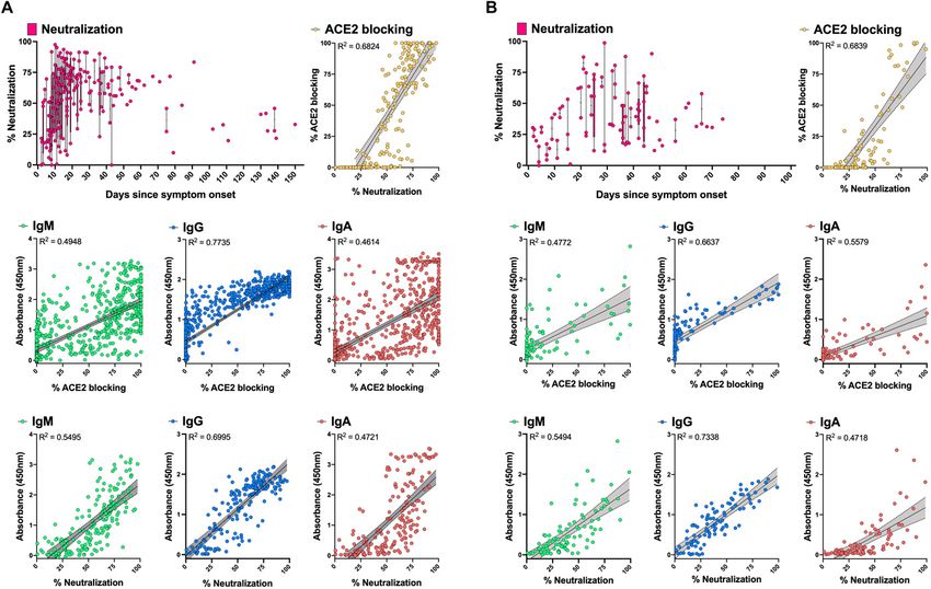
spike and N proteins, which are therefore used as antigens in      asymptomatic individuals or outpatients), or after they re-
clinical serology assays. The spike protein is an important tar-   ported to Stanford Health Care-associated clinical sites with
get for neutralizing antibodies, as they can prevent viral entry   symptoms of COVID-19. This included 24 outpatients, 35 hos-
into host cells (12, 13). Current information on the role of an-   pitalized patients who were not admitted to the intensive
tibodies in viral clearance and modulation of disease severity     care unit (ICU), and 20 ICU inpatients who survived their ill-
as well as the durability of these responses following primary     ness. To evaluate serological responses associated with pa-
infection is limited or controversial. Improved understanding      tient mortality, we also analyzed specimens from 25 patients
of humoral immunity to SARS-CoV-2 is needed to inform              who died of COVID-19 (one outpatient, seven admitted non-
strategies for vaccination and the use of therapeutics in the      ICU, and seventeen admitted ICU patients). Of patients who
form of neutralizing antibodies or convalescent plasma. Re-        were treated in the ICU, 26 of 37 (70%) required mechanical
ports about the longevity of antibody titers to SARS-CoV-2 are     ventilation, including 15 patients who died.
not in full agreement, with some finding a rapid waning of             Demographic and clinical characteristics of patients strat-
virus-specific IgG antibodies by approximately 3 months after      ified by disease status are presented in Table 1. The percent-

                                                                                                                                        Downloaded from http://immunology.sciencemag.org/ by guest on January 5, 2021
infection (14, 15), and others emphasizing stable titers de-       age of males, and those with comorbidities of hypertension
tected over several weeks or several months (16–18). Virus-        or diabetes mellitus increased with patient disease severity.
specific antibody responses appear to be elevated in COVID-        Outpatients and asymptomatic individuals were younger and
19 patients with severe disease as opposed to asymptomatic         had the lowest levels of obesity compared to more severely ill
or mildly ill individuals, raising concerns about the effective-   patients. Notably, levels of viral RNA measured by rRT-PCR
ness of antibody responses to SARS-CoV-2. A suggestion that        of nasopharyngeal swabs at diagnosis showed a progressive
the quality rather than quantity of antibodies may predict the     increase (lower rRT-PCR cycle threshold, Ct) with disease se-
outcome of infection is provided by a recent report applying       verity; patients who died had the highest viral loads (fig. S2).
a panel of serological assays to COVID-19 patients who con-        Plasma samples for in-depth serological testing were availa-
valesced or died (19). Nonhuman primates challenged with           ble from all inpatients and 86 of the 160 outpatient survivors.
SARS-CoV-2 after vaccination with spike-based DNA vaccines         Demographic and clinical characteristics of outpatients and
developed neutralizing antibodies and immune correlates of         asymptomatic individuals with and without plasma availabil-
protection, suggesting that antibody responses may be more         ity are presented in table S1. A total of 828 samples were an-
effective in preventing than resolving disease (20).               alyzed with ELISAs measuring IgM, IgG and IgA specific for
    We performed a comprehensive analysis of SARS-CoV-2            SARS-CoV-2 RBD, S1 or N, as well as the RBD-ACE2 blocking
RBD, S1 and N protein specific antibodies, and RBD-ACE2            assay (Fig. 1A-C). Representative samples were also tested for
blocking antibodies, spike-pseudotyped virus neutralization,       antibody cross-reactivity to SARS-CoV RBD, viral RNA in the
and viral RNA measurements in nasopharyngeal swab and              blood (RNAemia), and neutralization of spike-pseudotyped
plasma samples of individuals infected with SARS-CoV-2. A          lentivirus (Fig. 1D). We also tested 45 plasma specimens from
total of 254 individuals (79 inpatients and 175 outpatients or     a validation cohort of 14 asymptomatic and outpatient SARS-
asymptomatic individuals) were studied. RBD-ACE2 blocking          CoV-2 rRT-PCR-positive individuals with monthly prospec-
antibodies and spike-pseudotyped lentivirus neutralization         tive sample collection for up to four months post-enrollment.
were well-correlated with IgG specific for RBD. Notably,           Anti-RBD, S1 and N antibody responses and duration
higher ratios of IgG antibodies targeting S1 or RBD compared       are associated with disease severity
to N were strongly associated with clinically milder infection.        Lower antibody responses in patients with mild symp-
Viral RNAemia decreased to undetectable levels rapidly once        toms compared to those with more severe disease have been
plasma antibodies and RBD-ACE2 blocking activity appeared.         reported for other coronavirus infections, such as MERS-CoV
Outpatients and asymptomatic individuals show substantial          (21–23) as well as for SARS-CoV-2 (15, 24). The detection rate
and progressive decreases in SARS-CoV-2 specific antibodies        of RBD-binding antibodies at one-week intervals after symp-
after the first month of documented infection.                     tom onset are shown in table S2; most individuals serocon-
RESULTS                                                            verted by week 2 post-onset of symptoms (Fig. 2A, B).
                                                                   Positivity rates for RBD IgM, IgG and IgA reached their max-
Study design and patient demographics                              imum at weeks 4, 6 and 5, respectively, with most patients
    A total of 254 individuals with a positive SARS-CoV-2 real-    negative for IgM and IgA after 12 weeks, while RBD IgG levels
time RT-PCR (rRT-PCR) nasopharyngeal swab test were in-            showed a slower, but progressive decline, or a continued neg-
cluded in the study (fig. S1). Study subjects were identified      ative result in those who failed to generate IgG at earlier time
either after routine serology testing or occupational health       points (Fig. 2A, fig. S3, and table S2). Outpatients had lower
screening in the Stanford Health Care Clinical Laboratories        titers of RBD IgM, IgG, and IgA compared to inpatients, and
for anti-SARS-CoV-2 RBD IgM and IgG antibodies (136                a more rapid decline of titers (Fig. 2B, fig. S4). Patients who

First release: 7 December 2020              immunology.sciencemag.org           (Page numbers not final at time of first release)   2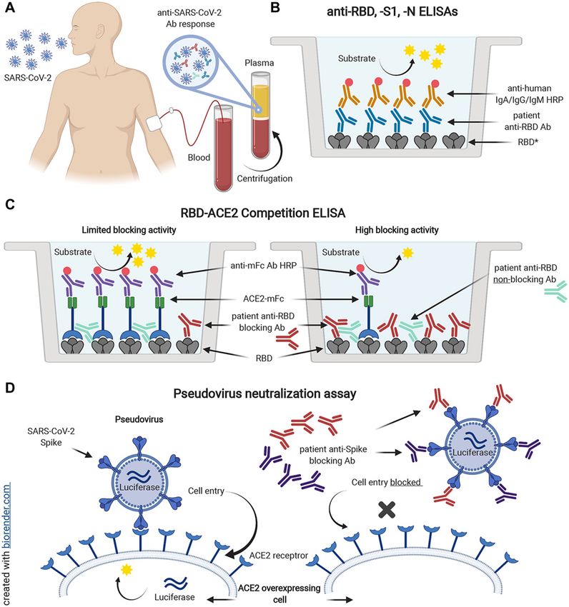
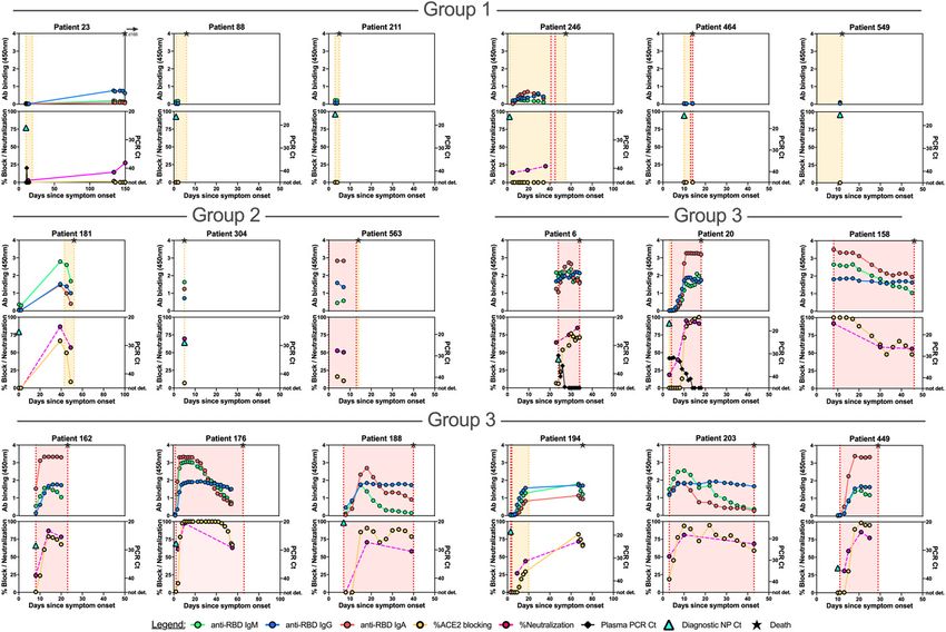
required ICU care, and those who died, developed and main-        Validation of decreasing antibody responses in mildly ill
tained the highest levels of IgM, IgG and IgA, as well as RBD-    and asymptomatic individuals
ACE2 blocking antibodies, throughout the time course (Fig.            Most individuals who become infected with SARS-CoV-2
2C).                                                              infection do not require hospitalization to recover from their
    S1-specific IgM, IgG, and IgA showed antibody kinetics        illness, and a sizeable fraction (approximately 40-45%) (29)
that were very similar to those seen for RBD (figs. S3 to S5      remain asymptomatic. We carried out further analysis of an-
and table S2). While N-specific IgG responses showed high         tibody responses in a larger set of samples from 136 outpa-
positivity rates with antibody kinetics similar to those for      tients and asymptomatic individuals who tested positive by
RBD and S1, IgM antibody responses to N were strikingly low       rRT-PCR for SARS-CoV-2 RNA in nasopharyngeal swabs and
in most patients (figs. S4 and S6 and table S2). There was no     had serological testing conducted in the Stanford Health Care
consistent difference in RBD, S1, and N antibody titers or        Clinical Laboratory. We also tested an independent valida-
RBD-ACE2 blocking antibodies between ICU patients who             tion set of 45 plasma samples from an additional 14 asymp-
survived compared to patients who died (fig. S7).                 tomatic or mildly ill individuals. As seen for outpatients in

                                                                                                                                       Downloaded from http://immunology.sciencemag.org/ by guest on January 5, 2021
    We further evaluated the breadth of antibody responses        Fig. 2B, the asymptomatic and outpatient individuals showed
in different disease severity categories by testing for SARS-     relatively low titers and rapid decline of RBD IgM and IgG
CoV RBD binding. Most monoclonal antibodies targeting             (Fig. 4A, table S3). The timing of infection in asymptomatic
SARS-CoV RBD fail to bind SARS-CoV-2 RBD, indicating dis-         individuals is less certain than for symptomatic individuals,
tinct antigenicity despite sequence and structural similarity     whose symptoms usually develop within 2 to 14 days after ex-
of the two proteins (25, 26). Nine of 13 ICU patients, three of   posure to the virus (30). Plotting of serological responses of
25 admitted non-ICU patients (fig. S8A) and five of 82 outpa-     outpatients and asymptomatic individuals relative to the date
tients (fig. S8B), developed SARS-CoV RBD IgG titers during       of their first positive rRT-PCR test for infection nonetheless
the course of their infection. The time course of anti-SARS-      showed a similar time course to that seen for outpatients
CoV RBD positivity in serial samples from individual patients     plotted as days post onset of symptoms (Fig. 2B). Notably, the
did not always mirror anti-SARS-CoV-2 RBD IgG responses,          amount of viral RNA detected in diagnostic nasopharyngeal
suggesting limited clonal or oligoclonal B cell responses with    swabs was correlated with the antibody titers measured in
this broader reactivity within the overall polyclonal anti-       these individuals (Fig. 4B).
SARS-CoV-2 serological response (figs. S8C, D).                       Plasma specimens from the independent validation co-
Neutralizing antibodies are increased in inpatients               hort of asymptomatic or outpatient individuals were col-
compared to outpatients, and correlate with RBD-ACE2              lected prospectively during monthly visits up to four months
blocking and RBD IgG titers                                       following recruitment (Fig. 4C, D, fig. S9). RBD IgM, IgG and
    Antibody neutralization of live SARS-CoV-2, or of pseudo-     IgA, RBD-ACE2 blocking and neutralization assays all
typed viruses such as lentiviruses expressing the SARS-CoV-2      showed a progressive decrease during the months sampled.
spike may represent the most physiologically relevant surro-      Neutralizing antibodies were best correlated with RBD IgG
gates for humoral immunity in vivo, but are poorly scalable       titers, and somewhat less well with RBD-ACE2 blocking
due to restrictive biosafety requirements for SARS-CoV-2 and      ELISA in these individuals (R2= 0.751 and 0.5221, respec-
less easily standardized assay components and protocols,          tively) (Fig. 4E).
compared to tests using purified proteins. We tested SARS-        Outpatients and non-ICU inpatients show preferential
CoV-2 spike-pseudotyped lentivirus neutralization (27) in         antibody targeting of RBD and S1 compared to N
HeLa cells overexpressing ACE2 (28) by inpatient and outpa-           It is an open question whether antibody responses in the
tient samples, and evaluated correlations with the more scal-     initial weeks of SARS-CoV-2 infection have a role in modulat-
able ELISAs for RBD, S1 and RBD-ACE2 blocking (Fig. 3A, B).       ing disease severity. Having found that patients with milder
As with RBD-ACE2 blocking, neutralizing antibody activity         illness or asymptomatic infection had lower levels of SARS-
was higher in inpatients compared to outpatients, and began       CoV-2 antibodies to RBD, S1 and N compared to severely ill
to decrease after about one month after symptom onset. Neu-       patients, we hypothesized that the relative targeting of the
tralization was well-correlated with RBD IgG titers and RBD-      antibody response between antigens might be associated
ACE2 blocking (linear regression coefficient of determination     with different disease severity. In a recent study, serological
R2 of 0.6995 and 0.6824 for inpatients; 0.7338 and 0.6839 for     analysis of samples from patients who died of COVID-19 com-
outpatients). The RBD-ACE2 blocking assay was less sensitive      pared to individuals who convalesced suggested an enrich-
than neutralization or RBD IgG ELISA. RBD IgM and IgA             ment of an aggregate measure of spike antibodies or antibody
ELISA results were much more variable and did not correlate       functional activities in the convalescent (19). We calculated
as well with neutralization or RBD-ACE2 blocking compared         the ratios of RBD to N (Fig. 5A) and S1 to N (Fig. 5B) ELISA
to RBD IgG.                                                       results for IgM, IgG and IgA for all specimens that had

First release: 7 December 2020             immunology.sciencemag.org           (Page numbers not final at time of first release)   3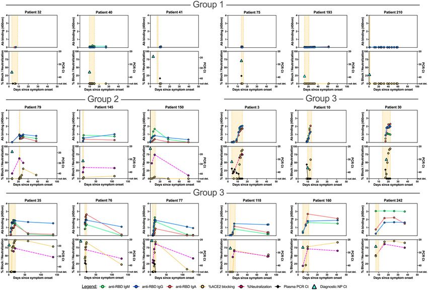
detectable antibodies. Notably, in weeks 1 and 2 post-onset of        0.47 for IgM, -0.43 for IgG, and -0.44 for IgA, p25% RBD-ACE2 blocking activity) admitted
                                                                      (this study and (32, 33)), although IgA timing is the most var-
non-ICU patients differed from ICU or deceased patients in
                                                                      iable. As we have recently reported, large polyclonal prolifer-
that they developed their robust antibody responses during
                                                                      ations of recently class-switched B cells expressing IgG and
short hospital stays prior to discharge, whereas ICU or de-
                                                                      IgA subtypes with low antibody somatic mutation frequen-
ceased patients typically had prolonged hospital courses. In
                                                                      cies appear in the circulation around the time of seroconver-
patients tested for neutralizing antibodies (13 admitted non-
                                                                      sion, likely including plasmablasts that contribute to the
ICU patients, 16 ICU surviving patients, and 16 deceased pa-
                                                                      observed serological responses (29).
tients), the results were closely correlated with IgG titers to
                                                                          Which features of patient antibody responses were asso-
RBD, or RBD-ACE2 blocking.
                                                                      ciated with disease severity? To our surprise, we found that
    Viral RNAemia is detected in up to a third of COVID-19
                                                                      outpatients with the mildest illness showed higher ratios of
patients, most often in patients with severe disease (3, 31).
                                                                      spike RBD and S1 domains compared to N antigen, beginning
RNAemia was evaluated in a subset of patients, and detected
                                                                      in the first two weeks post-onset of symptoms. These findings
in 15 of 25 admitted patients, 13 of 15 ICU patients who re-
                                                                      could suggest that early humoral immune responses focused
covered, and 2 of 2 patients who died of their disease. Reduc-
                                                                      on spike antigens help to constrain the viral infection, per-
tion in RNAemia was strongly correlated with the appearance
                                                                      haps even at times when titers are not yet high enough to be
of plasma antibodies (Spearman’s correlation coefficients of -

First release: 7 December 2020                immunology.sciencemag.org            (Page numbers not final at time of first release)   4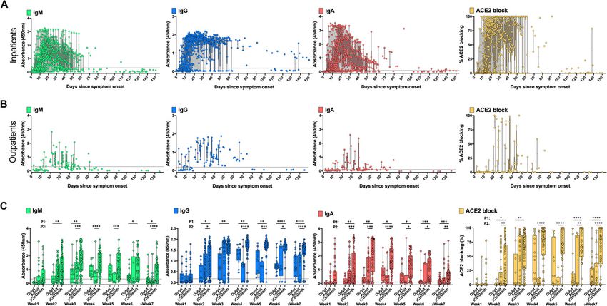
measured in the blood. Outpatients and admitted non-ICU                There is an urgent need to understand how long antibody
patients had the lowest viral loads in their nasopharyngeal        titers against the virus persist after infection, now that the
swabs, but whether this is due to their antibody responses         pandemic has been underway for more than half a year in
cannot be determined from this observational study. Associ-        many countries, and initial case reports of proven reinfection
ations of mortality in COVID-19 patients with SARS-CoV-2 vi-       by SARS-CoV-2 have begun to appear (37, 38). Studies differ-
ral load assessed by rRT-PCR applied to nasopharyngeal swab        ing in their patient populations, disease severity, and serolog-
specimens have also been found in other studies (34, 35). Our      ical assays, have disagreed on the duration of SARS-CoV-2
data are consistent with results reported from a panel of an-      antibody responses (14, 16, 32). Our data derived from inpa-
tibody assays applied to single time point samples from            tients, outpatients and asymptomatic individuals, with an ad-
COVID-19 patients who recovered or died of their disease,          ditional asymptomatic validation cohort, show clearly that
which found higher values of spike-targeting responses in the      not only IgM and IgA, but also IgG titers to RBD, S1 and N
convalescents (19). Patients with more severe illness in our       antigens, RBD-ACE2 blocking activity and spike pseudotyped
study eventually raised higher antibody titers than those with     viral neutralization titers all begin to decrease in patients af-

                                                                                                                                        Downloaded from http://immunology.sciencemag.org/ by guest on January 5, 2021
milder disease, consistent with prior publications (32, 36)        ter approximately the first month post-onset of symptoms.
and reports of other coronavirus infections (21–23). Patients      The decline in antibody titers is most evident in individuals
with more severe disease also had somewhat higher viral            who had asymptomatic infection or mild illness, who produce
loads than patients with milder illness, suggesting that larger    lower levels of antibodies at the peak of their responses. We
initial amounts of viral antigen may contribute to their           note that reported results failing to find a decrease in SARS-
greater serological responses. Functional blocking of RBD-         CoV-2 antibodies after several months post-infection have re-
ACE2 interaction, and spike-pseudotyped virus neutraliza-          lied on “pan-Ig” assays that cannot evaluate each isotype sep-
tion by patient antibodies appeared with a similar time            arately (16). Our data do not permit us to predict what
course to IgM, IgG, and IgA, but were most closely correlated      fraction of the population will be susceptible to reinfection at
in magnitude with IgG titers. Neutralization, RBD-ACE2             a given time after their initial illness, or whether individuals
blocking, and RBD-specific IgG were all highly correlated in       will maintain sustained plateaus of lower antibody levels af-
patients with high antibody levels, but RBD-ACE2 blocking          ter an initial decrease; additional time and follow-up will be
was less sensitive than the neutralization assay, potentially      required to obtain this information. A limitation of our study
because of antibodies that can neutralize by binding to non-       is that, apart from the prospective validation cohort, the sam-
RBD regions of the spike, or lower affinity antibodies that can    pling time points for each patient were determined by their
neutralize in the cell culture assay but do not compete as well    length of hospital stay and subsequent healthcare visits, ena-
with binding of ACE2 under the blocking assay conditions. It       bling more detailed analysis of serological responses in pa-
is currently unclear, however, which of these assays will be       tients with more severe illness. It is important to note that
the best predictor of in vivo immunological protection from        decreasing antibody levels do not necessarily indicate that all
SARS-CoV-2 infection or reinfection in vaccinated or previ-        immunity will be lost. It is possible that local mucosal anti-
ously infected individuals.                                        body production in the airways could help prevent or impede
    In the detailed serological time courses of the hospitalized   SARS-CoV-2-infection upon re-exposure (39). Even if serum
patients in this study it was evident that the patterns of anti-   antibodies wane to undetectable levels, memory B and T cells
body responses could not fully explain patient outcomes, in-       stimulated by infection could provide a faster or more effec-
cluding death. Substantial numbers of patients recovered           tive response following future exposure. Initial reinfection re-
from their illness and were discharged from hospital before        ports offer some hope that SARS-CoV-2 may behave similarly
they had formed detectable antibody responses, but minimal         to other community coronaviruses, with reinfection generally
serological responses were also seen in patients who died of       producing milder illness than the initial infection (40, 41).
COVID-19 at early times post-onset of symptoms. Similarly,             One implication of our finding of waning antibody levels
individuals with moderate antibody production were seen            is that seroprevalence studies may, over time, underestimate
across the full spectrum of inpatient disease severity, and        the proportion of the investigated population which has been
many patients who died of their disease generated high levels      previously infected with SARS-CoV-2. The decrease in anti-
of antibodies, RBD-ACE2 blocking activity, and neutralizing        bodies after infection also raises the question of how long an-
titers. Differences between individuals in other aspects of the    tibodies elicited by vaccination will last, and whether
immune response or disease course, such as production of in-       frequent boosting will be needed to maintain protection, as-
flammatory mediators, T cell responses, host cell and tissue       suming that safe and effective vaccines are identified. The
vulnerability to the damage during viral infection, coagulopa-     current vaccination strategies undergoing clinical trials differ
thy, and secondary infections, are all likely to contribute to     from natural infection in a variety of ways, including the
patient outcomes.                                                  method for generating or introducing viral antigens into the

First release: 7 December 2020              immunology.sciencemag.org           (Page numbers not final at time of first release)   5
body, the site of exposure, and the presence of adjuvants (42–    ACE2-mFc
45). It is possible that some of the vaccine approaches may           The SARS-CoV and SARS-CoV-2 RBD proteins were ex-
generate more potent and long-lasting antibodies than natu-       pressed in Expi293F cells and purified using Nickel-NTA
ral infection, in which the virus may have currently unknown      resin and size exclusion chromatography. The SARS-CoV con-
mechanisms for subverting humoral immune responses. Fur-          struct (RBD-His_pTT5, GenBank AAP13441.1) was synthe-
ther detailed study of the generation of memory B cell popu-      sized commercially by Twist Bioscience (San Francisco, CA);
lations, short- or long-lived plasma cells, and T cell memory     the SARS-CoV-2 construct (RBD-His_pCAGGS, GenBank
to SARS-CoV-2 as well as other coronaviruses should begin to      MN908947.3) was kindly provided by Dr. Florian Krammer
clarify some of these key mechanistic points.                     (48). SARS-CoV-2 S1 (spike residues 1-682) and ACE2-mFc,
                                                                  expressed in HEK293 cells, and the N protein, expressed in E.
MATERIALS AND METHODS                                             coli were produced by the ATUM contract research organiza-
Study design and participants                                     tion. Soluble human ACE2 fused to a mouse Fc tag was con-
    The objective of this study was to investigate correlations   structed by synthesizing a gene encoding ACE2 (residues 1-

                                                                                                                                       Downloaded from http://immunology.sciencemag.org/ by guest on January 5, 2021
between humoral immune responses to SARS-CoV-2, includ-           615) joined by a (G4S)x2 linker to mouse IgG2a Fc, and placed
ing antibodies blocking binding of RBD to the human ACE2          under control of a CMV promoter by cloning into a mamma-
receptor or neutralizing spike pseudovirus, and viral RNA         lian expression plasmid.
loads in the nasopharynx and blood in different COVID-19          ELISA to detect anti-SARS-CoV-2 Spike RBD antibod-
patient groups and individual patients. On March 4, 2020, the     ies in plasma samples
Stanford Health Care Clinical Virology Laboratory began               The ELISA procedure in this study was modified from a
rRT-PCR testing on nasopharyngeal specimens from sus-             protocol published by Stadlbauer (48).
pected COVID-19 patients using a laboratory-developed                 96-well Corning Costar high binding plates (catalog no.
SARS-CoV-2 rRT-PCR assay (46, 47). For this study, we in-         9018, Thermo Fisher) were coated with SARS-CoV RBD,
cluded specimens from patients with rRT-PCR-confirmed             SARS-CoV-2 RBD, S1, or N protein in phosphate-buffered sa-
SARS-CoV-2 infection who reported with symptoms of                line (PBS) at a concentration of 0.1 μg per well (0.025 μg per
COVID-19 to Stanford Healthcare-associated clinical sites be-     well for the nucleocapsid IgG assay) overnight at 4°C. On the
tween March 2020 and August 2020; and specimens from              next day, wells were washed 3x with PBS-0.1% Tween 20
rRT-PCR positive outpatients and asymptomatic individuals         (PBS-T) and blocked with PBS-T containing 3% non-fat milk
identified between April 2020 and May 2020 through occu-          powder for 1 hour at room temperature (RT). Wells were then
pational health screening including rRT-PCR and serology          incubated with plasma samples from COVID-19 patients at a
testing for RBD IgM/G at Stanford Clinical Laboratories. The      dilution of 1:100 in PBS-T containing 1% non-fat milk for 1
screening program that detected asymptomatic SARS-CoV-2           hour at 37°C. Two negative and two positive plasma pool
infected individuals was offered to all Stanford Healthcare       wells and two blank wells incubated with PBS-T containing
employees on a voluntary basis, and screened employees with       1% non-fat milk powder were included on each plate. After
nasopharyngeal swab rRT-PCR testing and serology. In addi-        washing 3x with PBS-T, horseradish peroxidase conjugated
tion, we included a validation cohort of 14 asymptomatic and      goat anti-human IgG (γ-chain specific, catalog no. 62-8420,
mildly ill individuals with prospective sample collection for     Thermo Fisher, 1:6,000 dilution), IgM (μ-chain specific, cata-
up to four months post-enrollment. This study was approved        log no. A6907, Sigma, 1:6,000 dilution), or IgA (α-chain spe-
by the Stanford University Institutional Review Board (Pro-       cific, catalog no. P0216, Agilent, 1:5,000 dilution) in PBS-T
tocols IRB-48973 and IRB-55689).                                  containing 1% non-fat milk was added and incubated for 1
Sample and data collection                                        hour at RT. Wells were washed 3x with PBS-T and dried by
    Venipuncture blood samples collected in sodium heparin-       vigorous tapping of plates on paper towels. 3,3′,5,5′-Tetra-
or K2EDTA-coated vacutainers were used for serology testing       methylbenzidine (TMB) substrate solution was added and the
and rRT-PCR detection of RNAemia, respectively. After cen-        reaction was stopped after 12 min by addition of 0.16 M sul-
trifugation for collection of plasma, samples were stored at -    furic acid. The optical density (OD) at 450 nanometers was
80°C.                                                             measured with an EMax Plus microplate reader (Molecular
    Retrospective chart review was performed on all study         Devices, San Jose, CA); values for blank wells were subtracted
participants. Collected data included age, gender, date of        from values obtained for plasma samples. The cutoff value for
symptom onset, length of hospital stay, length of time admit-     seroconversion was calculated by adding 3 standard devia-
ted in the ICU, date and Ct value for the diagnostic nasopha-     tions to mean ELISA ODs of 94 historical negative control
ryngeal swab rRT-PCR test result, the presence of underlying      samples from healthy blood donors (collected before the pan-
comorbidities, clinical symptoms, and mortality.                  demic for an unrelated seroprevalence study) obtained by
Production of SARS-CoV and SARS-CoV-2 proteins and                testing the samples in all protein/isotype assays (table S4).

First release: 7 December 2020             immunology.sciencemag.org           (Page numbers not final at time of first release)   6
Additional details for the manual and clinical lab instrument     dilution series shown in Figure S9 were performed in tech-
ELISA assay setup are provided in figs. S12 and S13.              nical duplicate; normalized % infectivity values were fit with
Competition ELISA to detect antibodies blocking bind-             a three-parameter non-linear regression inhibitor curve in
ing of ACE2 to RBD                                                GraphPad Prism 8.4.1 and fits were constrained to have a
    96-well Corning Costar high binding plates (Thermo            value of 0% at the bottom of the fit.
Fisher: cat. 9018) were coated with SARS-CoV-2 spike RBD          Real-time PCR to detect SARS-CoV-2 RNA in plasma
protein in PBS at a concentration of 0.1 μg per well overnight        A volume of 400 μL of EDTA-anticoagulated plasma was
at 4°C. All competition ELISA steps were carried out on the       extracted by Qiagen EZ1 Virus Mini Kit v2.0 (Qiagen German-
next day at RT. Wells were washed 3x with PBS-T and blocked       town, MD). Molecular testing for the presence of SARS-CoV-
with PBS-T containing 3% non-fat milk powder for 1 hour.          2 RNA in plasma was performed with a modification of a pub-
Wells were then incubated with plasma samples from                lished rRT-PCR assay targeting the envelope (E) gene (46, 47).
COVID-19 patients at a dilution of 1:10 in PBS-T containing       The standard Ct values of positive tests with this assay range

                                                                                                                                                             Downloaded from http://immunology.sciencemag.org/ by guest on January 5, 2021
1% non-fat milk for 1 hour. Two quality controls (Access          from Ct
Fig. S12. Checkerboard titration for serological RBD ELISA.                                         Virol. 3, 237–261 (2016). doi:10.1146/annurev-virology-110615-042301 Medline
Fig. S13. Validation of the Stanford Health Care Clinical Laboratory anti-RBD IgM/G             12. B. Ju, Q. Zhang, J. Ge, R. Wang, J. Sun, X. Ge, J. Yu, S. Shan, B. Zhou, S. Song, X.
     ELISA.                                                                                         Tang, J. Yu, J. Lan, J. Yuan, H. Wang, J. Zhao, S. Zhang, Y. Wang, X. Shi, L. Liu, J.
Fig. S14. Checkerboard titration for receptor blocking ELISA.                                       Zhao, X. Wang, Z. Zhang, L. Zhang, Human neutralizing antibodies elicited by
Fig. S15. RBD-ACE2 blocking ELISA optimization.                                                     SARS-CoV-2 infection. Nature 584, 115–119 (2020). doi:10.1038/s41586-020-
Table S1. Demographic and clinical characteristics of outpatients and asymptomatic                  2380-z Medline
     individuals with and without plasma availability.                                          13. M. Ho, Perspectives on the development of neutralizing antibodies against SARS-
Table S2. Inpatient seropositivity.                                                                 CoV-2. Antibody Ther 3, 109–114 (2020). doi:10.1093/abt/tbaa009 Medline
Table S3. Outpatient and asymptomatic individuals’ seropositivity.                              14. F. J. Ibarrondo, J. A. Fulcher, D. Goodman-Meza, J. Elliott, C. Hofmann, M. A.
Table S4. Determination of SARS-CoV-2 assay cutoff values and specificity.                          Hausner, K. G. Ferbas, N. H. Tobin, G. M. Aldrovandi, O. O. Yang, Rapid Decay of
Table S5. Raw data file (Excel spreadsheet)                                                         Anti-SARS-CoV-2 Antibodies in Persons with Mild Covid-19. N. Engl. J. Med. 383,
                                                                                                    1085–1087 (2020). doi:10.1056/NEJMc2025179 Medline
REFERENCES AND NOTES                                                                            15. Q.-X. Long, X.-J. Tang, Q.-L. Shi, Q. Li, H.-J. Deng, J. Yuan, J.-L. Hu, W. Xu, Y. Zhang,
1. P. Zhou, X.-L. Yang, X.-G. Wang, B. Hu, L. Zhang, W. Zhang, H.-R. Si, Y. Zhu, B. Li, C.-         F.-J. Lv, K. Su, F. Zhang, J. Gong, B. Wu, X.-M. Liu, J.-J. Li, J.-F. Qiu, J. Chen, A.-L.
     L. Huang, H.-D. Chen, J. Chen, Y. Luo, H. Guo, R.-D. Jiang, M.-Q. Liu, Y. Chen, X.-R.          Huang, Clinical and immunological assessment of asymptomatic SARS-CoV-2
     Shen, X. Wang, X.-S. Zheng, K. Zhao, Q.-J. Chen, F. Deng, L.-L. Liu, B. Yan, F.-X.             infections. Nat. Med. 26, 1200–1204 (2020). doi:10.1038/s41591-020-0965-6

                                                                                                                                                                                                Downloaded from http://immunology.sciencemag.org/ by guest on January 5, 2021
     Zhan, Y.-Y. Wang, G.-F. Xiao, Z.-L. Shi, A pneumonia outbreak associated with a                Medline
     new coronavirus of probable bat origin. Nature 579, 270–273 (2020).                        16. D. F. Gudbjartsson, G. L. Norddahl, P. Melsted, K. Gunnarsdottir, H. Holm, E.
     doi:10.1038/s41586-020-2012-7 Medline                                                          Eythorsson, A. O. Arnthorsson, D. Helgason, K. Bjarnadottir, R. F. Ingvarsson, B.
2. Z. Wu, J. M. McGoogan, Characteristics of and Important Lessons From the                         Thorsteinsdottir, S. Kristjansdottir, K. Birgisdottir, A. M. Kristinsdottir, M. I.
     Coronavirus Disease 2019 (COVID-19) Outbreak in China: Summary of a Report of                  Sigurdsson, G. A. Arnadottir, E. V. Ivarsdottir, M. Andresdottir, F. Jonsson, A. B.
     72 314 Cases From the Chinese Center for Disease Control and Prevention. JAMA                  Agustsdottir, J. Berglund, B. Eiriksdottir, R. Fridriksdottir, E. E. Gardarsdottir, M.
     323, 1239–1242 (2020). doi:10.1001/jama.2020.2648 Medline                                      Gottfredsson, O. S. Gretarsdottir, S. Gudmundsdottir, K. R. Gudmundsson, T. R.
3. C. Huang, Y. Wang, X. Li, L. Ren, J. Zhao, Y. Hu, L. Zhang, G. Fan, J. Xu, X. Gu, Z.             Gunnarsdottir, A. Gylfason, A. Helgason, B. O. Jensson, A. Jonasdottir, H. Jonsson,
     Cheng, T. Yu, J. Xia, Y. Wei, W. Wu, X. Xie, W. Yin, H. Li, M. Liu, Y. Xiao, H. Gao, L.        T. Kristjansson, K. G. Kristinsson, D. N. Magnusdottir, O. T. Magnusson, L. B.
     Guo, J. Xie, G. Wang, R. Jiang, Z. Gao, Q. Jin, J. Wang, B. Cao, Clinical features of          Olafsdottir, S. Rognvaldsson, L. le Roux, G. Sigmundsdottir, A. Sigurdsson, G.
     patients infected with 2019 novel coronavirus in Wuhan, China. Lancet 395, 497–                Sveinbjornsson, K. E. Sveinsdottir, M. Sveinsdottir, E. A. Thorarensen, B.
     506 (2020). doi:10.1016/S0140-6736(20)30183-5 Medline                                          Thorbjornsson, M. Thordardottir, J. Saemundsdottir, S. H. Kristjansson, K. S.
4. N. Chen, M. Zhou, X. Dong, J. Qu, F. Gong, Y. Han, Y. Qiu, J. Wang, Y. Liu, Y. Wei, J.           Josefsdottir, G. Masson, G. Georgsson, M. Kristjansson, A. Moller, R. Palsson, T.
     Xia, T. Yu, X. Zhang, L. Zhang, Epidemiological and clinical characteristics of 99             Gudnason, U. Thorsteinsdottir, I. Jonsdottir, P. Sulem, K. Stefansson, Humoral
     cases of 2019 novel coronavirus pneumonia in Wuhan, China: A descriptive study.                Immune Response to SARS-CoV-2 in Iceland. N. Engl. J. Med. 383, 1724–1734
     Lancet 395, 507–513 (2020). doi:10.1016/S0140-6736(20)30211-7 Medline                          (2020). doi:10.1056/NEJMoa2026116 Medline
5. F. Zhou, T. Yu, R. Du, G. Fan, Y. Liu, Z. Liu, J. Xiang, Y. Wang, B. Song, X. Gu, L. Guan,   17. Y. Wang, L. Zhang, L. Sang, F. Ye, S. Ruan, B. Zhong, T. Song, A. N. Alshukairi, R.
     Y. Wei, H. Li, X. Wu, J. Xu, S. Tu, Y. Zhang, H. Chen, B. Cao, Clinical course and risk        Chen, Z. Zhang, M. Gan, A. Zhu, Y. Huang, L. Luo, C. K. P. Mok, M. M. Al Gethamy,
     factors for mortality of adult inpatients with COVID-19 in Wuhan, China: A                     H. Tan, Z. Li, X. Huang, F. Li, J. Sun, Y. Zhang, L. Wen, Y. Li, Z. Chen, Z. Zhuang, J.
     retrospective cohort study. Lancet 395, 1054–1062 (2020). doi:10.1016/S0140-                   Zhuo, C. Chen, L. Kuang, J. Wang, H. Lv, Y. Jiang, M. Li, Y. Lin, Y. Deng, L. Tang, J.
     6736(20)30566-3 Medline                                                                        Liang, J. Huang, S. Perlman, N. Zhong, J. Zhao, J. S. Malik Peiris, Y. Li, J. Zhao,
6. S. Richardson, J. S. Hirsch, M. Narasimhan, J. M. Crawford, T. McGinn, K. W.                     Kinetics of viral load and antibody response in relation to COVID-19 severity. J.
     Davidson, D. P. Barnaby, L. B. Becker, J. D. Chelico, S. L. Cohen, J. Cookingham,              Clin. Invest. 130, 5235–5244 (2020). doi:10.1172/JCI138759 Medline
     K. Coppa, M. A. Diefenbach, A. J. Dominello, J. Duer-Hefele, L. Falzon, J. Gitlin, N.      18. B. Isho, K. T. Abe, M. Zuo, A. J. Jamal, B. Rathod, J. H. Wang, Z. Li, G. Chao, O. L.
     Hajizadeh, T. G. Harvin, D. A. Hirschwerk, E. J. Kim, Z. M. Kozel, L. M. Marrast, J. N.        Rojas, Y. M. Bang, A. Pu, N. Christie-Holmes, C. Gervais, D. Ceccarelli, P.
     Mogavero, G. A. Osorio, M. Qiu, T. P. Zanos; the Northwell COVID-19 Research                   Samavarchi-Tehrani, F. Guvenc, P. Budylowski, A. Li, A. Paterson, F. Y. Yue, L. M.
     Consortium, Presenting Characteristics, Comorbidities, and Outcomes Among                      Marin, L. Caldwell, J. L. Wrana, K. Colwill, F. Sicheri, S. Mubareka, S. D. Gray-Owen,
     5700 Patients Hospitalized With COVID-19 in the New York City Area. JAMA 323,                  S. J. Drews, W. L. Siqueira, M. Barrios-Rodiles, M. Ostrowski, J. M. Rini, Y.
     2052–2059 (2020). 10.1001/jama.2020.6775 Medline                                               Durocher, A. J. McGeer, J. L. Gommerman, A.-C. Gingras, Persistence of serum
7. T. Chen, D. Wu, H. Chen, W. Yan, D. Yang, G. Chen, K. Ma, D. Xu, H. Yu, H. Wang, T.              and saliva antibody responses to SARS-CoV-2 spike antigens in COVID-19
     Wang, W. Guo, J. Chen, C. Ding, X. Zhang, J. Huang, M. Han, S. Li, X. Luo, J. Zhao,            patients. Sci. Immunol. 5, eabe5511 (2020). 10.1126/sciimmunol.abe5511 Medline
     Q. Ning, Clinical characteristics of 113 deceased patients with coronavirus disease        19. C. Atyeo, S. Fischinger, T. Zohar, M. D. Slein, J. Burke, C. Loos, D. J. McCulloch, K.
     2019: Retrospective study. BMJ 368, m1091 (2020). doi:10.1136/bmj.m1091                        L. Newman, C. Wolf, J. Yu, K. Shuey, J. Feldman, B. M. Hauser, T. Caradonna, A. G.
     Medline                                                                                        Schmidt, T. J. Suscovich, C. Linde, Y. Cai, D. Barouch, E. T. Ryan, R. C. Charles, D.
8. J. Perez-Saez, S. A. Lauer, L. Kaiser, S. Regard, E. Delaporte, I. Guessous, S.                  Lauffenburger, H. Chu, G. Alter, Distinct Early Serological Signatures Track with
     Stringhini, A. S. Azman; Serocov-POP Study Group, Serology-informed estimates                  SARS-CoV-2           Survival.      Immunity        53,      524–532.e4          (2020).
     of SARS-CoV-2 infection fatality risk in Geneva, Switzerland. Lancet Infect. Dis. 0,           doi:10.1016/j.immuni.2020.07.020 Medline
     S1473-3099(20)30584-3 (2020). doi:10.1016/S1473-3099(20)30584-3 Medline                    20. J. Yu, L. H. Tostanoski, L. Peter, N. B. Mercado, K. McMahan, S. H. Mahrokhian, J.
9. A. E. Gorbalenya, S. C. Baker, R. S. Baric, R. J. de Groot, C. Drosten, A. A. Gulyaeva,          P. Nkolola, J. Liu, Z. Li, A. Chandrashekar, D. R. Martinez, C. Loos, C. Atyeo, S.
     B. L. Haagmans, C. Lauber, A. M. Leontovich, B. W. Neuman, D. Penzar, S.                       Fischinger, J. S. Burke, M. D. Slein, Y. Chen, A. Zuiani, F. J. N. Lelis, M. Travers, S.
     Perlman, L. L. M. Poon, D. V. Samborskiy, I. A. Sidorov, I. Sola, J. Ziebuhr;                  Habibi, L. Pessaint, A. Van Ry, K. Blade, R. Brown, A. Cook, B. Finneyfrock, A.
     Coronaviridae Study Group of the International Committee on Taxonomy of                        Dodson, E. Teow, J. Velasco, R. Zahn, F. Wegmann, E. A. Bondzie, G. Dagotto, M.
     Viruses, The species Severe acute respiratory syndrome-related coronavirus:                    S. Gebre, X. He, C. Jacob-Dolan, M. Kirilova, N. Kordana, Z. Lin, L. F. Maxfield, F.
     Classifying 2019-nCoV and naming it SARS-CoV-2. Nat. Microbiol. 5, 536–544                     Nampanya, R. Nityanandam, J. D. Ventura, H. Wan, Y. Cai, B. Chen, A. G. Schmidt,
     (2020). doi:10.1038/s41564-020-0695-z Medline                                                  D. R. Wesemann, R. S. Baric, G. Alter, H. Andersen, M. G. Lewis, D. H. Barouch,
10. W. Tai, L. He, X. Zhang, J. Pu, D. Voronin, S. Jiang, Y. Zhou, L. Du, Characterization          DNA vaccine protection against SARS-CoV-2 in rhesus macaques. Science 369,
     of the receptor-binding domain (RBD) of 2019 novel coronavirus: Implication for                806–811 (2020). doi:10.1126/science.abc6284 Medline
     development of RBD protein as a viral attachment inhibitor and vaccine. Cell. Mol.         21. N. M. A. Okba, V. S. Raj, I. Widjaja, C. H. GeurtsvanKessel, E. de Bruin, F. D.
     Immunol. 17, 613–620 (2020). doi:10.1038/s41423-020-0400-4 Medline                             Chandler, W. B. Park, N.-J. Kim, E. A. B. A. Farag, M. Al-Hajri, B.-J. Bosch, M. D. Oh,
11. F. Li, Structure, Function, and Evolution of Coronavirus Spike Proteins. Annu. Rev.

First release: 7 December 2020                                  immunology.sciencemag.org                          (Page numbers not final at time of first release)                       8
M. P. G. Koopmans, C. B. E. M. Reusken, B. L. Haagmans, Sensitive and Specific 19 patients. Sci. Immunol. 5, eabe0367 (2020). doi:10.1126/sciimmunol.abe0367
Detection of Low-Level Antibody Responses in Mild Middle East Respiratory Medline
Syndrome Coronavirus Infections. Emerg. Infect. Dis. 25, 1868–1877 (2019). 34. L. F. Westblade, G. Brar, L. C. Pinheiro, D. Paidoussis, M. Rajan, P. Martin, P. Goyal,
doi:10.3201/eid2510.190051 Medline J. L. Sepulveda, L. Zhang, G. George, D. Liu, S. Whittier, M. Plate, C. B. Small, J. H.
22. A. N. Alshukairi, I. Khalid, W. A. Ahmed, A. M. Dada, D. T. Bayumi, L. S. Malic, S. Rand, M. M. Cushing, T. J. Walsh, J. Cooke, M. M. Safford, M. Loda, M. J. Satlin,
Althawadi, K. Ignacio, H. S. Alsalmi, H. M. Al-Abdely, G. Y. Wali, I. A. Qushmaq, B. SARS-CoV-2 Viral Load Predicts Mortality in Patients with and without Cancer
M. Alraddadi, S. Perlman, Antibody Response and Disease Severity in Healthcare Who Are Hospitalized with COVID-19. Cancer Cell 38, 661–671.e2 (2020).
Worker MERS Survivors. Emerg. Infect. Dis. 22, (2016). doi:10.1016/j.ccell.2020.09.007 Medline
doi:10.3201/eid2206.160010 Medline 35. R. Magleby, L. F. Westblade, A. Trzebucki, M. S. Simon, M. Rajan, J. Park, P. Goyal,
23. C. Drosten, B. Meyer, M. A. Müller, V. M. Corman, M. Al-Masri, R. Hossain, H. M. M. Safford, M. J. Satlin, Impact of SARS-CoV-2 Viral Load on Risk of Intubation
Madani, A. Sieberg, B. J. Bosch, E. Lattwein, R. F. Alhakeem, A. M. Assiri, W. and Mortality Among Hospitalized Patients with Coronavirus Disease 2019.
Hajomar, A. M. Albarrak, J. A. Al-Tawfiq, A. I. Zumla, Z. A. Memish, Transmission Nephrol. Dial. Transplant. ciaa851 (2020). 10.1093/cid/ciaa851 Medline
of MERS-coronavirus in household contacts. N. Engl. J. Med. 371, 828–835 36. A. Casadevall, L. A. Pirofski, The convalescent sera option for containing COVID-
(2014). doi:10.1056/NEJMoa1405858 Medline 19. J. Clin. Invest. 130, 1545–1548 (2020). doi:10.1172/JCI138003 Medline
24. X. Chen, Z. Pan, S. Yue, F. Yu, J. Zhang, Y. Yang, R. Li, B. Liu, X. Yang, L. Gao, Z. Li, 37. J. D. Goldman, K. Wang, K. Roltgen, S. C. A. Nielsen, J. C. Roach, S. N. Naccache,
Y. Lin, Q. Huang, L. Xu, J. Tang, L. Hu, J. Zhao, P. Liu, G. Zhang, Y. Chen, K. Deng, F. Yang, O. F. Wirz, K. E. Yost, J.-Y. Lee, K. Chun, T. Wrin, C. J. Petropoulos, I. Lee,
Downloaded from http://immunology.sciencemag.org/ by guest on January 5, 2021
L. Ye, Disease severity dictates SARS-CoV-2-specific neutralizing antibody S. Fallen, P. M. Manner, J. A. Wallick, H. A. Algren, K. M. Murray, Y. Su, J. Hadlock,
responses in COVID-19. Signal Transduct. Target. Ther. 5, 180 (2020). J. Jeharajah, W. R. Berrington, G. P. Pappas, S. T. Nyatsatsang, A. L. Greninger, A.
doi:10.1038/s41392-020-00301-9 Medline T. Satpathy, J. S. Pauk, S. D. Boyd, J. R. Heath, Reinfection with SARS-CoV-2 and
25. X. Tian, C. Li, A. Huang, S. Xia, S. Lu, Z. Shi, L. Lu, S. Jiang, Z. Yang, Y. Wu, T. Ying, Failure of Humoral Immunity: A case report. medRxiv 2020.09.22.20192443
Potent binding of 2019 novel coronavirus spike protein by a SARS coronavirus- (2020). 10.1101/2020.09.22.20192443 Medline
specific human monoclonal antibody. Emerg. Microbes Infect. 9, 382–385 (2020). 38. K. K.-W. To, I. F.-N. Hung, J. D. Ip, A. W.-H. Chu, W.-M. Chan, A. R. Tam, C. H.-Y.
doi:10.1080/22221751.2020.1729069 Medline Fong, S. Yuan, H.-W. Tsoi, A. C.-K. Ng, L. L.-Y. Lee, P. Wan, E. Tso, W.-K. To, D.
26. D. Wrapp, N. Wang, K. S. Corbett, J. A. Goldsmith, C.-L. Hsieh, O. Abiona, B. S. Tsang, K.-H. Chan, J.-D. Huang, K.-H. Kok, V. C.-C. Cheng, K.-Y. Yuen, COVID-19
Graham, J. S. McLellan, Cryo-EM structure of the 2019-nCoV spike in the re-infection by a phylogenetically distinct SARS-coronavirus-2 strain confirmed
prefusion conformation. Science 367, 1260–1263 (2020). by whole genome sequencing. Nephrol. Dial. Transplant. ciaa1275 (2020).
doi:10.1126/science.abb2507 Medline 10.1093/cid/ciaa1275 Medline
27. K. H. D. Crawford, R. Eguia, A. S. Dingens, A. N. Loes, K. D. Malone, C. R. Wolf, H. Y. 39. C. Cervia, J. Nilsson, Y. Zurbuchen, A. Valaperti, J. Schreiner, A. Wolfensberger, M.
Chu, M. A. Tortorici, D. Veesler, M. Murphy, D. Pettie, N. P. King, A. B. Balazs, J. D. E. Raeber, S. Adamo, S. Weigang, M. Emmenegger, S. Hasler, P. P. Bosshard, E.
Bloom, Protocol and Reagents for Pseudotyping Lentiviral Particles with SARS- De Cecco, E. Bächli, A. Rudiger, M. Stüssi-Helbling, L. C. Huber, A. S. Zinkernagel,
CoV-2 Spike Protein for Neutralization Assays. Viruses 12, 513 (2020). D. J. Schaer, A. Aguzzi, G. Kochs, U. Held, E. Probst-Müller, S. K. Rampini, O.
doi:10.3390/v12050513 Medline Boyman, Systemic and mucosal antibody responses specific to SARS-CoV-2
28. T. F. Rogers, F. Zhao, D. Huang, N. Beutler, A. Burns, W. T. He, O. Limbo, C. Smith, during mild versus severe COVID-19. J. Allergy Clin. Immunol. S0091-
G. Song, J. Woehl, L. Yang, R. K. Abbott, S. Callaghan, E. Garcia, J. Hurtado, M. 6749(20)31623-7 (2020). doi:10.1016/j.jaci.2020.10.040 Medline
Parren, L. Peng, S. Ramirez, J. Ricketts, M. J. Ricciardi, S. A. Rawlings, N. C. Wu, 40. A. W. D. Edridge, J. Kaczorowska, A. C. R. Hoste, M. Bakker, M. Klein, K. Loens, M.
M. Yuan, D. M. Smith, D. Nemazee, J. R. Teijaro, J. E. Voss, I. A. Wilson, R. Andrabi, F. Jebbink, A. Matser, C. M. Kinsella, P. Rueda, M. Ieven, H. Goossens, M. Prins, P.
B. Briney, E. Landais, D. Sok, J. G. Jardine, D. R. Burton, Isolation of potent SARS- Sastre, M. Deijs, L. van der Hoek, Seasonal coronavirus protective immunity is
CoV-2 neutralizing antibodies and protection from disease in a small animal short-lasting. Nat. Med. 26, 1691–1693 (2020). Medline
model. Science 369, 956–963 (2020). doi:10.1126/science.abc7520 Medline 41. K. A. Callow, H. F. Parry, M. Sergeant, D. A. Tyrrell, The time course of the immune
29. D. P. Oran, E. J. Topol, Prevalence of Asymptomatic SARS-CoV-2 Infection : A response to experimental coronavirus infection of man. Epidemiol. Infect. 105,
Narrative Review. Ann. Intern. Med. 173, 362–367 (2020). doi:10.7326/M20-3012 435–446 (1990). doi:10.1017/S0950268800048019 Medline
Medline 42. L. A. Jackson, E. J. Anderson, N. G. Rouphael, P. C. Roberts, M. Makhene, R. N.
30. CDC, Coronavirus Disease 2019 (COVID-19) – SymptomsCenters for Disease Coler, M. P. McCullough, J. D. Chappell, M. R. Denison, L. J. Stevens, A. J.
Control and Prevention (2020) (available at Pruijssers, A. McDermott, B. Flach, N. A. Doria-Rose, K. S. Corbett, K. M. Morabito,
https://www.cdc.gov/coronavirus/2019-ncov/symptoms- S. O’Dell, S. D. Schmidt, P. A. Swanson 2nd, M. Padilla, J. R. Mascola, K. M. Neuzil,
testing/symptoms.html). H. Bennett, W. Sun, E. Peters, M. Makowski, J. Albert, K. Cross, W. Buchanan, R.
31. C. A. Hogan, B. A. Stevens, M. K. Sahoo, C. Huang, N. Garamani, S. Gombar, F. Pikaart-Tautges, J. E. Ledgerwood, B. S. Graham, J. H. Beigel; mRNA-1273 Study
Yamamoto, K. Murugesan, J. Kurzer, J. Zehnder, B. A. Pinsky, High Frequency of Group, An mRNA Vaccine against SARS-CoV-2 - Preliminary Report. N. Engl. J.
SARS-CoV-2 RNAemia and Association With Severe Disease. Clin. Infect. Dis. Med. 383, 1920–1931 (2020). Medline
ciaa1054 (2020). doi:10.1093/cid/ciaa1054 Medline 43. M. J. Mulligan, K. E. Lyke, N. Kitchin, J. Absalon, A. Gurtman, S. Lockhart, K. Neuzil,
32. Q.-X. Long, B.-Z. Liu, H.-J. Deng, G.-C. Wu, K. Deng, Y.-K. Chen, P. Liao, J.-F. Qiu, V. Raabe, R. Bailey, K. A. Swanson, P. Li, K. Koury, W. Kalina, D. Cooper, C. Fontes-
Y. Lin, X.-F. Cai, D.-Q. Wang, Y. Hu, J.-H. Ren, N. Tang, Y.-Y. Xu, L.-H. Yu, Z. Mo, F. Garfias, P.-Y. Shi, Ö. Türeci, K. R. Tompkins, E. E. Walsh, R. Frenck, A. R. Falsey, P.
Gong, X.-L. Zhang, W.-G. Tian, L. Hu, X.-X. Zhang, J.-L. Xiang, H.-X. Du, H.-W. Liu, R. Dormitzer, W. C. Gruber, U. Şahin, K. U. Jansen, Phase I/II study of COVID-19
C.-H. Lang, X.-H. Luo, S.-B. Wu, X.-P. Cui, Z. Zhou, M.-M. Zhu, J. Wang, C.-J. Xue, RNA vaccine BNT162b1 in adults. Nature 586, 589–593 (2020).
X.-F. Li, L. Wang, Z.-J. Li, K. Wang, C.-C. Niu, Q.-J. Yang, X.-J. Tang, Y. Zhang, X.- doi:10.1038/s41586-020-2639-4 Medline
M. Liu, J.-J. Li, D.-C. Zhang, F. Zhang, P. Liu, J. Yuan, Q. Li, J.-L. Hu, J. Chen, A.-L. 44. P. M. Folegatti, K. J. Ewer, P. K. Aley, B. Angus, S. Becker, S. Belij-Rammerstorfer,
Huang, Antibody responses to SARS-CoV-2 in patients with COVID-19. Nat. Med. D. Bellamy, S. Bibi, M. Bittaye, E. A. Clutterbuck, C. Dold, S. N. Faust, A. Finn, A. L.
26, 845–848 (2020). doi:10.1038/s41591-020-0897-1 Medline Flaxman, B. Hallis, P. Heath, D. Jenkin, R. Lazarus, R. Makinson, A. M. Minassian,
33. A. S. Iyer, F. K. Jones, A. Nodoushani, M. Kelly, M. Becker, D. Slater, R. Mills, E. K. M. Pollock, M. Ramasamy, H. Robinson, M. Snape, R. Tarrant, M. Voysey, C.
Teng, M. Kamruzzaman, W. F. Garcia-Beltran, M. Astudillo, D. Yang, T. E. Miller, E. Green, A. D. Douglas, A. V. S. Hill, T. Lambe, S. C. Gilbert, A. J. Pollard; Oxford
Oliver, S. Fischinger, C. Atyeo, A. J. Iafrate, S. B. Calderwood, S. A. Lauer, J. Yu, Z. COVID Vaccine Trial Group, Safety and immunogenicity of the ChAdOx1 nCoV-19
Li, J. Feldman, B. M. Hauser, T. M. Caradonna, J. A. Branda, S. E. Turbett, R. C. vaccine against SARS-CoV-2: A preliminary report of a phase 1/2, single-blind,
LaRocque, G. Mellon, D. H. Barouch, A. G. Schmidt, A. S. Azman, G. Alter, E. T. randomised controlled trial. Lancet 396, 467–478 (2020). doi:10.1016/S0140-
Ryan, J. B. Harris, R. C. Charles, Persistence and decay of human antibody 6736(20)31604-4 Medline
responses to the receptor binding domain of SARS-CoV-2 spike protein in COVID- 45. C. Keech, G. Albert, I. Cho, A. Robertson, P. Reed, S. Neal, J. S. Plested, M. Zhu, S.
First release: 7 December 2020 immunology.sciencemag.org (Page numbers not final at time of first release) 9Cloney-Clark, H. Zhou, G. Smith, N. Patel, M. B. Frieman, R. E. Haupt, J. Logue, M. Supplementary Materials. This work is licensed under a Creative Commons
McGrath, S. Weston, P. A. Piedra, C. Desai, K. Callahan, M. Lewis, P. Price-Abbott, Attribution 4.0 International (CC BY 4.0) license, which permits unrestricted
N. Formica, V. Shinde, L. Fries, J. D. Lickliter, P. Griffin, B. Wilkinson, G. M. Glenn, use, distribution, and reproduction in any medium, provided the original work is
Phase 1-2 Trial of a SARS-CoV-2 Recombinant Spike Protein Nanoparticle properly cited. To view a copy of this license, visit
Vaccine. N. Engl. J. Med. (2020). Medline https://creativecommons.org/licenses/by/4.0/. This license does not apply to
46. C. A. Hogan, M. K. Sahoo, B. A. Pinsky, Sample Pooling as a Strategy to Detect figures/photos/artwork or other content included in the article that is credited
Community Transmission of SARS-CoV-2. JAMA 323, 1967–1969 (2020). to a third party; obtain authorization from the rights holder before using such
doi:10.1001/jama.2020.5445 Medline material.
47. V. M. Corman, O. Landt, M. Kaiser, R. Molenkamp, A. Meijer, D. K. Chu, T. Bleicker,
S. Brünink, J. Schneider, M. L. Schmidt, D. G. Mulders, B. L. Haagmans, B. van der Submitted 27 July 2020
Veer, S. van den Brink, L. Wijsman, G. Goderski, J.-L. Romette, J. Ellis, M. Zambon, Resubmitted 5 October 2020
M. Peiris, H. Goossens, C. Reusken, M. P. Koopmans, C. Drosten, Detection of Accepted 3 December 2020
2019 novel coronavirus (2019-nCoV) by real-time RT-PCR. Euro Surveill. 25, Published First Release 7 December 2020
(2020). doi:10.2807/1560-7917.ES.2020.25.3.2000045 Medline 10.1126/sciimmunol.abe0240
48. D. Stadlbauer, F. Amanat, V. Chromikova, K. Jiang, S. Strohmeier, G. A.
Arunkumar, J. Tan, D. Bhavsar, C. Capuano, E. Kirkpatrick, P. Meade, R. N. Brito,
Downloaded from http://immunology.sciencemag.org/ by guest on January 5, 2021
C. Teo, M. McMahon, V. Simon, F. Krammer, SARS-CoV-2 Seroconversion in
Humans: A Detailed Protocol for a Serological Assay, Antigen Production, and
Test Setup. Curr. Protoc. Microbiol. 57, e100 (2020). doi:10.1002/cpmc.100
Medline
49. A. E. Powell, K. Zhang, M. Sanyal, S. Tang, P. A. Weidenbacher, S. Li, T. D. Pham, J.
E. Pak, W. Chiu, P. S. Kim, A single immunization with spike-functionalized ferritin
vaccines elicits neutralizing antibody responses against SARS-CoV-2 in mice.
bioRxiv 2020.08.28.272518 (2020). 10.1101/2020.08.28.272518 Medline
50. R Core Team, (2013). R: A language and environment for statistical computing. R
Foundation for Statistical Computing, Vienna, Austria. URL: http://www.R-
project.org/.
Acknowledgments: The authors thank Dana Anderson for helpful advice about
protein production, and the ATUM Bio team for optimization and production of
antigens. We thank Dr. Jesse Bloom, Kate Crawford, Dr. Dennis Burton, and Dr.
Deli Huang for sharing the plasmids, cells, and invaluable advice for
implementation of the spike-pseudotyped lentiviral neutralization assay.
Funding: This work was supported by NIH/NIAID R01AI127877 (S.D.B.),
NIH/NIAID R01AI130398 (S.D.B.), NIH 1U54CA260517 (S.D.B.), NIH/NIAID
T32AI007502-23 (A.R.), NIH/NIAID U19AI111825 (T.T.W.), NIH/NCATS
UL1TR003142 (B.N.), an endowment to S.D.B. from the Crown Family
Foundation, a postdoctoral fellowship to A.E.P from the Stanford Maternal and
Child Health Research Institute, an Early Postdoc.Mobility Fellowship Stipend to
O.F.W. from the Swiss National Science Foundation (SNSF), and a Coulter
COVID-19 Rapid Response Award to J.R.C. and S.D.B. Author contributions: K.R,
A.E.P., O.F.W., B.A.S., B.A.P., S.D.B. conceptualized and designed the study. K.R.,
A.E.P., O.F.W., C.A.H., M.K.S., C.H.H., F.Y., J.M. performed the experiments.
B.A.S., J.N., M.H., H.W., M.K.S., M.M., A.R.O, T.D.P., A.R., A.J.R., C.A.B., J.R.C.,
K.C.N., B.A.P., S.G., N.H.S. collected data and/or contributed samples/reagents
or EHR processing methods. C.A.H., J.M., T.S.J., J.L.Z., T.T.W., K.C.N., P.S.K.,
provided intellectual contributions throughout the study. K.R., B.N., R.T,
performed statistical analyses. K.R., A.E.P., O.F.W., B.A.P., C.A.H., S.G., R.T.,
S.D.B. analyzed the data. K.R., S.D.B. wrote the manuscript. All authors edited
and approved the manuscript. Competing interests: M.H. is an employee of
ATUM. S.D.B. has consulted for Regeneron, Sanofi, and Novartis on topics
unrelated to this study. S.D.B., K.R., P.S.K and A.E.P. have filed provisional
patent applications related to serological tests for SARS-CoV-2 antibodies.
K.C.N. reports grants from National Institute of Allergy and Infectious Diseases
(NIAID), Food Allergy Research & Education (FARE), End Allergies Together
(EAT); National Heart, Lung, and Blood Institute (NHLBI), and National Institute
of Environmental Health Sciences (NIEHS). K.C.N. is Director of the FARE and
World Allergy Organization (WAO) Center of Excellence at Stanford University;
Advisor at Cour Pharmaceuticals; Cofounder of Before Brands, Alladapt,
Latitude, and IgGenix; National Scientific Committee member for the Immune
Tolerance Network (ITN) of NIAID; recipient of a Research Sponsorship from
Nestle; Consultant and Advisory Board Member at Before Brands, Alladapt,
IgGenix, NHLBI, and ProBio; and Data and Safety Monitoring Board member at
NHLBI. C.A.B. is on the board of Catamaran Bio. The remaining authors declare
that they have no competing interests. Data and materials availability: All data
needed to evaluate the conclusions in the paper are present in the paper or the
First release: 7 December 2020 immunology.sciencemag.org (Page numbers not final at time of first release) 10You can also read