Cleavage of proteoglycans, plasma proteins and the platelet derived growth factor receptor in the hemorrhagic process induced by snake venom ...
←
→
Page content transcription
If your browser does not render page correctly, please read the page content below
www.nature.com/scientificreports

            OPEN            Cleavage of proteoglycans, plasma
                            proteins and the platelet‑derived
                            growth factor receptor
                            in the hemorrhagic process
                            induced by snake venom
                            metalloproteinases
                            Amanda F. Asega1, Milene C. Menezes1, Dilza Trevisan‑Silva1, Daniela Cajado‑Carvalho1,
                            Luciana Bertholim1, Ana K. Oliveira1,3, André Zelanis2 & Solange M. T. Serrano1*

                            Envenoming by viperid snakes results in a complex pattern of tissue damage, including hemorrhage,
                            which in severe cases may lead to permanent sequelae. Snake venom metalloproteinases (SVMPs)
                            are main players in this pathogenesis, acting synergistically upon different mammalian proteomes.
                            Hemorrhagic Factor 3 (HF3), a P-III class SVMP from Bothrops jararaca, induces severe local
                            hemorrhage at pmol doses in a murine model. Our hypothesis is that in a complex scenario of tissue
                            damage, HF3 triggers proteolytic cascades by acting on a partially known substrate repertoire.
                            Here, we focused on the hypothesis that different proteoglycans, plasma proteins, and the platelet
                            derived growth factor receptor (PDGFR) could be involved in the HF3-induced hemorrhagic process.
                            In surface plasmon resonance assays, various proteoglycans were demonstrated to interact with
                            HF3, and their incubation with HF3 showed degradation or limited proteolysis. Likewise, Western
                            blot analysis showed in vivo degradation of biglycan, decorin, glypican, lumican and syndecan in the
                            HF3-induced hemorrhagic process. Moreover, antithrombin III, complement components C3 and C4,
                            factor II and plasminogen were cleaved in vitro by HF3. Notably, HF3 cleaved PDGFR (alpha and beta)
                            and PDGF in vitro, while both receptor forms were detected as cleaved in vivo in the hemorrhagic
                            process induced by HF3. These findings outline the multifactorial character of SVMP-induced tissue
                            damage, including the transient activation of tissue proteinases, and underscore for the first time
                            that endothelial glycocalyx proteoglycans and PDGFR are targets of SVMPs in the disruption of
                            microvasculature integrity and generation of hemorrhage.

                            Abbreviations
                            BM	Basement membrane
                            ECM	Extracellular matrix
                            MMP	Matrix metalloproteinase
                            MHD	Minimum hemorrhagic dose
                            PDGF	Platelet-derived growth factor
                            PDGFR	Platelet-derived growth factor receptor

                            1
                             Laboratório de Toxinologia Aplicada, Center of Toxins, Immune‑Response and Cell Signaling (CeTICS), Instituto
                            Butantan, Av. Vital Brasil 1500, São Paulo, SP 05503‑900, Brazil. 2Departamento de Ciência e Tecnologia,
                            Universidade Federal de São Paulo, São José dos Campos, SP, Brazil. 3Present address: Laboratório Nacional
                            de Biociências (LNBio), Centro Nacional de Pesquisa em Energia e Materiais (CNPEM), Campinas, São Paulo,
                            Brazil. *email: solange.serrano@butantan.gov.br

Scientific Reports |   (2020) 10:12912               | https://doi.org/10.1038/s41598-020-69396-y                                        1

                                                                                                                                    Vol.:(0123456789)
www.nature.com/scientificreports/
SPR Surface plasmon resonance
SVMP Snake venom metalloproteinase
Envenoming resulting from snakebites is a neglected public health problem, involving death and disability, in
many tropical and subtropical countries. Local or systemic hemorrhage and coagulopathy are primary manifes-
tations of viperid envenoming, involving the synergistic effects of snake venom metalloproteinases (SVMPs) on
plasma proteins, platelets and blood vessels. SVMPs are zinc-dependent enzymes classified in the M12B subfamily
of metalloproteinases, in which the P-III class is distinguished by being comprised of proproteinase, proteinase,
disintegrin-like, and cysteine-rich d omains1. The proteinase domain of hemorrhagic SVMPs plays a role in the
hydrolysis of specific capillary basement membranes, cell surface proteins, and extracellular matrix (ECM) pro-
teins, promoting capillary rupture and content extravasation, and resulting in local or systemic h emorrhage1–5.
However, various studies have shown that the disintegrin-like/cysteine-rich domains of SVMPs play a role in the
engagement of the metalloproteinase with its s ubstrates6–8. The mechanism proposed to explain the generation
of hemorrhage by SVMPs involves the hydrolysis of specific proteins in the microvessel basement membrane
causing its mechanical instability and culminating with disruption and e xtravasation4,9. A number of studies
employing different molecular technologies have revealed some important substrates of SVMPs both in vitro
and in vivo2,5,10,11, nevertheless, the key components of different cell membranes that are susceptible to limited
proteolysis or degradation by SVMPs remain elusive.
The ECM is generally divided into the cell-adjacent basement membrane (BM) and the interstitial matrix. Two
main classes of proteins are present in the ECM: proteoglycans, glycoproteins and fibrous proteins12–15. Capillary
ECM provides structural support to blood vessels and regulates properties of endothelial cells and pericytes. The
major components of the BM are collagens IV and XVIII, laminins, heparan sulfate proteoglycans and nidogen,
whose organization and scaffolding provide proper capillary integrity and maintenance of vascular homeostasis15.
Among the components of BM, proteoglycans are heavily glycosylated proteins containing at least one attached
glycosaminoglycan chain16. In vascular endothelium tissue, permeability is controlled by the microvascular wall,
comprised of the endothelial glycocalyx, the endothelium, BM, interstitial membrane and the cells surround-
ing the outer surface of the microvessel (pericytes and smooth muscle cells)17–19. In this context, the mesh-like
structure of the glycocalyx, composed of a complex mixture of proteoglycans, glycoproteins, enzymes, heparan
sulphate and hyaluronic acid, plays an important role in endothelial cell mechanotransduction of shear stress
and is a key regulator of vascular permeability, cell adhesion, and i nflammation19,20. Furthermore, degradation
or shedding of endothelial glycocalyx components has been associated with bleeding and microvascular barrier
dysregulation21–23.
Once snake venoms reach the blood circulation, plasma proteins are direct targets of their proteinases, includ-
ing the coagulation cascade and complement components, proteinase inhibitors and kinin precursors24–28. In
mammals, a number of SVMPs have been shown to clearly escape inhibition by plasma proteinase inhibitors, such
as alpha-2-macroglobulin, and are capable of exerting their catalytic activities on a variety of macromolecular
substrates29–32. In this context, SVMPs contribute to the coagulopathy characteristic of viperid envenomings, and
a number of P-III SVMPs were shown to activate or degrade some components of the coagulant cascade, such as
fibrinogen, factor II (prothrombin) and factor X, tissue factor and von Willebrand factor27,33,34.
Platelet-derived growth factor (PDGF), a potent mitogen synthesized and released by platelets, endothelial
cells and macrophages, is a glycoprotein occurring in five isoforms (PDGF-AA, PDGF-AB, PDGF-BB, PDGF-
CC and PDGF-DD), which participate in the regulation of cell growth and d ivision35–38. The PDGF receptors
(PDGFR-alpha and -beta) are cell surface class III tyrosine kinase proteins characterized by a 5-Ig-domain
extracellular segment (designated D1–D5) and a split intracellular kinase domain39. Upon binding of ligand, the
receptor subunits alpha and beta homodimerize or heterodimerize, undergo autophosphorylation, and activate
downstream signal transduction involving SH-2-domain-containing molecules, with further internalization
and degradation to quench the signal40,41. PDGFR-alpha signaling controls the development of various organs,
whereas PDGFR-beta signaling plays a role in early hematopoiesis and blood vessel f ormation42. Knockout
studies in mice revealed the importance of PDGFR-beta signaling in embryonic development, particularly in
vasculogenesis and angiogenesis, as PDGFR-beta knockout mice died due to widespread hemorrhage and edema
formation43,44. Similarly, it has been shown that the increase in PDGF signaling by a single point mutation in
PDGFR-beta was sufficient to increase the early proliferative phase of injury r esponse45.
HF3 is a potent hemorrhagic SVMP present in Bothrops jararaca venom. It shows a minimum hemorrhagic
dose (MHD) of 15 ng (0.2 pmol) on the rabbit skin and 160 ng (2.3 pmol) on the mouse skin46,47. The mature form
of HF3 is composed of 416 amino acid residues, including five putative N-glycosylation sites, with a calculated
molecular mass of 46,317.2 Da implying that ~ 35% of its molecular mass as assessed by SDS-PAGE (~ 70 kDa)
is accounted for as glycan chains2,47,48. The non-catalytic domains (disintegrin-like and cysteine-rich) of HF3 are
involved in its functional activities displayed upon cells and tissues, such as the inhibition of collagen-induced
platelet aggregation, the activation of macrophage phagocytosis mediated by integrin αMβ2, and the promotion
of inflammation by increasing leukocyte rolling in the m icrocirculation2,48–51. Furthermore, HF3 was shown to
degrade fibrinogen, fibronectin, vitronectin, von Willebrand factor, collagens IV and VI, laminin and Matrigel
in vitro2, while the determination of its primary specificity using the Proteomic Identification of Cleavage Sites
(PICS) approach showed that it prefers Leu residues at P1′ position52,53. The proteomic analysis of the hemor-
rhagic process generated by HF3 on the mouse skin revealed the hydrolysis of intracellular, extracellular, and
plasma proteins, including some p roteoglycans11. Moreover, the proteolytic activity of HF3 is not affected by
plasma inhibitors, whereas α2-macroglobulin is cleaved by H F331. Recently, the analysis of proteomic and pep-
tidomic profiles of C2C12 myotubes treated with a sub-cytotoxic dose of HF3 revealed differential abundance of
cell proteins involved in oxidative stress and inflammation, and proteolysis in the culture supernatant, indicating
Scientific Reports | (2020) 10:12912 | https://doi.org/10.1038/s41598-020-69396-y 2
Vol:.(1234567890)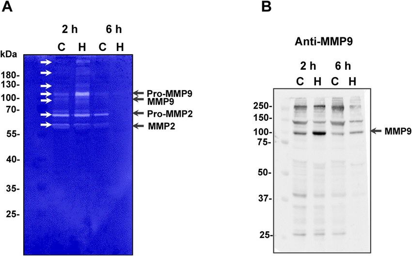
www.nature.com/scientificreports/
F354. Furthermore, we showed that in the secretome of mouse embryonic fibroblasts,
potential new substrates of H
HF3 alters the N-terminome by promoting the cleavage of proteins of the ECM and of focal adhesions and the
cysteine protease inhibitor cystatin-C53. The aim of this study was to gain new insights into the mechanisms of
hemorrhage production by HF3 by expanding the analysis of the substrate repertoire of this SVMP.
Results
Blood vessel damage resulting in extravasation contributes to local tissue damage and poor tissue regeneration
observed upon viperid envenoming. This pathogenesis involves the disruption of hemostasis and hemorrhage,
where metalloproteinases play key roles in different pathways. SVMPs may exert pro- or anti-coagulation effects
by limited proteolysis or degradation of plasma components. Besides, SVMPs may cleave ECM proteins and
microvasculature BM proteins leading to fluid extravasation into the surrounding tissue. The main objective
of this study was to evaluate the role of proteins involved in the stability of ECM and integrity of glycocalyx
(proteoglycans) and in hemostasis (blood coagulation proteins and PDGF/PDGFR) in the hemorrhage induced
by HF3, thereby providing new insights into the mechanisms for the typical pathological effects observed upon
envenoming.
Interaction of HF3 with proteoglycans in vitro. Proteoglycans are a complex group of proteins
encoded by 43 g enes55. Our aim with these studies was to investigate further the interaction of P-III class SVMPs
with proteoglycans, including some components of the endothelial glycocalyx. Initially we tried to immobilize
different proteoglycans to the BIAcore CM-5 sensorchip, however, without success (data not shown). To over-
come this issue, HF3 was covalently immobilized on the BIAcore CM-5 sensorchip and assayed to assess its
ability to bind to biglycan, brevican, glypican-1, lumican, mimecan and syndecan-1 using surface plasmon reso-
nance (SPR). As observed in Fig. 1, these proteoglycans were capable of interacting with immobilized HF3 in a
concentration-dependent fashion (75–1200 nM). On the other hand, although decorin was shown to be cleaved
by HF3 in a previous study, using a decorin sample from a different source11, here it did not show interaction
with HF3 at 1 µM and 5 µM (Supplementary Figure S1). In the case of aggrecan, we did succeed in covalently
immobilizing it to a CM-5 sensorchip and HF3 was assayed for interaction to this proteoglycan at increasing
concentrations (15–1000 nM), showing a concentration-dependent binding profile (Supplementary Figure S1).
Proteolytic effect of HF3 on proteoglycans in vitro and in vivo. Proteolysis of extracellular matrix
and basement membrane components is considered to be a critical factor in the production of hemorrhage by
certain SVMPs. Since HF3 showed interaction with various proteoglycans, we evaluated the ability of HF3 to
catalyze their hydrolysis. For this purpose, HF3 was incubated with aggrecan, brevican, glypican-1, syndecan-1,
biglycan, decorin, mimecan and lumican, as described in Experimental Procedures, and the reactions were fur-
ther evaluated by SDS-PAGE (Fig. 2). Aggrecan, a chondroitin sulfate proteoglycan, is a major structural proteo-
glycan of the cartilage extracellular matrix, but is also expressed in the brain55,56. A recombinant aggrecan form
composed of domains G1-IGD-G2 was seen as a band of ~ 120 kDa on the gel, and HF3 cleaved it generating
fragments of 30 kDa, 50 kDa and 70 kDa. Brevican, another chondroitin sulfate proteoglycan, is one of the
most important hyaluronan- and lectin-binding proteoglycans of the central nervous s ystem55,57. It was nearly
refractory to the proteolytic activity of HF3, however, a faint band of ~ 65 kDa was observed and may represent a
hydrolysis product. Glypican-1 is a cell surface proteoglycan that contains heparan s ulfate55,58 and was observed
on the gel as a main ~ 60 kDa protein band and a secondary band of ~ 40 kDa. Upon incubation with HF3 it
resulted in a single hydrolysis product of ~ 50 kDa while the band of ~ 40 kDa appeared with higher intensity
indicating that it might also contain an additional hydrolysis product of similar molecular mass. Syndecan-1 is
also a cell surface proteoglycan that contains both heparan and chondroitin sulfate and participates in the link-
age of the cytoskeleton to the interstitial matrix55,59. The protein was observed on the gel at a molecular mass
of ~ 60 kDa which nearly disappeared after incubation with HF3. Biglycan is a small leucine-rich repeat proteo-
glycan found in a variety of extracellular matrix tissues, which binds TGFβ60 and modulates its b ioactivity55,61.
Biglycan showed a molecular mass of ~ 45 kDa on the gel and was partially cleaved by HF3 resulting in products
of ~ 24, 20, and 18 kDa. Decorin, a small leucine-rich proteoglycan that has the ability to bind and decorate
fibrillar collagen in a periodic f ashion55,62, migrated as a broad band of 80 kDa, which upon incubation with HF3
resulted in a product of ~ 75 kDa. Mimecan, or osteoglycin, is a small leucine-rich proteoglycan that induces
ectopic bone formation in conjunction with transforming growth factor beta55,63. The 45 kDa band of mimecan
was almost completely degraded to generate a main hydrolysis fragment of ~ 30 kDa and minor fragments of
13–15 kDa. Lumican is also a member of the small leucine-rich proteoglycan family55,64. The 55–60 kDa band
of lumican showed a slight reduction of molecular mass to ~ 50 kDa indicating that HF3 may have promoted its
limited proteolysis.
We next examined whether endothelial glycocalyx-related proteoglycans could be degraded in vivo, in the
hemorrhagic process generated after 2 h and 6 h of HF3 injection (1 µg; 14 pmol) in the mouse skin (Fig. 3).
To this end, proteins extracted from mouse control and hemorrhagic skin were submitted to SDS-PAGE
and immunostained with biglycan, decorin, glypican-1, lumican, and syndecan-1 antibodies. As seen in Fig. 4,
in most cases, the degradation of these proteoglycans in vivo was more pronounced after 6 h of HF3 injection.
Anti-biglycan antibody recognized protein bands of nearly 250 and 120 kDa, whose intensity clearly decreased
in the hemorrhagic skin. In the control skin, anti-decorin antibody weakly immunostained two protein bands
of ~ 170 and 120 kDa, while two strongly recognized bands showed 70 and 40 kDa. All bands recognized by
anti-decorin either disappeared or diminished in intensity in the hemorrhagic skin. Immunodetection with an
anti-glypican-1 antibody revealed protein bands of ~ 150 and 50 kDa in the control samples, which were not
recognized in the hemorrhagic skin after 6 h of HF3 injection. In the case of lumican, a large smear of 60–75 kDa
Scientific Reports | (2020) 10:12912 | https://doi.org/10.1038/s41598-020-69396-y 3
Vol.:(0123456789)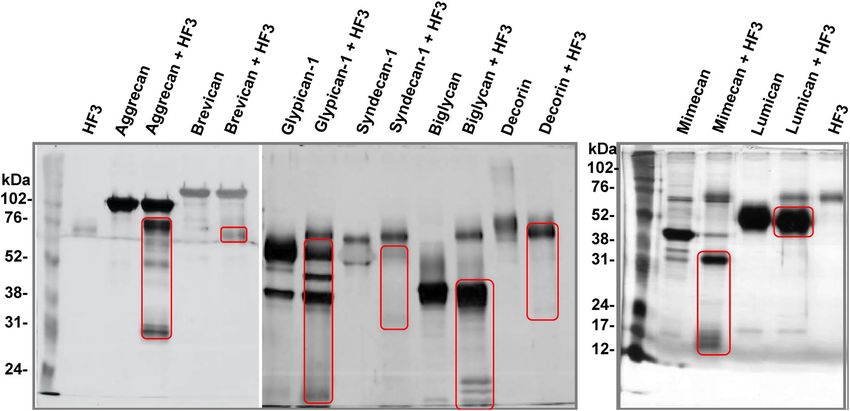
www.nature.com/scientificreports/
Biglycan on HF3 Brevican on HF3
150 0 nM
0 nM 18.75 nM
130 18.75 nM 150
37.5 nM
37.5 nM 130 75 nM
110 75 nM
110 150 nM
90 150 nM 300 nM
300 nM 90 600 nM
70 600 nM 1200 nM
70
1200 nM
RU
50
RU
50
30 30
10 10
-10 0 50 100 150 200 250 300 350 -50 -10 50 150 250 350
-30 -30
-50
-50 Seconds
Seconds
Glypican-1 on HF3 Lumican on HF3
0 nM 90 0 nM
18.75 nM 75 nM
150
37.5 nM 150 nM
130 70
75 nM 300 nM
110
RU
150 nM 600 nM
90 50 1200 nM
300 nM
70 600 nM
RU
50 1200 nM 30
30
10 10
-10 0 50 100 150 200 250 300 350
-30
-10
-50 0 50 100 150 200 250 300 350
Seconds Seconds
30
Mimecan on HF3 Syndecan-1 on HF3
25 0 nM
0 nM
150 18.75 nM
20 75 nM
130 37.5 nM
150 nM
75 nM
15 300 nM 110
150 nM
600 nM 90
RU
10 300 nM
1200 nM 70 600 nM
5
RU
50 1200 nM
0 30
10
-5
-10 0 50 100 150 200 250 300 350
-10 -30
0 50 100 150 200 250 300 350
-50
Seconds Seconds
Figure 1. Interaction of HF3 with proteoglycans by surface plasmon resonance, using a Biacore T100
system. Proteoglycans were injected over immobilized HF3, at the indicated concentrations, as described in
experimental procedures.
was recognized by the antibody, however with a core stained band of ~ 60 kDa, which decreased upon the 6 h
hemorrhagic process and showed a slight molecular mass shift to ~ 58 kDa, as observed in Fig. 2. Anti-syndecan-1
recognized different bands (250, 150, 70–75 and 50–60 kDa), which nearly disappeared after 6 h of HF3 injection.
Although aggrecan, brevican, and mimecan were cleaved in vitro by HF3, our results using antibodies to probe
their abundance in the hemorrhagic skin were inconclusive to suggest a clear degradation of these proteoglycans
in vivo (Supplementary Figure S2). Therefore, while it is not possible to make a direct comparison of the results
on the cleavage of recombinant proteoglycans in vitro by HF3 and the analysis by Western blot using specific
antibodies to assess the abundance of the same proteoglycans in the hemorrhagic skin, our results show a good
agreement between the two approaches, and point out the hydrolysis of proteoglycans as a relevant mechanism
for microvasculature destabilization and generation of hemorrhage.
Scientific Reports | (2020) 10:12912 | https://doi.org/10.1038/s41598-020-69396-y 4
Vol:.(1234567890)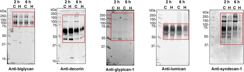
www.nature.com/scientificreports/
Figure 2. Activity of HF3 upon proteoglycans in vitro. Proteoglycans were incubated with HF3, as described in
“Materials and methods”, and submitted SDS-PAGE. Proteins were stained with silver. Red rectangles indicate
main degradation products.
Gelatinolytic activity is generated in the mouse skin injected with HF3. In
a recent investigation on the effects of HF3 upon the secretome of mouse embry-
onic fibroblasts using terminal amine isotopic labeling of substrates (TAILS)
analysis, we identified 190 unique peptide cleavages which suggested the participation of other proteinases acti-
vated upon incubation with HF3, including the matrix metalloproteinases (MMPs) 2 and 9 53. Here, we assessed the
possibility that these proteinases play a role in the proteolytic process affecting the ECM and endothelial glycocalyx
proteoglycans in the hemorrhagic mouse skin (Fig. 3). Figure 5A shows the profile of gelatinolytic activity of skin
proteins analyzed by zymography. Interestingly, the intensity of bands of ~ 68 kDa and ~ 58 kDa of gelatinolytic
activity corresponding to, respectively, pro-MMP2 and active MMP2, were nearly similar in control and in the
hemorrhagic skin after 2 h of HF3 injection, and disappeared after 6 h. However, a gelatinolytic band of ~ 105 kDa,
corresponding to the zymogen of mouse MMP9, was detected as clearly more intense in the 2 h hemorrhagic pro-
cess compared to the control, as well as and in its cleaved, activated form of 97 kDa. Interestingly, both bands were
also absent in the skin within 6 h of hemorrhage, therefore, both MMP2 and MMP9 did not show any gelatinolytic
activity in the hemorrhagic skin after 6 h of HF3 injection.
Immunostaining of the mouse skin proteins with an anti-MMP9 antibody revealed a band of ~ 100 kDa,
corresponding to the recognition of MMP9, which was only more intense in the hemorrhagic skin 2 h after HF3
injection, in agreement with the gelatin zymography (Fig. 5B). This antibody also recognized protein bands
of ~ 250 and 130 kDa which showed lower intensity in the 6 h hemorrhagic skin, however it is unclear whether
these bands correspond to other molecular, possibly complexed, forms of MMP9.
Proteolytic effect of HF3 on plasma proteins. The activity of SVMPs on plasma proteins is involved in
the severe coagulopathy and local effects observed upon viperid envenoming. Among the plasma proteins, HF3
had previously shown to cleave alpha-2-macroglobulin, fibrinogen, fibronectin, vitronectin, and von Willebrand
factor2,31. Here, we extended our analysis of plasma proteins to identify new substrates for HF3 by incubat-
ing antithrombin III, complement components C3 and C4, coagulation factors II (prothrombin), XI and XIII,
plasminogen, and protein S with HF3 at a 1:10 (w/w) enzyme:substrate ratio and the reactions were further
evaluated by SDS-PAGE (Fig. 6). Antithrombin III is a serine proteinase-inhibiting plasma protein (serpin)
of 58 kDa that targets thrombin and other coagulation cascade components of the contact activation and tis-
sue factor pathways65. HF3 promoted limited proteolysis of antithrombin III generating a product of ~ 50 kDa.
Complement component C3 plays a central role in the activation of the complement system and its activation
is required for both classical and alternative complement activation pathways66. Component C4 is cleaved by
upstream proteinases of the complement cascade and converted into fragments C4a and C4b. C4b is a protein
of 193 kDa which forms a complex with C2b, which in turn cleaves C3 into C3a and C3b as part of the events of
the complement cascade. HF3 cleaved the complement C3α chain and generated various degradation products
of ~ 25 to 50 kDa, while the C3β chain remained intact. As for component C4, after the incubation with HF3,
although its C4α, C4β, and C4γ chains apparently remained intact, a main degradation product of ~ 85 kDa
and two faint degradation products of ~ 40 kDa e 55 kDa were observed. Factor II (prothrombin) is a vitamin
K-dependent coagulation component that upon activation is proteolytically cleaved to form thrombin, a serine
brin67. The 72 kDa protein chain of prothrombin was almost completed
proteinase that converts fibrinogen into fi
Scientific Reports | (2020) 10:12912 | https://doi.org/10.1038/s41598-020-69396-y 5
Vol.:(0123456789)www.nature.com/scientificreports/
Figure 3. Representative images of hemorrhage generated in the mouse skin 2 h and 6 h after HF3 injection
(1.0 μg in 50 mM HEPES, 1 mM C aCl2, pH 7.5), as described in “Materials and methods”.
Figure 4. Degradation of glycocalyx-related proteoglycans in the mouse skin injected with HF3, as shown
by Western blot analysis. Protein samples (30 μg) extracted from control skin (C) or skin injected with HF3
(H) after 2 h and 6 h, as described in “Materials and methods”, were immunostained with anti-biglycan, anti-
decorin, anti-glypican-1, anti-lumican and anti-syndecan antibodies. Red rectangles indicate main protein
bands recognized by the antibodies.
Scientific Reports | (2020) 10:12912 | https://doi.org/10.1038/s41598-020-69396-y 6
Vol:.(1234567890)www.nature.com/scientificreports/
Figure 5. Activation of mouse skin proteinases. (A) Gelatin zymography of mouse skin proteins. Protein
samples (30 μg) from control skin proteins (C) or hemorrhagic skin proteins (H), prepared under non-reducing
conditions, were submitted to electrophoresis on 10% SDS–polyacrylamide copolymerized with gelatin. Gels
were stained with Coomassie blue. Proteins with activity were identified as clear zones of lysis against a dark
background. White arrows indicate bands of gelatinolytic activity. (B) Detection of the presence of MMP9 in
the mouse skin, as shown by Western blot analysis. Protein samples (30 μg) from control skin proteins (C) or
hemorrhagic skin proteins (H) were immunostained with anti-MMP9 antibodies. Numbers on the left indicate
molecular mass marker mobility.
degraded by HF3 to generate four stable products of ~ 28, 30, 35 and 50 kDa, of which the bands of 50 kDa and
30 kDa likely correspond to, respectively meizothrombin and thrombin, in a profile similar to that obtained by
Morita and colleagues68 for the activation of prothrombin with the prothrombin activator from Echis carinatus
venom. Factor XI (thromboplastin), which acts by cleaving factor IX in the intrinsic blood coagulation pathway,
is a homodimer consisting of two identical subunits of 80 kDa held together by disulfide bonds69. Factor XIII,
an enzyme with transglutaminase activity that crosslinks fibrin, is a tetramer of noncovalently associated pairs
of subunits of 75 and 88 kDa70. Both factors XI and XIII were not cleaved upon incubation with HF3. Plasmino-
gen is the inactive zymogen form of the serine proteinase plasmin, a fibrinolytic component of the coagulation
cascade. After incubation with HF3, the 92 kDa band of plasminogen appeared with reduced intensity and two
main products of 38 and 55 kDa were observed on the gel. The 38 kDa protein band might correspond to angio-
statin, an internal proteolytic fragment of plasminogen that is an inhibitor of proliferation of endothelial c ells71.
Protein S, a single-chain, 69 kDa protein that plays a role in the anticoagulation pathway by acting as a cofactor
to protein C in the inactivation of factors Va and VIIIa72 was not cleaved by HF3.
Proteolytic effect of HF3 on PDGFR and PDGF in vitro and in vivo. The PDGF/PDGFR axis has
been shown to participate in signaling events that control a spectrum of cellular responses in both physiological
and pathological s cenarios41. To examine whether PDGF/PDGFR could be involved in the hemorrhagic process
induced by HF3, we incubated PDGFR-α chimera, a recombinant mouse protein composed of mouse PDGFRα
(Leu25Glu524) linked via the peptide IEGRMD to the human IgG1 (Pro100Lys330), and PDGFR-β chimera, a
recombinant mouse protein composed of mouse PDGFRβ (Leu32Glu530) linked via the peptide IEGRMD to
the human IgG1 (Pro100Lys330), with HF3 at a 1:10 (w/w) enzyme:substrate ratio. As seen in Fig. 7A, PDGFR-α
chimera and PDGFRβ-chimera showed specific, limited proteolysis by HF3. Cleavage products from both pro-
teins following incubation with HF3 when subjected to mass spectrometric analysis indicated that the sites of
cleavage in the PDGFR-chimeras were localized to regions within the PDGFRs, while products originated from
the IgG1 region were not detected (Supplementary Tables S1 and S2). HF3 was also incubated with human
PDGF, a protein consisting predominantly of PDGF-AB heterodimers, at a 1:10 enzyme:substrate ratio. The pro-
tein migrated on the gel as close bands of ~ 14 and 12 kDa and was cleaved by HF3 generating products of ~ 10
and 8 kDa (Fig. 7A). We next examined whether the PDGFRs could be degraded in vivo, in the hemorrhagic
process generated by HF3 in the mouse skin (Fig. 3). As seen in Fig. 7B, PDGFRα and PDGFRβ were degraded
in the mouse skin, as shown by Western blot analysis of skin proteins after 2 h and 6 h of injection with HF3, with
more pronounced effect observed after 6 h.
Scientific Reports | (2020) 10:12912 | https://doi.org/10.1038/s41598-020-69396-y 7
Vol.:(0123456789)www.nature.com/scientificreports/
Figure 6. Activity of HF3 upon plasma proteins. Proteins were incubated with HF3 at a 1:10 (w/w)
enzyme:substrate, as described in “Materials and methods”, and submitted SDS-PAGE. Greek letters indicate
protein chains. P: prothrombin; A: albumin. Proteins were stained with silver.
Discussion
Over the past several years there has been mounting evidence showing the correlation of snake venom-induced
pathologies with the activity of metalloproteinases and pointing out to these venom components as potent
factors for the perturbation of the prey’s or human victim circulatory system, with the endothelium and the
blood components being directly affected by the venom. In this context, the hemorrhagic process generated by
SVMPs is a complex phenomenon resulting in capillary disruption and blood extravasation. Here, we focused
on the hypothesis that various proteoglycans, especially those of the endothelium glycocalyx, plasma proteins,
Scientific Reports | (2020) 10:12912 | https://doi.org/10.1038/s41598-020-69396-y 8
Vol:.(1234567890)www.nature.com/scientificreports/
Figure 7. Activity of HF3 upon PDGFR and PDGF. (A) HF3 cleaves PDGFR (alpha and beta forms) and PDGF
in vitro. PDGFR-α (recombinant mouse PDGFR-α-Fc chimera), PDGFR-β (recombinant mouse PDGFR-
β-Fc chimera) and PDGF were incubated with HF3, as described in “Materials and methods”, and submitted
SDS-PAGE. White rectangles indicate proteins bands identified by mass spectrometry. Letters A and B indicate
the subunits of the disulfidelinked PDGF heterodimer. Arrows indicate cleavage products generated by HF3.
Proteins were stained with silver. (B) Degradation of PDGFR-α and PDGFR-β in the mouse skin injected with
HF3, as shown by Western blot analysis. Protein samples (30 μg) from control skin (C) or skin injected with
HF3 (H), and PDGFR-α (recombinant mouse PDGFR-α-Fc chimera) and PDGFR-β (recombinant mouse
PDGFR-β-Fc chimera) (10 ng) were immunostained with anti-PDGFR-α and anti-PDGFR-β antibodies. Full-
length images of Western blots and SDS-PAGE profiles of mouse proteins from control and hemorrhagic skins
in comparison with PDGFRα and PDGFRβ are shown in Supplementary Figure S3.
Scientific Reports | (2020) 10:12912 | https://doi.org/10.1038/s41598-020-69396-y 9
Vol.:(0123456789)www.nature.com/scientificreports/
and PDGFR/PDGF could be involved in the hemorrhagic process induced by HF3 and hence carried out both
in vivo and in vitro assays.
Basement membrane proteoglycans are found at the periphery of cells and those associated with vascular
endothelial cells play important roles in endothelial functions, such as in the inhibition of intravascular coagu-
lation and extravasation of plasma proteins and blood cells73. The endothelium is lined with a glycocalyx layer
that functions as a barrier between circulating blood and the vessel wall. SVMPs have been shown to be active
upon a variety of substrates, including plasma proteins, extracellular matrix components located in the basement
membrane of the microvasculature, endothelial cells and components involved on platelet-aggregation. In the
scenario of local hemorrhage induced by SVMPs there appears to be a clearing of regions of capillary basement
membrane with a subsequent dissolution of the endothelial lining, ultimately allowing escape of capillary contents
into the stroma74. In this context, the ability of SVMPs to degrade proteoglycans may be a relevant mechanism
to induce hemorrhage. Tortorella and c olleagues75 showed for the first time that a P-I class SVMP (atrolysin C,
from Crotalus atrox venom) was able to cleave aggrecan in vitro. Moreover, Escalante and c olleagues76 have shown
that perlecan was present in the exudate collected from muscle tissue injected with BaPI, from B. asper venom.
Using proteomic analysis, we have shown that in the hemorrhage generated by HF3 in the mouse skin there is
degradation of decorin, lumican and mimecan11. Here, using SPR we show that HF3 dose-dependently inter-
acted with proteoglycans aggrecan, brevican, glypican-1, syndecan-1, biglycan, mimecan and lumican, but not
with decorin. Nevertheless, decorin, and all the other proteoglycans were susceptible to degradation or apparent
limited proteolysis by HF3. Furthermore, immunostaining with specific antibodies showed the degradation of
proteoglycans composing the endothelial glycocalyx (biglycan, decorin, glypican-1, lumican, and syndecan-1)
in the hemorrhagic process generated by HF3 in the mouse skin.
Although at different levels, all proteoglycans tested in this study are detected in the skin either as mRNA or
protein (https://www.proteinatlas.org/)77. Therefore, their direct in vitro degradation by HF3, or in vivo, in the
hemorrhagic process generated by HF3, suggests a critical role of the destabilization of the mouse skin integrity
promoted by HF3, as noticed by the extreme friability of the hemorrhagic skin tissue observed upon cryostat
sectioning for histological analysis (not shown). Notably, the results of this study underscore the role of degrada-
tion of biglycan (extracellular matrix), decorin (pericellular matrix), glypican (cell surface), lumican (extracel-
lular matrix) and syndecan-1 (cell surface) in the local hemorrhagic process by SVMPs. Biglycan degradation
in the hemorrhagic process would have twofold implications, as this proteoglycan (1) participates in different
proinflammatory signaling pathways in conjunction with multi-receptor crosstalk in macrophages78, and (2) can
be released from the ECM by proteolytic enzymes of the MMP family, or by granzyme B 79–82 then acting as a
Damage-Associated Molecular Pattern (DAMP) m olecule78,83,84. Decorin plays a role in the regulation of collagen
fibrillogenesis and functions as a reservoir of transforming growth factor beta 1 (TGF-beta) in the extracellular
matrix85–87 implying that in the hemorrhagic process induced by HF3 decorin cleavage might induce tissue reac-
tions mediated by TGF-beta released in the connective tissue. Glypicans are bound to the outer surface of the
cell membrane by a glycosyl-phosphatidylinositol anchor, while their heparan sulfate chains bind growth factors
and participate in cell signaling, proliferation and matrix production88. For instance, glypican-1, by means of
its heparan sulfate chains, regulates the stability of the interaction of fibroblast growth factor 2 (FGF2) and its
receptor, protecting FGF2 from degradation89. The shedding of glypican-1 by ADAM 17 was recently reported90,
and its degradation in the hemorrhagic process induced by HF3 suggests that it is susceptible to proteolysis with
implications to growth factors functions in physiological and pathological conditions. Lumican has been shown
to be cleaved by MT-MMP191 and MMP1379. The interaction of lumican with collagen fibers at sites prone to
their cleavage by collagenases was shown to sterically retards or protects against p roteolysis92, suggesting that
in the hemorrhagic process induced by HF3, collagens would be more susceptible to degradation. Cell-surface
syndecans links the cytoskeleton to the interstitial matrix by means of heparan sulfate chains and is expressed
on leukocytes and endothelial cells. Syndecans regulate many cellular processes, such as adhesion, proliferation,
and migration, and are susceptible to shedding by proteinases, resulting in the release of ectodomains that can
act as competitive inhibitors of their cell surface-linked counterparts93–95 and modulate the onset of different
pathological processes96–98. Considering the role of syndecan-1 in the endothelial glycocalyx, its degradation
in the hemorrhagic process generated by HF3 in the mouse skin suggests the impairment of its functions with
consequences related to glycocalyx permeability and mechanotransduction of fluid shear stress, as well as to
leukocyte access to endothelial tissue.
While we show that HF3 can directly interact with and cleave proteoglycans in vitro, the possibility that tis-
sue proteinases may also contribute to their degradation in the HF3-induced hemorrhage cannot be ruled out.
Indeed, proteoglycans are susceptible to shed or degradation by different metalloproteinases (MMP, MT-MMP,
ADAM and ADAMTS)99–103 and therefore the increased MMP9-like gelatinolytic activity detected among pro-
teins extracted from the hemorrhagic tissue 2 h after HF3 injection points out to the dysregulation of homeostasis
and proteolytic balance triggered by protein degradation products, cytokines and other inflammation mediators
such as DAMP molecules, with modulation of the tissue proteolytic signature. Because of their potential for tissue
damage, there are different ways to regulate MMPs activity, including gene expression, zymogen activation, and
enzyme inactivation by specific inhibitors. Interestingly, the increased gelatinolytic activity in the 2 h hemorrhagic
process was a transient phenomenon, which took a turn and evolved to an unexpected absence of activity within
6 h of hemorrhagic process, indicating a clear tissue response to proteolytic damage.
Regarding the hemorrhagic process induced by SVMPs, it has been suggested that the degradation of capil-
lary basement membrane results in the weakening of the mechanical stability of the capillary wall, causing its
distention due to the action of hemodynamic biophysical forces operating in the circulation and resulting in
the capillary wall disruption and extravasation of blood5,9. As the glycocalyx is suggested to play a role in the
regulation of vascular permeability, in the prevention of the adhesion of blood cells to the vessel wall, and in
the transmission of shear stress, our findings suggest that in vivo the activity of SVMPs, not only on basement
Scientific Reports | (2020) 10:12912 | https://doi.org/10.1038/s41598-020-69396-y 10
Vol:.(1234567890)www.nature.com/scientificreports/
membrane components but also on the endothelial glycocalyx would result in disruption of microvessel integrity
and dynamics, and contribute to local inflammation and hemorrhage. Besides, it is also tempting to speculate
that the degradation products originated from the activity of SVMPs in the endothelial glycocalyx microenviron-
ment could interfere with the microvascular barrier regulation and have a direct effect upon the permeability
of endothelial cells.
The mechanisms involved in the hemorrhagic process generated by HF3, particularly the role of the cleavage
of plasma proteins in the context of the hemorrhage, remain not fully understood. HF3 had been previously
shown to directly cleave alpha-2-macroglobulin, fibrinogen, fibronectin, vitronectin, and von Willebrand factor
in vitro2,31. Moreover, we showed that alpha-1-antitrypsin, apolipoprotein AII, fibrinogen and fibronectin are
degraded in the hemorrhagic process induced by HF3 in the mouse s kin11. Here we show that HF3 is also able to
cleavage antithrombin III, components C3 and C4 of the complement system, factor II and plasminogen, but not
factors XI and XIII and protein S in vitro. Among these substrates, there are proteins of the coagulation cascade
and of the complement system, as well as plasma proteinase inhibitors, indicating that HF3 escapes inhibition
and may act in an unregulated fashion causing the imbalance of hemostasis. Cleavage of fibrinogen, factor II
and plasminogen by HF3 indicate a clear interference of this SVMP in three strategic points of the coagulation
cascade, i.e., the generation of fibrin, thrombin and plasmin. Pidde-Queiroz et al.104 showed the cleavage of
complement components C3 and C4 by a P-I class SVMP from B. pirajai venom. The cleavage of these pro-
teins in vitro by HF3 suggests the ability of HF3 to impair the proper immune response provided by activation
the classical and alternative pathways of the complement cascade. Antithrombin is synthesized by endothelial
cells and is found within the glycocalyx, where it exerts specific anti-inflammatory effects105,106. Furthermore,
antithrombin III was shown to play a role in the preservation of the endothelial glycocalyx from shedding upon
ischaemia/reperfusion106,107. A previous report had shown the proteolytic inactivation of antithrombin III by a
P-I class SVMP from Crotalus adamanteus (adamalysin) in the presence or absence of h eparin108, generating a
stable hydrolysis product similar to that observed with HF3. Our finding on the cleavage of antithrombin III by
HF3 adds a novel mechanism by which SVMPs interfere with endothelial glycocalyx integrity contributing to
hemorrhage and inflammation.
To the best of our knowledge, a role for the PDGF/PDGFR axis has not been assessed in the hemorrhagic
process induced by SVMPs. Among the physiological processes in which the PDGF/PDGFR family participates,
the migration and proliferation of pericytes contribute to the formation and proper function of blood vessels109.
Likewise, a link between the activity and inhibition of PDGFR has been shown in different endothelial pathol-
ogy scenarios. For instance, the PDGF signaling through PDGFRα and PDGFRβ induces angiogenesis by up-
regulating vascular endothelial growth factor production and modulating the proliferation and recruitment of
perivascular cells110. Furthermore, it has been shown that the disruption of PDGF-B/PDGFRβ paracrine signaling
affects perivascular cells and endothelial cells proliferate irregularly, leading to improper vessel formation and
hemorrhage111. And concerning the crucial role of PDGFRβ–mediated interactions in recruitment of mural
cells by newly formed vessels, genetic-induced disruption of PDGF-B or PDGFRβ leads to the development
of microvascular leakage, lethal hemorrhage, and edema in late embryogenesis in mice112,113. Recently, using a
rat model, Hall et al.114 showed that the use of specific PDGFRβ inhibitors resulted in pericyte depletion and
hemorrhage into the corpus luteum of the ovary. In line with these findings, here we show that HF3 is able to
cleave PDGFR (alpha and beta forms) and PDGF in vitro. Interestingly, immunostaining of proteins extracted
from the mouse hemorrhagic skin also showed the degradation of PDGFR (alpha and beta forms), suggesting
that the concerted function of the PDGF-mediated signaling upon PDGFR may be impaired in the hemorrhagic
process induced by HF3.
Conclusions
This investigation serves to underscores new targets of an extremely potent snake venom metalloproteinase
that induces hemorrhage in the mouse and rabbit skins at pmolar, hormonal-like doses. Components of the
endothelial glycocalyx, plasma proteins and PDGF/PDGFR are shown for the first time to be potentially involved
in the generation of hemorrhage. Taken together, our results suggest that the effects of HF3 in the mouse skin
and plasma are rather complex and involve the degradation or limited proteolysis of a range of substrates that
might be directly hydrolyzed by HF3 or by tissue proteinases activated in the inflammatory/hemorrhagic sce-
nario generated in vivo by HF3. In consonance with the two-step model proposed by Escalante and c olleagues4,
these events generate the triggering of tissue and plasma pathological responses, derived from the degradation
of proteins and from their degradation products, that culminate in the disturbance of hemostasis, and in the
destabilization of the microvasculature via disruption of glycocalyx integrity, of cell–cell and cell–ECM interac-
tions (Fig. 8).
Materials and methods
HF3. HF348 (Uniprot entry Q98UF9) was purified as described previously47 from B. jararaca venom provided
by the Laboratory of Herpetology of Butantan Institute (São Paulo, Brazil). Briefly, the venom (900 mg) was dis-
solved in 90 mL of 0.15 M NaCl, centrifuged (4 °C, 10,000 g, 20 min), and submitted to saturation with ammo-
nium sulfate. The protein fraction obtained between 30 and 60% of ammonium sulfate was dissolved in 20 mL
water, dialyzed against 0.02 M Tris–HCl buffer containing 1 mM CaCl2 and 0.02% sodium azide (pH 7.5), at
4 °C, and chromatographed on a DEAE-cellulose DE-52 column previously equilibrated with the dialysis buffer,
at 4 °C. The bound fraction, eluted with 0.15 M–0.20 M NaCl, contained HF3 and was concentrated by lyophi-
lization, and resuspended with 0.25 M ammonium bicarbonate containing 1 mM CaCl2 and chromatographed
in aliquots of 0.25 mL on a Superose 12 h 10/30 column (GE Healthcare), at 0.1 mL/min, using an Äkta Purifier
Scientific Reports | (2020) 10:12912 | https://doi.org/10.1038/s41598-020-69396-y 11
Vol.:(0123456789)www.nature.com/scientificreports/
Figure 8. Schematic illustration of different proteins and proteoglycans that are cleaved in vitro by HF3.
Components of the ECM, of the endothelial cell membrane, of the endothelial glycocalyx, and of plasma are
potential targets of HF3, as reported in this and in former studies, and cited throughout this article. Some of
these proteins and proteoglycans have also been shown to be cleaved in vivo, either due to the direct activity
of HF3 or by tissue proteinases activated in the inflammatory/hemorrhagic scenario generated by HF3 in the
mouse skin, culminating in the disturbance of hemostasis, and in the destabilization of the microvasculature via
disruption of glycocalyx integrity, of cell–cell and cell–ECM interactions.
system (GE Healthcare). HF3 was obtained in different peaks of the Superose 12 chromatography and identified
by trypsin digestion and mass spectrometric (LC–MS/MS) analysis, as described b efore47.
Proteolytic effect of HF3 on proteoglycans in vitro. Aggrecan (domains G1-IGD-G2), brevican,
glypican-1, syndecan-1, byglican (R&D Systems) and decorin (Sigma-Aldrich) (2 µg) were incubated with
100 ng HF3 in 50 mM Tris–HCl, pH 8.0, containing 1 mM CaCl2 [1:20 (w/w) enzyme:substrate ratio; final
volume 20 µL], for 3 h at 37 °C. Mimecan and lumican (R&D Systems) (2 µg) were incubated with 200 ng HF3
in 50 mM Tris–HCl, pH 8.0, containing 1 mM C aCl2 [1:10 (w/w) enzyme:substrate ratio; final volume 20 µL],
for 3 h at 37 °C. A sample of each protein was incubated without HF3 under identical conditions. Reactions
were interrupted by adding Laemmli sample b uffer115 and submitted to SDS-PAGE under reducing conditions
using different polyacrylamide concentrations: 10% (aggrecan, brevican, glypican-1, syndecan-1, byglican and
decorin) and 12% (mimecan and lumican).
Proteolytic effect of HF3 on plasma proteins in vitro. Antithrombin III (Abcam), complement com-
ponents C3 and C4 (Complement Technology), coagulation factors II (prothrombin; Hyphen Biomed), XI and
XIII (Abcam), plasminogen (Calbiochem), and protein S (Abcam) (2 µg) were incubated with HF3 (200 ng) in
50 mM Tris–HCl, pH 8.0, containing 1 mM CaCl2 [1:10 (w/w) enzyme:substrate ratio; final volume 20 µL], for
uffer115 and submitted to SDS-PAGE under
2 h at 37 °C. Reactions were interrupted by adding Laemmli sample b
Scientific Reports | (2020) 10:12912 | https://doi.org/10.1038/s41598-020-69396-y 12
Vol:.(1234567890)www.nature.com/scientificreports/
reducing conditions using different polyacrylamide concentrations (C3 and plasminogen, 10%; antithrombin
III, C4, factors II, XI and XIII, and protein S, 12%).
Proteolytic effect of HF3 on PDGFR and PDGF in vitro. PDGFR-α [recombinant mouse protein
composed of mouse PDGFRα (Leu25Glu524) linked via the peptide IEGRMD to the human IgG1 (Pro100
Lys330)], PDGFR-β [recombinant mouse protein composed of mouse PDGFRβ (Leu32Glu530) linked via the
peptide IEGRMD to the human IgG1 (Pro100Lys330)] (2 µg), and PDGF (200 ng) (R&D Systems) were incu-
bated with HF3 in 50 mM Tris–HCl, pH 8.0, containing 1 mM CaCl2 [1:10 (w/w) enzyme:substrate ratio; final
volume 20 µL], for 3 h at 37 °C. Reactions were interrupted by adding Laemmli sample buffer115 and submitted
to SDS-PAGE under reducing conditions using different polyacrylamide concentrations: 9% (PDGFR) and 12%
(PDGF).
Identification of PDGFR degradation products by mass spectrometry. Protein bands were
excised, and in-gel trypsin digestion was performed according to Hanna and c olleagues116. An aliquot (4.5 µL)
of the resulting peptide mixture was desalinized using a C-18 column (180 μm × 20 mm) and separated in a C18
column (8 cm × 75 μm) (Waters) by RP-HPLC (Eksigent) coupled with a LTQ-XL mass spectrometer (Thermo
Scientific) at a flow rate of 500 nL/min. The gradient was 2–80% acetonitrile in 0.1% formic acid over 30 min.
The instrument was operated in the top ten mode, in which one MS spectrum is acquired followed by MS/MS
of the top ten most-intense peaks detected, using 2.5 kV and 200 °C as source voltage and temperature, respec-
tively. Full dynamic exclusion was used to enhance dynamic range—one spectrum before exclusion for 120 s.
The resulting fragment spectra were searched using Mascot (version 2.2) search engine (Matrix Science) against
nrNCBI Databank (www.ncbi.nlm.nih.gov) restricted to “Mus” taxonomy with a parent tolerance of 1.5 Da and
fragment tolerance of 1.0 Da. Iodoacetamide derivative of cysteine and oxidation of methionine were specified
in Mascot as fixed and variable modifications, respectively. Mascot identifications required ion scores greater
than the associated identity scores and 20, 30, 40 and 40 for singly, doubly, triply, and quadruply charged pep-
tides, respectively.
Analysis of the interaction of HF3 with proteoglycans in vitro. Protein–protein interactions were
assessed by surface plasmon resonance with a BIAcoreT100 system (GE Healthcare), as previously d escribed2.
HF3 and aggrecan were covalently immobilized on the BIAcore CM-5 sensorchip (carboxylated dextran matrix;
GE Healthcare) according to the manufacturer’s instructions. Briefly, the CM-5 chip was activated with a 1:1
mixture of 0.4 M 1-ethyl-3-(3-dimethylaminopropyl) carbodiimide and 0.1 M N-hydroxysuccinimide for 7 min.
HF3 (1 µM) and aggrecan (0.2 µM) in 10 mM sodium acetate, pH 4.0, were injected over the activated CM-5 chip
at 25 °C. Remaining active groups on the matrix were blocked with 1 M ethanolamine/HCl, pH 8.5. Immobiliza-
tion of HF3 and aggrecan on CM-5 sensorchip resulted in average surface concentrations of 2.45 ng/mm2 and
1.7 ng/mm2, respectively. Samples of proteoglycans biglycan, syndecan-1, glypican and brevican (0, 18.75, 37.5,
75, 150, 300, 600 and 1200 nM), mimecan and lumican (0, 75, 150, 300, 600 and 1200 nM) in HBS-EP buffer
(10 mM HEPES pH 7.4, containing 150 mM NaCl, 3 mM EDTA and 0.005% surfactant P-20), were injected
over immobilized HF3 at a flow rate of 30 µL/min. HF3 (0; 15,62; 31,25; 62,5; 125; 250; 500 and 1000 nM) in
HBS-EP buffer was injected over immobilized aggrecan at a flow rate of 30 µL/min. Results were analyzed using
BIAevaluation software version 1.1.1.
Animals. Male Swiss mice were housed in temperature-controlled rooms and received water and food
ad libitum. These studies were approved by the Experimental Animals Committee of Butantan Institute, São
Paulo, Brazil; (Protocol 688-09), in accordance with guidelines of the National law for Laboratory Animal Exper-
imentation Control (Law No. 11.794, October 8, 2008).
Injection of HF3 in the mouse skin. Generation of hemorrhage in the mouse skin was carried out as
previously described2. Briefly, male Swiss mice (n = 3) weighing 18–22 g were injected intradermally on the dor-
sal region with 100 μL of a control solution (50 mM HEPES, 1 mM C aCl2, pH 7.5) or with 100 μL of a solution
containing 1.0 μg of HF3 in 50 mM HEPES, 1 mM CaCl2, pH 7.5. After 2 h and 6 h, animals were euthanized,
and the dorsal skin was sectioned and homogenized with a tissue homogenizer (Polytron PT3100; Kinematica)
using lysis buffer (50 mM HEPES, 200 mM NaCl, 2% CHAPS, pH 7.5) containing the HALT protease inhibitor
cocktail (Thermo Fisher) and 5 mM EDTA (final concentration), with 18,000 rpm cycles, for 30 s, in ice bath.
Homogenates were clarified by centrifugation at 14,000 × g, for 10 min, at 4 °C, to remove debris and the clear
supernatant was carefully removed for analysis.
Gelatin zymography of mouse skin proteins. Protein samples (30 μg) from mouse control skin or
skin injected with HF3, prepared under nonreducing conditions, as described before117, were submitted to elec-
trophoresis on 10% SDS-PAGE copolymerized with gelatin (1 mg/mL; Sigma-Aldrich). Gels were stained with
Coomassie blue and destained. Gelatin digestion was identified as clear zones of lysis against a blue background.
Western blot analysis of proteoglycans, MMP9 and PDGFRs in the mouse skin. Skin proteins
from control and HF3-treated animals (30 μg), PDGFR-α (recombinant mouse PDGFR-α-Fc chimera), and
PDGFR-β (recombinant mouse PDGFR-β-Fc chimera) (10 ng) were submitted to SDS-PAGE (10% SDS–poly-
acrylamide gel) under reducing conditions and transferred onto nitrocellulose membranes (GE Healthcare)118.
Membranes were blocked for 3 h in PBS-T buffer (150 mM sodium chloride, 20 mM sodium phosphate, pH 7.1,
Scientific Reports | (2020) 10:12912 | https://doi.org/10.1038/s41598-020-69396-y 13
Vol.:(0123456789)www.nature.com/scientificreports/
and 0.1% Tween 20) containing 5% nonfat milk and were then incubated for 16 h, according to manufacturer’s
instructions, with antibodies recognizing mouse lumican (AF2745; R&D Systems), mimecan (AF2949; R&D
Systems), syndecan-1 (AF3190; R&D Systems), PDGFR-α (AF1062; R&D Systems), PDGFR-β (AF1042; R&D
Systems) and MMP9 (ab38898; Abcam) or human aggrecan (AF1220; R&D Systems), brevican (AF4009, R&D
Systems), biglycan (AF2667; R&D Systems), decorin (AF143; R&D Systems), and glypican (AF4519; R&D Sys-
tems). After incubation for 1 h with specific anti-IgG secondary antibodies (Sigma-Aldrich, Vector Laboratories
or R&D systems), conjugated to peroxidase, immunoreactive signals were visualized by enhanced chemilumi-
nescence using ECL prime Western Blotting detection reagent (GE Healthcare) and a chemiluminescence docu-
mentation system (Uvitec). As glyceraldehyde 3-phosphate dehydrogenase (GAPDH) and actin are cleaved by
HF311,54 and hence could not be used as protein loading controls we show the electrophoretic profile of the skin
proteins by SDS-PAGE (30 μg) run under the same conditions used for Western blot analysis and stained with
colloidal Coomassie blue or Ponceau S (Supplementary Figure S4).
Data availability
The datasets generated during and/or analyzed during the current study are available from the corresponding
author on reasonable request.
Received: 22 February 2020; Accepted: 7 July 2020
References
1. Fox, J. W. & Serrano, S. M. Insights into and speculations about snake venom metalloproteinase (SVMP) synthesis, folding and
disulfide bond formation and their contribution to venom complexity. FEBS J. 275, 3016–3030 (2008).
2. Oliveira, A. K. et al. New insights into the structural elements involved in the skin haemorrhage induced by snake venom metal-
loproteinases. Thromb. Haemost. 104, 485–497 (2010).
3. Baldo, C., Jamora, C., Yamanouye, N., Zorn, T. M. & Moura-da-Silva, A. M. Mechanisms of vascular damage by hemorrhagic
snake venom metalloproteinases: tissue distribution and in situ hydrolysis. PLoS Negl. Trop. Dis. 4(6), e727 (2010).
4. Escalante, T., Rucavado, A., Fox, J. W. & Gutiérrez, J. M. Key events in microvascular damage induced by snake venom hemor-
rhagic metalloproteinases. J. Proteom. 74, 1781–1794 (2011).
5. Gutiérrez, J. M., Escalante, T., Rucavado, A., Herrera, C. & Fox, J. W. A comprehensive view of the structural and functional
alterations of extracellular matrix by snake venom metalloproteinases (SVMPs): novel perspectives on the pathophysiology of
envenoming. Toxins (Basel) 8(10), E304 (2016).
6. Moura-da-Silva, A. M. et al. Collagen binding is a key factor for the hemorrhagic activity of snake venom metalloproteinases.
Biochimie 90, 484–492 (2008).
7. Serrano, S. M. et al. The cysteine-rich domain of snake venom metalloproteinases is a ligand for von Willebrand factor A domains:
role in substrate targeting. J. Biol. Chem. 281, 39746–39756 (2006).
8. Serrano, S. M. T. et al. Interaction of the cysteine-rich domain of snake venom metalloproteinases with the A1 domain of von
Willebrand factor promotes site-specific proteolysis of von Willebrand factor and inhibition of von Willebrand factor-mediated
platelet aggregation. FEBS J. 274, 3611–3621 (2007).
9. Gutiérrez, J. M., Rucavado, A., Escalante, T. & Díaz, C. Hemorrhage induced by snake venom metalloproteinases: biochemical
and biophysical mechanisms involved in microvessel damage. Toxicon 45, 997–1011 (2005).
10. Escalante, T. et al. Wound exudate as a proteomic window to reveal different mechanisms of tissue damage by snake venom
toxins. J. Proteome Res. 8, 5120–5131 (2009).
11. Paes Leme, A. F. et al. Hemorrhagic activity of HF3, a snake venom metalloproteinase: insights from the proteomic analysis of
mouse skin and blood plasma. J. Proteome Res. 11, 279–291 (2012).
12. Jarvelainen, H., Sainio, A., Koulu, M., Wight, T. N. & Penttinen, R. Extracellular matrix molecules: potential targets in pharma-
cotherapy. Pharmacol. Rev. 61, 198–223 (2009).
13. Iozzo, R. V. Basement membrane proteoglycans: from cellar to ceiling. Nat. Rev. Mol. Cell Biol. 6, 646–656 (2005).
14. Eble, J. A. & Niland, S. The extracellular matrix of blood vessels. Curr. Pharm. Des. 15, 1385–1400 (2009).
15. Marchand, M., Monnot, C., Muller, L. & Germain, S. Extracellular matrix scaffolding in angiogenesis and capillary homeostasis.
Semin. Cell Dev. Biol. 89, 147–156 (2019).
16. Halper, J. Proteoglycans and diseases of soft tissues. In Progress in heritable soft connective tissue diseases. Advances in experimental
medicine and biology Vol. 802 (ed. Halper, J.) 49–58 (Springer, Dordrecht, 2014).
17. Reitsma, S., Slaaf, D. W., Vink, H., van Zandvoort, M. A. & Oude Egbrink, M. G. The endothelial glycocalyx: composition,
functions, and visualization. Pflugers Arch. 454, 345–359 (2007).
18. Manon-Jensen, T., Kjeld, N. G. & Karsdal, M. A. Collagen-mediated hemostasis. J. Thromb. Haemost. 14, 438–448 (2016).
19. Zeng, Y. Endothelial glycocalyx as a critical signalling platform integrating the extracellular haemodynamic forces and chemical
signalling. J. Cell. Mol. Med. 21, 1457–1462 (2017).
20. Tarbell, J. M. & Cancel, L. M. The glycocalyx and its significance in human medicine. J. Intern. Med. 280, 97–113 (2016).
21. Larsen, A. M., Leinøe, E. B., Johansson, P. I., Birgens, H. & Ostrowski, S. R. High syndecan-1 levels in acute myeloid leukemia
are associated with bleeding, thrombocytopathy, endothelial cell damage, and leukocytosis. Leuk. Res. 37, 777–783 (2013).
22. Rahbar, E. et al. Endothelial glycocalyx shedding and vascular permeability in severely injured trauma patients. J. Transl. Med.
13, 117 (2015).
23. Kottke, M. A. & Walters, T. J. Where’s the leak in vascular barriers? A review. Shock 46(3 Suppl 1), 20–36 (2016).
24. Farsky, S. H. et al. Bothrops asper snake venom and its metalloproteinase BaP-1 activate the complement system. Role in leucocyte
recruitment. Mediat. Inflamm. 9, 213–221 (2000).
25. Serrano, S. M. & Maroun, R. C. Snake venom serine proteinases: sequence homology vs. substrate specificity, a paradox to be
solved. Toxicon. 45, 1115–1132 (2005).
26. Fox, J. W. & Serrano, S. M. Structural considerations of the snake venom metalloproteinases, key members of the M12 reprolysin
family of metalloproteinases. Toxicon 45, 969–985 (2005).
27. Kini, R. M. Toxins in thrombosis and haemostasis: potential beyond imagination. J. Thromb. Haemost. 9(Suppl 1), 195–208
(2011).
28. Markland, F. S. Jr. & Swenson, S. Snake venom metalloproteinases. Toxicon 62, 3–18 (2013).
29. Baramova, E. N., Shannon, J. D., Bjarnason, J. B., Gonias, S. L. & Fox, J. W. Interaction of hemorrhagic metalloproteinases with
human alpha 2-macroglobulin. Biochemistry 29, 1069–1074 (1990).
Scientific Reports | (2020) 10:12912 | https://doi.org/10.1038/s41598-020-69396-y 14
Vol:.(1234567890)You can also read