Activation of proline biosynthesis is critical to maintain glutamate homeostasis during acute methamphetamine exposure - Nature
←
→
Page content transcription
If your browser does not render page correctly, please read the page content below
www.nature.com/scientificreports
OPEN Activation of proline biosynthesis
is critical to maintain glutamate
homeostasis during acute
methamphetamine exposure
Bobby Jones1,2,3,4, Muthukumar Balasubramaniam1,5, Joseph J. Lebowitz6,
Anne Taylor7, Fernando Villalta1,5, Habibeh Khoshbouei6, Carrie Grueter7, Brad Grueter7,
Chandravanu Dash1,2,5,8* & Jui Pandhare1,2,3,4,9*
Methamphetamine (METH) is a highly addictive psychostimulant that causes long-lasting effects in
the brain and increases the risk of developing neurodegenerative diseases. The cellular and molecular
effects of METH in the brain are functionally linked to alterations in glutamate levels. Despite the
well-documented effects of METH on glutamate neurotransmission, the underlying mechanism by
which METH alters glutamate levels is not clearly understood. In this study, we report an essential role
of proline biosynthesis in maintaining METH-induced glutamate homeostasis. We observed that acute
METH exposure resulted in the induction of proline biosynthetic enzymes in both undifferentiated
and differentiated neuronal cells. Proline level was also increased in these cells after METH exposure.
Surprisingly, METH treatment did not increase glutamate levels nor caused neuronal excitotoxicity.
However, METH exposure resulted in a significant upregulation of pyrroline-5-carboxylate synthase
(P5CS), the key enzyme that catalyzes synthesis of proline from glutamate. Interestingly, depletion
of P5CS by CRISPR/Cas9 resulted in a significant increase in glutamate levels upon METH exposure.
METH exposure also increased glutamate levels in P5CS-deficient proline-auxotropic cells. Conversely,
restoration of P5CS expression in P5CS-deficient cells abrogated the effect of METH on glutamate
levels. Consistent with these findings, P5CS expression was significantly enhanced in the cortical brain
region of mice administered with METH and in the slices of cortical brain tissues treated with METH.
Collectively, these results uncover a key role of P5CS for the molecular effects of METH and highlight
that excess glutamate can be sequestered for proline biosynthesis as a protective mechanism to
maintain glutamate homeostasis during drug exposure.
Methamphetamine (METH) is a powerful and highly addictive p sychostimulant1. It belongs to a larger group of
drugs called Amphetamine-Type Stimulants (ATS)1–3. Use of ATS generate a sense of euphoria, increase energy
and concentration as well as decrease appetite, induce weight loss, leading to a variety of emotional, cognitive,
and physical e ffects4,5. Among the ATS, METH has the greatest potential for abuse because of easy availability,
low cost, and longer duration of action1–3. Therefore, METH use and abuse continues to be a significant public
health problem in the United States and all over the world1–3.
METH is a lipophilic compound that crosses the blood–brain barrier easily to cause long lasting effects in
the brain4,5. Since METH’s chemical structure is similar to monoamines, it targets both dopaminergic (DA) and
1
Center for AIDS Health Disparities Research, Meharry Medical College, Nashville, TN 37208, USA. 2Center for
Molecular and Behavioral Neuroscience, Meharry Medical College, Nashville, TN 37208, USA. 3School of Graduate
Studies and Research, Meharry Medical College, Nashville, TN 37208, USA. 4Department of Microbiology,
Immunology, and Physiology, Meharry Medical College, Nashville, TN 37208, USA. 5Department of
Biochemistry, Cancer Biology, Pharmacology and Neuroscience, Meharry Medical College, Nashville,
TN 37208, USA. 6Department of Neuroscience, University of Florida, Gainesville, FL 32611, USA. 7Department
of Anesthesiology, Vanderbilt University Medical Center, Nashville, TN 37232, USA. 8Center for AIDS Health
Disparities Research, Meharry Medical College, Old Hospital Bldg‑CAHDR, Room 5027, 1005 Dr. DB Todd Jr Blvd.,
Nashville, TN 37208, USA. 9Center for AIDS Health Disparities Research, Meharry Medical College, Old Hospital
Bldg‑CAHDR, Room 5023, 1005 Dr. DB Todd Jr Blvd., Nashville, TN 37208, USA. *email: cdash@mmc.edu;
jpandhare@mmc.edu
Scientific Reports | (2021) 11:1422 | https://doi.org/10.1038/s41598-020-80917-7 1
Vol.:(0123456789)www.nature.com/scientificreports/
serotonergic (5HT) neurons in the brain. METH enters these neurons by binding to the membrane-bound DA
transporter (DAT) and/or 5HT t ransporter3. There is also evidence that METH can cross neuronal membranes
by passive diffusion mechanism6. Once inside the neuron, METH can alter the function of the vesicular mono-
amine transporter 2 (VMAT-2) and affect cytoplasmic monoamine concentrations and DA r elease6. METH
administration is also known to increase DAT internalization and affect 5HT transporter functionality6–8. In
addition, METH prevents degradation of neurotransmitters, reduces neurotransmitter reuptake, and causes
an efflux of DAT and 5HT t ransporters6–8. These biochemical and cellular effects result in an increased neuro-
transmitter availability in the synapse6–8 and cause continuous stimulation of neurons to manifest the euphoric
effects among METH users9–11.
In addition to its effects on DA and 5HT neurons, METH has been shown to affect glutamate
neurotransmission12,13. There is evidence that METH’s effect on glutamate neurotransmission is mediated by
increasing levels of extracellular g lutamate13,14. However, the mechanism of glutamate homeostasis during METH
exposure has not been clearly delineated. Glutamate is the principal excitatory neurotransmitter and is ubiq-
uitously distributed in the b rain15–17. Due to its critical role in neuronal plasticity, glutamate is involved in a
number of critical brain f unctions15–17 including learning and m emory18. It is important to note that glutamate is
produced in the nerve terminals predominantly from two sources: (1) the tricarboxylic acid (TCA) cycle, and (2)
glutamine produced by the glial c ells19,20. Glutamate produced in the nerve terminals is packaged into vesicles by
the vesicular glutamate transporters (vGLUT) and is released to the extracellular space upon stimulus of an action
potential20. After release, the glial cells clear glutamate from the synapse via the glutamate transporters, GLAST
and GLT-1, to maintain sub-neurotoxic levels of extracellular glutamate21–23. Maintaining glutamate homeostasis
is critical for proper neuronal function since excessive glutamate has been implicated in neuronal e xcitoxicity24.
The link between glutamate metabolism and carbohydrate/amino acid metabolism has been well established.
Specifically, glutamate is channeled to the TCA cycle—a common pathway in carbohydrate and amino acid
metabolism25. However, the metabolic link between glutamate and p roline26,27 remains largely understudied.
Proline, unlike other amino acids, has its α-amino group within a pyrrolidine ring, and thus it is the sole pro-
teinogenic secondary (imino) amino acid27. Proline is also unique since it has its own set of metabolic enzymes28.
It is known that glutamate can be converted to proline through Δ1-pyrroline-5-carboxylate (P5C) by the enzy-
matic activity of P5C synthase (P5CS) and P5C reductase (PYCR)26,29 (Fig. 1A). Conversely, proline can also
be converted to glutamate through the catabolic pathway catalyzed by proline oxidase/dehydrogenase (POX/
PRODH) and P5C dehydrogenase (P5CDH)25,26,29. Given this important metabolic link, proline metabolism has
been reported to play key roles in normal brain function and various neurological disorders.
Proline is abundantly found in the central nervous system (CNS)30. The presence of high affinity proline
transporters has also been reported in a subset of glutamatergic neurons in rodent brain31. In animal models,
elevated proline levels affect glutamatergic t ransmission32, depolarize n eurons33,34, increase synaptic a ctivity35,
36,37
alter cognitive tasks, sensorimotor gating, and locomotor a ctivity . In humans, hyperprolinemia—a condition
associated with abnormally elevated levels of proline has been linked to epilepsy, seizures and impaired cognitive
function38. Hyperprolinemia is also linked to schizoaffective d isorders39 and s chizophrenia40,41. Even though
these studies and others demonstrate key roles of proline in brain function and neurological disorders, the role
of proline metabolism during drug exposure remains largely unknown.
In this study, we report a functional link between proline and glutamate metabolism during acute METH
exposure. First, we utilized the SH-SY5Y neuronal cell model, that has been widely used to study neuronal
biology42,43. SH-SY5Y cells are positive for tyrosine hydroxylase (TH) and dopamine-β-hydroxylase characteristic
of catecholaminergic neurons that are known to be affected by METH44. In this model, we tested the functional
contribution of proline metabolism in METH-exposure mediated alterations in glutamate levels. Interestingly,
acute treatment with METH markedly induced the enzymes of the proline biosynthetic pathway. Specifically,
a marked induction in the expression of P5CS and PYCRs was observed in METH treated neuronal cells. This
induction was accompanied by a concomitant increase in intracellular proline levels. Surprisingly, METH expo-
sure neither increased extracellular glutamate levels or caused neuronal excitotoxicity. However, blocking the
proline synthetic arm by knocking-out P5CS increased both intracellular and extracellular glutamate levels in
METH treated cells. Furthermore, treatment of proline auxotrophic-Chinese hamster ovary (CHO-K1) cells with
METH significantly enhanced extracellular glutamate levels. These results suggested a functional link between
biosynthesis of proline and glutamate during METH exposure. Finally, levels of proline biosynthetic enzymes,
specifically, P5CS and PYCR2 were also significantly induced in the cortex of animals administered with METH
and in the slices of cortical brain tissues exposed to METH. Collectively, our studies demonstrate activation of
proline biosynthesis to sequester increased glutamate during acute METH exposure and imply an essential role
of proline metabolism in limiting neuronal glutamate excitotoxicity.
Results
Acute METH exposure activates the proline synthetic pathway. METH exposure affects gluta-
mate neurotransmission primarily by altering glutamate levels12,13. While, glutamine serves as the major source
of glutamate21–23, proline metabolism is also closely linked to g lutamate25 (Fig. 1A). However, the functional link
between proline and glutamate during drug exposure induced glutamate neurotransmission is poorly under-
stood. Therefore, we examined the effects of METH exposure on proline metabolism using the SH-SY5Y neu-
ronal cell model. First, we measured the levels of enzymes of the proline catabolic arm- POX and P5CDH- which
sequentially convert proline to P5C and then to glutamate (Fig. 1A). We treated SH-SY5Y cells with METH in
a dose dependent manner using concentrations of 1 mM and lower. Although higher concentrations of METH
have been used in published studies45,46, we chose concentrations below 1 mM since higher levels of the drug
are rarely achieved in METH associated d isorders47,48. Lysates of METH-treated and control cells were analyzed
Scientific Reports | (2021) 11:1422 | https://doi.org/10.1038/s41598-020-80917-7 2
Vol:.(1234567890)www.nature.com/scientificreports/
Figure 1. Acute METH exposure upregulates enzymes of proline synthetic pathway from glutamate. (A)
Schematic depicting the metabolic link between glutamate and proline. Synthesis of proline begins with the
conversion of glutamine to glutamate by the enzyme glutaminase (GLS). Glutamate is then converted by P5C
synthase (P5CS) to glutamic-γ-semialdehyde that spontaneously cycles to Δ1-pyrroline-5-carboxylate (P5C).
P5C is subsequently catalyzed to l-proline by P5C-reductase (PYCR). Conversely, catabolism of proline to
glutamate is catalyzed in two consecutive steps by proline oxidase (POX) and P5C dehydrogenase (P5CDH).
Glutamate is finally converted to glutamine by glutamine synthase (GS). (B–D) Effect of acute METH treatment
on proline catabolic enzymes. SH-SY5Y cells were treated with increasing concentrations of METH for
24 h. Post-treatment cells were harvested and lysed. Cell lysates were subjected to immunoblot analyses. (B)
Representative immunoblot of enzymes involved in catabolism of proline to glutamate (n = 3) (Fig. S1). (C,D)
Densitometry analysis of POX and P5CDH, respectively normalized to β-actin. (E–H) Effect of acute METH
treatment on proline metabolic enzymes. (E) Representative immunoblot of enzymes, P5CS, PYCR1 and
PYCR2, which are essential for proline biosynthesis from glutamate (n = 3). (F–H) Densitometry analysis of
P5CS, PYCR1, and PYCR2, respectively normalized to β-actin. Data presented in panels (C,D,F–H) are mean
values of three independent experiments with error bars representing SEM. *p < 0.05, **p < 0.005 represent
statistical comparison of untreated vs METH-treated cells.
by immunoblot to measure the levels of POX and P5CDH. These analysis revealed that SH-SY5Y cells endog-
enously express all the key metabolic enzymes of proline (Fig. 1B,D). Interestingly, METH exposure minimally
altered the expression of POX and P5CDH when compared to the untreated controls (Fig. 1B–D). Even at 1 mM
concentration of METH, expression of these two enzymes were not significantly altered (Fig. 1B–D), suggest-
ing that acute METH exposure minimally alters the proline catabolic arm that converts proline to glutamate
(Fig. 1A).
We next measured the expression of enzymes of the proline biosynthetic arm (Fig. 1A), P5C synthase (P5CS)
and P5C reductase (PYCR) that convert glutamate to proline by sequential c atalysis26. Notably, the expression of
P5CS was significantly increased in cells exposed to METH (Fig. 1E,F). Densitometry analysis illustrated a dose-
dependent increase in P5CS expression in cells treated with METH relative to untreated cells (Fig. 1F). Then, we
measured the levels of PYCRs- PYCR1, and PYCR2, which are involved in biosynthesis of proline (Fig. 1A)27,29,49.
As presented in Fig. 1E,G,H, expression of the PYCR1 and PYCR2 was significantly increased in the METH
treated cells. While the expression of PYCR1 was increased ~ 1.7-fold, the levels of PYCR2 was ~ sixfold higher in
cells treated with 1 mM METH compared to the untreated control cells (Fig. 1E,G,H). Collectively, these results
demonstrated that acute METH exposure activates the proline synthetic arm without altering the catabolic arm.
METH exposure upregulates key proline metabolic enzymes in differentiated neuronal cells
and cortical brain slices. Our results in Fig. 1 showed upregulation of proline synthetic enzymes by METH
exposure in undifferentiated SH-SY5Y cells. Even though the SH-SY5Y neuroblastoma cells show some char-
acteristics of neurons, we have previously shown that these cells acquire a neuronal phenotype by differentia-
tion utilizing ATRA50. Therefore, we used differentiated SH-SY5Y cells and treated them with METH in a dose
dependent manner. 24 h post treatment cellular extracts were prepared to measure the expression of P5CS and
Scientific Reports | (2021) 11:1422 | https://doi.org/10.1038/s41598-020-80917-7 3
Vol.:(0123456789)www.nature.com/scientificreports/
Scientific Reports | (2021) 11:1422 | https://doi.org/10.1038/s41598-020-80917-7 4
Vol:.(1234567890)www.nature.com/scientificreports/
◂Figure 2. METH exposure upregulates P5CS and PYCR2 in differentiated neuronal cells and slices of cortical
brain. (A–C) Differentiated SH-SY5Y cells were treated with increasing concentrations of METH for 24 h.
Post-treatment cellular lysates were subjected to immunoblot analyses. (A) Representative immunoblot of P5CS
and PYCR2 (n = 3). (B,C) Densitometry analysis of P5CS and PYCR2, respectively normalized to β-actin. (D–F)
Slices of the frontal cortex were prepared from whole brain and were incubated in 200 µM METH (n = 4) or
vehicle (n = 2). Tissue lysates were subjected to western blot analysis. (D) Representative immunoblot of P5CS
and PYCR2 (n = 3). (E,F) Densitometry analysis of P5CS and PYCR2, respectively normalized to β-actin. (G–I)
SH-SY5Y cells were treated with increasing concentrations of AMPH for 24 h. After treatment, cellular lysates
were analyzed by immunoblot analyses. (G) Representative immunoblot of P5CS and PYCR2 (n = 3). (H,I)
Densitometry analysis of P5CS and PYCR2, respectively normalized to β-actin. (J–L) Differentiated SH-SY5Y
cells were treated with increasing concentrations of AMPH for 24 h and the cellular lysates were analyzed by
immunoblot. (J) Representative immunoblot of P5CS and PYCR2 (n = 3). (K,L) Densitometry analysis of P5CS
and PYCR2, respectively normalized to β-actin. (M–O) Brain slices of the frontal cortex were prepared from
whole brain and were incubated in 200 µM AMPH (n = 4) or vehicle (n = 2). Lysates of the cortical tissues were
analyzed by immunoblot. (M) Representative immunoblot of P5CS and PYCR2 (n = 3). (N–O) Densitometry
analysis of P5CS and PYCR2, respectively normalized to β-actin. Data in panels (B,C,E,F,H,I,K,L,N,O) are
presented as the mean ± SEM of at least three independent experiments. *p < 0.05, **p < 0.005 represent statistical
comparison of untreated/saline-treated vs METH-treatment.
PYCR2, as representatives of proline synthetic enzymes. Immunoblot analysis showed that METH treatment
resulted in a significant upregulation of both P5CS and PYCR2 when compared to untreated controls (Fig. 2A–
C). These observations are similar to the results obtained with the undifferentiated cells (Fig. 1). To further probe
the physiological relevance of these cell-based observations, we carried out METH exposure experiments using
slices of cortical regions of rodent brain. Given the technical challenges associated with brain slice experiments,
200 µM METH was selected based on the significant upregulation of both P5CS and PYCR2 in neuronal cells at
this concentration (Figs. 1E–H, 2A–C). Brain slices were incubated in METH or vehicle (saline) for a 6-h period.
Then cortical enriched tissue was isolated and immunoblot analysis was carried out of the tissue lysates. Data
from these studies illustrated that P5CS and PYCR2 levels were significantly upregulated in the cortical brain
slices upon METH exposure (Fig. 2D–F). Taken together, results in Figs. 1 and 2A–F establish that acute METH
exposure upregulates the proline metabolic enzymes in neuronal cells and cortical brain slices.
Next, we determined whether activation of proline synthetic arm is specific to METH exposure or a general
mechanism utilized by other ALS. To test this, we first treated undifferentiated SHSY-5Y cells with increasing
concentration of Amphetamine (AMPH). Immunoblot analysis of cellular lysates showed a minimal but non-
significant increase in P5CS and PYCR2 protein expression by AMPH exposure when compared to untreated
controls (Fig. 2G–I). Interestingly, AMPH exposure of differentiated SH-SY5Y cells illustrated a modest increase
in the expression of P5CS at 1000 µM METH. However, PYCR2 expression was minimally changed in the dif-
ferentiated cells exposed to increasing concentrations of AMPH (Fig. 2J–L). Finally, exposure of brain slices to
AMPH (200 µM) showed a lack of significant increase in the expression of both P5CS and PYCR2 (Fig. 2L–O).
Collectively, results described in Fig. 2 strongly suggested that acute METH exposure activates the proline cata-
bolic arm, whereas AMPH exposure lacks such activation.
Acute METH exposure enhances proline levels but does not increase glutamate levels. Results
in Figs. 1 and 2 demonstrated that METH exposure upregulated the expression of P5CS and PYCR2. Since these
enzymes catalyze the synthesis of proline from glutamate, we measured intracellular proline levels in METH
treated SH-SY5Y cells. We chose 200, 500 and 1000 μM of METH for this experiment since both P5CS and
PYCR2 were significantly upregulated at these drug concentrations (Figs. 1, 2). To quantify proline levels, we
utilized the acid-ninhydrin assay that specifically detects p roline51,52. Results from this assay illustrate that the
levels of intracellular proline were significantly elevated in METH treated cells when compared to the untreated
control cells (Fig. 3A). Specifically, treatment of cells with 200 μM METH resulted in a ~ 100 μM increase in pro-
line levels relative to the untreated cells. Similarly, treatment with 500 μM METH further increased the proline
levels up to ~ 130 μM and 1000 μM METH enhanced proline levels up to ~ 155 μM. These results indicate that
acute METH exposure significantly enhances proline biosynthesis in accordance with the induction of proline
synthetic enzymes.
A number of studies have shown that METH exposure alters glutamate levels53–55. Therefore, we tested
whether the increased proline levels were due to higher glutamate in METH-treated cells. We measured the
levels of both intracellular and extracellular glutamate in SH-SY5Y cells. We treated these cells with 50–1000 µM
METH for 24 h. Following that, we measured glutamate levels in the cellular lysates and supernatants by a col-
orimetric assay. As shown in Fig. 3B,C, treatment of SH-SY5Y cells with METH at concentrations of 0–1000 µM
did not significantly increase either intracellular (Fig. 3B) or extracellular (Fig. 3C) glutamate levels. Surprisingly,
there was a slight decrease in both intracellular and extracellular glutamate levels (~ 20–30%) in cells treated
with METH at the concentrations 200–1000 µM. Collectively these results suggest that acute METH treatment
at concentrations ≤ 1 mM minimally affects intracellular and extracellular glutamate levels.
A lack of increased glutamate in METH treated cells could be due to reduced glutamate synthesis from
glutamine-the major source of glutamate in neurons (19,20). To test this hypothesis, we measured the levels
of glutaminase (GLS) enzyme that converts glutamine to glutamate in neurons (22–24). Western blot analysis
revealed that METH treatment did not reduce GLS levels in SH-SY5Y cells, (Fig. 3D,E). Interestingly, a modest
increase in GLS expression was observed in METH treated cells (Fig. 2D,E). These observations suggested that
Scientific Reports | (2021) 11:1422 | https://doi.org/10.1038/s41598-020-80917-7 5
Vol.:(0123456789)www.nature.com/scientificreports/
Figure 3. Acute METH exposure increases levels of proline without increasing glutamate levels. (A) Effect of
acute METH treatment on proline levels-SH-SY5Y cells were treated acutely with METH at concentrations 200,
500, and 1000 µM. After treatment, the cells were harvested and the intracellular proline levels were measured
by the acid-ninhydrin assay, that specifically detects proline. (B,C) Effect of Acute METH treatment on
glutamate levels. SH-SY5Y cells were treated with increasing concentrations of METH for 24 h. Post-treatment
cells were centrifuged and the cell extracts were used to measure intracellular glutamate while the cell-free
supernatants were used to measure extracellular glutamate. Data are plotted as fold change in glutamate levels
in METH-treated cells compared to control cells. (D–F) Effects of acute METH treatment on GLS and vGLUT1
expression—SH-SY5Y cells were treated with varying concentrations of METH under acute conditions.
Cells were then harvested and cellular lysates were subjected to immunoblot analyses. (D) Representative
immunoblot of GLS and vGLUT1 expression (n = 3). Densitometry analysis of GLS-1 expression in (E) and
vGLUT1 expression in (F) normalized to β-actin. (G) Acute METH treatment does not induce cytotoxicity—
SH-SY5Y cells were treated with increasing concentrations of METH for 24 h, following which the cells were
centrifuged and culture supernatant were collected. Cytotoxicity was measured by LDH release assay (n = 3).
Data presented in (A–C,E–G) are mean values of (n = 3) independent experiments conducted in triplicates with
error bars representing SEM. **p < 0.005 represents statistical comparison of untreated vs METH-treated cells.
glutamate synthesis is not reduced in METH treated cells. Thus, a lack of increased glutamate during acute METH
exposure was most likely not due to reduced synthesis from glutamine.
Extracellular glutamate levels are also dependent on packaging of intracellular glutamate by the vesicular
glutamate transporters: vGLUT1, vGLUT2, and vGLUT3 (22–24). Among these transporters, the functional
characteristics of vGLUT1 and vGLUT2 for glutamate transport is similar56. Interestingly, vGLUT3 has been
reported to be found in cholinergic neurons but not in glutamatergic cell populations57. Therefore, we measured
the levels of vGLUT1 as the selected marker of glutamate release in METH treated cells. Surprisingly, west-
ern blot analysis revealed a dose-dependent decrease in vGLUT1 expression with increasing concentration of
METH (Fig. 3D,F). The reduction in vGLUT1 expression suggested that the packaging of glutamate was most
likely reduced in METH treated cells. Interestingly, the reduction in vGLUT1 expression is correlated with
the decrease in extracellular glutamate levels in cells treated with METH at concentrations of 200–1000 µM
(Fig. 3C). Finally, we also measured neuronal cytotoxicity by LDH assay in the supernatants of METH-treated
cells. METH treatment did not cause any cytotoxicity even at 1 mM concentration when compared to untreated
control cells (Fig. 3G). Collectively, these results demonstrate that acute METH exposure at concentrations below
1 mM significantly enhances proline levels but minimally increases extracellular glutamate and lack the ability
to cause neuronal cytotoxicity.
P5CS is the key enzyme that regulates glutamate levels during acute METH exposure. Our
results in Fig. 3A demonstrated that acute METH exposure activates proline biosynthesis. However, a lack of
alterations in glutamate levels in these cells (Fig. 3B,C), led us to hypothesize that activation of proline biosynthe-
sis may aid in regulating glutamate levels during acute METH exposure. To test this, we created SH-SY5Y cells
Scientific Reports | (2021) 11:1422 | https://doi.org/10.1038/s41598-020-80917-7 6
Vol:.(1234567890)www.nature.com/scientificreports/
Figure 4. METH treatment of P5CS knockout (P5CS-KO) SH-SY5Y cells significantly increases glutamate
levels. The P5CS-KO SH-SY5Y cells were generated from wild-type SH-SY5Y cells using CRISPR/Cas9
system. Following which the P5CS-KO cells were treated with METH for 24 h and cellular lysates and cell-free
supernatants were collected. (A) Western blot showing a lack of P5CS expression in P5CS-KO vs. WT SH-SY5Y
cells (n = 3). β-actin was used as a loading control. (B,C) Effect of METH on glutamate in P5CS-KO cells. (B)
The cell extracts were used to measure intracellular glutamate while (C) the cell-free supernatants were used
to measure extracellular glutamate (n = 3). A marked increase in levels of both intracellular and extracellular
glutamate was obtained in P5CS-KO cells after METH treatment. (D,E) Effect of acute METH treatment on
PYCR2 in P5CS-KO cells. Representative western blot of PYCR2 in (D) following acute METH treatment
(n = 3). β-actin was used as a loading control. (E) Densitometric analyses of PYCR2 normalized to β-actin.
(F–H) Effect of acute METH treatment on GLS and vGlut1 in P5CS-KO cells. (F) Representative western blot
of GLS and vGLUT1 in METH-treated P5CS-KO SH-SY5Y cells (n = 3). β-actin was used as a loading control.
(G,H) Densitometric analyses of GLS and vGLUT1 western blot normalized to β-actin. Data are presented
as the mean ± SEM of at least three independent experiments. *p < 0.05, **p < 0.005 represents statistical
comparison of untreated vs METH-treated cells.
that are deficient in proline biosynthesis from glutamate. We specifically depleted the P5CS enzyme since syn-
thesis of proline from glutamate is regulated by P5CS (Fig. 1A)26 and METH significantly increased P5CS levels
(Figs. 1, 2). Even though PYCR2 was also upregulated by METH, blocking the reductase step required depletion
of both PYCR1 and PYCR2 due to the functional redundancy of these two enzymes. Therefore, we generated
P5CS knockout SH-SY5Y cells (P5CS-KO) using CRISPR/Cas9 genome-editing a pproach58. We employed a len-
tiviral vector-based expression of nuclease-active Cas9 and P5CS gRNAs and selected single clones of P5CS-KO.
Western blot analysis confirmed lack of P5CS expression in the P5CS-KO cells when compared to the wild type
cells (Fig. 4A). We then treated the P5CS-KO cells with METH in a dose-dependent manner for 24 h. Measure-
ment of glutamate levels showed a significant increase in intracellular glutamate levels in the P5CS-KO cells upon
METH treatment (Fig. 4B). For instance, METH exposure at 50 µM resulted in ~ 1.8-fold increase in intracel-
lular glutamate levels in P5CS-KO cells compared to the untreated control. This increase was further enhanced
to ~ 2.6-fold with 1 mM METH treatment P5CS-KO cells (Fig. 4B). Interestingly, acute METH exposure also
significantly elevated extracellular glutamate levels in the P5CS-KO cells (Fig. 4C). These results are in contrast
to the minimal effect of METH exposure on glutamate levels in the wild type cells that express P5CS (Fig. 3B,C).
Scientific Reports | (2021) 11:1422 | https://doi.org/10.1038/s41598-020-80917-7 7
Vol.:(0123456789)www.nature.com/scientificreports/
Figure 5. METH treatment increases glutamate levels in proline-auxotrophic (CHO-K1) cells. CHO-K1 cells
are proline auxotrophs that lack P5CS and PYCR2. (A) Western blot showing the absence of P5CS and PYCR2
in CHO-K1 cells (n = 3). β-actin was used as loading control. HEK293T cells that express both P5CS and PYCR2
was used as positive control. (B) Overexpression of P5CS in CHO-K1 cells. An expression construct of P5CS
was generated using pcDNA 3.1. CHO-K1 cells were transfected with either pcDNA-empty vector or pcDNA-
P5CS and post-transfection western blot was performed to confirm the expression of P5CS (n = 3). β-actin
was used as a loading control. (C) Effect of METH treatment on glutamate levels in CHO-K1 in the presence
and absence of P5CS expression. CHO-K1 cells were transfected with either pcDNA-empty vector or pcDNA-
P5CS. Post-transfection the cells were treated with varying concentrations of METH under acute conditions.
Glutamate levels were measured in the cell-free supernatants after 24 h (n = 3). Data represent the increase in
glutamate levels over control untreated cells and are the mean ± SEM of at least three independent experiments.
In (C), *p < 0.05, **p < 0.005 represents statistical comparison of untreated vs METH-treated cells shown in black
bars, whereas *p < 0.05, **p < 0.005 represents comparison of METH-treated cells in the absence and presence of
P5CS shown in gray bars.
To further solidify the role of P5CS in regulating glutamate levels, we probed the expression of PYCR2 in the
P5CS-KO cells, since METH treatment increased PYCR2 expression in wild type cells (Figs. 1, 2). Results pre-
sented in Fig. 4D,E clearly show that METH exposure minimally alters the expression of PYCR2 in the P5CS-KO
cells. Moreover, in P5CS-KO cells treatment of METH minimally affected GLS expression (Fig. 4F,G), suggesting
that depletion of P5CS does not affect glutamate synthesis from glutamine. Interestingly, a marginal increase in
vGLUT1 expression was observed in the P5CS-KO cells upon METH treatment above 500 µM (Fig. 4H). This
increase is consistent with the increased glutamate levels observed in the P5CS-KO cells after METH exposure.
Collectively, these results establish that depletion of P5CS causes accumulation of glutamate and strongly suggest
the involvement of proline biosynthetic arm in maintaining METH-induced glutamate homeostasis.
METH exposure increases glutamate levels in proline‑auxotrophic Chinese hamster ovary
(CHO) cell line. Our results demonstrated that P5CS expression is associated with alterations in glutamate
Scientific Reports | (2021) 11:1422 | https://doi.org/10.1038/s41598-020-80917-7 8
Vol:.(1234567890)www.nature.com/scientificreports/
Figure 6. METH treatment activates proline synthetic pathway in cortical regions of mice brain-Mice were
injected intra-peritoneally (IP) with a single dose of METH (2 mg/kg) for acute METH treatment. Then the
animals were euthanized after 24 h and whole brain was isolated from METH treated (n = 4) and saline-treated
(n = 4) animals. Cortical regions were then sliced and homogenized for measuring the levels of proline synthetic
enzymes. (A) Representative western blot probing P5CS levels in the cortices of saline treated mice and METH
treated mice with β-actin used as a loading control (n = 3). (B) Quantitative representation of P5CS western
blot of either saline or METH treated mice. (C) Western blot probing for PYCR2 in the cortical region of
saline treated mice and METH treated mice with β-actin used as a loading control (n = 3). (D), Quantitative
representation of PYCR2 western blot of either saline or METH treated mice. Data are presented as the
mean ± SEM of at least three independent experiments. *p < 0.05 represents statistical comparison of untreated
vs METH-treated samples.
levels during acute METH exposure (Fig. 4). To further probe a functional link between proline synthetic arm
and glutamate, we exploited the proline auxotrophic nature of Chinese Hamster Ovary (CHO) cells. CHO cells
lack the proline biosynthetic arm that is needed to catalyze proline from g lutamate59,60. Specifically, these cells
do not express P5CS and PYCR2 as measured by western blot analysis (Fig. 5A). Therefore, to further strengthen
the role of P5CS during glutamate homeostasis, CHO cells were treated with increasing doses of METH for 24 h.
Then extracellular glutamate levels were measured in the culture supernatants of treated and untreated cells.
As shown in Fig. 5C, a dose-dependent increase in glutamate levels was detected in the presence of increasing
concentrations of METH.
Next, we tested whether the higher levels of glutamate in METH-treated CHO cells is a consequence of lack
of proline synthesis from glutamate. We exogenously expressed P5CS in the CHO cells by transfecting a P5CS
expression construct. Post-transfection, western blot analysis of the cellular lysates of these cells confirmed
the expression of P5CS compared to the lack of P5CS expression in wild type cells (Fig. 5B). Then, the P5CS-
expressing CHO cells were exposed to increasing concentrations of METH for 24 h and extracellular glutamate
levels were measured in the supernatants. Results from these assays show that P5CS expression significantly
abrogates the increase in extracellular glutamate upon METH exposure as compared to the P5CS-lacking wild
type cells (Fig. 5C). Taken together, these observations provide strong evidence that P5CS regulates glutamate
levels during acute METH exposure.
Acute METH exposure activates proline biosynthetic pathway in the cortical regions of mice
brain. Our studies using neuronal cell model and proline auxotrophic cells (Figs. 1, 2, 3, 4, 5) demonstrated a
key role of proline biosynthetic pathway in regulating glutamate levels during acute METH exposure. To deter-
mine the in vivo significance of these observations, we investigated the effects of METH exposure on the proline
synthetic pathway in a rodent model. Mice were administered with either METH (2 mg/kg IP) or saline under
acute conditions for 24 h. Then, the cortical regions of the brain were isolated and total protein from these
brain tissues were extracted and subjected to western blot analyses. We focused on the cortical regions, since
Scientific Reports | (2021) 11:1422 | https://doi.org/10.1038/s41598-020-80917-7 9
Vol.:(0123456789)www.nature.com/scientificreports/
glutamatergic neuronal projections originating in the prefrontal cortex (PFC) extend to the striatum, nucleus
accumbens (NAc), ventral tegmental area (VTA) and substantia nigra of the midbrain61,62 and METH has been
shown to affect glutamate in the PFC of mice brain63. Data in Fig. 6A show that P5CS is constitutively expressed
in the cortical region of mouse brain. Upon acute METH treatment of these mice, a significant increase in P5CS
levels was detected when compared to the control animals, (Fig. 6A,B), suggesting upregulation of P5CS during
METH exposure. Furthermore, protein levels of the PYCR2 were also increased in the cortical tissues of METH
administered animals (Fig. 6C,D). However, the level of increase in PYCR2 was lower when compared to the
significant increase in P5CS levels. These data are in accordance with the results obtained with the neuronal cell
model (Figs. 1, 2). Collectively, these observations support the activation of the proline synthetic arm in the
cortical region of mice brain during acute METH exposure.
Discussion
Glutamate is the key excitatory neurotransmitter in the b rain15–17. During glutamatergic neurotransmission,
glutamate is packaged in presynaptic neurons and released into the synapse15–17. The released glutamate binds to
ionotropic and metabotropic receptors on postsynaptic neurons to propagate the incoming s ignals15–17. Subse-
quently, high-affinity transporters present on the glial cells and neurons rapidly sequester the glutamate to facili-
tate a continuous cycle of neurotransmitter a ctivity64. Given the central role of glutamate in neurotransmission,
it is involved in most aspects of normal brain function including cognition, memory, and l earning18. Glutamate
also plays a critical role in drug reward, reinforcement, and r elapse65–68. Importantly, recent studies suggest that in
individuals with substance use disorder, glutamate is associated with synaptic plasticity65–68. Specifically, METH
has been reported to affect glutamate neurotransmission by altering glutamate l evels14. However, the mechanism
by which METH exposure affects glutamate homeostasis is poorly understood.
METH has been shown to alter the extracellular concentrations of both glutamate and dopamine in the
brain13. For example, chronic treatment with high doses of METH has been reported to increase glutamate
efflux in the s triatum14. Depletion of striatal dopamine levels with low dose METH has been shown to enhance
glutamate release69. Similarly, a low dose of METH has been demonstrated to increase extracellular dopamine
but not glutamate in PFC and striatum63. Additionally, repeated METH administrations were shown to enhance
cortical glutamate e fflux63. Interestingly, a single injection of a high dose of METH (30 mg/kg, i.p.), was reported
to cause glutamate depletion70. These observations suggest that alterations in the levels of glutamate are dependent
on the duration and dose of METH exposure. Accordingly, our results with a neuronal cell model demonstrated
that acute METH exposure at concentrations below 1 mM minimally altered extracellular glutamate levels. It
is noteworthy that majority of the published studies examined the effects of long-term exposure and relatively
high doses of METH on glutamate47,71. Therefore, based on our results we speculate that a single dose of METH
below 1 mM concentrations causes minimal changes in glutamate homeostasis.
Neuronal glutamate levels are regulated by a multi-prong mechanism that involve glutamate biosynthesis
from glutamine and synaptic vesicle loading of intracellular glutamate. Glutamate synthesis is dependent on
the glutamine–glutamate cycle between astrocytes and n eurons72. In this metabolic cycle, GLS is primarily
responsible for synthesizing glutamate from glutamine to continuously replenish the neurotransmitter pool
(22–24). While, GLS regulates glutamate biosynthesis from glutamine (22–24), vGLUTs mediate the transport
of glutamate into synaptic vesicles73,74 and vGLUT expression determines the amount of glutamate packaged into
vesicles73,74. Specifically, in glutamatergic neurons the functionally similar vGLUT1 and vGLUT2 are responsible
for glutamate t ransport56. Interestingly, our results demonstrated that expression of GLS remains mostly unaf-
fected in METH treated cells (Fig. 3). Moreover, with acute METH treatment, a minimal to marginal reduction
in vGLUT-1 expression was detected (Fig. 3). These observations strongly suggest that acute METH exposure
minimally alters ongoing glutamate biosynthesis and vesicular packaging, consistent with the minimal effect of
METH on glutamate levels under acute exposure conditions.
The glutamine–glutamate biosynthetic cycle is not stoichiometric, but is an open biochemical pathway
wherein glutamate interfaces with several other metabolic pathways including carbohydrate and amino acid
metabolism through the TCA c ycle70. In this context, our results describing activation of proline synthetic arm
by METH exposure highlights the novel role of proline biosynthesis during METH exposure. Glutamate can be
channeled for proline synthesis through P5C. These biochemical reactions are sequentially catalyzed by P5CS
and PYCR (Fig. 1A). Remarkably, our data showed that acute METH treatment resulted in a marked enhance-
ment in both P5CS and PYCR2 expression (Figs. 1, 2). Concomitant increase in intracellular proline levels
(Fig. 3A) indicated that induction of these two enzymes are most likely responsible for synthesizing proline from
glutamate. Moreover, this increase was observed only with METH and not AMPH indicating the effect to be
specifically mediated by METH. Previously, a genome-wide study, analyzed METH-induced mRNA expression
in rodent brain to reveal the potential regulatory consequences in response to M ETH75. Pathways upregulated by
METH included proline metabolism especially the proline synthetic enzyme P5CS (encoded by ALDH18A1) and
PYCRs75. Similarly, a metabolomic study of brain tissues from METH affected animals uncovered neurochemi-
cal signatures related to the metabolism of amino acids including glutamate and p roline76. Similar to METH,
morphine is also known to affect glutamatergic neurotransmission77,78. In a proteomics analysis, PYCR2 was
identified as one of the proteins that was induced by morphine in mouse hippocampal postsynaptic density-
associated proteins (HPSD)79. These studies indicate an interplay between glutamate and proline metabolism.
However, the functional relevance of proline metabolism during METH exposure is unknown. Therefore, our
studies showing a functional association between glutamate and proline metabolism during METH exposure
are highly significant.
Our results provide strong evidence of glutamate efflux to proline biosynthesis. Specifically, a marked induc-
tion in the expression of P5CS was observed during acute METH exposure not only in the SH-SY5Y neuronal
Scientific Reports | (2021) 11:1422 | https://doi.org/10.1038/s41598-020-80917-7 10
Vol:.(1234567890)www.nature.com/scientificreports/
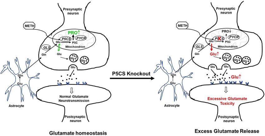
Figure 7. Proposed model demonstrating the key role of P5CS in maintaining glutamate homeostasis
during acute METH exposure. The schematic model shows presynaptic and postsynaptic neurons along with
astrocytes and highlights the pathways involved in glutamate homeostasis in the brain. The left panel depicts
the sequestration of excess glutamate in the neuron for proline biosynthesis during acute METH exposure.
Specifically, METH-induced enhancement of P5CS expression is associated with utilization of glutamate for
proline synthesis, thus maintaining glutamate homeostasis. Whereas, the right panel describes that depletion
of P5CS blocks the proline synthetic arm from glutamate and abrogates glutamate sequestration for proline
biosynthesis. This metabolic disruption results in an increased levels of glutamate that could be detrimental for
the neurons.
cells but also in brain cortical region of mice. Interestingly, disruption in proline biosynthesis by knocking out
of P5CS dramatically increased both intracellular and extracellular glutamate with METH treatment. The role of
P5CS was further confirmed using a proline auxotrophic cell line (CHO-K1) that lacks the proline biosynthetic
machinery. A marked enhancement in glutamate levels was also observed in these cells with METH treatment
and this effect was abrogated by overexpression of P5CS. Collectively, these results demonstrated sequestration
of neuronal glutamate pool towards proline biosynthesis. A lack of neuronal cell death by acute METH expo-
sure even at 1 mM concentrations supports the hypothesis that sequestration of glutamate to proline is a critical
mechanism activated to avert the accumulation of excess glutamate and prevent excitotoxicity.
Sequestration of glutamate to proline may have significant implications in the CNS. For instance, P5C formed
during proline synthesis from glutamate can serve as the source for carbon exchange between the TCA and urea
cycle28. Proline can also be deposited in collagen, the most abundant protein by weight in the human body80.
Approximately, 25% of proline residues make up collagen, therefore, collagen has been suggested to be both a
dump as well as a reservoir for proline80. This may be very relevant in the context of the extracellular matrix
(ECM) of neurons. For example, excess glutamate can be converted to proline and deposited into the ECM as
collagen without dramatically altering extracellular proline levels. High levels of proline have been shown to have
excitotoxic properties and studies have shown that addition of proline to hippocampal slices decreased glutamate
uptake causing glutamate to accumulate81. It is plausible that once the threshold for proline deposition is reached,
especially with chronic METH, it may result in the accumulation of proline, which may also contribute to the
glutamate excitotoxicity. Thus, the interplay between glutamate and proline may be an important regulator in
METH-induced glutamate excitotoxicity.
Importantly, we envision that increased conversion of glutamate to proline offers a bioenergetic metabolic
advantage. The bioenergetic cost of maintaining homeostatic levels of synaptic glutamate is e xpensive82. Glucose
via glycolysis provides the main source of energy for maintaining glutamate homeostasis in the brain. However,
for efficient glycolysis a sustained supply of electron acceptor nicotinamide adenine dinucleotide (NAD+) is
essential83,84. Proline biosynthesis has been shown to augment glycolysis by affecting the levels of NAD+, espe-
cially in cancer m etabolism85–87. Therefore, our results demonstrating METH-induced proline biosynthesis may
confer a metabolic bioenergetic advantage to neurons in response to increased neuronal stimulus by METH. In
summary, our results identify a key role of P5CS during METH exposure and highlight that sequestering excess
glutamate for proline biosynthesis is a key mechanism to maintain glutamate levels (Fig. 7).
Materials and methods
All the methods used in this study are in accordance with relevant guidelines and regulations.
Scientific Reports | (2021) 11:1422 | https://doi.org/10.1038/s41598-020-80917-7 11
Vol.:(0123456789)www.nature.com/scientificreports/
Reagents. Methamphetamine hydrochloride (METH), Amphetamine hydrochloride (AMPH), all-trans-
retinoic acid (ATRA) were obtained from Sigma-Aldrich Chemicals (USA). The primary antibodies used were as
follows: anti-P5CS (cat# NBP1-83324) was purchased from Novus Biologicals (USA), anti-PYCR1 (cat# 13108-
1-AP, anti-PYCR2 human (cat# 55060-1-AP, anti-PYCR2 mouse (cat #17146-1-AP, anti-P5CDH (cat# 11604-1-
AP), anti-GLS (cat# 12855-1-AP) were purchased from Proteintech (USA), anti-POX IgG was a gift from Dr.
James Phang (NCI-Frederick), anti-vGLUT1 (cat# 48-2400) was purchased from Thermo Fisher (USA), and
anti-β-actin was obtained from Sigma-Aldrich Chemicals (USA). The secondary antibodies used were goat anti-
rabbit or goat anti-mouse purchased from BioRad laboratories (USA).
Cell culture and METH treatment. Human neuroblastoma cells (SH-SY5Y), Chinese hamster ovary
(CHO-K1) cells were purchased from American Type Culture Collection (Manassas, VA). The SH-SY5Y cells
were maintained in Dulbecco’s modified Eagle’s medium (DMEM) (Gibco) supplemented with 10% fetal bovine
serum (FBS) (Gibco), 2 mM Glutamine (Gibco), and 1000 U/mL penicillin, and 100 mg/mL streptomycin
(Gibco). CHO-K1 cells were grown and maintained in Ham’s F-12K medium with 10% FBS, 2 mM Glutamine,
1000 U/mL penicillin, and 100 mg/mL streptomycin. SH-SY5Y were differentiated with ATRA as per published
methods (48). All cells were maintained at 37 °C and 5% CO2 before and during treatments. The cells were
treated acutely for 24 h with METH in a dose dependent manner at physiologically relevant concentrations of
50–1000 μM.
Cloning. All three P5CS-targeting sgRNA sequences used in this study were from Doench et al.88 and each
sgRNA was individually cloned into the TLCV2 plasmid vector (Addgene) by following the recommended pro-
tocol with modifications58. Briefly, the TLCV2 plasmid was first digested with BsmB1 by assembling a reaction
mixture containing the plasmid DNA, Buffer 3.1 (NEB), and BsmB1 (NEB) and incubating at 55 °C for 30 min,
then dephosphorylated by incubating the restriction digestion products with rSAP (NEB) at 37 °C for 30 min,
and finally gel-purified. Each pair of oligos corresponding to a sgRNA was phosphorylated and annealed by
assembling a reaction mixture containing the oligos, T4 ligation buffer (NEB), and T4 Polynucleotide Kinase
(NEB) and incubating at 37 °C for 30 min followed by incubation at 95 °C for 5 min and then ramping down to
25 °C at 5 °C/min. The annealed oligos were diluted 200 fold and then ligated to the BsmB1-digested and dephos-
phorylated TLCV2 in a ligation mixture containing T4 ligation buffer (NEB) and T4 DNA ligase (NEB) and
incubating at room temperature for 1 h. Subsequently, NEB-Stbl competent cells, prepared using the Mix&Go
Transformation Kit (Zymo Research, USA) were transformed with an aliquot of the ligation reaction products as
per manufacturer-recommended protocol and the bacterial recombinants were confirmed by Sanger sequencing
of the isolated plasmids.
To construct the P5CS overexpression plasmid, the P5CS ORF was obtained by RT-PCR (abm, Canada) from
SupT1 total RNA using P5CS-specific primers (forward: 5-TTA TAG GAT CCG CCA CCA TGT TGA GTC
AAG TTT ACC-3′ and reverse: 5′-TGA CAC TCG AGT CAG TTG GTG TTT CTC TGA G-3′) containing
BamH1 and Xho1 restriction enzyme sites. The BamH1 and Xho1 digested P5CS ORF amplicon was ligated to
compatible ends of the pcDNA 3.1 plasmid (Addgene). The recombinant plasmids were confirmed by colony
PCR and Sanger sequencing.
Lentivirus transduction. HEK293T cells (5 × 10⁵) seeded per well in 6-well plates and cultured overnight
were transfected with plasmid DNAs using the PEI transfection reagent (Polysciences Inc.). For transfecting
cells in each well, a 4:3:1 ratio of lentiviral transfer plasmid (described above), packaging plasmid psPAX2, and
pseudotyping envelope plasmid pMD2.G, and the PEI at a DNA:PEI ratio of 1:3 were used. After overnight cul-
turing, the culture medium was removed and the cells were replenished with fresh medium. Forty-eight hours
post transfection, the lentivirus-containing culture medium was collected, centrifuged at 500×g for 5 min at
room temperature to remove cell debris, and the supernatant was filtered through 0.45 μm filter. Unused virus
stocks were stored at − 80 °C for later use.
P5CS CRISPR/Cas9 knockout in SH‑SY5Y cells. For generation of stable cell lines, SH-SY5Y cells were
inoculated with lentivirus for 24–48 h, then replenished with fresh culture medium containing the selection
drug puromycin at 1 μg/mL. Cells were replenished with fresh culture medium containing puromycin every
2 days until majority of the uninfected control cells were eliminated (in ~ 7–10 days). To generate single cell
clones, cells were treated with 1 μg/mL DOX for 24 h and the GFP positive cells were sorted out by flow cytom-
etry. The GFP-positive single cells were then seeded into a 96-well plate and allowed to expand. The cell clones
that were depleted of the target protein as analyzed by western blot were used in functional experiments.
Lactate dehydrogenase (LDH) assay. To determine cytotoxicity, LDH release was measured with the
LDH Cytotoxicity Assay Kit (Pierce) according to the manufacturer’s protocol. 5 × 10⁵ SH-SY5Y cells were plated
into 6-well plates and maintained overnight before treatment, following which the cells were treated with METH
at various concentrations for 24 h. After treatment, the cell supernatants were collected by centrifugation. Super-
natants were assayed for LDH in triplicates as per the manufacturer’s protocol. Absorbances were measured at
490 nm and 680 nm using a spectrophotometer (Biotek). The percentage of cytotoxicity was calculated based on
the percentage difference compared with the LDH-positive control provided with the kit.
Glutamate assay. 5 × 10⁵ SH-SY5Y cells were plated in 6-well plate overnight and treated with METH for
24 h. Supernatants were collected, briefly centrifuged to remove cell debris, and analyzed by a Glutamate Col-
Scientific Reports | (2021) 11:1422 | https://doi.org/10.1038/s41598-020-80917-7 12
Vol:.(1234567890)www.nature.com/scientificreports/
orimetric Assay kit purchased from Cell Biolabs as per the instructions from the manufacturer. For measuring
intracellular glutamate, the cells were lysed by sonication in sucrose buffer. After lysis, the cell extracts were cen-
trifuged at 15,000×g for 5 min and the supernatants were subjected to glutamate assay using the kit. All the reac-
tions were performed in triplicates. The glutamate concentrations in the samples were determined by plotting
the data against the respective standard curve that was generated in parallel using a series of known glutamate
concentrations ranging from 6.25 to 400 µM.
Measurement of intracellular proline levels. 5 × 10⁵ SH-SY5Y cells were plated per well in 6-well plates
overnight and treated with METH for 24 h. After treatment cells were harvested and lysed in cold PBS with 1%
Triton X-100. The cellular debris were removed by centrifugation (10,000×g). The supernatants were transferred
to a boiling water bath, and intracellular amino acids were extracted by boiling for 10 min. After centrifuga-
tion (5 min, 4 °C, 15,000×g), the supernatant was free of proteins, and intracellular proline was determined as
described51,52. Briefly, 100 µL of the supernatant was incubated with 100 µl of acid-ninhydrin (0.25 g ninhydrin
dissolved in 6 mL glacial acetic acid and 4 mL 6 M phosphoric acid) and 100 µL of glacial acetic acid for 1 h at
100 °C. The reaction was stopped by incubation on ice for 5 min, and the mixture was extracted with 200 µL
toluene. The toluene phase was separated, and absorbance at 520 nm was used to determine the concentration
of proline. All the reactions were performed in triplicates and a standard curve ranging in concentrations of
0.05–1 mM proline was generated for determining proline concentration of samples.
Western blot analyses. Cells were treated with various concentrations of METH for 24 h, after which cells
were harvested and washed with PBS. Cell lysates were prepared as per our published m ethod50 and quantified
according to standard BCA protein assay (Pierce, USA). Equal amounts of cell lysates (20 µg) were resolved
on SDS–polyacrylamide gels and transferred to nitrocellulose membranes using a semi-dry blotter (Bio-Rad).
Membranes were blocked using Tris-buffered saline with 5% nonfat milk (pH 8.0; Sigma). Blots were then
probed with the appropriate primary antibody in blocking buffer overnight at 4 °C. Incubation with secondary
anti-mouse or anti-rabbit IgG antibodies conjugated to horseradish peroxidase (1:2000) was performed at room
temperature for 1 h. All blots were washed in Tris-buffered saline with Tween 20 (pH 8.0; Sigma) and developed
using the enhanced chemiluminescence (ECL) procedure (BioRad). Blots were routinely stripped by Restore
Plus Stripping Buffer (Pierce) and reprobed with anti-actin monoclonal antibody to serve as loading controls.
The density of the band was evaluated by Bio-Rad imaging-lab 4.0 software (https://www.bio-rad.com/en-pl/
product/image-lab-software?ID=KRE6P5E8Z).
Transfection of P5CS expression construct in CHO‑K1 cells. CHO-K1 cells (1 × 10⁵) were seeded
per well in 6-well plates for 24 h in complete F12K media. Prior to transfection, the cells were pretreated with
METH for 1 h and then transfected with pcDNA control vector or P5CS expression vector. Transfections were
performed with JetPrime (Polyplus) according to the manufacturer’s instructions. 24 h post transfection the
cells were harvested by scraping. The cell supernatants and pellets were collected by centrifugation. Supernatants
were assayed for glutamate and the cell pellets were analyzed by western blot as described before.
Animal studies. All protocols were approved by the University of Florida and Vanderbilt University Insti-
tutional Animal Care and Use Committee (IACUC) policies and adhered to the NIH guidelines. WT C57BL/6J
male mice were obtained from Jackson Laboratories (Vanderbilt; Stock No. 000664) or from the University of
Florida Animal Care Services and were maintained on a 12-h light/dark cycle with food and water available ad
libitum in their home cages. The mice were injected with a single dose of METH at 2 mg/kg body weight (acute
exposure). Control mice received saline injections. The animals were euthanized after 24 h and whole brain were
extracted, rapidly rinsed with ice-cold PBS and immediately flash frozen. Punches from the cortical regions were
taken for each sample and homogenized manually in a sucrose buffer system supplemented with protease inhibi-
tors. Samples were spun down at high speed for 5 min, the supernatants were transferred into separate tubes and
the protein concentration was estimated by BCA assay (Pierce).
For brain slices studies 6-week-old male C57BL/6J mice were euthanized under isoflurane anesthesia after
which 300-µm coronal brain slices containing the frontal cortex were prepared from whole brain tissue using a
Leica Vibratome in oxygenated (95% O2; 5% CO2) ice-cold artificial cerebrospinal fluid (aCSF, in mM: 119 NaCl,
2.5 KCl, 1.3 MgCl2–6H2O, 2.5 CaCl2–2H2O, 1.0 NaH2PO4–H2O, 26.2 NaHCO3, and 11 glucose). Following a
30-min recovery period, slices were then incubated in either 200 µM amphetamine, 200 µM methamphetamine
or vehicle (phosphate buffered saline, PBS). Following a 6-h incubation, cortical enriched tissue was isolated
and flash frozen until processing for immunoblot analysis.
Statistical analyses. Data are expressed as mean ± SEM obtained from at least three independent experi-
ments. Significance of differences between control and treated samples was determined by Student’s t test. Values
of p < 0.05 were considered to be statistically significant.
Received: 19 May 2020; Accepted: 30 December 2020
Scientific Reports | (2021) 11:1422 | https://doi.org/10.1038/s41598-020-80917-7 13
Vol.:(0123456789)You can also read