The role of the inferior parietal lobule in writer's cramp
←
→
Page content transcription
If your browser does not render page correctly, please read the page content below
doi:10.1093/brain/awaa138                                                                                                 BRAIN 2020: 143; 1766–1779   | 1766

The role of the inferior parietal lobule in

                                                                                                                                                                Downloaded from https://academic.oup.com/brain/article-abstract/143/6/1766/5840768 by I.N.M. NEUROMED user on 06 July 2020
writer’s cramp
  Shabbir Hussain I. Merchant,1,2 Eleni Frangos,3 Jacob Parker,1 Megan Bradson,3
Tianxia Wu,1 Felipe Vial-Undurraga,1,4 Giorgio Leodori,1,5 M.C. Bushnell,3
Silvina G. Horovitz,1 Mark Hallett1 and Traian Popa1,6,7

Humans have a distinguishing ability for fine motor control that is subserved by a highly evolved cortico-motor neuronal network.
The acquisition of a particular motor skill involves a long series of practice movements, trial and error, adjustment and refinement.
At the cortical level, this acquisition begins in the parieto-temporal sensory regions and is subsequently consolidated and stratified
in the premotor-motor cortex. Task-specific dystonia can be viewed as a corruption or loss of motor control confined to a single
motor skill. Using a multimodal experimental approach combining neuroimaging and non-invasive brain stimulation, we explored
interactions between the principal nodes of the fine motor control network in patients with writer’s cramp and healthy matched
controls. Patients and healthy volunteers underwent clinical assessment, diffusion-weighted MRI for tractography, and functional
MRI during a finger tapping task. Activation maps from the task-functional MRI scans were used for target selection and neuro-
navigation of the transcranial magnetic stimulation. Single- and double-pulse TMS evaluation included measurement of the input-
output recruitment curve, cortical silent period, and amplitude of the motor evoked potentials conditioned by cortico-cortical inter-
actions between premotor ventral (PMv)-motor cortex (M1), anterior inferior parietal lobule (aIPL)-M1, and dorsal inferior par-
ietal lobule (dIPL)-M1 before and after inducing a long term depression-like plastic change to dIPL node with continuous theta-
burst transcranial magnetic stimulation in a randomized, sham-controlled design. Baseline dIPL-M1 and aIPL-M1 cortico-cortical
interactions were facilitatory and inhibitory, respectively, in healthy volunteers, whereas the interactions were converse and signifi-
cantly different in writer’s cramp. Baseline PMv-M1 interactions were inhibitory and similar between the groups. The dIPL-PMv
resting state functional connectivity was increased in patients compared to controls, but no differences in structural connectivity be-
tween the nodes were observed. Cortical silent period was significantly prolonged in writer’s cramp. Making a long term depres-
sion-like plastic change to dIPL node transformed the aIPL-M1 interaction to inhibitory (similar to healthy volunteers) and can-
celled the PMv-M1 inhibition only in the writer’s cramp group. These findings suggest that the parietal multimodal sensory
association region could have an aberrant downstream influence on the fine motor control network in writer’s cramp, which could
be artificially restored to its normal function.

1 National Institute of Neurological Disorders and Stroke, National Institutes of Health, Bethesda, MD, USA
2 Department of Neurology, Medical University of South Carolina, Charleston, SC, USA
3 National Center for Complementary and Integrative Health, National Institutes of Health, Bethesda, MD, USA
4 Facultad de Medicina, Clı́nica Alemana Universidad del Desarrollo, Santiago, Chile
5 IRCCS Neuromed, Pozzilli, IS, Italy
6 Defitech Chair of Clinical Neuroengineering, Center for Neuroprosthetics (CNP) and Brain Mind Institute (BMI), Swiss Federal
  Institute of Technology (EPFL), 1202 Geneva, Switzerland
7 Defitech Chair of Clinical Neuroengineering, Center for Neuroprosthetics (CNP) and Brain Mind Institute (BMI), Swiss Federal
  Institute of Technology Valais (EPFL Valais), Clinique Romande de Réadaptation, 1951 Sion, Switzerland

Received November 28, 2019. Revised March 1, 2020. Accepted March 9, 2020. Advance access publication May 18, 2020
C The Author(s) (2020). Published by Oxford University Press on behalf of the Guarantors of Brain. All rights reserved.
V
For permissions, please email: journals.permissions@oup.com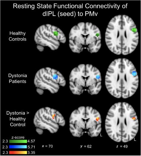
Pathophysiology of task-specific dystonia                                                   BRAIN 2020: 143; 1766–1779    | 1767

Correspondence to: Shabbir Hussain I. Merchant
Assistant Professor of Neurology
Division of Movement Disorders
Medical University of South Carolina
208 B Rutledge Avenue, MSC 108
Charleston, SC 29425, USA
E-mail: merchash@musc.edu

Keywords: task-specific dystonia; writer’s cramp; fine motor control; parietal lobe; cortical silent period
Abbreviations: a/dIPL = anterior/dorsal inferior parietal lobule; cSP = cortical silent period; CS-TS = conditioning stimulus-test

                                                                                                                                      Downloaded from https://academic.oup.com/brain/article-abstract/143/6/1766/5840768 by I.N.M. NEUROMED user on 06 July 2020
stimulus; cTBS = continuous theta-burst stimulation; FDI = first dorsal interossei muscle; M1 = primary motor cortex; MEP =
motor evoked potential; PMv = ventral premotor cortex; TMS = transcranial magnetic stimulation

                                                                      Task-specific dystonia (TSD) has been defined as ‘a collec-
Introduction                                                       tion of movement disorders that present with persistent mus-
The skilful handling of objects is a distinguishing human          cular incoordination or loss of motor control during skilled
ability. It is acquired over a long series of practice move-       movement’ (Albanese et al., 2013). Common forms include
ments adjusted via multimodal sensory feedback. Our under-         writer’s cramp and musician’s dystonia, both of which have
standing of human fine motor control is mainly indirect,           been noted to involve functional alterations in fine motor
deriving inferences from studies of non-human primates,            skill circuits (Sadnicka et al., 2016, 2018; Pirio Richardson
with additional insights gained from human neuroimaging            et al., 2017). Some of the pathophysiological mechanisms,
studies (Rizzolatti and Luppino, 2001; Rizzolatti and              such as loss of inhibition, abnormal plasticity, and abnormal-
Wolpert, 2005; Lemon, 2008a, b; Kristo et al., 2014;               ities in sensorimotor integration are shared with other types
Hamano et al., 2020). The network implicated in human              of dystonia (Hallett, 2011; Quartarone and Hallett, 2013).
fine motor control, associated with pincer grasp involves the      In the context of human motor control, TSD can be viewed
ventral premotor cortex (PMv) and anterior part of the in-         as a corruption of a specific aspect of a learned and perfected
ferior parietal lobule (aIPL) working in concert with the pri-     motor skill. This may initially manifest clinically as various
mary motor cortex (M1). The dorsal part of the inferior            degrees and patterns of difficulties in the performance of a
parietal lobule (dIPL) is the multimodal sensory association       particular task, which can potentially corrupt other learned
region, involved in the initial acquisition and learning of a      motor skills over time, on account of maladaptive plasticity
motor task, which is subsequently stratified downstream            (Sadnicka et al., 2018). It is also recognized today that ab-
in the fine motor control network composed of the                  normal input from subcortical structures contribute to the
aIPL-PMv-M1 (Rizzolatti and Luppino, 2001; Rizzolatti and          abnormal cortical plasticity and motor learning, which sup-
Wolpert, 2005; Karabanov et al., 2012). The selection of a         ports the view that TSD is a network disorder (Peterson
particular motor sequence accounting for the object mean-          et al., 2010; Shakkottai et al., 2017; Kaji et al., 2018).
ing, context and the desired goals of current actions are             Previous studies on TSD have reported abnormally
selected based on the inputs to PMv from the prefrontal cor-       increased activation in the dIPL region (Gallea et al., 2016;
tex and parietal-temporal regions (Fagg and Arbib 1998;            Battistella and Simonyan, 2019; Bianchi et al., 2019).
Grafton et al., 1998; Rizzolatti and Luppino, 2001;                Considering dIPL from the perspective of motor learning, it
Hamano et al., 2020). The superior longitudinal fasciculus is      could be considered a prime candidate for the introduction
the white matter pathway supporting these parietal-pre-            and maintenance of aberrancies within the fine motor con-
motor interactions (Lemon, 2008a, b; Schaffelhofer and             trol network (Karabanov et al., 2012; Gallea et al., 2016;
Scherberger, 2016).                                                Battistella et al., 2017; Battistella and Simonyan, 2019;
   The IPL is uniquely located and connected to many               Bianchi et al., 2019). If this were the case, transiently lower-
different brain regions, subserving several neurological           ing the responsiveness of this region could lead to the nor-
processes. There are also distinctive connections and or-          malization of its downstream influence on the fine motor
ganization within the parietal lobe; however, the function-        control network.
al and behavioural implications of these interactions                 In humans, network-level pathophysiological interactions
within the parietal lobe are poorly understood. The dIPL           can be probed, and excitability temporarily altered using
is posited to be the multimodal sensory association region         non-invasive brain stimulation techniques such as simple
and aIPL being one of the critical nodes of fine motor             and repetitive transcranial magnetic stimulation (TMS). The
control network (Bremmer et al., 2001; Rozzi et al.,               excitability of M1 can be quantified indirectly with the amp-
2008; Bonini et al., 2010; Ishibashi et al., 2011; Caspers         litude of motor evoked potentials (MEP) and the duration of
et al., 2013; Burks et al., 2017; Deroche et al., 2017;            cortical silent periods (cSP) by single pulses: the recruitment
Bruni et al., 2018).                                               curve can reveal the threshold, as well as the excitability
1768 | BRAIN 2020: 143; 1766–1779 S. H. I. Merchant et al.
profile of M1, while the cSP can give a measure of the speed control network readily accessible to TMS, leaving subcortical
with which the motor circuits can resume their normal inter- structures out of the model (Hubsch et al., 2013). To address
action after an artificially-induced focal disruption (Chen these exploratory aims we recruited participants with writer’s
et al., 1997; Classen et al., 2000; Cantello, 2002; Saisanen cramp and healthy volunteers, and asked the following ques-
et al., 2008). The functional influence of other cortical areas tions: (i) Do patients with writer’s cramp and control subjects
onto M1 can be quantified with MEP by paired TMS differ in structural and resting state functional connectivity be-
pulses—one pulse delivered over an area of interest influenc- tween the cortical hubs of fine motor control? (ii) Do patients
ing M1 followed by a second pulse delivered over M1. with writer’s cramp and control subjects differ in cortical ex-
Aberrancies in these interactions can provide useful patho- citability? (iii) Do patients with writer’s cramp have altered
Downloaded from https://academic.oup.com/brain/article-abstract/143/6/1766/5840768 by I.N.M. NEUROMED user on 06 July 2020
physiological insights into disorders of motor control physiological interactions within the fine motor control net-
(Rizzolatti and Luppino, 2001; Davare et al., 2008; Lemon work, while at rest? and (iv) Would transiently decreasing the
2008a, b). The preponderant normal and dystonic interac- excitability of the dIPL have any direct influence on motor
tions within the fine motor control network, probed using cortical excitability and/or downstream influence on the fine
paired pulse TMS are summarized in Fig. 1. Techniques uti- motor control network? To address these questions, we car-
lizing repetitive TMS can be used to influence cortical excit- ried out diffusion weighted imaging (DWI) for tractography,
ability by inducing a temporary long-term potentiation-like a resting state functional MRI to assess functional interactions
(LTP-like) or long-term depression-like (LTD-like) plastic at rest, a finger tapping task functional MRI scan for neuro-
change. Theta-burst stimulation (TBS) is one such high fre- navigation purposes, and explored the interactions within the
quency repetitive stimulation paradigm used to transiently cortical parieto-premotor-motor network (aIPL/dIPL-PMv-
alter the excitability of a brain region, which can potentially M1) at baseline and after modulating the excitability of dIPL
influence the involved network (Huang et al., 2005; by inducing an LTD-like plastic change with continuous TBS
Wischnewski and Schutter, 2016) (cTBS), in a randomized sham-controlled design.
In this exploratory study, we used a multimodal approach
combining structural and functional MRI, and TMS to quan-
tify the interactions between the principal cortical nodes of
fine motor control network in patients with writer’s cramp
Materials and methods
and matched healthy controls. We also explored whether arti-
ficially decreasing the excitability of the dIPL would have any Participants
direct influence on M1 excitability and its interactions within Nine patients with writer’s cramp based on clinical criteria
the fine motor control network. The focus of this study was (Albanese et al., 2013), affected on their right side (four females)
to explore the interactions within the cortical fine motor and 15 healthy volunteers (five females), without any other
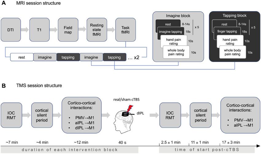
Figure 1 Schematic representation of the principal cortical nodes involved in human upper limb fine motor control, as they
emerge from previous studies. The fine motor control network involved in pincer grasp is composed of aIPL, PMv, and M1. Predominant
interactions between the different regions implicated in upper limb motor control using TMS-based cortico-cortical interactions using a condi-
tioning stimulus-test stimulus (CS-TS) paradigm as noted in healthy subjects and focal hand dystonia. Inhibitory interactions are reflected by a
reduced amplitude of the MEPs after a double pulse, while excitatory interactions are reflected by an increased amplitude of the MEPs, both
when compared with MEPs of single pulses delivered over M1. Inhibitory PMv-M1 interactions have been reported in healthy volunteers and loss
of PMv-M1 inhibition reported in focal hand dystonia. Inhibitory aIPL-M1 interactions have been reported in healthy volunteers and focal hand
dystonia. Facilitatory dIPL-M1 interactions have been reported in healthy volunteers and are unknown in focal hand dystonia.Pathophysiology of task-specific dystonia BRAIN 2020: 143; 1766–1779 | 1769
neurological or psychiatric disorders were enrolled in this ex- muscle (FDI). The right hand was positioned in a comfortable,
ploratory study. The clinical assessment included neurological rested, supinated position along the right side of the body.
examination and scoring of the dystonia on the Burke-Fahn-
Marsden Dystonia Rating Scale (BFMDRS) (Burke et al., 1985). Resting state and task scans
Subjects on chronic opioid, anticholinergic or GABA-ergic medi- A 6-min resting state scan was collected prior to the task scans.
cations, or reported alcohol consumption of 414 drinks/week Participants were instructed to relax, keep their eyes open, and
were excluded. Patients being treated with botulinum toxin fixate on a white cross hair on a black screen for the duration
injections were studied at least 3 months after their last injec- of the scan.
tions. The study was approved by the Combined Neurosciences Each of the two subsequent task scans began with a 1-min
Institutional Review Board of the National Institutes of Health rest period followed by counterbalanced 18-s blocks of ‘Tap’
Downloaded from https://academic.oup.com/brain/article-abstract/143/6/1766/5840768 by I.N.M. NEUROMED user on 06 July 2020
(NIH). and ‘Imagine tapping’ (five trials per block, two blocks per con-
dition), each followed by a ‘Rest’ block (jittered 8–14 s).
Participants tapped their right index finger to their right thumb
Experimental design (a pincer-like motion) at a rate of 1–2 Hz or imagined doing
Study overview the action, respectively. The fine motor ‘pincer’ tapping, the tap-
Subjects satisfying the study inclusion criteria underwent the ini- ping rate, and imagining the action were rehearsed prior to the
tial clinical assessment. Subsequently, they underwent a clinical scanning session. Following each block of trials, two pain inten-
MRI, to verify subjects had no pathological findings, T1- sity scales were presented for 10 s. They are described in the
weighted images for image registration, DWI for tractography Supplementary material and not used in the analysis.
and evaluation of structural connectivity, functional MRI during
rest to evaluate resting state functional connectivity, and func- Functional MRI preprocessing and analysis
tional MRI during a finger tapping task between the index and All data were preprocessed using FSL version 6.00 (FMRIB
thumb for target localization. Single subject functional MRI ac- Software Library, Oxford, UK). Briefly, preprocessing included
tivation maps of the motor control network mapped onto their removal of the skull and non-brain tissue, standard motion cor-
anatomical scans were used for target selection and TMS neuro- rection (six parameters), grand-mean intensity normalization of
navigation. Baseline TMS evaluation included measurement of all voxels across time, high-pass filtering, and spatial smoothing
input-output recruitment curve (IOC) and cSP to evaluate cor- using a Gaussian kernel of fixed-width at half-maximum
tical excitability, and the variation in the amplitude of MEPs (FWHM) = 5 mm and 6 mm for the task and resting state data,
conditioned by cortico-cortical interactions between PMv-M1, respectively. Further preprocessing of the resting state data
aIPL-M1 and dIPL-M1 to evaluate physiological connectivity. included single session independent components analysis (ICA)
After the baseline block, one cTBS block of 600 pulses was using MELODIC (version 3.14) with automatic dimensionality
applied to the dIPL node. The cTBS is a non-invasive brain estimation. Artefacts were identified using FMRIB’s ICA-based
stimulation technique capable of inducing a temporary LTD-like Xnoisefier (FIX 1.06;) and then regressed from each individual’s
plastic change in the superficial cortical areas (Huang et al., resting state data. The classification threshold was set to 20.
2005; Wischnewski and Schutter 2016). Either real or sham Registration of the task functional image to the individual’s ana-
cTBS (randomized and counterbalanced) was applied at two tomical was performed using a six parameter registration, fol-
separate sessions to all subjects, with at least 24 h between the lowed by a 12-parameter linear registration to the MNI152
two sessions. After the cTBS block, the same parameters standard space template. A non-linear registration to standard
recorded at baseline were re-evaluated to ascertain the influence space was carried out for the resting state functional data.
of making an LTD-like plastic change to dIPL on cortical excit- Details are provided in the Supplementary material.
ability and downstream physiological interactions within the
fine motor control (aIPL-PMv-M1) network. The dIPL-M1 Task scan analyses
interaction was not tested after cTBS, since the cTBS interven-
tion was applied to dIPL node and the overarching exploratory The task scan data were analysed prior to the TMS session and
hypothesis was to study the influence of a plastic change of this used for the functional neuro-navigation procedures. At the indi-
node downstream on the fine motor control network. The over- vidual level, explanatory variables (EV) for the ‘tapping’ and
view of the study design is presented in Fig. 2. ‘imagine’ conditions were created for each of the two runs and
modelled using a double-gamma haemodynamic response func-
tion. Rest periods were not included in the model. A fixed-
Structural and functional MRI effects, whole-brain analysis was used to average the ‘tapping’
conditions across both runs for each individual. The peak un-
MRI acquisition
corrected, linearly registered results observed in each individual’s
Brain images were acquired using a 3 T Siemens Skyra scanner finger tapping activation map were used to localize the four sub-
(Siemens) with a 32-channel head coil. Details of the MRI ac- ject-specific nodes (left dIPL, aIPL, M1, and PMv).
quisition parameters are noted in the Supplementary material.
Scanning procedures Seed-based resting state analysis
Participants completed a series of scans as indicated in Fig. 2A. To assess the functional connectivity between the four nodes of
Subjects wore earplugs throughout the session and their heads interest (left dIPL, aIPL, M1, and PMv) during resting state, we
were immobilized with foam padding. Finger movements were extracted the time series from 6 mm spheres (seeds) created
monitored during scans with functional MRI compatible EMG based on the peak activity observed in the finger tapping task.
recording electrodes placed on the right first dorsal interossei Each sphere was confirmed to be within the respective1770 | BRAIN 2020: 143; 1766–1779 S. H. I. Merchant et al.
Downloaded from https://academic.oup.com/brain/article-abstract/143/6/1766/5840768 by I.N.M. NEUROMED user on 06 July 2020
Figure 2 Experimental procedures. (A) Each scanning session included diffusion tensor imaging (DTI), high resolution anatomical MRI, field
mapping, resting state functional MRI (fMRI), and two task-based functional MRI scans to assess neural responses during tapping of the right index
finger to the thumb, or imagining this movement. The order in which the ‘tapping’ and ‘imagine’ blocks were presented was counterbalanced be-
tween the two scans. (B) TMS experiment involved baseline recording of input-output recruitment curve (IOC), resting motor threshold (RMT),
cortical silent period (cSP), and cortico-cortical interactions between PMv-M1, aIPL-M1, and dIPL-M1. A block of real/sham cTBS was applied to
the dIPL node followed by repeated measurements of IOC, RMT, cSP, and cortico-cortical interactions between PMv-M1 and aIPL-M1. The ap-
proximate timelines for the performance of the different TMS experimental blocks pre- and post-cTBS are also noted.
anatomical region based on the Juelich Histological Atlas iterations of whole brain tracking to estimate the likelihood of
(Supplementary Fig. 1). This atlas was used to create four structural white matter connections between all pairs of target
regions of interest for the small volume correction seed-based regions of interest within the predefined network (Saad and
analysis. The PFm and PGa parcellation of the IPL made up the Reynolds, 2012; Taylor and Saad, 2013). The same spheres of
dIPL mask, and the PFop parcellation of the IPL made up the M1, PMv, aIPL, and dIPL from the functional analysis were
aIPL mask (Economo and Koskinas, 1925; Caspers et al., used as the regions of interest for the tractography. Prior to trac-
2013). The M1 mask was restricted to the region of the hand tography, each region of interest was inflated by a maximum of
representation, and the PMv mask was restricted to the PMv re- two voxels until its surface was directly adjacent to the white
gion identified by the Mayka et al. (2006) meta-analysis. matter skeleton defined by a fractional anisotropy (FA) 4 0.2.
Tractography was performed between each possible region of
interest pair.
DTI preprocessing and data analysis
The DWIs were processed using TORTOISE and FATCAT soft-
ware with the default parameters (Pierpaoli, 2010; Taylor and
TMS neuro-navigational set-up
Saad, 2013; Irfanoglu et al., 2017). Raw volumes were visually Each individual set of MRI scans was uploaded to a frameless
inspected and removed if significant motion was present. stereotaxic neuro-navigation system (Brainsight2; Rogue
Motion and eddy current distortion corrections were performed Research, Montreal, Quebec, Canada), which allows simultan-
on the A-P and P-A datasets independently within the diffprep eous neuro-navigation of two TMS coils. The sets consisted of
module then corrected for EPI distortion using DR-BUDDI. The three images linearly registered to the MNI template space: the
two datasets were registered to an axial T2-weighted image and T1-weighted (with skull) for 3D head reconstruction, its skull
non-linear tensor estimation was carried out on the final DWIs stripped version for functional MRI registration, and the uncor-
using FATCAT. rected activation map from the finger tapping task for targeting
The probabilistic tractography was performed using the M1, PMv, aIPL, and dIPL. The functional MRI activation with-
3dTrackID program in FATCAT, which carries out repeated in each pre-defined area of interest from single subject dataPathophysiology of task-specific dystonia BRAIN 2020: 143; 1766–1779 | 1771
were used as TMS targets for each individual. The precise coor- Cortical silent period
dinates of these hotspots and the mismatch between the anatom- The cSP is defined as the time required for re-emergence of on-
ically and functionally defined stimulation targets can be found going tonic EMG activity in a muscle after interruption using a
in Supplementary Table 2 and Supplementary Fig. 2, respective- single TMS pulse producing a MEP. The duration of the cSP
ly. A clear activation of the aIPL, PMv and M1 nodes was has been associated with GABAergic inhibition and proposed to
noted in all subjects. In the rare cases when dIPL activation was correlate with cortical excitability (Saisanen et al., 2008); short-
not clearly noted, the centroids of the anatomical regions of ening of the silent period deemed to be reflective of loss of in-
interest were selected from the atlas mapped to the individual hibition. The subject’s right hand was secured on a
anatomy. The M1 hotspot (site and coil orientation giving most manipulandum in prone position, only allowing abduction of
consistently the highest MEP at the lowest stimulator output) FDI with the right index finger over a force transducer
Downloaded from https://academic.oup.com/brain/article-abstract/143/6/1766/5840768 by I.N.M. NEUROMED user on 06 July 2020
for FDI was determined empirically and the coil oriented to in- (Supplementary Fig. 3A). The subject’s maximal voluntary con-
duce the current in a posterior-anterior direction within M1. traction (MVC) was determined as the maximal force generated
The motor hotspot determined empirically with the TMS and by holding the FDI in tonic contraction for 5 s. The subject was
used for the experiment overlapped with the M1 hotspot indi- then asked to hold the FDI in tonic contraction at a constant
cated by the functional activation mask during finger tapping force between 30–50% of MVC while visual feedback was pro-
task in all subjects, further confirming good alignment of the vided on a potentiometer. For the measurement of cSP, the sub-
overlays used for neuro-navigation. jects maintained this constant tonic contraction of the right FDI
while 20 TMS pulses (divided into two blocks of 10 pulses)
were delivered over the motor hotspot at S50. The cSP was
measured as the duration of interruption of the ongoing EMG
EMG set-up motor activity after each individual pulse, beginning from the
TMS artefact and ending with the resumption of the tonic FDI
Participants were seated in a comfortable armchair with the activity. The individual 20 measurements were then averaged
right arm rested on a pillow. The EMG activity of the right FDI for each subject and submitted to the group statistical analysis.
(muscle of interest) was recorded throughout the experiment. Identical cSP measurements were performed before and after the
Ag-AgCl surface electrodes were placed in a belly-tendon mon- cTBS block (both real and sham).
tage, with impedances kept below 20 kX. Data were collected
using a Viking IV EMG machine (Nicolet Biomedical), bandpass
filtered at 20–2000 Hz with the amplified (1000) analogue Dual site TMS
outputs digitized at 5 kHz with the Signal software (version To compare the interactions within the fine motor control net-
5.09; Cambridge Electronic Design, Cambridge, UK) and stored work in writer’s cramp patients and healthy volunteers, we
in a computer for offline analysis. studied the physiological interactions between PMv-M1, aIPL-
M1 and dIPL-M1, with nodes defined functionally by each indi-
vidual’s finger tapping activation map (Supplementary Fig. 2).
Custom-made, figure-of-eight, ‘branding iron’ style coils with
TMS input-output recruitment 70-mm external diameter were used. Stimulation was delivered
curve and cortical silent period using two high-power, monophasic Magstim2002 stimulators
(Magstim Company Ltd.).
Input-output curve For the PMv-M1 stimulation block, conditioning stimulus
The resting motor threshold (RMT), S50 (stimulation intensity coil was initially placed centred on the functional PMv node,
required to obtain a peak-peak EMG response at 50% of the with the coil placed in an anterior-posterior and slight lateral-
maximum), and slope of an input-output curve (IOC) are varia- medial orientation towards M1. For the aIPL-M1 and the dIPL-
bles that can reflect motor cortical excitability. The slope of the M1 stimulation the conditioning stimulus coil was placed first
IOC can also reflect lability and variability of motor cortical ex- in a posterior-anterior orientation. The TS M1 coil was then
citability. To obtain these measures, TMS was applied tangen- placed to be centred on the motor hotspot with the coil oriented
tially to the scalp over the left M1 hand area, with the x-axis in the posterior-anterior direction; the coil trajectory modified in
parallel to the central sulcus and the y-axis oriented to induce a order to accommodate both the coils, which overlapped slightly
posterior-anterior current flow in the brain. The optimal site (Supplementary Fig. 3B). After the coil positions were secured,
and coil orientation for evoking MEPs from the contralateral trial pulses were delivered to ensure clear MEPs with the target
FDI muscle was identified as the motor hotspot. peak-peak amplitude close to the S50 amplitude. The stimulator
The IOC was obtained by giving 60 single pulses of different output intensity was increased to obtain the target MEP ampli-
intensities (three pulses for each 5% increment between 5% to tude if the coil orientation needed to be changed in order to ac-
100% of the maximal stimulator output), via a custom-made, commodate both coils. After the coil position and stimulator
‘branding ion’ style figure-of-eight coil (70 mm outer diameter) outputs were optimized, the coil trajectories were saved for the
linked to a monophasic Magstim 2002 module. The data were post-cTBS block.
fitted with a Boltzmann sigmoidal function. The S50 for FDI The interstimulus interval (ISI) used for studying these cor-
was determined from the fitted IOC and used as the test stimu- tico-cortical interactions were defined based on the nature of the
lus (TS) intensity throughout the experiment. The RMT was connections between these nodes; premotor cortex having a dir-
determined as the lowest intensity that induced 50 mV peak-to- ect projection to M1 whereas the parietal cortex mainly having
peak amplitude MEP in 5 of 10 trials from the motor hotspot. an indirect projection to M1 via the premotor cortex. They are
The conditioning stimulus (CS) intensity used for the experiment also explained based on the latency of the late I (indirect) waves
was 90% of the RMT. generated by TMS, where these cortico-cortical interactions1772 | BRAIN 2020: 143; 1766–1779 S. H. I. Merchant et al.
occur (Rizzolatti and Luppino, 2001; Cerri et al., 2003; Electrophysiology
Shimazu et al., 2004; Koch et al., 2008; Baumer et al., 2009; The conditioned MEP amplitudes of the dual-site paradigms
Karabanov et al., 2013; Schaffelhofer and Scherberger, 2016; were normalized to the mean amplitude of the unconditioned
Bruni et al., 2018). Therefore, for the PMv-M1 block, 30 MEPs MEPs during the same experimental block.
were collected: 10 after single pulses, 10 each after CS-TS pulse To evaluate the reliability of cSP measurement, intraclass cor-
pairs with 4 ms and 6 ms ISI. For the aIPL-M1 and dIPL-M1 relation coefficients were calculated for writer’s cramp and
blocks 30 MEPs were collected for each: 10 after single pulses, healthy volunteers separately using the three repeated measures
10 each after CS-TS pulse pairs with 6 ms and 8 ms ISI. Pulses from each subject: pre-intervention for both real and sham and
were delivered in a randomized order. post-intervention for sham only.
For each of the three outcome measures: cSP, PMV-M1
Downloaded from https://academic.oup.com/brain/article-abstract/143/6/1766/5840768 by I.N.M. NEUROMED user on 06 July 2020
and alPL-M1, repeated measures ANOVA was performed to
Inhibition of the dorsal inferior evaluate the effect of Group (writer’s cramp versus healthy
volunteers), Intervention (real versus sham), and Time (pre-
parietal lobule using continuous versus post-intervention), and their interactions (Group
TBS Intervention, Group Time, Intervention Time, and
Group Intervention Time). Group was a between-
To assess downstream effects of inducing LTD-like plastic
subject factor, and both intervention and time were within-
change (inhibition) in the dIPL onto cortical excitability and fine
subject factors. Since the study design involved two within-
motor control network, we used a cTBS 600 protocol involving
subject factors, covariance structure un@cs (un: unstructured
triplets of pulses at 50 Hz repeated at a frequency of 5 Hz
for intervention, cs: compound symmetry for time) was used
resulting in 600 pulses over 40 s (Huang et al., 2005). The cTBS
to address the covariance within the subject, where the time
was applied using the Magstim Rapid2 and AirFilm Coil [aver-
was nested within intervention.
age coil diameter of 2 (3 0.92 mm)] that delivered biphasic
pulses of 0.5 ms duration with an average rise time of 80 ms For the outcome measure of dIPL-M1, repeated measures
(Magstim Company Ltd.). The RMT was measured using this ANOVA was performed to evaluate the effect of group (writer’s
coil and 80% of RMT was used as the stimulation intensity for cramp versus healthy volunteers), where the means of the out-
cTBS. The same dIPL target used for the dIPL-M1 CS-TS block come measure from two sessions were used as dependent vari-
was used for cTBS. The coil was orientated tangentially over the able, since the repeated measures were similar (paired t-test
scalp to induce a PA current for the real cTBS block. For the P 4 0.5 for TSD and healthy volunteers group).
sham block, the coil edge was held on target with the coil centre The reported P-values were adjusted (Holm’s adaptive
facing posteriorly and perpendicular to the scalp. method) for multiple comparisons for each experiment, but not
for the multiple (n = 4) experiments. Significance level (a) of
0.05 was used for the comparison of means, and a = 0.1 for
interaction test. Statistical analyses were performed with SAS
Statistical analysis version 9.4.
Demographics
Differences between the writer’s cramp and healthy volunteer
groups for age, gender and handedness were examined using a Data availability
two-sample t-test (continuous variable with normal distribu- The data that support the findings of this study are available
tion), Mann-Whitney test (continuous variable with abnormal from the authors upon reasonable request.
distribution), or Fisher’s exact test (binary variable). The as-
sumption of normality was tested using the residuals and
Shapiro-Wilk method.
Results
Resting state seed-based
A mixed effects model (FLAME 1) was used to evaluate group Demographic characteristics and
connectivity differences between each node pair. The model experimental timelines
included the following contrasts: healthy volunteers 4 writer’s
cramp; writer’s cramp 4 healthy volunteers; mean healthy vol- Twelve healthy volunteers and nine patients with writer’s
unteers connectivity; and mean writer’s cramp connectivity. All cramp completed all the experiments and were included in
analyses were corrected for age (mean-centred across all partici- the final analyses. Of the 15 healthy volunteers recruited,
pants). Voxel-based thresholds were set to z 4 2.3 and were three were excluded from the final analysis; two did not re-
cluster corrected for multiple comparisons at P 5 0.05. turn for the second TMS session and one had high motor
Correlations between clinical measures and functional MRI data thresholds and could not tolerate the intensity of TMS. For
were investigated using Pearson correlations. one patient with writer’s cramp, cSP data for the post-cTBS
cSP block could not be correctly analysed on account of
Diffusion tensor imaging artefactual errors and were excluded. No significant unex-
A two-sample t-test was used to examine the difference in struc- pected adverse events were reported. Both groups were simi-
tural connectivity (fractional anisotropy). Age was not used as a lar in terms of age, gender, and handedness (Table 1). All
covariate considering the low correlation between age and frac- writer’s cramp patients had dystonia of the right hand only
tional anisotropy for the cohort. for the task of writing and no other fine motor dexterityPathophysiology of task-specific dystonia BRAIN 2020: 143; 1766–1779 | 1773
Table 1 Comparative demographic and clinical characteristics of healthy volunteers and writer’s cramp cohort
Healthy volunteers n = 12 Writer’s cramp n = 9 P-value
Age, median; 25–75th quartiles 43.3; 31.5–51.25 58; 48–60 0.11a
Gender, male/female 7/5 5/4 1.0b
Handedness, right/left/ambidextrous 11/0/1 7/1/1 0.70b
a
Mann-Whitney test.
b
Fisher’s exact test.
Downloaded from https://academic.oup.com/brain/article-abstract/143/6/1766/5840768 by I.N.M. NEUROMED user on 06 July 2020
Table 2 Demographic and clinical characteristics of the writer’s cramp cohort
Subject ID Age Sex Handedness Dystonic hand BFMDRS scores Disease duration, years
Right arm Left arm
SJ201 44 M Right Right 4 0 3
SJ202 65 M Right Right 4 0 25
SJ203 59 M Right Right 4 0 18
SJ204 66 F Left Both 4 6 26
SJ205 58 F Right Right 4 0 20
SJ206 37 F Ambidextrous Both 6 4 5
SJ207 48 M Right Right 4 0 11
SJ208 57 F Right Both 6 4 13
SJ209 60 M Right Right 6 0 8
BFMDRS = Burke-Fahn-Marsden Dystonia Rating Scale.
issues (Table 2). The average time between the two experi- were: M1-PMv (healthy volunteers 0.45 ± 0.02, writer’s
mental sessions when the real cTBS was performed first was cramp 0.45 ± 0.02; P = 0.8); PMv-aIPL (healthy volunteers
12 days (range 2–27 days). The TMS coordinates of the 0.43 ± 0.03, writer’s cramp 0.41 ± 0.03; P = 0.1); PMv-dIPL
nodes for the two groups and the average duration of the (healthy volunteers 0.47 ± 0.03, writer’s cramp 0.47 ± 0.03;
post-TBS experimental blocks are provided in P = 0.6).
Supplementary Table 1.
Prolonged cortical silent period and
Patients with writer’s cramp have normal recruitment curves in
altered functional and normal writer’s cramp
structural connectivity No significant differences were noted between the healthy
The seed-based resting state analysis indicated that each volunteers and writer’s cramp cohort in terms of baseline
nodal pair in both groups had a significant positive correl- S50 values (P = 0.78). This did not change after either the
ation (Supplementary Table 2). However, the functional real or sham cTBSdIPL in the healthy volunteers (P = 0.87) or
connectivity between dIPL-PMv was significantly greater in writer’s cramp (P = 0.98) group.
patients with writer’s cramp compared to healthy controls The cSP was found to be significantly longer in the writ-
(Fig. 3 and Supplementary Table 2). There was no relation- er’s cramp cohort compared to the healthy volunteers
ship between the dIPL-PMv connectivity in writer’s cramp [F(20.4); P 5 0.0001] (Fig. 4B). The intraclass correlation
patients and their symptom severity (r = 0.23, P = 0.55) or coefficient (ICC) for the three measures of cSP within the
their symptom duration (r = 0.06, P = 0.87). No other rest- same subjects for each group separately suggested good re-
ing state functional connectivity group differences were producibility and reliability [ICC = 0.60, 95% confidence
observed. interval (CI): 0.19–0.89, for writer’s cramp and ICC = 0.62,
The tractography algorithm found white matter connec- 95% CI: 0.30–0.86 for healthy volunteers]. Combining the
tions in at least 75% of subjects of each group (7/9 writer’s two groups for the entire cohort of total 20 subjects, ICC =
cramp and 12/15 healthy volunteers) only for the PMv-M1, 0.79, 95% CI: 0.62–0.90 (Shrout and Fleiss 1979). A mixed
aIPL-PMv, and dIPL-PMv node pairs, and in 550% for the model ANOVA revealed a slight increase in cSP for both the
other node pairs (aIPL-M1 and dIPL-M1). We found no sig- healthy volunteers and writer’s cramp groups after cTBSdIPL,
nificant group differences in the white matter integrity of the but no other new significant effects or interactions (Table 3).
pathways connecting the nodes of interest involved in fine This symmetrical prolongation of cSP in the two groups
motor control. The mean fractional anisotropy values ± post-cTBS could be an effect of time (i.e. fatigue) and not
standard deviation (SD) between the nodes for each group related to the intervention per se.1774 | BRAIN 2020: 143; 1766–1779 S. H. I. Merchant et al.
Using normalized MEP amplitudes evoked by the CS-TS
paradigm as a dependent variable, significant between-group
differences were noted for the baseline dIPL-M1 interactions
(F = 28.61; P 5 0.0001) compared to TS alone. The baseline
dIPL-M1 interaction was notable for significant facilitation
(baseline average normalized MEP ± SD = 1.13 ± 0.19;
t = 3.43; P = 0.0013) for the healthy volunteers group while
displaying significant inhibition for the writer’s cramp group
(baseline average normalized MEP ± SD = 0.849 ± 0.13; t =
Downloaded from https://academic.oup.com/brain/article-abstract/143/6/1766/5840768 by I.N.M. NEUROMED user on 06 July 2020
–4.69; P = 0.00004). The results are suggestive of opposite
influence of the dIPL on M1 for the two groups (t = 5.46;
P 5 0.00001) (Fig. 5C and Table 3).
Effect of continuous TBS of the
dIPL on the aIPL-PMv-M1 network
No changes were observed in the baseline PMv-M1 interac-
tions in the healthy volunteers group after cTBS of dIPL.
There was a loss of baseline PMv-M1 inhibition for the writ-
er’s cramp group, only for the real intervention (Fig. 5A).
ANOVA revealed significant effects of the factor Time
Figure 3 Results of dIPL connectivity to PMv. Each group (F = 6.5; P = 0.018) and significant interactions Group
had significant dIPL-PMv functional connectivity (green and blue). Time and Group Time Intervention (Table 3). The
However, patients with writer’s cramp had greater dIPL-PMv func- Bonferroni adjusted P-value (Holm’s correction) was signifi-
tional connectivity compared to healthy controls (red-yellow). cant only for the real intervention in the writer’s cramp group
Results are corrected for age and presented using a cluster-forming (P = 0.0012) and not for the sham intervention (P = 0.97).
threshold of z 4 2.3 and a cluster-corrected threshold of P 5 0.05. Continuous TBS of the dIPL node did not alter significant-
ly the baseline aIPL-M1 interactions for the healthy volun-
teers group (Fig. 5B). Mixed model ANOVA revealed a
significant change post-cTBS in the writer’s cramp group,
with aIPL-M1 interaction now being significantly inhibitory
Normal PMv-M1 inhibition in
compared to baseline. ANOVA revealed significant interac-
writer’s cramp tions for Group Time and Group Time
Using normalized MEP amplitudes evoked by the CS-TS Intervention (Table 3). The post hoc analysis, showed that
paradigm as a dependent variable, no significant between- the Bonferroni adjusted P-value (Holm’s correction) was sig-
group differences were noted for the baseline PMv-M1 nificant only for the real intervention in the writer’s cramp
interactions. A similar degree of inhibition was noted group (P = 0.045) and not for the sham intervention
for the CS-TS paradigm at baseline for the two groups (P = 0.83).
(baseline between group difference, t = 0.35; P = 0.73;
normalized MEP ± SD, healthy volunteers 0.87 ± 0.15; t =
–3.87; P = 0.0003; writer’s cramp 0.85 ± 0.16; t = –3.49;
P = 0.0013) (Fig. 5A).
Discussion
Using a multimodal experimental approach combining non-
invasive brain stimulation and neuroimaging, we explored
Loss of aIPL-M1 inhibition and the physiological, functional and structural interactions be-
dIPL-M1 facilitation in writer’s tween the principal cortical nodes involved in human fine
motor control in a cohort of task-specific dystonia with writ-
cramp er’s cramp compared to healthy volunteers. The evaluation
Using normalized MEP amplitudes evoked by the CS-TS of cortico-cortical interactions between the parietal and pre-
paradigm as a dependent variable, significant between-group motor areas implicated in fine motor control using neuro-
differences were noted for the baseline aIPL-M1 interactions. navigation based targeting of each individual’s relevant func-
The baseline aIPL-M1 interaction showed significant inhib- tional network is a novel personalized approach for move-
ition only for the healthy volunteers group (baseline between ment disorders. Our exploratory hypothesis was that
group difference, t = –2.3; P = 0.02; normalized MEP ± SD, possible abnormalities in the parietal sensorimotor integra-
healthy volunteers 0.89 ± 0.16; t = –2.5; P = 0.016; writer’s tion regions might have aberrant downstream influence on
cramp 1.01 ± 0.14; t = 0.62; P = 0.54) (Fig. 5B). the fine motor control network resulting in corruption of aPathophysiology of task-specific dystonia BRAIN 2020: 143; 1766–1779 | 1775
Downloaded from https://academic.oup.com/brain/article-abstract/143/6/1766/5840768 by I.N.M. NEUROMED user on 06 July 2020
Figure 4 Summary of cSP results. (A) Example of cSP in healthy volunteers (HV) and writer’s cramp (WC), demonstrating interruption in
ongoing muscle EMG activity after a TMS pulse resulting in an MEP with re-emergence of EMG activity noted after a period of EMG silence, which
is defined as the cSP. The duration of the cSP being notably longer in writer’s cramp. (B) Summary of cSP results at baseline and post-intervention
(real versus sham), demonstrating significantly prolonged cSP at baseline in writer’s cramp, with persistent significant differences between the
groups after both real and sham interventions. *Correlate with degree of statistical significant differences (increasing number of asterisks indi-
cates more significant differences). FHD = focal hand dystonia.
motor task. In the sections below, we discuss separately each Premotor-motor interactions
investigated parameter and the potential inferences.
The baseline premotor-motor (PMv-M1) interactions were
similar and predominantly inhibitory in both groups, con-
trary to previous reports for writer’s cramp (Houdayer
et al., 2012; Pirio Richardson et al., 2017). Our standardized
Cortical excitability and cortical methodology of neuro-navigated targeting of the functional-
silent period ly relevant PMv node during the fine finger tapping task
could perhaps explain the differences in the results.
We found a significantly prolonged cSP in patients with
Importantly, the real but not the sham cTBS of the dIPL
writer’s cramp, which is contrary to some of the previous
induced a change in the baseline PMv-M1 interaction in the
studies demonstrating shortening of cSP in dystonia, impli-
writer’s cramp group. Considering the major influence of
cating disinhibition of the motor cortex being a possible
the parietal lobe on motor cortex is indirect via the premotor
pathophysiological basis for dystonia (Chen et al., 1997,
cortex (Fagg and Arbib 1998; Bonini et al., 2010, 2012;
2017; Curra et al., 2000). Various factors such as stimulus
Schaffelhofer and Scherberger 2016), the change noted in
intensity, duration, location and sensory stimulation can in-
the PMv-M1 interaction after cTBS of dIPL only in patients
fluence the duration of cSP, apart from the disorder being
could suggest the presence of a labile parieto-premotor inter-
studied (Priori et al., 1994; Classen et al., 2000; Ertas et al.,
action and the possibility of restoring the normal interac-
2000; Cantello 2002; Saisanen et al., 2008). The notion of
tions within the aIPL-PMv-M1 network, as reflected by the
cSP reflective of GABAergic inhibition is based on pharma-
restoration of the normal inhibitory aIPL-M1 physiological
cological studies showing that drugs affecting GABAergic
interaction. In fact, we observed increased functional con-
receptors also change cSP (Priori et al., 1994; Inghilleri
nectivity between the parietal and premotor regions at rest
et al., 1996). However, a single suprathreshold TMS pulse
(dIPL-PMv) in the patients with writer’s cramp, before any
simultaneously depolarizes all impacted neurons (excitatory,
modulation of the dIPL output.
inhibitory, and pyramidal alike), acting as a ‘hard reset’ of
the ongoing subjacent activity. Therefore, the cSP measured
in a well controlled experimental set-up could reflect more Parietal-premotor-motor
specifically the time required by the motor circuits to recover
and get back to performing a finely regulated motor task
interactions
(maintaining tonic contraction of FDI muscle) after the artifi- The baseline influence of dIPL onto M1 was significantly
cial depolarization. The prolongation of cSP in writer’s different and opposite between the writer’s cramp and
cramp could thus indicate a longer duration required for re- healthy volunteers groups: inhibitory in writer’s cramp and
covery and re-initiation of a finely regulated motor task and facilitatory in healthy volunteers. The opposite was noted
not simply disinhibition of the motor cortex. for the baseline influence of aIPL onto M1: inhibitory in1776 | BRAIN 2020: 143; 1766–1779 S. H. I. Merchant et al.
Table 3 Group comparisons of cTBSdIPL effects in within the IPL, which can have downstream influence on the
patients with writer’s cramp and healthy volunteers fine motor control network.
Effect
An analogy can be made between our understanding of
the development of motor skills for the performance of vari-
cSP F P
ous tasks and development of language vocabulary, as the
Group 31.0 50.0001
development of a motor vocabulary. Task-specific dystonia
Intervention 0.1 ns
Time 5.2 0.047
such as writer’s cramp can be viewed as the inability to exe-
Group Intervention 1.4 ns cute a particular motor task secondary to the corruption of
Intervention Time 2.6 ns the motor vocabulary, although the concept and the skills
Downloaded from https://academic.oup.com/brain/article-abstract/143/6/1766/5840768 by I.N.M. NEUROMED user on 06 July 2020
Group Time 0.1 ns required for the performance of the task seem intact. The ab-
Group Intervention Time 0.0 ns errant downstream influence of the multimodal sensory inte-
PMv F P gration region is optimally positioned to corrupt the
Group 1.0 ns execution of a learned motor skill. The aberrancies intro-
Intervention 1.9 ns duced into the motor command can clinically manifest vari-
Time 6.5 0.018 ously as abnormal dystonic adaptations characterized by
Group Intervention 0.1 ns
dystonic posturing, cramping, difficulty to maintain a stable
Intervention Time 2.9 ns
grip, tremors or sudden interruptions or ‘blocks’ in ongoing
Group Time 5.6 0.027
Group Intervention Time 3.7 0.065
activity.
Post hoc P Padj
Writer’s cramp - Real stim: pre versus post 0.0003 0.0012 Limitations
Writer’s cramp - Sham stim: pre versus post ns ns
Healthy volunteers - Real stim: pre versus post ns ns Our study could have benefited from a larger cohort.
Healthy volunteers - Sham stim: pre versus post ns ns However, the results rely on a clinically homogenous pool
aIPL F P of patients, matched controls, and a highly personalized ap-
Group 1.04 ns proach. The precise targeting based on functionally relevant
Intervention 1.35 ns hubs and individual anatomy prevented the inherent prob-
Time 0.1 ns
lem of targeting based on aggregated atlas coordinates. Also,
Group Intervention 0.62 ns
the sham-controlled cross-over design of the study allowed
Intervention Time 2.46 ns
Group Time 5.84 0.023
us (i) to maximize the outcome by measuring real and sham
Group Intervention Time 5.33 0.030 stimulation effects in every subject; and (ii) to confirm the re-
Post hoc P Padj liability of electrophysiological parameters by measuring
Writer’s cramp, real stim: pre versus post 0.015 0.045 them in two separate sessions, before each intervention.
Writer’s cramp, sham stim: pre versus post ns ns Patients with writer’s cramp exhibit a complex psychopatho-
Healthy volunteers, real stim: pre versus post ns ns logical profile that could influence the detection of neural
Healthy volunteers, sham stim: pre versus post ns ns differences (Sadnicka et al., 2018), especially in the dynamic
dIPL F P state. We believe the resting state evaluation of the experi-
Group 29.8 50.0001 mental outcomes noted in this study is another major
For each of the three outcome measures: cSP, PMV-M1 and alPL-M1, repeated meas- strength, since the network abnormalities could be more re-
ures ANOVA was performed to evaluate the effect of group (writer’s cramp versus flective of the disease pathophysiology rather than an epi-
healthy volunteers), intervention (real versus sham) and time (pre- versus post-inter-
phenomenon of the dystonic state.
vention), and their interactions (Group Intervention, Group Time, Intervention
Time, and Group Intervention Time). The reported P-values were adjusted This study did not quantify changes in clinical scores for
(Holm’s adaptive method) for multiple comparisons for each experiment. For the out- two main reasons: (i) because of the limited time available to
come measure of dIPL-M1, repeated measures ANOVA was performed to evaluate
explore the electrophysiological interactions post-cTBS; and
the effect of group (writer’s cramp versus healthy volunteers). cTBSdIPL = continuous
theta burst stimulation of dIPL; ns = not significant; Padj = P adjusted with adaptive (ii) because we did not expect a priori any significant clinical
Bonferroni-Holm correction. effects. Early studies used single session inhibitory repetitive
TMS over the M1 or PMv as a proof of concept (Siebner
et al., 1999; Murase et al., 2005), and their conclusions
were subsequently leveraged in studies using stimulations
healthy volunteers, whereas no clear inhibition was found in over longer periods combined with other interventions
writer’s cramp. The interaction gradient of inhibition-to- (Borich et al., 2009; Kimberley et al., 2015; Pirio
facilitation of M1 described in healthy subjects as the condi- Richardson et al., 2017). A single session of TBS is unlikely
tioning pulse is moved dorsally along the inferior parietal to induce any lasting clinically-relevant effect due to the very
lobe (Karabanov et al., 2013) was absent or inverted in nature of this pathology (Meunier et al., 2015), in which
patients with writer’s cramp. After cTBS of dIPL node, the long abnormal training changes the circuitry supporting a
abnormal null influence of the aIPL onto M1 was restored specific fine motor task (Sadnicka et al., 2018).
towards the normal inhibitory interaction observed in In conclusion, these findings put the spotlight on the ab-
healthy volunteers, suggestive of functional interactions normal multilateral interactions between the parietal,Pathophysiology of task-specific dystonia BRAIN 2020: 143; 1766–1779 | 1777
Downloaded from https://academic.oup.com/brain/article-abstract/143/6/1766/5840768 by I.N.M. NEUROMED user on 06 July 2020
Figure 5 Summary of TMS results. (A) Changes in the PMv-M1 electrophysiological interaction (ISI 4 ms) introduced by real or sham
cTBSdIPL in healthy volunteers (HV) and writer’s cramp (WC) groups. The changes are represented as the difference between conditioned MEP
amplitudes normalized to the unconditioned MEP amplitudes after versus before the intervention. The changes were significant only in the writ-
er’s cramp group after the real cTBSdIPL. (B) Changes in the aIPL-M1 electrophysiological interaction (ISI 6 ms) introduced by real or sham
cTBSdIPL in healthy volunteers and writer’s cramp groups. The conditioned MEP amplitudes are represented normalized to the unconditioned
ones. At baseline, the aIPL-M1 was significantly inhibitory only in the healthy volunteers and without modulatory effect in writer’s cramp. The
interaction changed to inhibitory only after the real stimulation in writer’s cramp. No significant change was noted after either real or sham inter-
vention in healthy volunteers, or after sham intervention in writer’s cramp group. (C) The baseline dIPL-M1 interaction (influence of conditioning
stimulus to dIPL given 6 ms preceding M1 stimulation) was significantly facilitatory for the healthy volunteers group and significantly inhibitory for
the writer’s cramp group.You can also read