Anillin propels myosin-independent constriction of actin rings
←
→
Page content transcription
If your browser does not render page correctly, please read the page content below
bioRxiv preprint first posted online Jan. 23, 2020; doi: http://dx.doi.org/10.1101/2020.01.22.915256. The copyright holder for
this preprint (which was not peer-reviewed) is the author/funder, who has granted bioRxiv a license to display the preprint in
 perpetuity.
 All rights reserved. No reuse allowed without permission.

 Anillin propels myosin-independent constriction of actin rings

 Ondřej Kučera1,†, Daniel Janda1, Valerie Siahaan1, Sietske H. Dijkstra1, Eliška Pilátová1,

 Eva Zatecka1, Stefan Diez2,3,4, Marcus Braun1*, Zdenek Lansky1*

 1
 Institute of Biotechnology of the Czech Academy of Sciences, BIOCEV, Vestec, Prague
 West, Czechia.
 2
 B CUBE Center for Molecular Bioengineering, TU Dresden, Dresden, Germany.
 3
 Max Planck Institute of Molecular Cell Biology and Genetics, Dresden, Germany
 4
 Cluster of Excellence Physics of Life, Technische Universität Dresden, Dresden, Germany
 *Correspondence to: marcus.braun@ibt.cas.cz, zdenek.lansky@ibt.cas.cz.
 †Present address: CytoMorpho Lab, Laboratoire Physiologie Cellulaire & Végétale, Institut
 de recherche interdisciplinaire de Grenoble, Commissariat à l’énergie atomique et aux
 énergies alternatives (CEA), Grenoble, France.

 Abstract: Constriction of the actin cytokinetic ring is an essential step of cell division. In a
 generally accepted view, the constriction is driven by relative sliding of actin filaments
 propelled by myosin motors. However, in multiple organisms, the ring constriction is myosin
 independent. How actin rings constrict in the absence of motor activity remains unclear. Here,
 we demonstrate that actin contractility can be propelled by anillin, a diffusible non-motor actin
 crosslinker, localising to the cytokinetic ring. We in vitro observed the formation and
 constriction of rings comprising multiple actin filaments bundled by anillin. Rings constricted
 due to anillin-generated forces maximising the overlap lengths between the filaments. Actin
 disassembly promoted constriction. We propose that actin crosslinkers, generating forces
 complementary to molecular motors, contribute to the contractility of diverse actin structures,
 including the cytokinetic ring.

 1
bioRxiv preprint first posted online Jan. 23, 2020; doi: http://dx.doi.org/10.1101/2020.01.22.915256. The copyright holder for
this preprint (which was not peer-reviewed) is the author/funder, who has granted bioRxiv a license to display the preprint in
 perpetuity.
 All rights reserved. No reuse allowed without permission.

 Introduction
 Constriction of the cytokinetic actin contractile ring drives the division of most eukaryotic cells
 at the end of mitosis or meiosis (1, 2). The contractile ring is composed of bundles of actin
 filaments overlapping in mixed orientations, non-muscle myosin II motors, crosslinking
 proteins and scaffolding proteins (3–5). It was suggested that the ring constriction is driven by
 myosin-propelled relative sliding of actin filaments, analogous to muscle sarcomere contraction
 (6, 7). Unlike muscle sarcomeres, however, the orientation of actin filaments forming the ring
 is disordered (8). The sliding activity of myosin alone in this disordered system is thus equally
 likely to locally lead to contraction or extension and is insufficient to generate any net
 constriction of the ring (9). Additional factors are therefore required to locally break the
 symmetry of the system in order to favour contractile forces (3, 9–11). Strikingly, in multiple
 organisms, myosin-II activity or presence is not required for cytokinesis. In budding yeast, the
 rings constrict when myosin-II motor activity is disabled (12), budding yeast
 and Dictyostelium cells constrict when myosin-II is deleted (13, 14), and a myosin-II-
 independent phase of the ring closure was identified during Drosophila embryo cleavage (15).
 These findings demonstrate that distinct mechanisms, myosin-independent and myosin-
 dependent, drive the constriction of cytokinetic actin rings.

 Theoretical work and in vivo experiments suggest that possible sources of the driving force
 underlying myosin-independent constriction mechanisms depend on actin-crosslinking proteins
 and actin filament disassembly (16–20). In microtubule networks, similar force-generating
 mechanisms, independent of molecular motors, are directly experimentally observed.
 Confinement of diffusible crosslinkers in regions of overlap between microtubules, resulting in
 the generation of entropic forces, leads to directional microtubule-microtubule sliding (21, 22,
 24). Kinetochores couple to the disassembling microtubule tips (23) to drive the movement of
 chromosomes. Direct experimental evidence of crosslinker-propelled actin filament sliding is,
 however, missing, leaving the question of the myosin-independent mechanism of actin ring
 constriction unanswered.

 Here we show in a minimal reconstituted system that constriction of actin rings can be propelled
 by anillin, non-motor actin crosslinking and scaffolding protein highly enriched in the
 contractile cytokinetic ring during cytokinesis (25–27). We find that anillin directly drives the
 relative sliding of actin filaments and can couple with actin filament disassembly to generate

 2
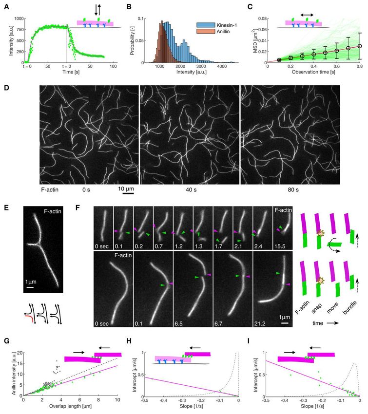
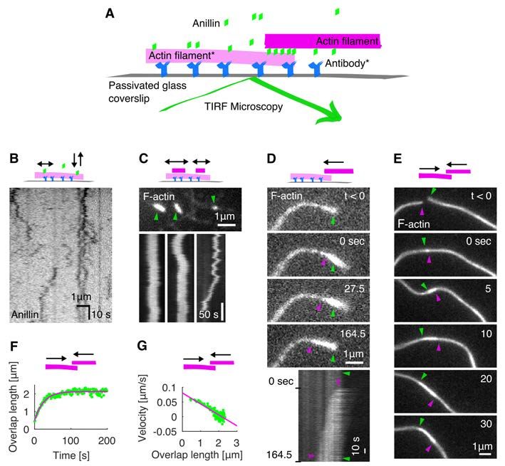
bioRxiv preprint first posted online Jan. 23, 2020; doi: http://dx.doi.org/10.1101/2020.01.22.915256. The copyright holder for
this preprint (which was not peer-reviewed) is the author/funder, who has granted bioRxiv a license to display the preprint in
 perpetuity.
 All rights reserved. No reuse allowed without permission.

 contractility. We thus demonstrate that filament crosslinkers, such as anillin, can generate
 contractile forces in actin networks, substituting for molecular motor activity.

 Results
 Anillin slides actin filaments to maximize their overlap
 To study the interactions between anillin and actin filaments in vitro, we specifically
 immobilised sparsely rhodamine-labelled, phalloidin-stabilised actin filaments to the coverslip
 surface (Methods). After the addition of GFP-labelled anillin to the experimental chamber,
 using TIRF microscopy, we observed anillin-GFP binding to actin filaments (Fig. S1A). At the
 anillin-GFP concentration of 0.12 nM, actin filaments were decorated by anillin-GFP molecules
 (Fig. 1 A, B), which in accordance with previously published data (26) we identified as
 monomers (Fig. S1 B). These single anillin-GFP molecules diffused along the actin filaments
 with a diffusion constant of 0.0088 ± 0.0006 µm2s-1 (linear fit coefficient ± 95% confidence
 bounds, 268 molecules in 2 experiments) (Fig. S1 C). When we increased the anillin-GFP
 concentration in solution to 12 nM and simultaneously added brightly rhodamine-labelled,
 phalloidin-stabilised actin filaments (mobile filaments), we observed mobile filaments landing
 from solution and length-wise crosslinking with the immobilised filaments forming filament
 bundles. In these bundles, we found mobile filaments moving diffusively along the immobilised
 filaments (Fig. 1 C, Movie S1), showing that anillin-GFP generates a diffusible link between
 actin filaments. Strikingly, when a mobile filament landed such that it overlapped partially with
 the immobilised filament, we observed these mobile filaments moving directionally along the
 immobilised filaments. Importantly, this movement was always in the direction increasing the
 length of the overlap between the two filaments and thus effectively contracting the elementary
 two-filament actin bundle (Fig. 1 D, Movie S2). We performed this experiment also in an
 alternative way, not immobilising any of the filaments, but imaging the bundle formation in
 solution in the presence of 0.1% (w/v) methylcellulose to facilitate the imaging at the plane
 near the coverslip surface (Methods). Also, in this set of experiments, we observed, upon an
 initial formation of a partially overlapping two-filament bundle, relative directional sliding of
 the two crosslinked filaments increasing their overlap length (Fig. 1 E, Movie S3). The presence
 of anillin was required for the formation of the actin bundles and their directional sliding (Fig.
 S1 D, Movie S4). We note that in both types of experiments, we neither observed directional
 sliding of filaments that overlapped fully nor directional sliding decreasing the overlap length.
 We also note that the directional sliding did not depend on the relative orientation (structural

 3
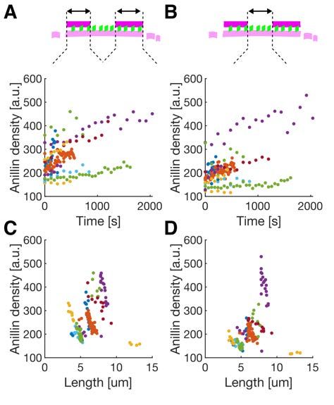
bioRxiv preprint first posted online Jan. 23, 2020; doi: http://dx.doi.org/10.1101/2020.01.22.915256. The copyright holder for
this preprint (which was not peer-reviewed) is the author/funder, who has granted bioRxiv a license to display the preprint in
 perpetuity.
 All rights reserved. No reuse allowed without permission.

 polarity) of the bundled filaments (Fig. S1 E, F). Analysing the dynamics of the sliding, we
 found that the overlap length started increasing immediately after the formation of the bundle.
 Concomitantly with the increase of the overlap length, anillin-GFP amount in the overlap
 increased (Fig. S1 G), indicating anillin-GFP binding into the newly forming overlap region.
 The sliding was slowing down as the overlap length, and the amount of anillin in the overlap
 increased, leading to the overlap length reaching an equilibrium value (Fig. 1 F, G, Fig. S1 H,
 I). The mean difference between the initial and final overlap length, i.e. the mean contraction
 length, was 1.20 ± 0.62 µm (± s.d., maximum 2.67 µm, n = 24 actin filament pairs). Combined,
 these data demonstrate that anillin drives directional sliding of actin filaments relative to each
 other, increasing the length of the overlap between the filaments and thus contracting the
 filament bundle.

 Anillin couples with actin disassembly to generate directed filament sliding
 As actin depolymerization is an important factor for the constriction of the cytokinetic ring, we
 wondered how will depolymerization of actin filaments affect the observed anillin-driven
 filament sliding. We employed the above described experimental setup using a mixture of
 rhodamine-labelled, phalloidin-stabilized actin filaments and unlabelled non-stabilized actin
 filaments in the presence of 12 nM anillin-GFP and not immobilising any of the filaments. To
 enhance the filament disassembly, we used 80-200 nM latrunculin (28). Similar to the
 experiment with stabilized actin, we observed anillin-dependent crosslinking of the filaments
 leading to the formation of actin bundles, which contracted over time. Strikingly, however, in
 this experiment, we did not observe only sliding of partially overlapping filaments as described
 above, but we also observed sliding of filaments that overlapped fully. This sliding was
 observed when the retreating tip of the disassembling non-stabilized actin filament reached a
 stabilized filament (Fig. 2 A, Movie S5). The stabilized filament then followed the retreating
 tip such that the velocity of sliding matched the rate of the filament disassembly (correlation
 coefficient 0.90 ± 0.06, mean ± s.d., n = 13 events in 9 experiments) (Figs. 2 B-D) and thus a
 full overlap between the two filaments was maintained (Fig. 2 A). During this sliding, the
 Anillin-GFP density in the overlaps increased with a faster rate than on single filaments (Fig.
 S2), suggesting that the overlap ends form boundaries for anillin diffusion. As the disassembly
 of one of the filaments works towards shortening the overlap length, the concurrent anillin-
 driven sliding compensates this shortening, maintaining the overlap length. This process,
 leading to the sliding of actin filaments along with the tip of a depolymerizing filament, is thus

 4
bioRxiv preprint first posted online Jan. 23, 2020; doi: http://dx.doi.org/10.1101/2020.01.22.915256. The copyright holder for
this preprint (which was not peer-reviewed) is the author/funder, who has granted bioRxiv a license to display the preprint in
 perpetuity.
 All rights reserved. No reuse allowed without permission.

 an efficient mechanism that couples actin filament depolymerization into sliding-driven
 contraction of actin bundles.

 Anillin promotes formation of actin rings and drives their constriction
 To study the anillin-driven actin filament sliding in structures of higher complexity, we
 increased the actin filament density in the solution, not immobilizing any of the filaments.
 Initially, we used non-stabilised actin filaments. We found that anillin, similar as septins (29),
 promotes the formation of rings of actin filaments several micrometres in diameter (Figs. 3 A,B,
 S3), which we observed forming throughout the volume of the microscopy chamber. The rings
 typically consisted of 3 - 8 actin filaments colocalising with anillin-GFP (Methods).
 Importantly, time-lapse imaging of these rings revealed that they constrict over time (Fig. 3
 B,C, Movie S6). The circumference of the rings decreased exponentially (Fig. 3 C-E), enabling
 us to quantify the asymptotical circumference of the maximally constricted ring. The
 constriction varied substantially, with maximum constriction after 300 s reaching 20 % of the
 initial circumference, maximum asymptotical constriction reaching 100 % and maximum
 constriction rate reaching 0.034 µm×s-1 (Fig. 3 F, G). The spread in the data likely reflects the
 uncertainty of the initial lengths of the overlaps between the filaments forming the rings, as
 well as the spread in the number of these filaments. While most of the rings (n = 18, out of total
 26 rings in 16 experiments) constricted during the time of the experiment, in some cases (n =
 8), we did not observe any significant constriction. We interpret these as rings that already
 reached its maximal constriction before the beginning of our observation. To test the
 contribution of actin disassembly in the constriction process, we studied the constriction
 dynamics of anillin-actin rings consisting of filaments stabilised by rhodamine-phalloidin. We
 found that the constriction was significantly slower (P = 0.0044), with maximum constriction
 after 300 s of about 5 % of the initial circumference and maximum rate of 0.006 µm×s-1 (Fig. 3
 F, G, n = 12 rings in 10 experiments). However, the maximum asymptotical constriction
 reached 100 %, similarly to the rings formed by disassembling actin. By contrast, the addition
 of latrunculin to rings of non-stabilised actin filaments led to higher asymptotic constriction
 (Fig. 3 G). These results are in agreement with our data on linear bundles, where we observed
 that depolymerisation of actin filaments promotes the contraction of the bundle. Combined
 these experiments show that rings of actin filaments can constrict without the action of
 nucleotide hydrolysing molecular motors, solely driven by forces generated by passive actin
 crosslinkers. This constriction is enhanced by the actin filament disassembly.

 5
bioRxiv preprint first posted online Jan. 23, 2020; doi: http://dx.doi.org/10.1101/2020.01.22.915256. The copyright holder for
this preprint (which was not peer-reviewed) is the author/funder, who has granted bioRxiv a license to display the preprint in
 perpetuity.
 All rights reserved. No reuse allowed without permission.

 Anillin generates tens of pico-Newton forces to slide actin filaments
 To directly measure the forces generated by anillin in the actin bundles, we specifically attached
 actin filaments to two microspheres (Methods). Holding the microspheres by two traps in an
 optical tweezers setup, we formed between them, in the presence of 12 nM anillin-GFP, a
 crosslinked actin bundle consisting on average of about 5 filaments (4.7 ± 1.3, mean ± s.d., n =
 35 bundles) (Fig. 4 A, B, Methods, Fig. S4 A). We then moved the microspheres apart from
 each other in 100 nm steps, stretching the bundle and thus sliding the filaments within the
 bundle in the direction shortening their overlaps (Fig. 4B). Simultaneously with each step-
 movement of the trap (each step taking approx. 33 µs), we observed a rapid increase in force,
 which then decayed within several seconds to a plateau reflecting the equilibrium between the
 anillin-generated force and the external load exerted by the optical trap (Fig. 4 C, D, see Fig.
 S4B for control experiment). Since the median time constant of the force decay (1.06 s, quartiles
 0.47s and 2.00 s, n = 326 steps in 43 experiments) together with the trap movement was an
 order of magnitude faster than the rate of anillin unbinding from actin (Fig. S1A, Methods), we
 interpret this dynamic response being due to the rearrangement of anillin molecules in the
 overlap. The equilibrium force values increased with increasing distance between the two
 microspheres and thus with decreasing overlaps between the filaments in the bundle, reaching
 up to tens of pico-Newtons, up to a point when the bundle ruptured (n = 326 steps in 43
 experiments, Fig. 4 E, F, Fig. S4 C). When we (before the rupture) decreased the external load
 on the bundle moving the microspheres closer together in steps of 100 nm, in a situation when
 the bundle was pre-stretched and the force equilibrium was non-zero, we immediately observed
 relative movement of the two microspheres in the direction increasing the overlap length (Fig.
 4 C, D) manifesting relative sliding of actin filaments as a direct consequence of the anillin-
 generated force. To test how the generated force depends on changes in the anillin
 concentration, we first pre-stretched the actin bundle by moving the microspheres apart such
 that the equilibrium force readout was non-zero, and then we decreased the concentration of
 anillin-GFP from 12 nM to 1 nM (keeping the distance between the microspheres and thus the
 external load constant, Methods). We then observed the force generated by anillin decreasing
 over time, presumably as anillin-GFP was unbinding from the overlap (Fig. 4 G, left panel, see
 Fig. S4D for control experiment). Conversely, increasing the anillin-GFP concentration in the
 measurement chamber resulted in a gradual increase in the anillin-generated force as anillin-
 GFP was binding into the overlap (Fig. 4 G, right panel). Combined, these results show that
 anillin generates substantial forces in the range of tens of pico-Newtons per bundle of about

 6
bioRxiv preprint first posted online Jan. 23, 2020; doi: http://dx.doi.org/10.1101/2020.01.22.915256. The copyright holder for
this preprint (which was not peer-reviewed) is the author/funder, who has granted bioRxiv a license to display the preprint in
 perpetuity.
 All rights reserved. No reuse allowed without permission.

 five filaments. These forces result in relative actin sliding increasing the overlaps between actin
 filaments in the bundle and, consequently, in the bundle contraction.

 Discussion
 We here show that passive, non-motor, actin crosslinker anillin can drive the contraction of
 actin bundles through directed filament sliding. Depletion interactions (30–33) did not
 contribute to this phenomenon, as filaments did neither bundle nor slide in the absence of anillin
 in our control experiments (Fig. S1 D). Anillin creates diffusible links between actin filaments
 (Fig. 1 B, C) and is retained in overlaps that transiently shorten (Fig. S2), suggesting that anillin
 molecules in the overlap can be described as confined gas, analogous to crosslinkers described
 in microtubule systems (21, 22). We observed that anillin generated force increased
 hyperbolically with decreasing overlap length (Fig. 4 E, F) and, as anillin unbinding on the
 experimental timescale was negligible, likely thus with increasing density of anillin in the actin
 overlap, as expected for an ideal gas (22). Increasing or decreasing the concentration of anillin
 in our trapping experiment resulting in increasing or decreasing anillin-generated force, can be
 interpreted as an increase or decrease in the anillin-gas pressure in the overlap. Combined, thus,
 our results suggest that the anillin-generated force is of an entropic origin (34) with an
 additional force component likely associated with energetically favourable binding of the
 anillin crosslinkers into the overlap, analogous to microtubule crosslinkers (21, 22). Anillin
 crosslinkers likely induce friction between the filaments that they bundle (22, 35), which might
 explain the slowdown in sliding or ring constriction observed with increasing overlap lengths.
 Circular constraints within the ring may also contribute to the deceleration of the constriction
 as the decrease of the diameter of the ring requires bending of its constituting filaments (29, 33,
 36). At anillin concentrations of 12 nM used in this study, anillin molecules, collectively, can
 generate forces in the order of 10 pN in a bundle of about five filaments. These forces are
 comparable to forces generated by myosins; single Myosin-II stall force is about 2 pN (37, 38),
 suggesting that anillin crosslinkers can generate forces relevant in the context of the cytokinetic
 ring.

 In a disordered actin array, such as in the cytokinetic ring, myosin motors alone are equally
 likely to locally generate contractile or extensile forces. Additional mechanisms are needed to
 break the symmetry of the process, locally favouring the contractile forces to drive the overall
 net constriction of the ring. In this context, various factors are currently under debate (reviewed
 extensively in (3, 10, 11)), such as the difference in behaviour of actin filaments under

 7
bioRxiv preprint first posted online Jan. 23, 2020; doi: http://dx.doi.org/10.1101/2020.01.22.915256. The copyright holder for
this preprint (which was not peer-reviewed) is the author/funder, who has granted bioRxiv a license to display the preprint in
 perpetuity.
 All rights reserved. No reuse allowed without permission.

 compressive and extensile forces (39, 40), inhomogeneous distribution of myosin motors (18,
 41–43) or membrane anchoring of actin filaments (38). Importantly, connectivity between actin
 filaments mediated by actin crosslinker -actinin facilitates myosin-driven contraction of actin
 rings in vitro (9). We here show that anillin-generated forces always act in the direction
 increasing the overlap length, contracting the actin bundle. Anillin will thus locally generate
 assisting forces in overlaps, which are locally contracted by myosin and resisting forces in
 overlaps, which myosin locally extends. Therefore, anillin generated force could be an element
 breaking the symmetry for myosin-dependent force generation in the cytokinetic ring and thus,
 in concert with myosin, drive its constriction. This notion is consistent with in vivo results
 showing that, in Drosophila embryos, lack of anillin results in a complete stall of myosin-driven
 contraction in the myosin-dependent phase of the ring constriction (15).

 Actin filament depolymerization in various, albeit not all, organisms (44) plays an important
 role during the constriction of the cytokinetic ring. Blocking actin depolymerization, e.g. with
 drugs or by impairing the actin-depolymerizing and severing enzyme cofilin, leads to defective
 ring constriction (20, 45–47). Moreover, decreased levels of cofilin or anillin resulted in similar
 effect in Drosophila embryos, namely a delayed switch to the myosin-independent phase of the
 constriction (15), suggesting that actin filament depolymerization, in concert with anillin might
 drive myosin-independent constriction of the cytokinetic ring. We here provide direct evidence
 that anillin crosslinkers efficiently couple with actin filament disassembly to generate
 contractile forces. This mechanism is reminiscent of the experimentally observed mechanism
 driving chromosome movement during the anaphase of cell division by coupling the
 chromosomes’ kinetochores to depolymerizing microtubule ends by biased diffusion (23, 48,
 49) as proposed earlier theoretically (50). Analogously, the diffusion of anillin is likely biased
 away from depolymerizing actin filament ends, resulting in directional sliding of crosslinked
 filaments with the depolymerizing end. We hypothesize that severing of actin filaments might
 promote this process, by increasing the number of filament ends, which can trigger at these
 positions additional anillin-mediated filament sliding and accelerate the bundle contraction.

 In summary, we show directly that actin crosslinkers can drive the contraction of actin bundles
 and can couple with actin disassembly to enhance this effect. We propose that this mechanism
 can i) underpin the in vivo observed myosin-independent constriction of cytokinetic rings and
 ii) break the symmetry of the randomly oriented actin-myosin system in the ring enabling

 8
bioRxiv preprint first posted online Jan. 23, 2020; doi: http://dx.doi.org/10.1101/2020.01.22.915256. The copyright holder for
this preprint (which was not peer-reviewed) is the author/funder, who has granted bioRxiv a license to display the preprint in
 perpetuity.
 All rights reserved. No reuse allowed without permission.

 myosin-dependent constriction of the ring. A recent finding that anillin promotes tensile forces
 in the apical actomyosin networks of X. laevis embryonal epithelium (51) suggests that
 analogous mechanism might also be employed in other actin structures. We propose that
 crosslinker-dependent contractility of filamentous networks is a fundamental mechanism
 readily available in actin-based cytoskeletal structures, likely contributing to various cellular
 movements.

 References
 1. T. E. Schroeder, Cytokinesis: Filaments in the cleavage furrow. Exp. Cell Res. 53, 272–
 318 (1968).
 2. K. Fujiwara, T. D. Pollard, Fluorescent antibody localization of myosin in the
 cytoplasm, cleavage furrow, and mitotic spindle of human cells. J. Cell Biol. 71, 848–
 875 (1976).
 3. T. D. Pollard, B. O’Shaughnessy, Annu. Rev. Biochem., 88, 12.1–12.29 (2019).
 4. C. DeKraker, E. Boucher, C. A. Mandato, Regulation and assembly of actomyosin
 contractile rings in cytokinesis and cell repair. Anat. Rec. 301, 2051–2066 (2018).
 5. M. C. Mangione, K. L. Gould, Molecular form and function of the cytokinetic ring. J.
 Cell Sci. 132, jcs226928 (2019).
 6. H. E. Huxley, J. Hanson, Changes in the cross-striations of muscle during contraction
 and stretch and their structural interpretation. Nature. 173, 973–976 (1954).
 7. A. F. Huxley, R. Niedergerke, Structural changes in muscle during contraction. Nature.
 4412, 971–973 (1954).
 8. T. Kamasaki, M. Osumi, I. Mabuchi, Three-dimensional arrangement of F-actin in the
 contractile ring of fission yeast. J. Cell Biol. 178, 765–771 (2007).
 9. H. Ennomani, G. Letort, C. Guérin, J. L. Martiel, W. Cao, F. Nédélec, E. M. De La
 Cruz, M. Théry, L. Blanchoin, Architecture and Connectivity Govern Actin Network
 Contractility. Curr. Biol. 26, 616–626 (2016).
 10. M. Murrell, P. W. Oakes, M. Lenz, M. L. Gardel, Forcing cells into shape: The
 mechanics of actomyosin contractility. Nat. Rev. Mol. Cell Biol. 16, 486–498 (2015).
 11. T. H. Cheffings, N. J. Burroughs, M. K. Balasubramanian, Actomyosin Ring
 Formation and Tension Generation in Eukaryotic Cytokinesis. Curr. Biol. 26, R719–
 R737 (2016).
 12. X. Ma, M. Kovacs, M. A. Conti, A. Wang, Y. Zhang, J. R. Sellers, R. S. Adelstein,
 Nonmuscle myosin II exerts tension but does not translocate actin in vertebrate
 cytokinesis. Proc. Natl. Acad. Sci. 109, 4509–4514 (2012).
 13. E. Bi, P. Maddox, D. J. Lew, E. D. Salmon, J. N. McMillan, E. Yeh, J. R. Pringle,
 Involvement of an actomyosin contractile ring in Saccharomyces cerevisiae
 cytokinesis. J. Cell Biol. 142, 1301–1312 (1998).
 14. R. Neujahr, C. Heizer, G. Gerisch, Myosin II-independent processes in mitotic cells of
 Dictyostelium discoideum: Redistribution of the nuclei, re-arrangement of the actin
 system and formation of the cleavage furrow. J. Cell Sci. 110, 123–137 (1997).
 15. Z. Xue, A. M. Sokac, Back-to-back mechanisms drive actomyosin ring closure during
 Drosophila embryo cleavage. J. Cell Biol. 215, 335–344 (2016).
 16. C. P. Descovich, D. B. Cortes, S. Ryan, J. Nash, L. Zhang, P. S. Maddox, F. Nedelec,
 A. S. Maddox, Crosslinkers both drive and brake cytoskeletal remodeling and
 furrowing in cytokinesis. Mol. Biol. Cell, 150813 (2018).

 9
bioRxiv preprint first posted online Jan. 23, 2020; doi: http://dx.doi.org/10.1101/2020.01.22.915256. The copyright holder for
this preprint (which was not peer-reviewed) is the author/funder, who has granted bioRxiv a license to display the preprint in
 perpetuity.
 All rights reserved. No reuse allowed without permission.

 17. P. Bun, S. Dmitrieff, J. M. Belmonte, F. J. Nédélec, P. Lénárt, A disassembly-driven
 mechanism explains F-actin-mediated chromosome transport in starfish oocytes. Elife.
 7, 1–27 (2018).
 18. D. B. Oelz, B. Y. Rubinstein, A. Mogilner, Article A Combination of Actin
 Treadmilling and Cross-Linking Drives Contraction of Random Actomyosin Arrays.
 Biophys. J. 109, 1818–1829 (2015).
 19. S. X. Sun, S. Walcott, C. W. Wolgemuth, Cytoskeletal cross-linking and bundling in
 motor-independent contraction. Curr. Biol. 20, R649–R654 (2010).
 20. I. Mendes Pinto, B. Rubinstein, A. Kucharavy, J. R. Unruh, R. Li, Actin
 Depolymerization Drives Actomyosin Ring Contraction during Budding Yeast
 Cytokinesis. Dev. Cell. 22, 1247–1260 (2012).
 21. M. Braun, Z. Lansky, A. Szuba, F. W. Schwarz, A. Mitra, M. Gao, A. Lüdecke, P. R.
 Ten Wolde, S. Diez, Changes in microtubule overlap length regulate kinesin-14-driven
 microtubule sliding. Nat. Chem. Biol. 13, 1245–1252 (2017).
 22. Z. Lansky, M. Braun, A. Luedecke, M. Schlierf, P. R. Ten Wolde, M. E. Janson, S.
 Diez, Diffusible Crosslinkers Generate Directed Forces in Microtubule Networks. Cell.
 160, 1159–1168 (2015).
 23. B. Akiyoshi, K. K. Sarangapani, A. F. Powers, C. R. Nelson, S. L. Reichow, H.
 Arellano-Santoyo, T. Gonen, J. A. Ranish, C. L. Asbury, S. Biggins, Tension directly
 stabilizes reconstituted kinetochore-microtubule attachments. Nature. 468, 576–579
 (2010).
 24. M. Braun, Z. Lansky, F. Hilitski, Z. Dogic, S. Diez, Entropic forces drive contraction
 of cytoskeletal networks. BioEssays. 38, 474–481 (2016).
 25. L. Zhang, A. S. Maddox, Anillin. Curr. Biol. 20, 135–136 (2010).
 26. C. M. Field, B. M. Alberts, Anillin, a contractile ring protein that cycles from the
 nucleus to the cell cortex. J. Cell Biol. 131, 165–178 (1995).
 27. P. Paolo D’Avino, How to scaffold the contractile ring for a safe cytokinesis - lessons
 from Anillin-related proteins. J. Cell Sci. 122, 1071–1079 (2009).
 28. H. Kueh, W. Brieher, T. Mitchison, Dynamic stabilization of actin filaments. Proc.
 Natl. Acad. Sci. USA. 105, 16531 (2008).
 29. M. Mavrakis, Y. Azou-Gros, F. C. Tsai, J. Alvarado, A. Bertin, F. Iv, A. Kress, S.
 Brasselet, G. H. Koenderink, T. Lecuit, Septins promote F-actin ring formation by
 crosslinking actin filaments into curved bundles. Nat. Cell Biol. 16, 322–334 (2014).
 30. A. W. C. Lau, A. Prasad, Z. Dogic, Condensation of isolated semi-flexible filaments
 driven by depletion interactions. Europhys. Lett. 87, 48006 (2009).
 31. A. Ward, F. Hilitski, W. Schwenger, D. Welch, A. W. C. Lau, V. Vitelli, L.
 Mahadevan, Z. Dogic, Solid friction between soft filaments. Nat. Mater. 14, 583–588
 (2015).
 32. F. Hilitski, A. R. Ward, L. Cajamarca, M. F. Hagan, G. M. Grason, Z. Dogic,
 Measuring cohesion between macromolecular filaments one pair at a time: Depletion-
 induced microtubule bundling. Phys. Rev. Lett. 114, 1–6 (2015).
 33. T. Sanchez, I. M. Kulic, Z. Dogic, Circularization, photomechanical switching, and a
 supercoiling transition of actin filaments. Phys. Rev. Lett. 104, 65–68 (2010).
 34. D. J. Odde, Previews and the Ideal Gas Law. Cell. 160, 1041–1043 (2015).
 35. V. Bormuth, V. Varga, J. Howard, E. Schäffer, Protein Friction Limits Diffusive and
 Directed Movements of Kinesin Motors on Microtubules. Science. 325, 870–873
 (2007).
 36. F. Gittes, B. Mickey, J. Nettleton, J. Howard, Flexural rigidity of microtubules and
 actin filaments measured from thermal fluctuations in shape. J. Cell Biol. 120, 923–934
 (1993).

 10
bioRxiv preprint first posted online Jan. 23, 2020; doi: http://dx.doi.org/10.1101/2020.01.22.915256. The copyright holder for
this preprint (which was not peer-reviewed) is the author/funder, who has granted bioRxiv a license to display the preprint in
 perpetuity.
 All rights reserved. No reuse allowed without permission.

 37. M. F. Norstrom, P. A. Smithback, R. S. Rock, Unconventional processive mechanics of
 non-muscle myosin IIB. J. Biol. Chem. 285, 26326–26334 (2010).
 38. M. R. Stachowiak, C. Laplante, H. F. Chin, B. Guirao, E. Karatekin, T. D. Pollard, B.
 O’Shaughnessy, Mechanism of cytokinetic contractile ring constriction in fission yeast.
 Dev. Cell. 29, 547–561 (2014).
 39. M. Lenz, T. Thoresen, M. L. Gardel, A. R. Dinner, Contractile units in disordered
 actomyosin bundles arise from f-actin buckling. Phys. Rev. Lett. 108, 1–5 (2012).
 40. M. P. Murrell, M. L. Gardel, F-actin buckling coordinates contractility and severing in
 a biomimetic actomyosin cortex. Proc. Natl. Acad. Sci. 109, 20820–20825 (2012).
 41. D. Vavylonis, J. Q. Wu, S. Hao, B. O’Shaughnessy, T. D. Pollard, Assembly
 mechanism of the contractile ring for cytokinesis by fission yeast. Science (80-. ). 319,
 97–100 (2008).
 42. K. Kruse, F. Jülicher, Actively contracting bundles of polar filaments. Phys. Rev. Lett.
 85, 1778–1781 (2000).
 43. T. B. Liverpool, M. C. Marchetti, Bridging the microscopic and the hydrodynamic in
 active filament solutions. Europhys. Lett. 69, 846–852 (2005).
 44. M. Mishra, J. Kashiwazaki, T. Takagi, R. Srinivasan, Y. Huang, M. K.
 Balasubramanian, I. Mabuchi, In vitro contraction of cytokinetic ring depends on
 myosin II but not on actin dynamics. Nat. Cell Biol. 15, 853–859 (2013).
 45. N. Kaji, K. Ohashi, M. Shuin, R. Niwa, T. Uemura, K. Mizuno, Cell cycle-associated
 changes in Slingshot phosphatase activity and roles in cytokinesis in animal cells. J.
 Biol. Chem. 278, 33450–33455 (2003).
 46. K. Ono, M. Parast, C. Alberico, G. M. Benian, S. Ono, Specific requirement for two
 ADF/cofilin isoforms in distinct actin-dependent processes in Caenorhabditis elegans.
 J. Cell Sci. 116, 2073–2085 (2003).
 47. M. P. Somma, B. Fasulo, G. Cenci, E. Cundari, M. Gatti, Molecular Dissection of
 Cytokinesis by RNA Interference in Drosophila Cultured Cells. Mol. Biol. Cell. 13,
 2448–2460 (2002).
 48. D. R. Gestaut, B. Graczyk, J. Cooper, P. O. Widlund, A. Zelter, L. Wordeman, C. L.
 Asbury, T. N. Davis, Phosphoregulation and depolymerization-driven movement of the
 Dam1 complex do not require ring formation. Nat. Cell Biol. 10, 407–414 (2008).
 49. M. K. Gardner, D. J. Odde, Dam1 complexes go it alone on disassembling
 microtubules. Nat. Cell Biol. 10, 379–381 (2008).
 50. T. L. Hill, Theoretical problems related to the attachment of microtubules to
 kinetochores. Proc. Natl. Acad. Sci. U. S. A. 82, 4404–4408 (1985).
 51. T. R. Arnold, J. H. Shawky, R. E. Stephenson, K. M. Dinshaw, T. Higashi, F. Huq, L.
 A. Davidson, A. L. Miller, Anillin regulates epithelial cell mechanics by structuring the
 medial-apical actomyosin network. Elife (2019), doi:10.7554/eLife.39065.

 11
bioRxiv preprint first posted online Jan. 23, 2020; doi: http://dx.doi.org/10.1101/2020.01.22.915256. The copyright holder for
this preprint (which was not peer-reviewed) is the author/funder, who has granted bioRxiv a license to display the preprint in
 perpetuity.
 All rights reserved. No reuse allowed without permission.

 Acknowledgments

 We thank Andrew Ward for hints on actin crosslinking in the optical tweezers, the Protein
 Facility of MPI-CBG and Yulia Bobrova for technical support and Laurent Blanchoin for
 critical reading of the manuscript. This study was supported by the projects nos. 19-27477X to
 Z.L. and 20-04068S to M.B. from the Czech Science Foundation, project Introduction of New
 Research Methods to BIOCEV (CZ.1.05/2.1.00/19.0390) from the European Regional
 Development Fund, and by the institutional support of the Czech Academy of Sciences (RVO:
 86652036). We acknowledge Biophysics facility of CMS supported by MEYS CR
 (LM2015043) and the Centre of Imaging Methods core facility, Faculty of Science, Charles
 University, supported by the Czech-BioImaging through MEYS CR (LM2015062 and
 CZ.02.1.01/0.0/0.0/16_013/0001775). DJ and EP carried out this research partly as a project
 work at the Faculty of Biomedical Engineering of the Czech Technical University in Prague.

 Author contributions

 Conceptualization, MB, ZL; Methodology, OK, MB, ZL; Investigation, OK, DJ, VS, SHD, EP;
 Formal Analysis, OK, DJ, EP; Data curation, OK, DJ, SHD; Validation, OK; Resources, OK,
 EZ, SD; Writing, OK, MB, ZL; Visualization, OK; Supervision, MB, ZL; Funding acquisition,
 MB, ZL.

 12
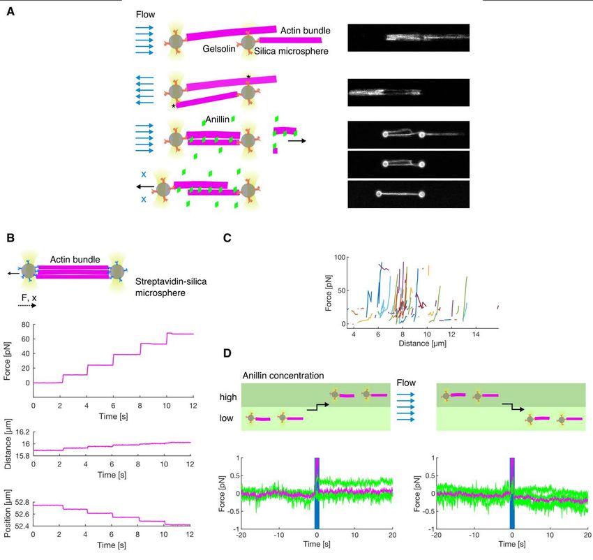
bioRxiv preprint first posted online Jan. 23, 2020; doi: http://dx.doi.org/10.1101/2020.01.22.915256. The copyright holder for
this preprint (which was not peer-reviewed) is the author/funder, who has granted bioRxiv a license to display the preprint in
 perpetuity.
 All rights reserved. No reuse allowed without permission.

 Fig. 1. Anillin slides actin filaments to maximize their overlap. (A) Schematic representation
 of the experimental setup. The asterisk denotes components used only in experiments with
 immobilised filaments. (B) Intensity-inverted kymograph showing single anillin-GFP
 molecules diffusing along an actin filament. (C) Fluorescence micrograph (top) showing three
 short mobile actin filaments (indicated by green arrowheads) crosslinked by anillin-GFP (not
 visualized) to long, sparsely fluorescently labelled, immobilised actin filaments (see Movie S1).
 Kymographs (bottom) show the diffusion of mobile actin filaments from the micrograph above
 along the immobilised filaments. (D) Time-lapse micrographs (top) and a kymograph (bottom)
 showing anillin-driven sliding of a mobile actin filament (bright) along an immobilised filament
 (dim), increasing the overlap between the two actin filaments (see Movie S2). Green and
 magenta arrowheads indicate the ends of the immobilised and mobile filaments, respectively.
 This experiment was repeated 5 times (8 events observed) with similar results. (E) Time-lapse
 micrographs showing anillin-driven sliding of two mobile actin filaments along each other,
 increasing the overlap between the two actin filaments (see Movie S3). Arrowheads indicate
 the filaments' ends. This experiment was repeated 15 times (24 events observed) with similar
 results. (F) A typical time trace of the overlap expansion (green dots) reaching an equilibrium
 value (magenta line represents an exponential fit (Methods) to the data). (G) Velocity of the
 overlap expansion decreases with increasing overlap length. Green points represent an
 exemplary event, magenta line is a linear fit to the data.

 13
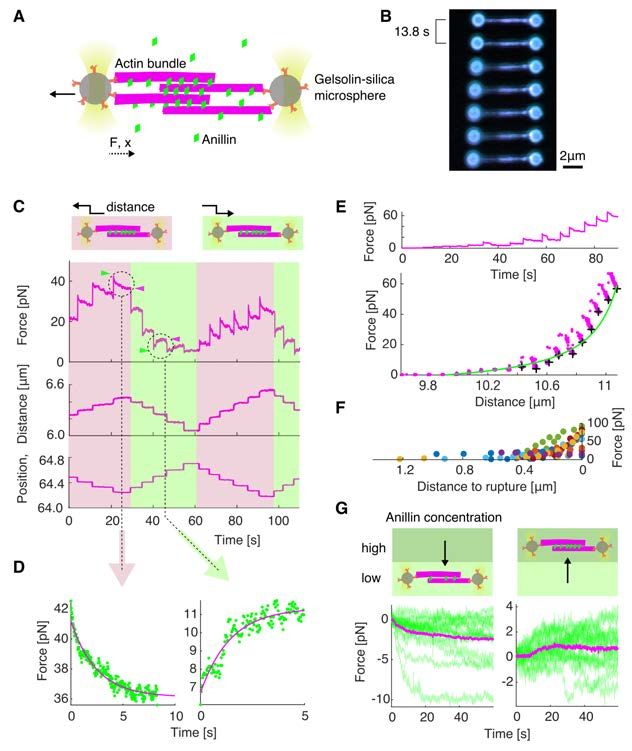
bioRxiv preprint first posted online Jan. 23, 2020; doi: http://dx.doi.org/10.1101/2020.01.22.915256. The copyright holder for
this preprint (which was not peer-reviewed) is the author/funder, who has granted bioRxiv a license to display the preprint in
 perpetuity.
 All rights reserved. No reuse allowed without permission.

 Fig. 2. Anillin couples with actin disassembly to generate directed filament sliding. (A)
 Fluorescence time-lapse micrographs showing two short phalloidin-stabilised filaments sliding
 along with the retreating ends of a non-stabilised filament disassembling in the presence of
 latrunculin (see Movie S5). The disassembling filament is not visualized directly, its length is
 visualized by the anillin-GFP signal. Arrowheads indicate the ends of the two stabilized actin
 filaments. (B) Typical time-traces of the length of the non-stabilised filament (green) and the
 distance between the two short stabilised filaments moving with the ends of the non-stabilised
 one. (C) The distance between the stabilized filaments decreases linearly with the length of the
 disassembling filament (n = 12 events in 9 experiments, colours denote individual events). (D)
 The contraction rate of the actin bundle scales linearly with the rate of disassembly of the non-
 stabilized filament (the same experimental data as shown in (C)).

 14
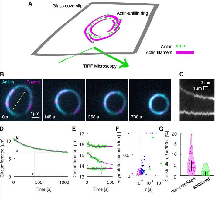
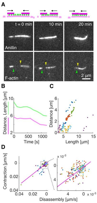
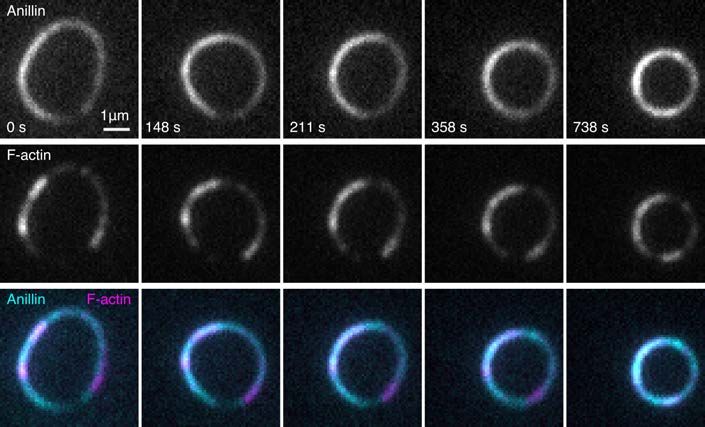
bioRxiv preprint first posted online Jan. 23, 2020; doi: http://dx.doi.org/10.1101/2020.01.22.915256. The copyright holder for
this preprint (which was not peer-reviewed) is the author/funder, who has granted bioRxiv a license to display the preprint in
 perpetuity.
 All rights reserved. No reuse allowed without permission.

 Fig. 3. Anillin promotes formation of actin rings and drives their constriction. (A)
 Schematic representation of the assay geometry. (B) Multicolour time-lapse micrographs of the
 anillin-actin ring, showing the ring constriction over time (see Movie S6). (C) Kymograph
 through the yellow dashed line in (B) of the constricting anillin-actin ring. (D) The time trace
 of the circumference of the ring shown in (B) and (C). Green dots represent experimental data,
 and the solid magenta curve an exponential fit. Dashed lines indicate the fit parameters (t –
 time constant, k – initial circumference, a – the asymptotical value of the circumference). (E)
 Exemplary time-traces of a fast (top), slow (centre), and not (bottom) constricting rings.
 Identical graphical representation as in (D). (F) Relation between the time constant and the
 relative asymptotic constriction (1 – (a/k)). Each dot represents one ring formed either by non-
 stabilised (magenta, n = 26 in 16 experiments) or stabilised (green, n = 12 in 10 experiments)
 actin, or non-stabilized actin in the presence of latrunculin (blue, n = 25 in 7 experiments). Fits
 leading to asymptotical constriction higher than 1 were cut off to 1 (n = 5). Events with higher
 time constant than 106 s, all having zero asymptotic constriction, are not shown (n = 5). t = 300
 s is highlighted by vertical dashed line. (G) Percentage of the constriction at t = 300 s for all
 the rings of stabilised and non-stabilised actin (n = 38). Individual data points are accompanied
 with violin plots, and black boxplots (horizontal tick – median, box – interquartile ranges,
 whiskers – 95% confidence intervals).

 15
bioRxiv preprint first posted online Jan. 23, 2020; doi: http://dx.doi.org/10.1101/2020.01.22.915256. The copyright holder for
this preprint (which was not peer-reviewed) is the author/funder, who has granted bioRxiv a license to display the preprint in
 perpetuity.
 All rights reserved. No reuse allowed without permission.

 Fig. 4. Anillin generates tens of pico-Newton forces to slide actin filaments. (A) Schematic
 representation of the experimental setup. (B) Multicolour time-lapse fluorescence micrographs
 showing an actin bundle attached between two silica microspheres (actin - green, anillin - blue).
 The bundle is being stretched as the left microsphere is pulled leftwards by an optical trap. (C)
 Typical temporal profile of the detected force (top) and the distance between the microspheres
 (middle) in response to the controlled step-wise movement of the left microsphere (bottom). A
 typical force response after stretching and relaxing steps are indicated by the light brown and
 light green background, respectively. The green arrowheads point to the initial peak force and
 the magenta arrowheads show the plateau force in exemplary steps. (D) Magnification of the
 two typical examples highlighted in (C) of the force response after a stretching (left) and
 relaxation (right) steps. Green dots represent individual time points, magenta curve is an
 exponential fit. (E) Typical force time-trace (top) and the force-distance curve (bottom)
 corresponding to stretching of an anillin-actin filament bundle (experimental data points -
 magenta). Asymptotic forces of individual stretching steps, calculated by fitting an exponential
 to the force decays (as shown in (D)), are indicated by black crosses. These increase
 hyperbolically with increasing distance between the microspheres, and thus with decreasing
 overlap length L. Green line represents ∼1/L fit to the data. (F) Detected force increases with
 the decreasing distance before a rupture of the bundle - with decreasing overlap length. All
 events longer than 6 steps are plotted (n = 11 experiments indicated by different colours). (G)
 Force response of a pre-stretched actin-anillin bundle to a change of anillin-GFP concentration.
 Schematic representation of the experiment (top) and temporal experimental data (bottom).
 Left: decrease of the concentration (n = 15 events in 14 experiments), right: increase of the
 concentration (n = 15 events in 14 experiments). Green curves are the experimental data, mean
 temporal profile is shown in magenta.

 16
bioRxiv preprint first posted online Jan. 23, 2020; doi: http://dx.doi.org/10.1101/2020.01.22.915256. The copyright holder for
this preprint (which was not peer-reviewed) is the author/funder, who has granted bioRxiv a license to display the preprint in
 perpetuity.
 All rights reserved. No reuse allowed without permission.

 Fig. S1.
 (A) Kinetics of the anillin-GFP binding/unbinding to/from actin filaments. A temporal profile
 of the fluorescence signal of the anillin-GFP along an actin filament shows a typical first-order
 growth upon addition of 6 nM anillin-GFP and first-order decay after the washout. The loading
 experiment was repeated 15 times (3 experiments) with similar result. The estimated unbinding
 rate of anillin from actin filaments is koff = 0.06 s-1, 95% confidence interval 0.03 – 0.10 s-1, 15
 measurements in 3 experiments. (B) Histograms of the fluorescence intensity of single dimeric
 GFP-labelled kinesin-1 molecules bound to a microtubule (blue) and anillin-GFP particles
 bound to an actin filament (red). The histogram of kinesin-1-GFP shows typical bimodal shape,
 while that of anillin-GFP is mono-modal suggesting that anillin is in monomeric state (128
 anillin-GFP molecules in 2 replicates). Note that the fluorescence intensities in (A) and (B) are
 not comparable as they were recorded with different acquisition times and using different
 excitation laser powers. (C) Mean-squared displacement (MSD) of anillin-GFP molecules

 17
bioRxiv preprint first posted online Jan. 23, 2020; doi: http://dx.doi.org/10.1101/2020.01.22.915256. The copyright holder for
this preprint (which was not peer-reviewed) is the author/funder, who has granted bioRxiv a license to display the preprint in
 perpetuity.
 All rights reserved. No reuse allowed without permission.

 diffusing on actin filaments. Green curves show MSD traces of individual molecules (n = 268
 molecules in 2 experiments), the error bars show mean ± SD. Red line represents a linear fit to
 the mean of the MSD. (D) Actin filaments do not bundle in the absence of anillin (see Movie
 S4). Time-lapse micrograph shows that actin filaments are free to move in the lateral plane, but
 do not form bundles when anillin is absent, showing that the concentration of methylcellulose
 used in experiments with no immobilised filaments is low enough to not induce actin bundling
 through depletion effects (8). (E, F) Anillin can bundle and slide both parallel and antiparallel
 actin filaments. (E) A construct of three filaments bundled by anillin-GFP in which every
 filament overlaps partially with each other. Below is a schematic illustration showing that both,
 parallel and antiparallel filament pairs are formed, in agreement with previously published data
 (9). (F) Time-lapse fluorescence micrographs showing initially a single actin filament in the
 presence of anillin (not visualized). After the filament broke due to thermal fluctuations, the
 two parts connected again in an antiparallel (top) and parallel (bottom) manner, respectively,
 and started sliding along each other. The arrowheads indicate the newly formed filament ends
 formed by the breaking of the initial filament. A schematic representation of the events is shown
 to the right. (G) Intensity of anillin-GFP in the overlap during relative sliding of unbound actin
 filaments. The amount of anillin-GFP in the overlap between two filaments scales linearly with
 the overlap length. The dots represent individual timepoints (n = 187 time points in 16 events,
 5 experiments). The data were fitted by a linear trend (dashed line). Two events with unusually
 higher intensity were marked as outliers (grey dots). The linear trend of remaining events (green
 dots, n = 142 time points in 14 events, 4 experiments) is shown in solid line (magenta). (H, I)
 Dependence between the parameters of the linear fit to the data, such as shown in Fig. 1 G
 (sliding velocity as a function of the overlap length) in experiments using (H) immobilised actin
 filaments (n = 8 bundles in 5 experiments) and (I) mobile actin filaments in the presence of
 methylcellulose (n = 24 bundles in 15 experiments). Although the events had random initial
 overlap length, the contraction in all events followed a regular pattern with linear dependence
 between the extrapolated maximum sliding velocity (intercept) and the deceleration rate
 (slope). Green dots show data from individual sliding events, solid magenta line is a linear
 trend, and the dotted curve shows an estimate of the exponential distribution of the slope of the
 fit.

 18
bioRxiv preprint first posted online Jan. 23, 2020; doi: http://dx.doi.org/10.1101/2020.01.22.915256. The copyright holder for
this preprint (which was not peer-reviewed) is the author/funder, who has granted bioRxiv a license to display the preprint in
 perpetuity.
 All rights reserved. No reuse allowed without permission.

 Fig. S2.
 The density of anillin (mean fluorescence intensity of anillin-GFP per unit length of the actin
 filament) as a function of time, in the overlaps (A), and along the single filament between the
 two overlaps (B) during the disassembling experiments (as shown in Fig. 2 A) and (C,D) as a
 function of the length of the bundle. Individual events are colour-coded (n = 181 time points in
 12 events, 9 experiments). As the bundle contracts, the density of anillin is increasing on
 average 1.39 times (95% confidence interval 1.34 – 1.46; fit to the temporal profiles when
 normalised to the initial density) faster in the overlaps compared to single filament.

 19
bioRxiv preprint first posted online Jan. 23, 2020; doi: http://dx.doi.org/10.1101/2020.01.22.915256. The copyright holder for
this preprint (which was not peer-reviewed) is the author/funder, who has granted bioRxiv a license to display the preprint in
 perpetuity.
 All rights reserved. No reuse allowed without permission.

 Fig. S3.
 Time-lapse micrograph of the constriction of the anillin-actin ring composed of non-stabilised
 actin filaments, showing the anillin-GFP and rhodamine-actin channels and their overlay (see
 Movie S5). The experiment was repeated 16 times with similar results.

 20
bioRxiv preprint first posted online Jan. 23, 2020; doi: http://dx.doi.org/10.1101/2020.01.22.915256. The copyright holder for
this preprint (which was not peer-reviewed) is the author/funder, who has granted bioRxiv a license to display the preprint in
 perpetuity.
 All rights reserved. No reuse allowed without permission.

 Fig. S4.
 (A) Schematics and fluorescence micrographs showing how the anillin-actin bundle was
 created in the optical tweezers. The actin-anillin bundle used in the optical tweezers experiment
 was created similarly as in (4). Briefly, the barbered ends of the filaments firstly bind gelsolin-
 coated microspheres. Then, by reversing the buffer flow, the filaments are transiently attached
 (indicated by asterisk) to the opposite microsphere. The construct is then moved to anillin-GFP
 channel where the higher concentration of CaCl2 triggers severing activity of gelsolin. As
 anillin binds the filaments at the same time, its bundling activity prevents the construct from
 breaking up (Methods). (B) Control experiment showing the force time trace (top) and the
 distance between the beads (middle) in response to step-wise movement of the left microsphere
 (bottom) when the actin bundle was bridging both microspheres (biotin-phalloidin stabilised
 actin filaments suspended between two streptavidin microspheres). In contrast to Fig. 4 C, the
 force time trace increases in steps concomitant with the microsphere position steps. The steps
 in detected force are not followed by relaxation, showing that the actin filaments in the bundle
 form a solid bridge between the two microspheres and cannot slide relative to each other. (C)
 Steady state asymptotical force values of all measured force-distance curves of anillin-bundled
 actin filaments. Individual experiments are colour-coded (n = 43 actin-anillin bundles in 43
 experiments). (D) Control experiments showing the force time trace when the actin-decorated
 but disjoined microspheres were moved between two channels with different concentration of
 anillin-GFP. Moving the disjoined construct between microfluidic channels with a stationary

 21
bioRxiv preprint first posted online Jan. 23, 2020; doi: http://dx.doi.org/10.1101/2020.01.22.915256. The copyright holder for
this preprint (which was not peer-reviewed) is the author/funder, who has granted bioRxiv a license to display the preprint in
 perpetuity.
 All rights reserved. No reuse allowed without permission.

 laminar flow (schematic depiction on the top) does not cause transient changes in the force
 response (bottom) - compare to Fig. 4 G. Green curves represent individual measurements (n =
 8 events in 3 experiments) with mean shown in magenta.

 Movie S1.
 Diffusive motion of mobile actin filaments crosslinked by anillin (not visualized) to long,
 sparsely fluorescently labelled, immobilised actin filaments.

 Movie S2.
 Anillin-driven sliding of a mobile actin filament (bright) along an immobilised filament
 (dim).

 Movie S3.
 Anillin-driven sliding of two mobile actin filaments.

 Movie S4.
 Actin filaments do not bundle in the absence of anillin.

 Movie S5.
 Anillin couples with actin disassembly to generate directed filament sliding.

 Movie S6.
 Constriction of the anillin-actin ring composed of non-stabilised actin filaments. Overlay of
 the anillin-GFP (cyan) and rhodamine-actin (magenta) fluorescence channels.

 22
bioRxiv preprint first posted online Jan. 23, 2020; doi: http://dx.doi.org/10.1101/2020.01.22.915256. The copyright holder for
this preprint (which was not peer-reviewed) is the author/funder, who has granted bioRxiv a license to display the preprint in
 perpetuity.
 All rights reserved. No reuse allowed without permission.

 Materials and Methods
 Proteins
 Homo sapiens anillin (GeneBank accession number: BC070066) cDNA was purchased
 (BC070066-seq-TCHS1003-GVO-TRI, BioCat GmbH Heidelberg, Germany), PCR amplified
 and ligated into an AscI-NotI-digested pOCC destination vector (1) containing a C-terminal
 GFP tag followed by a 3C PreScission Protease cleavage site and a 6xHis-tag. The protein was
 expressed in SF9 insect cells using an opensource FlexiBAC baculovirus vector system (1).
 The insect cells were harvested after 4 days by centrifugation at 300 g for 10 min at 4 ºC in an
 Avanti J-26S ultracentrifuge (JLA-9.1000 rotor, Beckman Coulter). The cell pellet was
 resuspended in 5 ml ice-cold phosphate buffered saline (PBS) and stored at - 80 ºC for further
 use. For cell lysis, the insect cells were homogenized in 30 ml ice-cold His-Trap buffer (50 mM
 Na-phosphate buffer, pH 7.5, 5% glycerol, 300 mM KCl, 1 mM MgCl2, 0.1% tween-20, 10
 mM BME, 0.1 mM ATP) supplemented with 30 mM imidazole, Protease Inhibitor Cocktail
 (cOmplete, EDTA free, Roche) and benzonase to the final concentration of 25 units/ml, and
 centrifuged at 45000 x g for 60 min at 4 ºC in the Avanti J-26S ultracentrifuge (JA-30.50Ti
 rotor, Beckman Coulter). The cleared cell lysate was incubated in a lysis buffer-equilibrated
 Ni-NTA column (HisPur Ni-NTA Superflow Agarose, Pierce, VWR) for 2 h at 4 ºC on a rotator.
 The Ni-NTA column was washed with the wash buffer (His-Trap buffer supplemented with 60
 mM imidazole) and the protein was eluted with the elution buffer (His-Trap buffer
 supplemented with 300 mM Imidazole). The fractions containing anillin-GFP were pooled,
 diluted 1:10 in the His-Trap buffer and the purification tag was cleaved overnight with 3C
 PreScisson protease. The solution was reloaded onto a Ni-NTA column to further separate the
 cleaved protein from the 6xHis-tag. The protein was concentrated using an Amicon
 ultracentrifuge filter and flash frozen in liquid nitrogen. Protein concentration was measured
 using Bradford assay (23236, Thermo Scientific) and microplate reader spectrometer
 (CLARIOstar, BMG Labtech, Germany). Kinesin-1 was expressed and purified as described
 previously (2).

 Unlabelled rabbit muscle actin (AKL99, Cytoskeleton, Inc., USA) and rhodamine labelled
 rabbit muscle actin (AR05, Cytoskeleton, Inc., USA) was resuspended to final concentration of
 10 mg/ml in general actin buffer (GAB; 5 mM TRIS-HCl pH 8, 0.2 mM CaCl2) supplemented
 with 0.2 mM ATP, 5% (w/v) sucrose and 1% (w/v) dextran. For experiments with fluorescently
 labelled non-stabilized filaments, rhodamine labelled actin was diluted with the unlabelled actin
 above in a 1:10 ratio. The actin was then aliquoted, flash-frozen and stored in -80 ºC. Actin
 filaments were polymerised by mixing the aliquoted actin (0.32 mg/ml final concentration) with
 polymerisation buffer (5 mM Tris-HCl pH 8.0, 0.2 mM CaCl2, 50 mM KCl, 2 mM MgCl2, 1
 mM ATP) and, optionally, phalloidin (15 µM final concentration) for filament stabilisation and
 additional labelling. Biotinylated phalloidin (biotin-XX phalloidin, B7474, Thermo Fisher
 Scientific), rhodamine-labelled phalloidin (R415, Thermo Fisher Scientific), or a 3:4 mixture
 of both were used in the experiments. Phalloidin-stabilised filaments were polymerised
 overnight at 4° C and remained stable for at least two months. Non-stabilised filaments were
 grown in the polymerisation solution in the absence of phalloidin for a period of 5 minutes to 5
 hours and were used immediately after polymerization. For use in the assays, actin filaments
 were diluted in a ratio between 1:10 and 1:300, depending on the experiment.

 Fluorescently-labelled microtubules were polymerized from 4 mg/ml porcine tubulin
 (80% unlabelled and 20% Alexa Fluor 647 NHS ester-labelled; Thermo Fisher Scientific) for
 2 h at 37 ºC in BRB80 (80 mM PIPES, 1 mM EGTA, 1 mM MgCl2, pH 6.9) supplemented with
 1 mM MgCl2 and 1 mM GMPCPP (Jena Bioscience, Jena, Germany). The polymerized
 microtubules were centrifuged for 30 min at 18000 x g in a Microfuge 18 Centrifuge (Beckman

 23
You can also read