Contribution of macrophage migration inhibitory factor
←
→
Page content transcription
If your browser does not render page correctly, please read the page content below
The FASEB Journal • Research Communication

Contribution of macrophage migration inhibitory factor
  to the pathogenesis of dengue virus infection
           Iranaia Assunção-Miranda,*,†,1 Flavio A. Amaral,‡,1 Fernando A. Bozza,§
           Caio T. Fagundes,‡ Lirlandia P. Sousa,储 Danielle G. Souza,‡ Patrícia Pacheco,¶
           Giselle Barbosa-Lima,¶ Rachel N. Gomes,¶ Patrícia T. Bozza,¶ Andrea T. Da Poian,*
           Mauro M. Teixeira,储,2 and Marcelo T. Bozza†,2
           *Programa de Biologia Estrutural, Instituto de Bioquímica Médica, and †Departamento de
           Imunologia, Instituto de Microbiologia, Universidade Federal do Rio de Janeiro (UFRJ), Rio de
           Janeiro, Brazil; ‡Departamento de Microbiologia and 储Departamento de Bioquímica e Imunologia,
           Universidade Federal de Minas Gerais, Minas Gerais, Brazil; §Intensive Care Unit, Instituto de
           Pesquisa Clinica Evandro Chagas, and ¶Laboratório de Imunofarmacologia, Instituto Oswaldo Cruz,
           Fundação Oswaldo Cruz, Rio de Janeiro, Brazil

ABSTRACT        Dengue fever is an emerging viral dis-       cal and subtropical regions of the world, with an
ease transmitted by arthropods to humans in tropical         estimated occurrence of 50 –100 million cases annually
countries. Dengue hemorrhagic fever (DHF) is escalat-        (1– 4). The prevalence of dengue fever (DF) has in-
ing in frequency and mortality rates. Here we studied        creased dramatically over the past few years, and ac-
the involvement of macrophage migration inhibitory           cording to the World Health Organization, ⬃500,000
factor (MIF) in dengue virus (DENV) infection and its        patients develop the severe forms of the disease, den-
pathogenesis. Patients with DHF had elevated plasma          gue hemorrhagic fever (DHF) and dengue shock syn-
concentrations of MIF. Both leukocytes from these            drome (DSS), with 20,000 deaths each year (5). The
patients and macrophages from healthy donors in-             situation with DF in the Americas has worsened since
fected in vitro with DENV showed a substantial amount        the detection of a new serotype of the virus (DENV3),
of MIF within lipid droplets. The secretion of MIF by        which caused the mortality rate to escalate to 4%.
macrophages and hepatocytes required a productive            According to the Pan-American Health Organization,
infection and occurred without an increase in gene           the total cases of infection reported in the Americas in
transcription or cell death, thus indicating active secre-   2007 was 850,769, with an increase of 46% of severe
tion from preformed stocks. In vivo infection of wild-       forms and 84% of deaths (6).
type and mif-deficient (Mif ⴚ/ⴚ) mice demonstrated a            The causes of disease progression and the mecha-
role of MIF in dengue pathogenesis. Clinical disease         nisms involved in the physiopathology and lethal out-
was less severe in Mif ⴚ/ⴚ mice, and they exhibited a        come of DENV infection have not been clearly defined,
significant delay in lethality, lower viremia, and lower     but it is believed that viral, host, and environmental
viral load in the spleen than wild-type mice. This           factors contribute to the pathogenesis and progression
reduction in all parameters of severity on DENV infec-       of the disease (4). The lack of adequate therapeutic
tion in Mif ⴚ/ⴚ mice correlated with reduced proinflam-      approaches for the treatment of DF is a consequence of
matory cytokine concentrations. These results demon-         many factors, including our limited understanding of
strated the contribution of MIF to the pathogenesis of       the molecular mechanisms that underlie the interac-
dengue and pointed to a possible beneficial role of          tion between DENV and the human host. One impor-
neutralizing MIF as an adjunctive therapeutic approach       tant reason was the lack, until recently, of an animal
to treat the severe forms of the disease.—Assunção-        model that could reflect the complex pathogenesis of
Miranda, I., Amaral, F. A., Bozza, F. A., Fagundes,          severe dengue. Such an animal model has been described
C. T., Sousa, L. P., Souza, D. G., Pacheco, P., Barbosa-     and displays the hallmarks of severe disease (7, 8).
Lima, G., Gomes, R. N., Bozza, P. T., Da Poian, A. T.,          An increase in proinflammatory cytokine production
Teixeira, M. M., Bozza, M. T. Contribution of macro-
phage migration inhibitory factor to the pathogenesis          1
of dengue virus infection. FASEB J. 24, 218 –228 (2010).         These authors contributed equally to this work.
                                                               2
                                                                  Correspondence: M.T.B., Departamento de Imunologia,
www.fasebj.org                                               Instituto de Microbiologia, CCS Bloco I, UFRJ, Avenida Carlos
                                                             Chagas Filho, 373 Cidade Universitária, Rio de Janeiro, RJ,
Key Words: MIF 䡠 cytokine 䡠 lipid droplets 䡠 inflammation    21941-902 Brasil. E-mail: mtbozza@gmail.com; M.M.T., Depar-
 䡠 hemorrhagic fever 䡠 sepsis                                tamento de Bioquímica e Imunologia, Instituto de Ciências
                                                             Biológicas, Universidade Federal de Minas Gerais, Avenida
                                                             Antônio Carlos, 6627 Pampulha, 31270-901 Belo Horizonte,
Dengue virus (DENV) infection causes the most im-            Minas Gerais, Brazil. E-mail: mmtex@icb.ufmg.br
portant arthropod-borne human viral disease in tropi-          doi: 10.1096/fj.09-139469

218                                                                                       0892-6638/10/0024-0218 © FASEB
by cells of patients with DF or DHF and cells infected collected between 10 and 12 AM using an arterial line or a
in vitro with DENV was documented previously (9 –14). peripheral vein. Blood was put on ice, and plasma was
Macrophage migration inhibitory factor (MIF) is collected by centrifugation at 800 g for 15 min at 4°C,
portioned into aliquots, and stored at ⫺70°C until the day of
among the cytokines found to be increased in the analysis. All patients had DENV3 infection confirmed either
plasma from patients with DF (15). MIF is a proinflam- by anti-DENV ELISA-IgM or serotype-specific RT-PCR. Pa-
matory mediator expressed in a variety of cell types, not tients and volunteers were recruited after protocol approval
only from the immune system, and released in response by the institutional review board for human studies (Comitê
to a number of stimuli, such as cytokines, microbial de Ética em Pesquisas do Instituto Oswaldo Cruz, Fiocruz, Rio
molecules, glucocorticoids, and the immune complex de Janeiro, Brazil), and an informed consent signature was
obtained from the patients themselves or their official repre-
(16 –20). The proinflammatory activities of MIF include
sentatives.
the induction of inflammatory mediators and the ex-
pression of Toll-like receptors and adhesion molecules, In vitro DENV infection
counteracting the effect of glucocorticoids, acting as
chemoattractants, and increasing the survival of leuko- Human monocytes were isolated from peripheral blood
cytes (17, 21–25). The effect of MIF is mediated at least mononuclear cells of healthy donors by density gradient
in part by activation of the CD74-CD44 receptor com- centrifugation on Histopaque (Sigma-Aldrich, St. Louis, MO,
plex (26, 27) and CXCR2 and CXCR4 chemokine USA) and cultured as described previously (19). HepG2, a
receptors (25). As observed in septic patients, MIF human hepatocarcinoma cell lineage, was obtained from
concentrations positively correlated with gravity and American Type Cell Collection (Manassas, VA, USA) and
cultured in minimal essential medium (MEM) supplemented
poor outcome in DENV infection (15, 28, 29). The
with 10% FBS (Invitrogen, Carlsbad, CA, USA) at 37°C in a
results indicating that MIF participates in the pathogen- 5% CO2 atmosphere. DENV3 strain 16562 and DENV2 strain
esis of bacterial sepsis suggest that it would be worth 16881 were propagated in C6/36 Aedes albopictus mosquito
examining the role of MIF as potentially playing an cells. The cells were grown in L-15 medium supplemented
important role in severe forms of dengue. In fact, with 0.3% tryptose phosphate broth, 0.75 g/L sodium bicar-
treatment with neutralizing anti-MIF antibodies or tar- bonate, 1.4 mM glutamine, and nonessential amino acids.
geted disruption of the mif gene protected mice in After 6 d of propagation, cell debris were removed by
centrifugation at 1000 g for 5 min, and the supernatant
several relevant experimental models of sepsis and containing the virus was collected, titrated by a plaque assay
septic shock, in most cases by inhibiting the production on BHK cells, and used for cell infection. Macrophage culture
of inflammatory mediators such as TNF-␣ (16, 21, 30). medium was replaced by fresh DMEM without serum and
In addition, it has been shown that MIF also affects the infected at a multiplicity of infection (MOI) of 4 plaque-
host response to viral, protozoan, and helminthic infec- forming units (PFU)/cell for 2 h at 37°C. After this period,
tions (31–36). the medium with nonadsorbed virus was changed to DMEM
The cell sources, the mechanisms of MIF production, supplemented with 5% heat-inactivated human serum and
maintained at 37°C in 5% CO2. The supernatants of mac-
and the role of MIF in the pathogenesis of DENV rophage-infected cultures were collected for cytokine analyses
infection are largely unknown. Here, we show that 24 and 48 h postinfection (p.i.). In HepG2 infection, semi-
patients with DHF have increased MIF concentrations confluent cultures were incubated with MEM without serum
in their plasma, characterize the mechanisms of MIF and infected with DENV at a MOI of 4 PFU/cell for 1 h. After
production by human macrophages and hepatocytes adsorption, the medium was replaced by MEM with 5%
infected with DENV in vitro, and show that Mif ⫺/⫺ heat-inactivated FCS, and cells were cultured at 37°C in 5%
CO2. After 24 and 48 h of infection, the cell culture super-
mice have reduced pathogenesis in a model of severe
natants were collected for virus titration and cytokine analy-
dengue. ses, and cellular extracts were used for total RNA extraction
for real-time PCR analyses. MIF was inhibited by adding to the
assay medium a purified goat IgG against human MIF (anti-
MATERIALS AND METHODS hMIF; R&D Systems, Minneapolis, MN, USA) to a final concen-
tration of 50 g/ml or the inhibitor compound (S,R)-3-(4-
hydroxyphenyl)-4,5-dihydro-5-isoxazole acetic acid methyl ester
Patients (ISO-1) (Calbiochem EMD Biosciences, San Diego, CA, USA) to
a final concentration of 100 M. DENV3 replication in human
We prospectively enrolled patients admitted recently (48 h) macrophages was assessed by quantification of infectious viral
to the Hospital de Clínicas de Niterói, Niterói, Brazil, who particles in culture supernatants collected at different time
had a strong clinical suspicion of a severe form of DENV points after infection by plaque assay in BHK-21 cells. In
infection. Patient inclusions occurred during epidemic peri- addition, an RT-PCR assay was used to amplify the virus RNA.
ods of DENV serotype 3 (DENV3) in the region. Patients with The reaction was performed using a high-capacity cDNA reverse
severe forms of dengue were those presenting hemodynamic transcription kit (Applied Biosystems, Foster City, CA, USA),
instability (postural hypotension, reduction of systolic arterial according to the manufacturer’s instructions, with 4 g of total
pressure on 20 mmHg in the supine position, or systolic RNA extracted with TRIzol (Invitrogen Life Technologies, Carls-
arterial pressure ⬍90 mmHg), hemorrhagic phenomenon bad, CA, USA). The amount of RNA was determined by real-
(positive tourniquet test, petechiae, equimoses or purpura, time PCR using TaqMan reagents. Determination of cell viability
mucosal bleeding, digestive hemorrhage, puncture bleeding during infection was performed using 3-(4,5-dimethylthiazol-2-yl)-
points), thrombocytopenia (platelet count ⬍50,000/mm3), 2,5-diphenyl tetrazolium bromide (MTT) and lactate dehydro-
dehydration/hemoconcentration (increase in hematocrit of genase assays with a CytoTox96 nonradioactive cytotoxicity assay
20% or more or plasma extravasation signs, such as ascites, kit (Promega Corp. Madison, WI, USA) following the manufac-
pleural effusion, or hypoproteinemia). Blood samples were turer’s instructions.
CONTRIBUTION OF MIF TO PATHOGENESIS OF DENV INFECTION 219In vivo DENV infection ples were subjected to 45 amplification cycles consisting of 95°C
for 30 s and 60°C for 1 min. The expression of the glycerol
3-phosphate dehydrogenase (GPDH) gene was used to
Eight- to 10-wk-old BALB/c [wild-type (WT)] and Mif ⫺/⫺
normalize the results, which are presented as fold induc-
mice, backcrossed into the BALB/c genetic background
tion of mRNA expression relative to control samples. The
(generation N10), were bred and maintained at the Bio-
analyses of relative gene expression data were performed
science Unit of Instituto de Ciências Biológicas [Universidade
by the 2⫺⌬⌬CT method (39).
Federal de Minas Gerais (UFMG), Minas Gerais, Brazil].
Animals were housed under specific pathogen-free condi- MIF immunolocalization
tions and had free access to commercial chow and water. All
procedures had prior approval from the UFMG animal ethics Human leukocytes obtained from DENV-infected patients
committee. DENV2 strain P23085 was obtained from the State were cytospun onto slides, fixed with 3.7% formaldehyde in
Collection of Viruses (Moscow, Russia). The virus was adapted PBS (pH 7.4) for 10 min, and permeabilized with 0.05%
to adult BALB/c mice by a number of sequential passages of saponin/HBSS solution (5 min). After washing, cytospin
mice of different age infected i.p. (7, 8). For the evaluation of preparations were incubated for 1 h at room temperature
lethality, mice were inoculated i.p. with DENV2, and lethality with goat polyclonal serum anti-hMIF or nonimmune goat
rates were assessed every 12 h during 14 d. Platelets were IgG diluted in 0.05% saponin/HBSS. After washes in 0.05%
counted in a Coulter counter (Beckman Coulter, Inc., Fuller- saponin/HBSS, the preparations were incubated with biotin-
ton, CA, USA). For determination of hematocrit, a sample of conjugated rabbit anti-goat IgG (Sigma-Aldrich). The MIF
blood was collected into heparinized capillary tubes and immunoreactivity was shown using an ABC Vectastatin glu-
centrifuged for 10 min in a hematocrit centrifuge (Fanem, cose-oxidase kit according to the manufacturer’s instructions
São Paulo, Brazil). For viral titration, mice were killed, and (Vector Laboratories, Burlingame, CA, USA) and then ob-
blood was collected immediately. For virus recovery, spleens served under light microscopy.
were collected aseptically and stored at ⫺70°C until assayed To determine the subcellular sites of MIF localization within
for DENV2. Viral load in the supernatants of tissue homoge- in vitro DENV3-stimulated human monocyte derived-macroph-
nates and blood samples was assessed by direct plaque assays ages, the cell preparations were fixed with 3.7% formaldehyde in
using LLC-MK2 cells with an agarose overlay plaque assay (7, PBS (pH 7.4) for 10 min and then permeabilized with 0.2%
8). The neutrophil accumulation in the lung tissue was Triton X-100 for 10 min. After cell fixation and permeabiliza-
measured by assaying myeloperoxidase activity, as described tion, human macrophages were blocked with 2% normal don-
previously (37). key serum-PBS for 15 min. The cells were then incubated with
goat anti-hMIF polyclonal antibody (pAb) (R&D Systems) for 45
Quantification of cytokines min, washed in PBS, and incubated with Alexa Fluor 546-labeled
anti-goat IgG (Molecular Probes, Eugene, OR, USA) along with
1 M BODIPY 493/503 (4,4-difluoro-1,3,5,7,8-pentamethyl-4-
MIF concentrations in the human plasma and in cell culture bora-3a,4a-diaza-s-indacene) for 1 h to highlight cytoplasmic
supernatants were measured by ELISA (R&D Systems) ac- lipid droplets (LDs) within macrophages. The specificity of the
cording to the manufacturer’s recommendations. A standard MIF immunolabeling within macrophages was ascertained with
curve was generated using a 2-fold dilution series of recom- normal goat serum (1:100 final dilution; Jackson Immuno-
binant human MIF starting at 2 ng/ml up to 30 pg/ml. A Research Laboratories, Inc., West Grove, PA, USA) used as an
multiplex cytokine kit was used to measure TNF-␣, IL-6, and irrelevant control to anti-MIF pAb. Slides were then washed with
IFN-␥ in the human plasma, and the assay was performed PBS, and an aqueous mounting medium (Polysciences, War-
according to the manufacturer’s instructions (Bio-Rad Labo- rington, PA, USA) was applied to each slide before coverslip
ratories, Hercules, CA, USA) as described previously (14, 38). attachment. Slides were observed under phase-contrast and
Data analyses of all assays were performed with Bio-Plex fluorescent microscopy. Digital photos were taken with a Cool
Manager software (Bio-Rad). Snap camera (Roper Scientific, GmbH, Ottobrun, Germany)
Cytokines in the cell culture supernatants from human and processed using Image Pro Express (Media Cybernetics,
macrophages [TNF-␣ (PeproTech, Inc., Rocky Hill, NJ) and Silver Spring, MD, USA).
IL-6 (R&D Systems)], and cytokines and chemokines [TNF-␣,
IFN-␥, IL-6, keratinocyte chemoattractant (KC), and macro- LD staining and enumeration
phage inflammatory protein (MIP)-2; R&D Systems] in serum
and tissue samples from mice were quantified by ELISA using LDs were stained as described previously (39). In brief,
commercially available antibodies according to the proce- leukocytes on cytospin slides were fixed in 3.7% formalde-
dures supplied by the manufacturer. Prostaglandin (PG) E2 hyde in Ca2⫹-Mg2⫹-free HBSS (pH 7.4) for 30 min and were
concentrations in the cell culture supernatants from human stained with osmium tetroxide or BODIPY 493/503. For
macrophages were determined by an enzyme immunoassay BODIPY labeling, which reflects the accumulation of neutral
(EIA) kit according to the procedures supplied by the man- lipids in LDs, cells were incubated with 1 M BODIPY for 1 h
ufacturer (Cayman Chemical, Ann Arbor, MI, USA). at 37°C. For osmium staining, the slides were rinsed in 0.1 M
Alterations in the expression of cytokines in infected macro- cacodylate buffer, incubated with 1.5% OsO4 (30 min),
phages were evaluated by real-time PCR. Total RNA (4 g) rinsed in H2O, immersed in 1.0% thiocarbohydrazide (5
extracted from the macrophages with TRIzol reagent was re- min), rinsed in 0.1 M cacodylate buffer, reincubated in 1.5%
verse transcribed using a high-capacity cDNA reverse transcrip- OsO4 (3 min), rinsed in distilled water, and then dried and
tion kit, and each sample was submitted to real-time PCR using mounted. The morphology of fixed cells was observed, and
Power SYBR Green PCR Master Mix (Applied Biosystems). The osmium-stained LDs were enumerated by light microscopy
reactions were carried out using specific primers for the following with an ⫻100 objective lens in 50 consecutively scanned
genes: human MIF (forward, 5⬘-GTTCCTCTCCGAGCTCAC- leukocytes.
CCAGCAGC-3⬘; reverse, 5⬘-GCAGCTTGCTGTAGGAGCGGT-
TCTG-3⬘); TNF-␣ (forward, 5⬘-CAGAGGGAAGAGTTCCCC- Statistical analysis
AGGGACC-3⬘; reverse, 5⬘-CCTTGGTCTGGTAGGAGACGG-3⬘);
and IL-6 (forward, 5⬘-TGTGAAAGCAGCAAAGAGGCACTG-3⬘; Statistical analyses were performed using GraphPad Prism 4.0
reverse, 5⬘-ACAGCTCTGGCTTGTTCCTCACTA-3⬘). The sam- for Windows (GraphPad Software, San Diego, CA, USA).
220 Vol. 24 January 2010 The FASEB Journal 䡠 www.fasebj.org ASSUNÇÃO-MIRANDA ET AL.Analysis of cytokine concentrations was performed using a These results confirm that MIF concentrations increase
Mann-Whitney U test or Student’s t test. Multiple group after acute infection with DENV and suggest a correla-
differences were compared using ANOVA followed by Stu- tion between the increase in MIF secretion and the
dent-Newman-Keuls post hoc analysis. Survival after DENV2
challenge was tested using the log-rank test (GraphPad Prism production of other inflammatory mediators during
4.0). Results with P ⬍ 0.05 were considered significant. dengue disease.
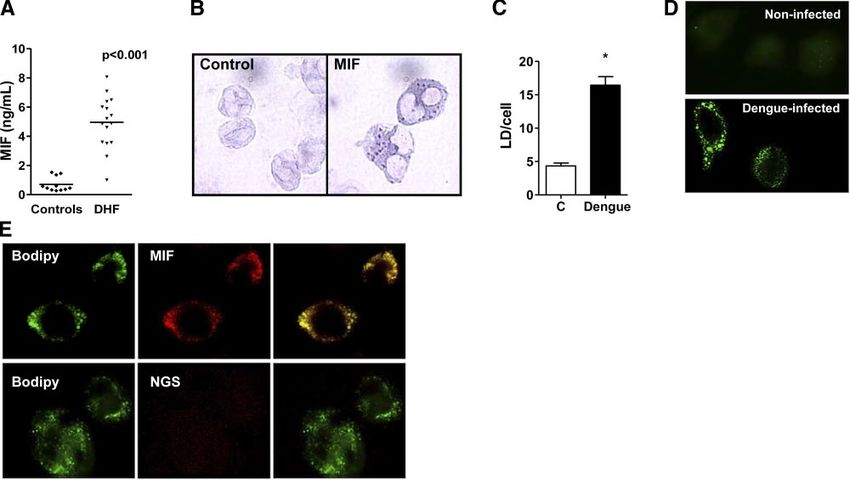
MIF is stored in LDs accumulated in leukocytes from
RESULTS patients with DHF
MIF concentration is increased in plasma of patients LDs are non-membrane-bound, lipid-rich cytoplasmic
with DHF inclusions that are candidates for playing a major role
in the formation of eicosanoid mediators and in the
An increase in the MIF concentration in plasma has storage of inflammatory mediators including cytokines
been documented in a number of inflammatory disor- in inflammatory processes (41). MIF staining of leuko-
ders, including noninfectious and infectious diseases cytes from patients with DHF revealed a punctated
(18, 19). Recent clinical studies indicated that patients cytoplasmic pattern, suggestive of MIF localization in
with viral infections, such as those caused by hepatitis B LDs (Fig. 1B). Quantification of LDs in leukocytes
virus, West Nile virus, or DENV, have higher MIF revealed a significant 3-fold increase in LD accumula-
plasma concentrations than control subjects (15, 34, tion in cells obtained from patients with DHF com-
40). In agreement with these data, we found a signifi- pared with those of healthy subjects (Fig. 1C). Accord-
cant 5-fold increase in MIF concentrations among ingly, increased LD formation was observed in human
patients with DHF compared with those in control macrophages infected with either DENV2 or DENV3
subjects (Fig. 1A). virus but not with heat-inactivated virus compared with
Previous studies demonstrated an increase in inflam- control noninfected cells (Fig. 1D and not shown). LDs
matory cytokines such as IL-6, TNF-␣, and IFN-␥ in were further visualized by intracellular labeling with
patients with DHF (9, 10, 14). Accordingly, we also BODIPY, an LD marker that showed colocalization of
observed a significant increase in plasma concentra- MIF and LDs in human macrophages infected in vitro
tions of these cytokines in patients with DHF compared with DENV3 (Fig. 1E). These results indicate that
with those in control subjects (Supplemental Fig. 1). DENV infection induces the appearance of MIF-posi-
Figure 1. DENV infection induces MIF secretion and com-
partmentalization at LDs. A) Increased plasma concentra-
tions of MIF determined by ELISA were observed in patients
with DHF (n⫽21) compared with healthy volunteers
(n⫽11). B) Peripheral leukocytes from patients with DHF
exhibited punctate cytoplasmic MIF staining, detected by
immunocytochemistry. Right panel: representative MIF
staining (goat pAb anti-hMIF). Left panel: control staining
using normal goat serum (NGS) instead of the specific
primary antibody. C) Quantification of lipid droplets in
osmium-stained peripheral leukocytes from patients with
DHF and healthy volunteers. Bars represent mean ⫾ se
LD/cell from 50 scanned leukocytes from 8 patients with
DHF and 7 volunteers. *P ⱕ 0.05. D) In vitro DENV3
infection induced LD formation on human macrophages.
LDs were labeled with BODIPY 24 h after infective DENV3 at a MOI of 4 PFU in cultures of 24 h p.i. E) MIF colocalizes with
BODIPY-labeled LDs in DENV3-infected human macrophages. Human macrophages infected in vitro with DENV3 (MOI of
4 PFU in cultures of 24 h p.i.) were incubated with anti-MIF (top panel) or nonimmune goat serum (bottom panel).
Cytoplasmic lipid droplets were visualized by BODIPY 493/503 staining (green). Merged image (right panel) shows
colocalization of MIF in BODIPY-labeled LDs.
CONTRIBUTION OF MIF TO PATHOGENESIS OF DENV INFECTION 221tive LDs, which are seen in increased numbers in human hepatome cell line HepG2 has generally been
patients with DHF. used to characterize hepatocyte responses to infection
(46 – 48). Thus, the putative involvement of hepatocytes
Human macrophages and hepatocytes secreted MIF in MIF production during DENV infection was evaluated
on DENV infection using HepG2 cells. The in vitro infection of HepG2 with
DENV3 caused a significant increase in MIF concentra-
Because macrophages are known to produce high tions in the supernatants that peaked at 48 h p.i. (Supple-
amounts of MIF and are susceptible to DENV infection mental Fig. 3). Again, inactivated virus was unable to
(42– 44), we investigated whether infection would pro- induce MIF secretion, indicating that a productive
mote MIF secretion by these cells. In vitro infection of infection is required to promote MIF secretion by
human macrophages with DENV2 or DENV3 caused a hepatocytes. In addition, in these cells the transcrip-
significant 4-fold increase in MIF concentrations in cell tion of MIF was barely affected by the infection, and
culture supernatants that peaked at 24 h after infection no change in cell viability was observed at the time
(Fig. 2A, Supplemental Fig. 2). A similar result was points analyzed (Supplemental Fig. 3). These data
obtained when secretion of TNF-␣ and IL-6 by these indicate that similar to macrophages, DENV infec-
cell cultures was analyzed (Fig. 2B, C). The results also tion in hepatocytes causes the secretion of preformed
showed that secretion of these cytokines requires a MIF irrespective of cell death.
productive infection, because inactivated DENV was
unable to induce it. Interestingly, the expression of MIF
In vitro blockade of MIF reduced production of
mRNA was marginally affected by infection, whereas a
inflammatory mediators during infection
marked induction of TNF-␣ and IL-6 mRNAs synthesis
could be observed as early as 14 h after infection (Fig.
2D–F). In addition, DENV infection induced PGE2 To examine the involvement of MIF in macrophage
production (Fig. 2G). activation on DENV infection, we used a MIF-neutral-
Although MIF release independent of gene transcrip- izing antibody and a selective antagonist of MIF action,
tion has been shown to occur concurrently with cell ISO-1 (49). Both treatments did not affect viral repli-
necrosis in influenza A virus-infected epithelial cells cation, as analyzed by plaque assay and quantitative
(45), this was not the case for MIF secretion by DENV- PCR (Fig. 3A, B). On the other hand, blockade of MIF
infected macrophages, because at 24 h p.i. viability was inhibited the secretion of TNF-␣ and IL-6 and affected
similar in both infected and noninfected cells (Fig. the mRNA expression of these cytokines (Fig. 3C, F).
2H). These results indicate that a productive virus Inhibition of MIF also reduced the production of PGE2
infection was required to promote MIF secretion, prob- (Fig. 3G). These results suggest that MIF secretion
ably from preformed stocks. Moreover, this effect was induces an amplification of macrophage inflammatory
independent of cell death. response due to infection and may play an important
The liver is an important target for the DENV, and the role in the pathogenesis of DENV infection.
Figure 2. DENV3 infection induces the produc-
tion of inflammatory mediators by human mac-
rophages. MIF (A), TNF-␣ (B) IL-6 (C), and PGE2
(G) concentrations were determined by ELISA or
EIA in the supernatants of control macrophages
(mock, C), macrophages incubated with heat-
inactivated (HI) DENV3, or infective DENV3
(D3) at a MOI of 4 PFU collected from cultures at
24 h p.i. Content of mRNA for MIF (D), TNF-␣
(E), and IL-6 (F) was determined by real time
RT-PCR at 5 and 14 h p.i. Results were normal-
ized by GPDH expression and are presented as
fold induction of mRNA expression relative to control samples. Cell viability at 24 h p.i. was analyzed using MTT assays (H).
Results are presented as means ⫾ se. *P ⱕ 0.05. Results are representative of ⱖ3 independent experiments.
222 Vol. 24 January 2010 The FASEB Journal 䡠 www.fasebj.org ASSUNÇÃO-MIRANDA ET AL.Figure 3. MIF contributes to the proinflammatory response during macrophage infection with DENV3. Production of infectious
virions measured by plaque assay (A) and viral replication measured by real time RT-PCR (B) were determined in macrophages at
24 h p.i. TNF-␣ (C) and IL-6 (E) concentrations in the supernatants of macrophage cultures, at 24 h p.i. with DENV3 at a MOI of 4
PFU, were determined by ELISA, and PGE2 (G) was quantified by EIA. Expression of mRNA for TNF-␣ (D) and IL-6 (F) was
determined by real time RT-PCR in cellular extracts. Results of real time RT-PCR were normalized by GPDH expression and are
represented as fold induction of mRNA expression relative to control samples. Total goat IgG and DMSO alone (vehicle of ISO-1)
were used as control for anti-hMIF and ISO-1 effects. Results are presented as means ⫾ se. *P ⱕ 0.05. Results are representative of
ⱖ2 independent experiments. C, control; HI, heat-inactivated.
Mif ⴚ/ⴚ mice had delayed mortality and reduced mice had significantly lower viremia and viral load in
viral load the spleen at all time points analyzed compared with
those in WT mice (Fig. 4B, C). These results suggest
A recent study demonstrated an important role for MIF that MIF contributes to lethality and viral burden by
in the pathogenesis of West Nile virus infection, affect- increasing viral spreading or hampering viral control.
ing the survival and virus invasion of the central ner-
vous system (34). Thus, to directly address whether MIF Reduced thrombocytopenia and inflammation in
is involved in the pathogenesis of DENV infection, we Mif ⴚ/ⴚ mice
used a DENV2 strain adapted to the mouse as an in vivo
model of DHF (7, 8). In the absence of MIF, lethality We have shown previously that infection of mice with
was significantly delayed (Fig. 4A). In addition, Mif ⫺/⫺ this DENV strain causes hemoconcentration and a
Figure 4. Mif⫺/⫺ mice had delayed mortality and reduced viral load after infection. A) Mif ⫺/⫺ mice showed a delay in lethality
after DENV2 infection compared with WT littermates; n ⫽ 9. B, C) Mif ⫺/⫺ mice showed a reduction in viremia in serum 7 d
after infection (B) and lower viremia in spleen on all days analyzed (C) compared with WT mice; n ⫽ 9. Results are presented
as means ⫾ se. *P ⱕ 0.05, **P ⱕ 0.01, ***P ⱕ 0.001 vs. Mif ⫺/⫺.
CONTRIBUTION OF MIF TO PATHOGENESIS OF DENV INFECTION 223Figure 5. Reduced thrombocytopenia and inflammation in Mif ⫺/⫺ mice. A, B) After DENV2 infection, Mif ⫺/⫺ mice retained basal levels of hematocrit (A) and platelets (B) compared with noninfected mice, whereas WT mice showed hemoconcentration (A) and a substantial drop of platelets (B). C) After 7 d p.i., an increase of IFN-␥ concentration was observed in WT mice in both serum and spleen, whereas Mif ⫺/⫺ mice retained values at the basal level. D) Similarly, no increase in IL-6 production in spleen was detected in Mif ⫺/⫺ mice compared with elevated concentrations 7 d p.i. in WT mice. NI, noninfected WT mice. Results are presented as means ⫾ se; n ⫽ 5. **P ⱕ 0.01; ***P ⱕ 0.001 vs. Mif ⫺/⫺. marked thrombocytopenia, similar to that observed in increase in the numbers of lung neutrophils as an patients with DHF (8). At 5 d p.i., WT animals pre- inflammatory response to infection [as assessed by sented hemoconcentration and a marked drop in plate- myeloperoxidase (MPO) activity], whereas in Mif ⫺/⫺ let number, whereas Mif ⫺/⫺ mice were protected from mice the number remained similar to that of nonin- these abnormalities (Fig. 5A, B). Cytokine storm plays a fected controls at 7 d p.i. (Fig. 6A). This increase in critical role in sepsis and is likely to contribute to the tissue neutrophils correlated with higher amounts of severity of DHF (44, 50). Quantification of cytokines the neutrophil-attracting chemokines KC and MIP-2 in demonstrated that Mif ⫺/⫺ mice had reduced concen- the lungs of WT mice at 7 d p.i. (Fig. 6B, C). Again, no trations of IFN-␥ and IL-6 compared with those in WT such increase in the concentration of chemokines was infected animals (Fig. 5C, D). WT mice showed an observed in the lungs of Mif ⫺/⫺ mice. Taken together, Figure 6. Lungs of Mif ⫺/⫺ mice are protected by DENV2 infection. Neutrophils in the lungs were determined by MPO. A) In WT mice, there was a great neutrophil accumulation in the lungs 7 d p.i., whereas no increase in this parameter was seen in Mif ⫺/⫺ mice. B, C) CXCL1 (KC) (B) and CXCL2 (MIP-2) (C) concentrations in lung tissue macerates were determined by ELISA. NI, noninfected WT mice. Results are presented as means ⫾ se; n ⫽ 5. ***P ⱕ 0.001 vs. Mif ⫺/⫺. 224 Vol. 24 January 2010 The FASEB Journal 䡠 www.fasebj.org ASSUNÇÃO-MIRANDA ET AL.
these results indicate that MIF participates in the          phages with DENV also caused an increase in numbers
pathogenesis of DENV infection, affecting the survival,      of LDs, together with an increase in MIF, TNF-␣, IL-6,
the coagulation system, and the inflammatory response        and PGE2 secretion. Blockade of MIF inhibited produc-
in a mouse model of severe disease.                          tion of these inflammatory mediators induced by DENV
                                                             infection. Considering the involvement of LDs in eico-
                                                             sanoid production and the role of MIF in inducing
DISCUSSION                                                   PGE2 synthesis and release (22, 26, 41), one could
                                                             envisage that MIF localization within LDs might be
MIF is a cytokine involved in several aspects of inflam-     important for lipid mediator production. Alternatively,
matory and immune responses, participating in the            the localization of MIF within LDs could be an inter-
pathogenesis of autoimmune, allergic, and infectious         mediary step in MIF secretion pathway. However, no
diseases (18, 19). A recent study demonstrated in-           formal evidence for this hypothesis is presently avail-
creased plasma concentrations of MIF in patients with        able, and future studies will be required to define the
severe forms of dengue (15), but the cells involved in       functional relationship between MIF and LDs. It has
MIF production as well as the role of MIF in the             been shown previously that MIF secretion requires the
pathogenesis of DENV infection have not been previ-          ABCA1 transporter (52), and, more recently, that p115,
ously evaluated. In the present study, to shed light on      a Golgi-associated protein, associates with MIF and is
the role of MIF in the pathogenesis of DENV infection,       involved in MIF secretion (53). Thus, it will be interest-
we combined data from patients with DENV3 infection          ing to analyze whether these proteins colocalize with
collected during an epidemic in Brazil, in vitro infection   MIF at the LD.
of human macrophages and hepatocytes with DENV2                 Human macrophages and hepatocytes infected with
and DENV3, and an experimental mouse model of                DENV showed a significant increase in secretion of
severe dengue. We showed that 1) patients with DHF           MIF, making these cells candidates to act as sources of
had elevated plasma concentrations of MIF, which was         proinflammatory cytokines during infection of patients
stored in LDs accumulated in the leukocytes; 2) in-          with DENV. The secretion of MIF occurred without a
fected human macrophages and hepatocytes secreted            significant change in its gene transcription or in cell
MIF, which is involved in the production of other            viability, suggesting that infection triggers a signaling
inflammatory cytokines; and 3) endogenous MIF con-           pathway that induces MIF release from preformed
tributed to the pathogenesis of experimental dengue          stocks. Previous studies have shown the production of
infection.                                                   MIF due to viral infection, although the mechanisms
   As found for patients with DHF in a DENV2 outbreak        involved in each case seem to be particular (34, 45,
in southern Taiwan in 2002 (15), we observed that MIF        54 –57). For example, infection of lung epithelial cells
concentration was elevated in the plasma of patients         with influenza A virus does not induce MIF gene
with the severe form of DENV3 infection in the epi-          transcription but causes the release of preformed MIF
demics that occurred in Rio de Janeiro, Brazil, also in      probably dependent of necrotic cell death (45). On the
2002. All patients included in our study had DHF as          other hand, infection of fibroblasts with human cyto-
diagnosed by multiple criteria, including confirmation       megalovirus triggers an early and sustained induction
of DENV3 infection, hemodynamic instability, hemor-          of MIF mRNA and protein production, with subsequent
rhagic phenomenon, reduction in platelet numbers,            MIF secretion (55, 57). Moreover, in vivo infection with
and dehydration/hemoconcentration. These patients            West Nile virus or Japanese encephalitis virus, both
also showed a significant increase in plasma concentra-      flaviviruses, cause a significant increase in MIF mRNA
tions of TNF-␣, IL-6, and IFN-␥. We and others have          in mouse brains (34, 54). Much like the results shown
previously shown a positive correlation of increased         here, macrophage infection with Sindbis virus resulted
plasma concentrations of MIF with disease severity in        in MIF secretion from intracellular stocks, without an
patients with bacterial sepsis and with DENV infection       increase in MIF gene expression or an effect on cell
(15, 28).                                                    viability (58). Thus, the mechanisms of MIF production
   Leukocytes from patients with DHF had most of the         and secretion in general and those due to viral infec-
MIF labeling located in cytoplasmic inclusions, compat-      tion in particular clearly require further investigation.
ible with LD localization. The compartmentalization of          MIF secretion during DENV infection followed a
MIF to LDs was analyzed by immunocytochemistry               pattern different from that of TNF-␣ and IL-6, whose
using conditions of cell fixation and permeabilization       production was clearly induced on the transcriptional
that avoid dissolution of these organelles. The require-     level. Blockade of MIF reduced the production of these
ment of these conditions might have prevented others         inflammatory mediators without affecting viral replica-
from identifying MIF in these structures. LDs, although      tion in macrophages. The sharp reduction in the
in reduced number, are normally present in leukocytes        production of TNF-␣ and IL-6 on blockage of MIF
and are increased in size and number on cell activation      indicates that secreted MIF acts in an autocrine/para-
(41). In fact, leukocytes from patients with DHF showed      crine fashion, regulating the production of these cyto-
a significant increase in LD number, similar to our          kines at the transcriptional level. Thus, MIF secreted on
previously described observation in leukocytes from          DENV infection induces the production of inflamma-
septic patients (51). The in vitro infection of macro-       tory mediators. These results suggest that MIF secretion

CONTRIBUTION OF MIF TO PATHOGENESIS OF DENV INFECTION                                                              225
precedes the amplification of the inflammatory re- tion, MIF may act directly as a chemoattractant for
sponse observed in severe cases of dengue and point to granulocytes (25, 36).
MIF blockage as a strategy for a therapeutic approach In conclusion, we presented evidence for significant
to DENV infection. involvement of MIF in the response to DENV infection
To investigate the role of MIF in the pathogenesis of and its pathogenesis. These results suggest that block-
dengue, we used an experimental model of severe DENV ade of MIF might constitute an adjunctive therapeutic
infection characterized by increased vascular permeabil- approach in severe cases of dengue.
ity, altered number and function of leukocytes, increased
hematocrit, thrombocytopenia, and varying degrees of This work was supported by Conselho de Desenvolvimento
hemorrhage (8). Mif ⫺/⫺ mice had a significant delay in Científico e Tecnológico (CNPq; Brazil), Fundação de Amparo
à Pesquisa do Estado do Rio de Janeiro (FAPERJ; Brazil), and
lethality and reduction in all parameters of severity on National Institute of Science and Technology in Dengue (INCT-
DENV infection compared with WT mice, reinforcing the Dengue; Brazil).
role of MIF in the pathogenesis of dengue. The mild
pathological condition of Mif ⫺/⫺ mice might reflect both
the reduced viral load observed in the initial days and the
lower production of inflammatory mediators. The reduc- REFERENCES
tion of viral load could be related to the better hemody- 1. Gibbons, R. V., and Vaughn, D. W. (2002) Dengue: an escalating
namic status of Mif ⫺/⫺ mice, thus facilitating leukocyte problem. BMJ 324, 1563–1566
circulation. At later time points, however, the viremia 2. Guzman, M. G., and Kouri, G. (2002) Dengue: an update. Lancet
Infect. Dis. 2, 33– 42
became similar to that of the WT mice, and eventually 3. Mackenzie, J. S., Gubler, D. J., and Petersen, L. R. (2004)
Mif ⫺/⫺ mice died. Previous studies demonstrated that Emerging flaviviruses: the spread and resurgence of Japanese
MIF blockade had no effect on hepatitis B virus control encephalitis, West Nile and dengue viruses. Nat. Med. 10,
but reduced liver injury (33). Similarly, abrogation of MIF S98 –S109
4. Guzman, M. G., and Kouri, G. (2008) Dengue haemorrhagic
reduced the cerebral pathogenesis in a model of West fever integral hypothesis: confirming observations, 1987–2007.
Nile virus infection without affecting the capacity to Trans. R. Soc. Trop. Med. Hyg. 102, 522–523
control the virus in the periphery (33). Lack of MIF 5. World Health Organization. (2002) Dengue and dengue hemor-
rhagic fever. Fact sheet 117/.2002. http://www.who.int/mediacentre/
benefits the clearance of certain bacterial infections but factsheets/fs117/en
impairs the control of protozoan parasites and Salmonella 6. Pan American Health Organization. (2007) Number of re-
typhimurium bacterial infection (31, 35, 59). Thus, as ported cases of dengue and dengue hemorrhagic fever (DHF),
shown by the results of in vitro studies of MIF production region of the Americas. http://www.paho.org/English/AD/
DPC/CD/dengue-cases-2007.htm
and secretion during infection, the role of MIF in the 7. Atrasheuskaya, A., Petzelbauer, P., Fredeking, T. M., and Igna-
pathogenesis of different infection seems to be specific to tyev, G. (2003) Anti-TNF antibody treatment reduces mortality
each case. in experimental dengue virus infection. FEMS Immunol. Med.
Microbiol. 35, 33– 42
We observed a striking reduction in the concentra- 8. Souza, D. G., Fagundes, C. T., Sousa, L. P., Amaral, F. A., Souza,
tions of cytokines in infected Mif ⫺/⫺ mice compared R. S., Souza, A. L., Kroon, E. G., Sachs, D., Cunha, F. Q., Bukin,
with those observed in WT animals. A central role for E., Atrasheuskaya, A., Ignatyev, G., and Teixeira, M. M. (2009)
MIF in tuning the production of cytokines is a common Essential role of platelet-activating factor receptor in the patho-
genesis of Dengue virus infection. Proc. Natl. Acad. Sci. U. S. A.
feature in many inflammatory and infectious models 106, 14138 –14143
and is considered important to the reduced pathogen- 9. Hober, D., Poli, L., Roblin, B., Gestas, P., Chungue, E., Granic,
esis observed when MIF is absent by genetic manipula- G., Imbert, P., Pecarere, J. L., Vergez-Pascal, R., Wattre, P., and
Maniez-Montreuil, M. (1993) Serum levels of tumor necrosis
tion, neutralizing antibody, or drug treatment (21, 30, factor-␣ (TNF-␣), interleukin-6 (IL-6), and interleukin-1  (IL-
49, 60, 61). Also, in consideration of the role of 1) in dengue-infected patients. Am. J. Trop. Med. Hyg. 48,
cytokines on coagulation and hemodynamic abnormal- 324 –331
ities found in dengue, it is conceivable that the reduced 10. Braga, E. L., Moura, P., Pinto, L. M., Ignacio, S. R., Oliveira,
M. J., Cordeiro, M. T., and Kubelka, C. F. (2001) Detection of
production of cytokines observed in Mif ⫺/⫺ mice circulant tumor necrosis factor-␣, soluble tumor necrosis factor
might have been beneficial (7, 44). In fact, we recently p75 and interferon-␥ in Brazilian patients with dengue fever and
observed a positive correlation between IFN-␥ concen- dengue hemorrhagic fever. Mem. Inst. Oswaldo Cruz 96, 229 –232
11. Suharti, C., van Gorp, E. C., Setiati, T. E., Dolmans, W. M.,
trations and disease severity in patients with dengue Djokomoeljanto, R. J., Hack, C. E., ten Cate, H., and van der
(14). Finally, neutrophil recruitment to the lungs was Meer, J. W. (2002) The role of cytokines in activation of
impaired in Mif ⫺/⫺ mice compared with that in WT coagulation and fibrinolysis in dengue shock syndrome. Thromb.
mice, and this was associated with reduced production Haemost. 87, 42– 46
12. Bosch, I., Xhaja, K., Estevez, L., Raines, G., Melichar, H., Warke,
of the chemoattractants KC and MIP-2. The involve- R. V., Fournier, M. V., Ennis, F. A., and Rothman, A. L. (2002)
ment of MIF in neutrophil recruitment in the DENV Increased production of interleukin-8 in primary human mono-
infection is likely to comprise multifactorial effects. In cytes and in human epithelial and endothelial cell lines after
dengue virus challenge. J. Virol. 76, 5588 –5597
fact, besides induction of chemoattractants, these fac- 13. Green, S., and Rothman, A. L. (2006) Immunopathological
tors could include controlling the expression of adhe- mechanisms in dengue and dengue hemorrhagic fever. Curr.
sion molecules, because MIF has been shown to mod- Opin. Infect. Dis. 19, 429 – 436
ulate intercellular adhesion molecule and vascular 14. Bozza, F. A., Cruz, O. G., Zagne, S. M., Azeredo, E. L., Nogueira,
R. M., Assis, E. F., Bozza, P. T., and Kubelka, C. F. (2008)
cell adhesion molecule expression on endothelial Multiplex cytokine profile from dengue patients: MIP-1 and
cells and chemokine production (20, 62). In addi- IFN-␥ as predictive factors for severity. BMC Infect. Dis. 8, 86 –97
226 Vol. 24 January 2010 The FASEB Journal 䡠 www.fasebj.org ASSUNÇÃO-MIRANDA ET AL.15. Chen, L. C., Lei, H. Y., Liu, C. C., Shiesh, S. C., Chen, S. H., Liu, inhibitory factor in the pathogenesis of malarial anemia. J. Exp.
H. S., Lin, Y. S., Wang, S. T., Shyu, H. W., and Yeh, T. M. (2006) Med. 203, 1185–1196
Correlation of serum levels of macrophage migration inhibitory 33. Kimura, K., Nagaki, M., Nishihira, J., Satake, S., Kuwata, K., and
factor with disease severity and clinical outcome in dengue Moriwaki, H. (2006) Role of macrophage migration inhibitory
patients. Am. J. Trop. Med. Hyg. 74, 142–147 factor in hepatitis B virus-specific cytotoxic-T-lymphocyte-in-
16. Bernhagen, J., Calandra, T., Mitchell, R. A., Martin, S. B., duced liver injury. Clin. Vaccine Immunol. 13, 415– 419
Tracey, K. J., Voelter, W., Manogue, K. R., Cerami, A., and 34. Arjona, A., Foellmer, H. G., Town, T., Leng, L., McDonald, C.,
Bucala, R. (1993) MIF is a pituitary-derived cytokine that Wang, T., Wong, S. J., Montgomery, R. R., Fikrig, E., and Bucala,
potentiates lethal endotoxaemia. Nature 365, 756 –759 R. (2007) Abrogation of macrophage migration inhibitory fac-
17. Calandra, T., Bernhagen, J., Metz, C. N., Spiegel, L. A., Bacher, tor decreases West Nile virus lethality by limiting viral neuroin-
M., Donnelly, T., Cerami, A., and Bucala, R. (1995) MIF as a vasion. J. Clin. Invest. 117, 3059 –3066
glucocorticoid-induced modulator of cytokine production. Na- 35. Flores, M., Saavedra, R., Bautista, R., Viedma, R., Tenorio, E. P.,
ture 377, 68 –71 Leng, L., Sánchez, Y., Juárez, I., Satoskar, A. A., Shenoy, A. S.,
18. Calandra, T., and Roger, T. (2003) Macrophage migration Terrazas, L. I., Bucala, R., Barbi, J., Satoskar, A. R., and
inhibitory factor: a regulator of innate immunity. Nat. Rev. Rodriguez-Sosa, M. (2008) Macrophage migration inhibitory
Immunol. 3, 791– 800 factor (MIF) is critical for the host resistance against Toxoplasma
19. Leng, L., and Bucala, R. (2005) Macrophage migration inhibi- gondii. FASEB J. 22, 3661–3671
tory factor. Crit. Care Med. 33, S475– 477 36. Magalhães, E. S., Paiva, C. N., Souza, H. S., Pyrrho, A. S.,
20. Paiva, C. N., Arras, R. H., Magalhães, E. S., Alves, L. S., Lessa, Mourão-Sá, D., Figueiredo, R. T., Vieira-de-Abreu, A., Dutra,
L. P., Silva, M. H., Ejzemberg, R., Canetti, C., and Bozza, M. T. H. S., Silveira, M. S., Gaspar-Elsas, M. I., Xavier-Elsas, P., Bozza,
(2009) Migration inhibitory factor (MIF) released by macro- P. T., and Bozza, M. T. (2009) Macrophage migration inhibitory
phages upon recognition of immune complexes is critical to factor is critical to interleukin-5-driven eosinophilopoiesis and
inflammation in Arthus reaction. J. Leukoc. Biol. 85, 855– 861 tissue eosinophilia triggered by Schistosoma mansoni infection.
21. Bozza, M., Satoskar, A. R., Lin, G., Lu, B., Humbles, A. A., FASEB J. 23, 1262–1271
Gerard, C., and David, J. R. (1999) Targeted disruption of 37. Souza, D. G., Soares, A. C., Pinho, V., Torloni, H., Reis, L. F.,
migration inhibitory factor gene reveals its critical role in sepsis. Teixeira, M. M., Dias, A. A., and Martins, M. T. (2002) Increased
J. Exp. Med. 189, 341–346 mortality and inflammation in tumor necrosis factor-stimulated
22. Mitchell, R. A., Metz, C. N., Peng, T., and Bucala, R. (1999) gene-14 transgenic mice after ischemia and reperfusion injury.
Sustained mitogen-activated protein kinase (MAPK) and cyto- Am. J. Pathol. 160, 1755–1765
plasmic phospholipase A2 activation by macrophage migration 38. Bozza, F. A., Salluh, J. I., Japiassu, A. M., Soares, M., Assis, E. F.,
inhibitory factor (MIF): regulatory role in cell proliferation and Gomes, R. N., Bozza, M. T., Castro-Faria-Neto, H. C., and Bozza,
glucocorticoid action. J. Biol. Chem. 274, 18100 –18106 P. T. (2007) Cytokine profiles as markers of disease severity in
23. Roger, T., David, J., Glauser, M. P., and Calandra, T. (2001) MIF sepsis: a multiplex analysis. Crit. Care 11, R49 –R56
regulates innate immune responses through modulation of 39. Livak, K. J., and Schmittgen, T. D. (2001) Analysis of relative
Toll-like receptor 4. Nature 414, 920 –924 gene expression data using real-time quantitative PCR and the
24. Mitchell, R. A., Liao, H., Chesney, J., Fingerle-Rowson, G., 2⫺⌬⌬CT. Methods 25, 402– 408
Baugh, J., David, J., and Bucala, R. (2002) Macrophage migra- 40. Zhang, W., Yue, B., Wang, G. Q., and Lu, S. L. (2002) Serum and
tion inhibitory factor (MIF) sustains macrophage proinflamma- ascites levels of macrophage migration inhibitory factor, TNF-␣
tory function by inhibiting p53: regulatory role in the innate and IL-6 in patients with chronic virus hepatitis B and hepatitis
immune response. Proc. Natl. Acad. Sci. U. S. A. 99, 345–350 cirrhosis. Hepatobiliary Pancreat. Dis. Int. 1, 577–580
25. Bernhagen, J., Krohn, R., Lue, H., Gregory, J. L., Zernecke, A., 41. Bozza, P. T., Magalhaes, K. G., and Weller, P. F. (2009)
Koenen, R. R., Dewor, M., Georgiev, I., Schober, A., Leng, L., Leukocyte lipid bodies— biogenesis and functions in inflamma-
Kooistra, T., Fingerle-Rowson, G., Ghezzi, P., Kleemann, R., tion. Biochim. Biophys. Acta 1791, 40 –51
McColl, S. R., Bucala, R., Hickey, M. J., and Weber, C. (2007) MIF 42. Calandra, T., Bernhagen, J., Mitchell, R. A., and Bucala, R.
is a noncognate ligand of CXC chemokine receptors in inflamma- (1994) The macrophage is an important and previously unrec-
tory and atherogenic cell recruitment. Nat. Med. 13, 587–596 ognized source of macrophage migration inhibitory factor. J.
26. Leng, L., Metz, C. N., Fang, Y., Xu, J., Donnelly, S., Baugh, J., Exp. Med. 179, 1895–1902
Delohery, T., Chen, Y., Mitchell, R. A., and Bucala, R. (2003) 43. Chen, Y. C., and Wang, S. Y. (2002) Activation of terminally
MIF signal transduction initiated by binding to CD74. J. Exp. differentiated human monocytes/macrophages by dengue vi-
Med. 197, 1467–1476 rus: productive infection, hierarchical production of innate
27. Shi, X., Leng, L., Wang, T., Wang, W., Du, X., Li, J., McDonald, cytokines and chemokines, and the synergistic effect of lipopoly-
C., Chen, Z., Murphy, J. W., Lolis, E., Noble, P., Knudson, W., saccharide. J. Virol. 76, 9877–9887
and Bucala, R. (2006) CD44 is the signaling component of the 44. Chen, S. T., Lin, Y. L., Huang, M. T., Wu, M. F., Cheng, S. C.,
macrophage migration inhibitory factor-CD74 receptor com- Lei, H. Y., Lee, C. K., Chiou, T. W., Wong, C. H., and Hsieh, S. L.
plex. Immunity 25, 595– 606 (2008) CLEC5A is critical for dengue-virus-induced lethal dis-
28. Bozza, F. A., Gomes, R. N., Japiassú, A. M., Soares, M., Castro- ease. Nature 453, 672– 676
Faria-Neto, H. C., Bozza, P. T., and Bozza, M. T. (2004) 45. Arndt, U., Wennemuth, G., Barth, P., Nain, M., Al-Abed, Y.,
Macrophage migration inhibitory factor levels correlate with Meinhardt, A., Gemsa, D., and Bacher, M. (2002) Release of
fatal outcome in sepsis. Shock 22, 309 –313 macrophage migration inhibitory factor and CXCL8/interleu-
29. Sprong, T., Pickkers, P., Geurts-Moespot, A., van der Ven- kin-8 from lung epithelial cells rendered necrotic by influenza A
Jongekrijg, J., Neeleman, C., Knaup, M., Leroy, D., Calandra, T., virus infection. J. Virol. 76, 9298 –9306
van der Meer, J. W., Sweep, F., and van Deuren, M. (2007) 46. El-Bacha, T., Midlej, V., Pereira da Silva, A. P., Silva da Costa, L.,
Macrophage migration inhibitory factor (MIF) in meningococ- Benchimol, M., Galina, A., and Da Poian, A. T. (2007) Mito-
cal septic shock and experimental human endotoxemia. Shock chondrial and bioenergetic dysfunction in human hepatic cells
27, 482– 487 infected with dengue 2 virus. Biochim. Biophys. Acta 1772, 1158 –
30. Calandra, T., Echtenacher, B., Roy, D. L., Pugin, J., Metz, C. N., 1166
Hültner, L., Heumann, D., Männel, D., Bucala, R., and Glauser, 47. Higa, L. M., Caruso, M. B., Canellas, F., Soares, M. R., Oliveira-
M. P. (2000) Protection from septic shock by neutralization of Carvalho, A. L., Chapeaurouge, D. A., Almeida, P. M., Perales, J.,
macrophage migration inhibitory factor. Nat. Med. 6, 164 –170 Zingali, R. B., and Da Poian, A. T. (2008) Secretome of HepG2
31. Satoskar, A. R., Bozza, M., Rodriguez Sosa, M., Lin, G., and cells infected with dengue virus: implications for pathogenesis.
David, J. R. (2001) Migration-inhibitory factor gene-deficient Biochim. Biophys. Acta 1784, 1607–1616
mice are susceptible to cutaneous Leishmania major infection. 48. Umareddy, I., Tang, K. F., Vasudevan, S. G., Devi, S., Hibberd,
Infect. Immun. 69, 906 –911 M. L., and Gu, F. (2008) Dengue virus regulates type I inter-
32. McDevitt, M. A., Xie, J., Shanmugasundaram, G., Griffith, J., feron signalling in a strain-dependent manner in human cell
Liu, A., McDonald, C., Thuma. P., Gordeuk, V. R., Metz, C. N., lines. J. Gen. Virol. 89, 3052–3062
Mitchell, R., Keefer, J., David, J., Leng, L., and Bucala, R. (2006) 49. Lubetsky, J. B., Dios, A., Han, J., Aljabari, B., Ruzsicska, B.,
A critical role for the host mediator macrophage migration Mitchell, R., Lolis, E., and Al-Abed, Y. (2002) The tautomerase
CONTRIBUTION OF MIF TO PATHOGENESIS OF DENV INFECTION 227active site of macrophage migration inhibitory factor is a 57. Frascaroli, G., Varani, S., Blankenhorn, N., Pretsch, R., Bacher,
potential target for discovery of novel anti-inflammatory agents. M., Leng, L., Bucala, R., Landini, M. P., and Mertens, T. (2009)
J. Biol. Chem. 277, 24976 –24982 Human cytomegalovirus paralyzes macrophage motility through
50. Pang, T., Cardosa, M. J., and Guzman, M. G. (2007) Of cascades down-regulation of chemokine receptors, reorganization of the
and perfect storms: the immunopathogenesis of dengue haem- cytoskeleton, and release of macrophage migration inhibitory
orrhagic fever-dengue shock syndrome (DHF/DSS). Immunol. factor. J. Immunol. 182, 477– 488
Cell Biol. 85, 43– 45 58. Assunção-Miranda, I., Bozza, M. T., and Da Poian, A. (2009)
51. Pacheco, P., Bozza, F. A., Gomes, R. N., Bozza, M., Weller, P. F., Pro-inflammatory response resulting from Sindbis virus infec-
Castro-Faria-Neto, H. C., and Bozza, P. T. (2002) Lipopolysac- tion of human macrophages: Implications for the pathogenesis
charide-induced leukocyte lipid body formation in vivo: innate of viral arthritis. J. Med. Virol. 82, 164 –174
immunity elicited intracellular loci involved in eicosanoid me- 59. Koebernick, H., Grode, L., David, J. R., Rohde, W., Rolph, M. S.,
tabolism. J. Immunol. 169, 6498 – 6506 Mittrücker, H. W., and Kaufmann, S. H. (2002) Macrophage
52. Flieger, O., Engling, A., Bucala, R., Lue, H., Nickel, W., and migration inhibitory factor (MIF) plays a pivotal role in immu-
Bernhagen, J. (2003) Regulated secretion of macrophage mi- nity against Salmonella typhimurium. Proc. Natl. Acad. Sci. U. S. A.
gration inhibitory factor is mediated by a non-classical pathway 99, 13681–13686
involving an ABC transporter. FEBS Lett. 551, 78 – 86 60. Mizue, Y., Ghani, S., Leng, L., McDonald, C., Kong, P., Baugh,
53. Merk, M., Baugh, J., Zierow, S., Leng, L., Pal, U., Lee, S. J., J., Lane, S. J., Craft, J., Nishihira, J., Donnelly, S. C., Zhu, Z., and
Ebert, A. D., Mizue, Y., Trent, J. O., Mitchell, R., Nickel, W., Bucala, R. (2005) Role for macrophage migration inhibitory
Kavathas, P. B., Bernhagen, J., and Bucala, R. (2009) The factor in asthma. Proc. Natl. Acad. Sci. U. S. A. 102, 14410 –14415
Golgi-associated protein p115 mediates the secretion of macro- 61. Magalhães, E. S., Mourao-Sa, D. S., Vieira-de-Abreu, A., Figueir-
phage migration inhibitory factor. J. Immunol. 182, 6896 – 6906 edo, R. T., Pires, A. L., Farias-Filho, F. A., Fonseca, B. P., Viola,
54. Suzuki, T., Ogata, A., Tashiro, K., Nagashima, K., Tamura, M., J. P., Metz, C., Martins, M. A., Castro-Faria-Neto, H. C., Bozza,
Yasui, K., and Nishihira, J. (2000) Japanese encephalitis virus P. T., and Bozza, M. T. (2007) Macrophage migration inhibitory
up-regulates expression of macrophage migration inhibitory factor factor is essential for allergic asthma but not for Th2 differen-
(MIF) mRNA in the mouse brain. Biochim. Biophys. Acta 1517, 100–106 tiation. Eur. J. Immunol. 37, 1097–1106
55. Bacher, M., Eickmann, M., Schrader, J., Gemsa, D., and Heiske, 62. Amin, M. A., Haas, C. S., Zhu, K., Mansfield, P. J., Kim, M. J.,
A. (2002) Human cytomegalovirus-mediated induction of MIF Lackowski, N. P., and Koch, A. E. (2006) Migration inhibitory
in fibroblasts. Virology 299, 32–37 factor up-regulates vascular cell adhesion molecule-1 and inter-
56. Armstrong, M. E., Gantier, M., Li, L., Chung, W. Y., McCann, A., cellular adhesion molecule-1 via Src, PI3 kinase, and NFB.
Baugh, J. A., and Donnelly, S. C. (2008) Small interfering RNAs Blood 107, 2252–2261
induce macrophage migration inhibitory factor production and pro-
liferation in breast cancer cells via a double-stranded RNA-dependent Received for publication June 22, 2009.
protein kinase-dependent mechanism. J. Immunol. 180, 7125–7133 Accepted for publication August 27, 2009.
228 Vol. 24 January 2010 The FASEB Journal 䡠 www.fasebj.org ASSUNÇÃO-MIRANDA ET AL.You can also read