Extracellular matrix components as diagnostic tools in inflam-matory bowel disease
←
→
Page content transcription
If your browser does not render page correctly, please read the page content below
Preprints (www.preprints.org) | NOT PEER-REVIEWED | Posted: 23 August 2021                             doi:10.20944/preprints202108.0439.v1

     Review

     Extracellular matrix components as diagnostic tools in inflam-
     matory bowel disease
     Laura Golusda 1,2, Anja A. Kühl 1, Britta Siegmund 2 and Daniela Paclik 1,2*

                                    1 Charité – Universitätsmedizin Berlin, corporate member of Freie Universität Berlin and Humboldt
                                      Universität zu Berlin, iPATH.Berlin, Campus Benjamin Franklin
                                    2 Charité – Universitätsmedizin Berlin, corporate member of Freie Universität Berlin and Humboldt

                                      Universität zu Berlin, Medizinische Klinik für Gastroenterologie, Infektiologie und Rheumatologie (einschl.
                                      Arbeitsbereich Ernährungsmedizin), Campus Benjamin Franklin
                                    * Correspondence: daniela.paclik@charite.de; Hindenburgdamm 30, D-12200 Berlin Tel.: +49 30-450-514-345

                                    Simple Summary: For decades, the extracellular matrix (ECM) has been defined as a structure com-
                                    ponent playing a rather neglected role in the human body. In the recent years, research has shed
                                    light on the role of ECM within cellular processes including proliferation, migration, and differen-
                                    tiation as well as in inflammation. In inflammation, ECM composition is constantly being remodeled
                                    and undergoes dynamic and rapid changes. Tracking these changes could serve as a novel diagnos-
                                    tic tool. Inflammatory bowel disease is accompanied by complications like fibrosis, stenosis and
                                    fistulas. All these structural complications involve excessive synthesis or degradation of ECM. With
                                    this review we explored whether the analysis of ECM composition can be of support in diagnosing
                                    inflammatory bowel disease and whether changes within ECM can help to predict early on a com-
                                    plicated disease course.

                                    Abstract: Work from the last years indicate that the extracellular matrix (ECM) plays a direct role in
                                    various cellular processes including proliferation, migration and differentiation. Besides homeo-
                                    static processes, its regulatory function in inflammation becomes more and more evident. In inflam-
                                    mation like inflammatory bowel disease, the ECM composition is constantly remodeled which can
                                    result in a structuring of fistulizing disease course. Thus, tracking early ECM changes might bear
                                    the potential to predict the disease course. In this review, we will provide an overview of relevant
                                    diagnostic methods focusing on ECM changes.

                                    Keywords: extracellular matrix; glycosaminoglycans; inflammatory bowel disease; ulcerative coli-
                                    tis; Crohn´s disease; fibrosis; stenosis; magnetic resonance imaging; elastography; histopathology

                                    1. Introduction
                                         The main forms of inflammatory bowel disease (IBD) are ulcerative colitis (UC) and
                                    Crohn´s disease (CD). Both are chronic inflammatory conditions with an altered extracel-
                                    lular matrix (ECM). The diagnosis of UC and CD is a lifelong threat as the available ther-
                                    apies treat and ease the symptoms but do not cure the disease. It is accepted that IBD
                                    results from an exaggerated mucosal immune response in genetically predisposed indi-
                                    viduals. Environmental factors trigger this response and a leaky epithelial barrier is either
                                    0cause or consequence. The onset of IBD occurs in late adolescence and early adulthood
                                    affecting all aspects of life. The incidence and prevalence of IBD is increasing worldwide
                                    just as the total number of related deaths 1. The western Europe region had the highest
                                    age-standardized death rate in 2017 1. Overall, an estimated 1.3 million people in Europe

            © 2021 by the author(s). Distributed under a Creative Commons CC BY license.
Preprints (www.preprints.org) | NOT PEER-REVIEWED | Posted: 23 August 2021 doi:10.20944/preprints202108.0439.v1
suffer from IBD, which equals 0.2% of the European population 2. The amount of direct
healthcare costs per patient per year reach up to €2,000 (UC) and €3,500 (CD), respectively
2. It is still not clear whether changes in ECM occur at an early disease stage triggering
inflammation and contributing to chronicity or whether changes develop at later disease
course that are caused by chronic and excessive inflammation. Complicated disease (stric-
turing or penetrating disease behavior) is a consequence of altered ECM requiring inter-
vention like balloon dilation or surgery including strictureplasty or resection 3. Over 50%
of CD patients and up to 11% of UC patients experience fibrostenotic complications 4.
These complications are accompanied by changes in the ECM. Currently the diagnosis of
IBD is based on a multitude of parameters from clinics, laboratory, imaging, endoscopy
and histopathology 5, 6 . However, the currently available tools to predict disease course
have not entered clinical routine yet 7. Thus, an in-depth analysis of ECM over the course
of the disease might provide a novel tool to fill this gap.
The core components of the ECM are fibronectin, collagens, laminins and proteogly-
cans. Proteoglycans have a protein core to which sulfated glycosaminoglycans (s-GAGs)
are attached. The attached s-GAGs are linear polysaccharides, which are highly negatively
charged. They are build out of disaccharide building blocks. Based on the degree of sul-
fation, the position of sulfation, the linkage between each and the type of monomeric unit,
they are classified into the following groups: chondroitin sulfate/dermatan sulfate
(CS/DS), keratan sulfate (KS) and heparin/heparan sulfate (HS) 8, 9. Hyaluronic acid is the
only non-sulfated GAG present in the ECM. GAGs take part in cell-matrix interactions
and s-GAGs are strongly expressed in the ECM of intestinal tissues. Throughout the gas-
trointestinal tract, s-GAG are found in the subepithelial basal membrane, the vascular en-
dothelium and the ECM of the (sub)mucosa 10. Naba et al. characterized the matrisome
that defines all colon tissue-specific proteins in the mouse colon that are part of or associ-
ated with the ECM 11, 12.
This review presents the latest findings on ECM changes in IBD and by this illustrates
how these could not only serve as a tool to monitor but rather to predict the disease course.
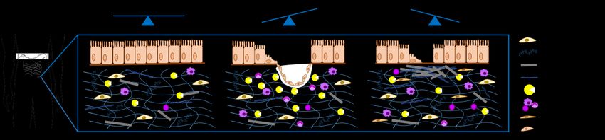
Figure 1: Changes in ECM composition during a chronic disease course. The panels shown illustrate
homeostasis (left), fistulizing disease (middle) and structuring disease (right).
2. Materials and Methods
A literature search in MEDLINE using the electronic database PubMed was conducted to
identify GAG/ECM-associated diagnostic tools in IBD. The following search terms were
included: ((inflammatory bowel disease) OR (ulcerative colitis) OR (Crohn´s disease))
AND (diagnosis) AND ((magnetic resonance imaging) OR (sonography) OR (ultrasound)
OR (elastography) OR (histology) OR (histopathology)) AND ((extracellular matrix) OR
(glycosaminoglycan)) AND (fibrosis OR stenosis OR stricture OR fistula) as well as (con-
trast-enhanced mri) AND (bowel) OR (intestine). Included were studies in adult humans
of the past 10 years.Preprints (www.preprints.org) | NOT PEER-REVIEWED | Posted: 23 August 2021 doi:10.20944/preprints202108.0439.v1
3. Results
3.1. In-vivo Imaging
Several types of imaging are required for diagnosing UC and CD. This generally in-
cludes colonoscopy as well as initially also small bowel magnetic resonance imaging
(MRI) as well as upper endoscopy to determine the disease pattern and the type of dis-
ease in the individual patient. Computed tomography (CT) should be restricted to emer-
gency situations to limit radiation exposure. Furthermore ultrasound (US) is nowadays
frequently used for follow-up evaluation 13, 14. Employing CT enterography in order to
predict small intestinal fibrosis in CD patients revealed that the only predictive parame-
ter was mesenteric hypervascularity although this parameter correlates better with in-
flammation than fibrosis 15. Combining CT enterography with positron emission tomog-
raphy [PET] (PET/CT) was as accurate as PET/magentic resonance-enterography in de-
tecting strictures and bowel wall thickening 16. However, for both techniques, the accu-
racy depended on the bowel segment. The accuracy was low for duodenum and colon
and highest for the terminal ileum and cecum as well as ileocolonic anastomosis 16. For
the detection of fistulas, the sensitivity, specificity and accuracy of small intestine con-
trast ultrasonography (SICUS) is comparable to CT enteroclysis and surgical findings 17.
SICUS is also accurate in detecting strictures and their extension in the proximal and
distal small intestine as well as fistulas and abscesses when compared to surgical and
histological findings 18. Compared to intraoperative findings, SICUS is more accurate in
assessing fistulas, abscesses and strictures than dilatations and bowel wall thickening 19.
The treatment response was monitored in a multicenter study including patients with
active CD by US assessing bowel wall thickness, vascularization, and contrast-agent up-
take as well as fistulas, abscesses and stenoses 20. For determining CD activity, Novak et
al. developed and validated an US score (simple sonographic score) 21. This score might
have been too simple and was not widely accepted. Besides this the Limberg score is
widely used to assess disease activity with US 22. A recent study proposed a regression
model based on multimodal multi-parametric ultrasound to assess CD activity 23. In or-
der to differentiate between inflammation and fibrosis, Bhatnagar et al. compared so-
nographic features with histomorphology in CD patients 24. Their study revealed that
thickness of the mucosal layer rather than bowel wall thickness correlates with acute
inflammation, chronic inflammation and fibrosis 24. Fibrosis was associated with reduced
submucosal echogenicity, increased submucosal echogenicity with hypoechoic bands
and an ill-defined submucosa 24.
The first contrast-enhanced MRI was performed in 1981 25. Oral ferric chloride and in-
haled oxygen was used. Resulting in altered spin-lattice relaxation time of the fundus
and was suggested to be useful as a bowel-labeling agent 25. Since 1988, the paramag-
netic contrast agent Gadolinum-diethylene triamine pentaacetic acid (Gd-DTPA) has
been in clinical application in Germany, the US and Japan 26. Laniado et al. were able to
demonstrate safety as well as effective imaging by orally administered Gd-DTPA and
Mannitol 27. In 1992, the first patient received Gd-DTPA intravenously (i.v.) 28. Intrave-
nous Gd-DPTA administration improved MRI with regard to detecting disease compli-
cations and extent of bowel involvement 29. Distribution of i.v. injected Gd-based con-
trast agents is associated with blood supply but there is no tissue targeting or specificity
30. Contrast agents targeting extracellular matrix for detection of aberrant matrix for-
mation are still in the preclinical phase. Stable plaque formation in atherosclerosis is as-
sociated with ECM accumulation 31, and has been visualized by very small superpara-
magnetic nanoparticles 32. ECM components like collagen or fibrin-fibronectin com-
plexes were targeted in models of liver fibrosis and colorectal cancer 33, 34. To our
knowledge, there is no data available for ECM-targeting contrast agents in models of
intestinal fibrosis. A prospective multicenter study compared the diagnostic accuracy of
MR enterography and US in small bowel CD and concluded MR enterography more
sensitive and more specific 35. Compared to SICUS, MR enterography is more accurate inPreprints (www.preprints.org) | NOT PEER-REVIEWED | Posted: 23 August 2021 doi:10.20944/preprints202108.0439.v1
assessing fistulas and strictures while comparably accurate in assessing abscesses and
dilatations 19. Additionally, contrast-enhanced MR enterography is preferred to bio-
chemical markers, as a significant number of patients with quiescent disease have high
fecal calprotectin levels 36. Furthermore, MR imaging could accurately detect and distin-
guish varying degrees of bowel fibrosis with or without coexisting inflammation. Mag-
netization transfer MRI could accurately detect the severity of bowel fibrosis in strictur-
ing CD but not the inflammatory component within the stricture 37, whereas fibrotic and
inflammatory strictures could be differentiated from purely inflammatory strictures 37.
With reasonable accuracy, an area under the ROC curve of > 0.7, MRI could distinguish
inflammation/edema and muscular hypertrophy from fibrosis in ileal CD in a retrospec-
tive study 38.
Table 1. Diagnostic imaging methods to detect inflammation and complications in IBD.
Method Diagnosis References
Bowel wall thickening, lesions, hypervascularity,
Ultrasound 39-41
strictures, dilatation
Computed tomography Mesenteric hypervascularity, fistulae, abscesses 15, 39
Strictures, bowel wall thickening, inflammatory activ-
PET/CT 16, 42
ity
Strictures, fistulae, abscesses, dilatations, edema, mus-
Magnetic resonance imaging 19, 38
cular hypertrophy
SICUS Strictures, fistulae, abscesses 18, 19
Ten years ago, a small number of CD patients underwent ultrasound elastography (US-
E) prior to elective resection of small intestinal strictures in a pilot study. US-E was
guided by MRI and CT and the scanning included diseased stenotic and adjacent unaf-
fected small intestine. This pilot study revealed that lower strain values indicate stiffer
tissue in stenotic than unaffected bowel 43. Histopathology confirmed predominantly
fibrotic strictures with submucosal collagen depositions, while stenotic tissue was char-
acterized by fibrosis and mild to moderate inflammation 43. US-E is a feasible and repro-
ducible technique for assessing ileal wall fibrosis in CD patients as the strain ratio corre-
lates significantly with the severity of fibrosis 44. US-E detects fibrosis in ileal/ileocolonic
segments of CD patients via increased muscular layer thickness and collagen deposition
45, but is still not able to differentiate fibrosis from inflammation even when contrast en-
hanced 46, 47. Shear-wave elastography (SWE) is a novel technique that allows for quanti-
tative estimation of tissue stiffness. While resected bowel segments showed ex vivo a
higher mean shear-wave speed in high-grade fibrosis than low-grade fibrosis with mini-
mal overlap 48, there was no relationship of fibrosis and SWE in vivo 49. Lu et al. found
that fibrosis was not the major component in bowel wall thickening and strictured bowel
segments but rather muscular hypertrophy 49. When combining SWE and the Limberg
score for bowel vascularization high- and low-grade inflammation as well as high- and
low-grade fibrosis could be discriminated 50. SWE seems also feasible for determining
disease activity in UC while it is rather discriminative in the left- than in the right-sided
colon 51.
Frequent clinical applications of magnetic resonance elastography (MRE) in the abdo-
men are liver diseases, whereas the bowel is technically challenging owing to its loca-
tion, mobility, and physiological motion 52. These challenges were tackled in a prospec-
tive pilot study. Here, shear-wave speed and loss angle representing stiffness and solid-
fluid behavior were studied in IBD patients and healthy controls 53. Both were increased
in IBD patients compared to controls, however, there was no significant difference be-
tween UC and CD 53. Further studies are needed for an assessment of intestinal fibrosis.Preprints (www.preprints.org) | NOT PEER-REVIEWED | Posted: 23 August 2021 doi:10.20944/preprints202108.0439.v1
3.2. Histopathology
In UC, histopathology is required for diagnosis, assessment of disease activity, and iden-
tification of dysplasia and cancer 14. Microscopic features include mucosal architecture,
lamina propria cellularity, neutrophil granulocyte infiltration, and epithelial abnormal-
ity 14. Microscopic features in diagnosing CD are discontinuous chronic inflammation
and crypt distortion as well as granulomas 13. In UC, any colonic stricture is suspicious
of cancer and requires evaluations as such 14. In contrast, structuring disease in CD can
occur even in the absence of cancer. Distinguishing structuring disease from cancer can
be a diagnostic challenge 13. However, an international consortium developed and vali-
dated a stricture histopathology scoring system, in order to enable the development of
novel biomarkers and support the construction of imaging endpoints for clinical trials in
stricturing CD 54. The consensus was reached for evaluating tissue sections stained with
hematoxylin and eosin (H&E) but not using Movat or elastin stains. No consensus could
be reached for the need of trichrome stain 54. The Movat stain is a pentachrome stain and
differentiates elastic, collagen and reticular fibers as well as muscle and fibrin 55, whereas
a trichrome staining allows for differentiation of muscle fibers, collagen and keratin 56.
Hence, ECM components or glycosaminoglycans are not yet part of the clinical routine
for diagnosis or follow-up but are potential candidates in histopathological assessment.
For example, serum levels of matrix metalloproteinase (MMP)-9 have been shown to be
increased in UC patients. In line, within the mucosa immunohistochemically detected
MMP-9 expression increases with severity 57. The advantage of histopathology is the spa-
tial allocation of protein expression. For instance, Fonseca-Camarillo et al. identified up-
regulation of extracellular matrix metalloproteinase inducer (EMMPRIN, CD147) in pa-
tients with active UC and mononuclear and endothelial cells being the main producers
58. Numbers of CD147-positive cells expressing MMP-23 and MMP-10 are increased in
active UC compared to active CD 58. Analyzing stenotic and inflamed ileum from CD
patients revealed enhanced CD3-positivity in the inflamed region and increased colla-
gen-positivity in the stenotic region 59. The increased collagen content was accompanied
by increased losyl oxidase (LOX) involved in the process of collagen deposition linking
collagen to elastin but also interacting with fibronectin 59. Also, CD20-positive cells were
increased in fistulizing versus stenosing CD 44. The ECM glycoprotein tenascin C was
also upregulated in the mucosa of patients with UC and CD compared to controls 60.
Tenasin C is mainly expressed in the lamina propria 60. Additionally, mucosal mRNA
expression has been associated with treatment response, tenascin C mRNA expression
was higher in UC patients nonresponsive to infliximab therapy 60.
3.3. Serological markers
As the balance of degradation and production of ECM is disrupted, components of the
degraded ECM can be found in the peripheral blood. Therefore, assessment of degraded
ECM components in the serum has diagnostic potential. Assessing s-GAGs, hyaluronan
and soluble CD138 in the serum revealed no changes in CD patients compared to
healthy controls whereas hyaluronan was significantly increased in UC patients and cor-
related with Mayo score and thus disease severity 61. Hence, this difference might help to
distinguish UC from CD. However, the data are currently limited to treatment-naïve
patients. Steroid therapy resulted in an increase of hyaluronan and was statistically not
significantly different from untreated UC or adalimumab-treated UC patients 61. Specific
ECM degradation proteins with diagnostic value like MMP-9 degraded type III collagen
fragment C3M has been associated with penetrating CD 62. Serum levels of C3M were
increased in patients with penetrating CD when compared to healthy controls but nei-
ther to non-penetrating or stricturing disease nor to perianal fistula 62. Mortensen et al.
also defined biomarker combinations to discriminate CD from UC and UC from non-
IBD controls 63. Serum levels of VICM (MMP-2/8 degraded and citrullinated-vimentin),
C3M and C4M (MMP-9 degraded collagen type IV) discriminate CD from UC, and C1M
(MMP-9 degraded collagen type I) as well as C3M discriminate UC from non-IBD 63.Preprints (www.preprints.org) | NOT PEER-REVIEWED | Posted: 23 August 2021 doi:10.20944/preprints202108.0439.v1
Compared to healthy controls, the tissue inhibitor of metalloproteinase 1 TIMP-1 was
increased in the serum of patients with UC or CD and higher in active disease allowing
also for disease activity assessment 64. Using serum glycoproteome profiles, Stidham et
al. identified two biomarkers which distinguish inflammatory from fibrostenotic pheno-
types of CD 65. Both cartilage oligomeric matrix protein (COMP) and hepatocyte growth
factor activator (HGFA) showed ≥ 20% change in relative abundance between fibrotic
and inflammatory disease types 65. Van Haaften et al. identified that serum levels of for-
mation and degradation products of collagens can serve to differentiate penetrating and
non-stricturing/non-penetrating as well as stricturing CD in the terminal ileum 66. Other
studies comparing different ECM components found that a strong increase of extracellu-
lar matrix protein 1 (ECM1) in CD patients is correlated with a higher risk to change
from inflammatory phenotype to stricturing phenotype 67. Unfortunately, for none of the
markers exists a standard value which clearly defines the disease status or activity.
Another group of serum makers are anti-microbial antibodies against specific bacteria,
bacterial membrane components or anti-glycans, like anti-Saccaromyces-cervisiae, anti-
Escherichia-coli outer membrane porine C, anti-flagellin, anti-laminaribioside carbohy-
drate antibodies, antimannobioside carbohydrate antibodies, antichitobioside carbohy-
drate antibodies, antichitin antibody, and antilaminarin antibody 68-70. Unfortunately,
none of the markers is directly associated and correlated with fibrostenotic stricture for-
mation 71.
Additionally, some growth factors have been studied as possible biomarkers to detect
early ECM alterations and fibrosis. Among them are vascular endothelial growth factor
(VEGF), platelet-derived growth factor (PDGF), fibroblast growth factor (bFGF) and hu-
man chitinase 3-like 1, influencing angiogenesis, fibrogenesis, myofibroblast prolifera-
tion and myofibroblast-induced collagen secretion 72. VEGF and bFGF both promoting
angiogenesis, tissue repair and fibroblast proliferation are significantly increased in se-
rum of CD patients and correlate with bowel wall thickness 73. The expression of PDGF
is enhanced at sites of inflammation and fibrosis 74 as well as in the serum of CD patients
75. None of the markers can be used to predict fibrostenotic risk but are rather markers
for the severity of fibrotic changes.
Besides components of ECM, growth factors and bacterial components, a potential tool
to diagnose fibrosis is the detection of epigenetic markers like microRNA (miR). These
short, non-coding RNAs are regulating expression of target genes at a post-transcrip-
tional level. One of those, miR-200b was shown to be increased in liver fibrosis 76. When
comparing CD patients with and without fibrostenotic complications, there is a signifi-
cant difference in serum levels of miR-200b between the groups 76. Mehta et al. revealed
that a down-regulation of miR-200b in intestinal epithelial cells is associated with epithe-
lial to mesenchymal transition 77. Furthermore, low serum levels of miR-19 78 and miR-
29b 79 could be correlated with a stricturing phenotype in CD patients. Currently there
are no epigenetic biomarkers which allow the early prediction of a high risk to develop
fibrostenotic complications and further studies to characterize their role are essential. To
summarize this paragraph, a number of potential biomarkers have been described to
identify fibrostenotic complications. Still, due to a low sensitivity and specificity none of
them has entered clinical routine.
4. Discussion
Changes in the ECM trigger inflammation and contribute to chronicity in IBD. This
is reflected by the presence of fibrosis and stricturing disease in about 11% of UC patients
as well as over 50% of CD patients. These complications often require surgical interven-
tion. We here review the latest developments in diagnosing ECM changes in order to as-
sess complicated disease, but also to monitor mucosal healing or differentiate CD from
UC.Preprints (www.preprints.org) | NOT PEER-REVIEWED | Posted: 23 August 2021 doi:10.20944/preprints202108.0439.v1
Early and accurate diagnosis of a complicated disease course is crucial for assessment
and management of these patients. For example, intestinal fibrosis negatively influences
the response to therapy with biologicals 80, 81.
The gold standard for diagnosing disease remains endoscopy. Cross-sectional imag-
ing techniques add in particular information with regards to the small bowel as well as to
complications including abscess and fistulizing disease. There is European consent, that
the use of CT should be limited to emergency due to radiation exposure. Thus, MRI should
present the standard technique and has a high contrast resolution providing anatomical
details without ionizing radiation, but is time- and cost-intensive. Additionally, most con-
trast agents used in MRI contain gadolinium which can accumulate in tissues regardless
of renal function 82. Against a background of IBD patients having an increased risk to de-
velop chronic kidney disease 83 and of patients with impaired renal function developing
in rare cases gadolinium-associated systemic fibrosis 84 radiologist have to balance risks
and benefits of gadolinium-enhanced MRI.
The technique of US is rapid, safe and easy to use. Recent studies have provided con-
vincing evidence that US can be performed in a reproducible manner, thus, the former
argument that it strongly dependent on the examiner is outdated 85. Elastography is also
non-invasive and the advantages of US apply to elastography. Shear wave elastography
requires fasting of the patients in order to reduce bowel content and blood flow. US-E
strain ratio not only depends on the degree of pressure exerted by the US probe 44, but the
mesenteric tissue surrounding the bowel wall serves as control. This control might be mis-
leading as hyperplasia of mesenteric fat itself already affects the strain ratio. Hyperplastic
mesenteric fat wrapping around the circumference of the intestine (creeping fat) is a com-
mon feature in CD 86, 87. Additionally, mesenteric and creeping fat is inflamed in CD, 88
providing misleading strain ratios 44. The application field of elastography is limited to
selected bowel segments and allows no cross-sectional imaging 89. Multimodal imaging
would be optimal for assessing the disease, the disease activity and complications, but one
has to keep in mind that bowel peristaltic negatively influences all imaging techniques.
There is no reference standard in the diagnosis of IBD but as initial diagnostic tool radio-
logic visualization combined with a follow-up via US are widely used to diagnose and
evaluate IBD 5.
In-situ imaging using histopathology provides a clear picture of the intestinal tissue
but is only a snapshot, and is limited to the surface layers when taken as biopsy. Histo-
pathology in combination with endoscopy gives a good overview of stricturing, mucosal
surface and gut motion and is one of the most important diagnostic strategies. One disad-
vantage is that not all segments can be reached and the view is restricted to the luminal
surface. In this regard adding insult to injury during diagnosis, noninvasive diagnostic
tools are favorable.
Especially using serological biomarkers present a minimal invasive and fast ap-
proach. Various ECM-related biomarkers not only diagnose IBD but also differentiate CD
from UC. But due to a low sensitivity and specificity none of them has entered clinical
routine. Besides this, serum biomarkers bear the risk of capturing ECM changes in other
organs. Since, IBD is associated with extraintestinal manifestations like rheumatological,
musculoskeletal, hepatological and dermatological manifestations but also arthropathies
and uveitis are frequent 90. Nevertheless, additional assessment of ECM changes provides
a great potential tool in IBD diagnosis.
5. Conclusions
Various methods and techniques are available for diagnosis of UC and CD as well as
the assessment of disease activity and complications. Every technique has its advantages
and accuracy but also implies disadvantages and inaccuracies. There is no one-size-fits-
all. The optimal treatment of IBD patients should aim at a multimodal approach. Some
techniques and approaches have not made it into the clinic yet and need further develop-
ment and validation. Involving the extracellular matrix and its synthesis, changes andPreprints (www.preprints.org) | NOT PEER-REVIEWED | Posted: 23 August 2021 doi:10.20944/preprints202108.0439.v1
degradation provides a potential toolbox to monitor disease course and phenotype over
time.
Funding: This research was funded by German Research Foundation, grant number SFB1340-TP
B06 to AAK and BS as well as SFB 1449 to BS.
Conflicts of Interest: BS has served as consultant for Abbvie, Arena, BMS, Boehringer, Celgene,
Falk, Janssen, Lilly, Pfizer, Prometheus and Takeda and received speaker’s fees from Abbvie, CED
Service GmbH, Falk, Ferring, Janssen, Novartis, Pfizer, Takeda [served as representative of the
Charité].
References
1. Collaborators, G. B. D. I. B. D., The global, regional, and national burden of inflammatory bowel disease in 195 countries and
territories, 1990-2017: a systematic analysis for the Global Burden of Disease Study 2017. Lancet Gastroenterol Hepatol 2020, 5 (1), 17-
30.
2. Zhao, M.; Gonczi, L.; Lakatos, P. L.; Burisch, J., The burden of inflammatory bowel disease in Europe in 2020. J Crohns Colitis
2021.
3. Bemelman, W. A.; Warusavitarne, J.; Sampietro, G. M.; Serclova, Z.; Zmora, O.; Luglio, G.; de Buck van Overstraeten,
A.; Burke, J. P.; Buskens, C. J.; Colombo, F.; Dias, J. A.; Eliakim, R.; Elosua, T.; Gecim, I. E.; Kolacek, S.; Kierkus, J.;
Kolho, K. L.; Lefevre, J. H.; Millan, M.; Panis, Y.; Pinkney, T.; Russell, R. K.; Shwaartz, C.; Vaizey, C.; Yassin, N.;
D'Hoore, A., ECCO-ESCP Consensus on Surgery for Crohn's Disease. J Crohns Colitis 2018, 12 (1), 1-16.
4. Rieder, F.; Fiocchi, C.; Rogler, G., Mechanisms, Management, and Treatment of Fibrosis in Patients With Inflammatory Bowel
Diseases. Gastroenterology 2017, 152 (2), 340-350 e6.
5. Maaser, C.; Sturm, A.; Vavricka, S. R.; Kucharzik, T.; Fiorino, G.; Annese, V.; Calabrese, E.; Baumgart, D. C.;
Bettenworth, D.; Borralho Nunes, P.; Burisch, J.; Castiglione, F.; Eliakim, R.; Ellul, P.; Gonzalez-Lama, Y.; Gordon, H.;
Halligan, S.; Katsanos, K.; Kopylov, U.; Kotze, P. G.; Krustins, E.; Laghi, A.; Limdi, J. K.; Rieder, F.; Rimola, J.; Taylor,
S. A.; Tolan, D.; van Rheenen, P.; Verstockt, B.; Stoker, J.; European, C. s.; Colitis, O.; the European Society of, G.;
Abdominal, R., ECCO-ESGAR Guideline for Diagnostic Assessment in IBD Part 1: Initial diagnosis, monitoring of known IBD,
detection of complications. J Crohns Colitis 2019, 13 (2), 144-164.
6. Sturm, A.; Maaser, C.; Calabrese, E.; Annese, V.; Fiorino, G.; Kucharzik, T.; Vavricka, S. R.; Verstockt, B.; van
Rheenen, P.; Tolan, D.; Taylor, S. A.; Rimola, J.; Rieder, F.; Limdi, J. K.; Laghi, A.; Krustins, E.; Kotze, P. G.; Kopylov,
U.; Katsanos, K.; Halligan, S.; Gordon, H.; Gonzalez Lama, Y.; Ellul, P.; Eliakim, R.; Castiglione, F.; Burisch, J.;
Borralho Nunes, P.; Bettenworth, D.; Baumgart, D. C.; Stoker, J.; European, C. s.; Colitis, O.; the European Society of, G.;
Abdominal, R., ECCO-ESGAR Guideline for Diagnostic Assessment in IBD Part 2: IBD scores and general principles and technical
aspects. J Crohns Colitis 2019, 13 (3), 273-284.
7. Biasci, D.; Lee, J. C.; Noor, N. M.; Pombal, D. R.; Hou, M.; Lewis, N.; Ahmad, T.; Hart, A.; Parkes, M.;
McKinney, E. F.; Lyons, P. A.; Smith, K. G. C., A blood-based prognostic biomarker in IBD. Gut 2019, 68 (8), 1386-1395.
8. Casale, J.; Crane, J. S., Biochemistry, Glycosaminoglycans. In StatPearls, Treasure Island (FL), 2021.
9. Morla, S., Glycosaminoglycans and Glycosaminoglycan Mimetics in Cancer and Inflammation. Int J Mol Sci 2019, 20 (8).
10. Murch, S. H.; MacDonald, T. T.; Walker-Smith, J. A.; Levin, M.; Lionetti, P.; Klein, N. J., Disruption of sulphated
glycosaminoglycans in intestinal inflammation. Lancet 1993, 341 (8847), 711-4.
11. Naba, A.; Clauser, K. R.; Ding, H.; Whittaker, C. A.; Carr, S. A.; Hynes, R. O., The extracellular matrix: Tools and insights
for the "omics" era. Matrix Biol 2016, 49, 10-24.
12. Naba, A.; Clauser, K. R.; Hoersch, S.; Liu, H.; Carr, S. A.; Hynes, R. O., The matrisome: in silico definition and in vivo
characterization by proteomics of normal and tumor extracellular matrices. Mol Cell Proteomics 2012, 11 (4), M111 014647.Preprints (www.preprints.org) | NOT PEER-REVIEWED | Posted: 23 August 2021 doi:10.20944/preprints202108.0439.v1
13. Gomollon, F.; Dignass, A.; Annese, V.; Tilg, H.; Van Assche, G.; Lindsay, J. O.; Peyrin-Biroulet, L.; Cullen, G. J.;
Daperno, M.; Kucharzik, T.; Rieder, F.; Almer, S.; Armuzzi, A.; Harbord, M.; Langhorst, J.; Sans, M.; Chowers, Y.;
Fiorino, G.; Juillerat, P.; Mantzaris, G. J.; Rizzello, F.; Vavricka, S.; Gionchetti, P.; Ecco, 3rd European Evidence-based
Consensus on the Diagnosis and Management of Crohn's Disease 2016: Part 1: Diagnosis and Medical Management. J Crohns Colitis
2017, 11 (1), 3-25.
14. Magro, F.; Gionchetti, P.; Eliakim, R.; Ardizzone, S.; Armuzzi, A.; Barreiro-de Acosta, M.; Burisch, J.; Gecse, K. B.;
Hart, A. L.; Hindryckx, P.; Langner, C.; Limdi, J. K.; Pellino, G.; Zagorowicz, E.; Raine, T.; Harbord, M.; Rieder, F.;
European, C. s.; Colitis, O., Third European Evidence-based Consensus on Diagnosis and Management of Ulcerative Colitis. Part 1:
Definitions, Diagnosis, Extra-intestinal Manifestations, Pregnancy, Cancer Surveillance, Surgery, and Ileo-anal Pouch Disorders. J
Crohns Colitis 2017, 11 (6), 649-670.
15. Adler, J.; Punglia, D. R.; Dillman, J. R.; Polydorides, A. D.; Dave, M.; Al-Hawary, M. M.; Platt, J. F.; McKenna, B. J.;
Zimmermann, E. M., Computed tomography enterography findings correlate with tissue inflammation, not fibrosis in resected small
bowel Crohn's disease. Inflamm Bowel Dis 2012, 18 (5), 849-56.
16. Pellino, G.; Nicolai, E.; Catalano, O. A.; Campione, S.; D'Armiento, F. P.; Salvatore, M.; Cuocolo, A.; Selvaggi, F.,
PET/MR Versus PET/CT Imaging: Impact on the Clinical Management of Small-Bowel Crohn's Disease. J Crohns Colitis 2016, 10 (3),
277-85.
17. Onali, S.; Calabrese, E.; Petruzziello, C.; Zorzi, F.; Sica, G.; Fiori, R.; Ascolani, M.; Lolli, E.; Condino, G.;
Palmieri, G.; Simonetti, G.; Pallone, F.; Biancone, L., Small intestine contrast ultrasonography vs computed tomography
enteroclysis for assessing ileal Crohn's disease. World J Gastroenterol 2012, 18 (42), 6088-95.
18. Pallotta, N.; Vincoli, G.; Montesani, C.; Chirletti, P.; Pronio, A.; Caronna, R.; Ciccantelli, B.; Romeo, E.;
Marcheggiano, A.; Corazziari, E., Small intestine contrast ultrasonography (SICUS) for the detection of small bowel complications in
crohn's disease: a prospective comparative study versus intraoperative findings. Inflamm Bowel Dis 2012, 18 (1), 74-84.
19. Kumar, S.; Hakim, A.; Alexakis, C.; Chhaya, V.; Tzias, D.; Pilcher, J.; Vlahos, J.; Pollok, R., Small intestinal contrast
ultrasonography for the detection of small bowel complications in Crohn's disease: correlation with intraoperative findings and
magnetic resonance enterography. J Gastroenterol Hepatol 2015, 30 (1), 86-91.
20. Ripolles, T.; Paredes, J. M.; Martinez-Perez, M. J.; Rimola, J.; Jauregui-Amezaga, A.; Bouzas, R.; Martin, G.; Moreno-
Osset, E., Ultrasonographic Changes at 12 Weeks of Anti-TNF Drugs Predict 1-year Sonographic Response and Clinical Outcome in
Crohn's Disease: A Multicenter Study. Inflamm Bowel Dis 2016, 22 (10), 2465-73.
21. Novak, K. L.; Kaplan, G. G.; Panaccione, R.; Afshar, E. E.; Tanyingoh, D.; Swain, M.; Kellar, A.; Wilson, S., A Simple
Ultrasound Score for the Accurate Detection of Inflammatory Activity in Crohn's Disease. Inflamm Bowel Dis 2017, 23 (11), 2001-2010.
22. Limberg, B., [Diagnosis of chronic inflammatory bowel disease by ultrasonography]. Z Gastroenterol 1999, 37 (6), 495-508.
23. Jing, J.; Wu, Y.; Zhang, H.; Zhang, Y.; Mu, J.; Luo, Y.; Zhuang, H., The establishment of a regression model from four
modes of ultrasound to predict the activity of Crohn's disease. Sci Rep 2021, 11 (1), 77.
24. Bhatnagar, G.; Rodriguez-Justo, M.; Higginson, A.; Bassett, P.; Windsor, A.; Cohen, R.; Halligan, S.; Taylor, S. A.,
Inflammation and fibrosis in Crohn's disease: location-matched histological correlation of small bowel ultrasound features. Abdom
Radiol (NY) 2021, 46 (1), 144-155.
25. Young, I. R.; Clarke, G. J.; Bailes, D. R.; Pennock, J. M.; Doyle, F. H.; Bydder, G. M., Enhancement of relaxation rate with
paramagnetic contrast agents in NMR imaging. J Comput Tomogr 1981, 5 (6), 543-7.
26. Niendorf, H. P.; Haustein, J.; Cornelius, I.; Alhassan, A.; Clauss, W., Safety of gadolinium-DTPA: extended clinical
experience. Magn Reson Med 1991, 22 (2), 222-8; discussion 229-32.
27. Laniado, M.; Kornmesser, W.; Hamm, B.; Clauss, W.; Weinmann, H. J.; Felix, R., MR imaging of the gastrointestinal tract:
value of Gd-DTPA. AJR Am J Roentgenol 1988, 150 (4), 817-21.Preprints (www.preprints.org) | NOT PEER-REVIEWED | Posted: 23 August 2021 doi:10.20944/preprints202108.0439.v1
28. Vlahos, L.; Gouliamos, A.; Clauss, W.; Kalovidouris, A.; Athanasopoulou, A.; Petroulakis, A.; Hadjiioannou, A.;
Papavasiliou, C., Gd-DTPA as an intestinal contrast agent for MR imaging of the lower abdomen: phase III clinical trial. Gastrointest
Radiol 1992, 17 (4), 300-4.
29. Rollandi, G. A.; Martinoli, C.; Conzi, R.; Cittadini, G.; Molinari, F.; Bertolotto, M.; Talenti, A.; Curone, P., [Magnetic
resonance imaging of the small intestine and colon in Crohn's disease]. Radiol Med 1996, 91 (1-2), 81-5.
30. Aime, S.; Caravan, P., Biodistribution of gadolinium-based contrast agents, including gadolinium deposition. J Magn Reson
Imaging 2009, 30 (6), 1259-67.
31. Holm Nielsen, S.; Jonasson, L.; Kalogeropoulos, K.; Karsdal, M. A.; Reese-Petersen, A. L.; Auf dem Keller, U.;
Genovese, F.; Nilsson, J.; Goncalves, I., Exploring the role of extracellular matrix proteins to develop biomarkers of plaque
vulnerability and outcome. J Intern Med 2020, 287 (5), 493-513.
32. Uca, Y. O.; Hallmann, D.; Hesse, B.; Seim, C.; Stolzenburg, N.; Pietsch, H.; Schnorr, J.; Taupitz, M., Microdistribution
of Magnetic Resonance Imaging Contrast Agents in Atherosclerotic Plaques Determined by LA-ICP-MS and SR-muXRF Imaging.
Mol Imaging Biol 2021, 23 (3), 382-393.
33. Chow, A. M.; Tan, M.; Gao, D. S.; Fan, S. J.; Cheung, J. S.; Man, K.; Lu, Z. R.; Wu, E. X., Molecular MRI of liver fibrosis
by a peptide-targeted contrast agent in an experimental mouse model. Invest Radiol 2013, 48 (1), 46-54.
34. Ye, F.; Wu, X.; Jeong, E. K.; Jia, Z.; Yang, T.; Parker, D.; Lu, Z. R., A peptide targeted contrast agent specific to fibrin-
fibronectin complexes for cancer molecular imaging with MRI. Bioconjug Chem 2008, 19 (12), 2300-3.
35. Taylor, S. A.; Mallett, S.; Bhatnagar, G.; Baldwin-Cleland, R.; Bloom, S.; Gupta, A.; Hamlin, P. J.; Hart, A. L.;
Higginson, A.; Jacobs, I.; McCartney, S.; Miles, A.; Murray, C. D.; Plumb, A. A.; Pollok, R. C.; Punwani, S.; Quinn, L.;
Rodriguez-Justo, M.; Shabir, Z.; Slater, A.; Tolan, D.; Travis, S.; Windsor, A.; Wylie, P.; Zealley, I.; Halligan, S.;
investigators, M. s., Diagnostic accuracy of magnetic resonance enterography and small bowel ultrasound for the extent and activity
of newly diagnosed and relapsed Crohn's disease (METRIC): a multicentre trial. Lancet Gastroenterol Hepatol 2018, 3 (8), 548-558.
36. Quaia, E.; Cabibbo, B.; Sozzi, M.; Gennari, A. G.; Pontello, M.; Degrassi, F.; Cova, M. A., Biochemical markers and MR
imaging findings as predictors of crohn disease activity in patients scanned by contrast-enhanced MR enterography. Acad Radiol 2014,
21 (10), 1225-32.
37. Li, X. H.; Mao, R.; Huang, S. Y.; Sun, C. H.; Cao, Q. H.; Fang, Z. N.; Zhang, Z. W.; Huang, L.; Lin, J. J.; Chen, Y.
J.; Rimola, J.; Rieder, F.; Chen, M. H.; Feng, S. T.; Li, Z. P., Characterization of Degree of Intestinal Fibrosis in Patients with
Crohn Disease by Using Magnetization Transfer MR Imaging. Radiology 2018, 287 (2), 494-503.
38. Wagner, M.; Ko, H. M.; Chatterji, M.; Besa, C.; Torres, J.; Zhang, X.; Panchal, H.; Hectors, S.; Cho, J.; Colombel,
J. F.; Harpaz, N.; Taouli, B., Magnetic Resonance Imaging Predicts Histopathological Composition of Ileal Crohn's Disease. J Crohns
Colitis 2018, 12 (6), 718-729.
39. Biernacka, K. B.; Baranska, D.; Grzelak, P.; Czkwianianc, E.; Szabelska-Zakrzewska, K., Up-to-date overview of imaging
techniques in the diagnosis and management of inflammatory bowel diseases. Prz Gastroenterol 2019, 14 (1), 19-25.
40. Pallotta, N.; Tomei, E.; Viscido, A.; Calabrese, E.; Marcheggiano, A.; Caprilli, R.; Corazziari, E., Small intestine contrast
ultrasonography: an alternative to radiology in the assessment of small bowel disease. Inflamm Bowel Dis 2005, 11 (2), 146-53.
41. Novak, K. L.; Nylund, K.; Maaser, C.; Petersen, F.; Kucharzik, T.; Lu, C.; Allocca, M.; Maconi, G.; de Voogd, F.;
Christensen, B.; Vaughan, R.; Palmela, C.; Carter, D.; Wilkens, R., Expert Consensus on Optimal Acquisition and Development
of the International Bowel Ultrasound Segmental Activity Score [IBUS-SAS]: A Reliability and Inter-rater Variability Study on
Intestinal Ultrasonography in Crohn's Disease. J Crohns Colitis 2021, 15 (4), 609-616.
42. Basu, S.; Zhuang, H.; Torigian, D. A.; Rosenbaum, J.; Chen, W.; Alavi, A., Functional imaging of inflammatory diseases
using nuclear medicine techniques. Semin Nucl Med 2009, 39 (2), 124-45.
43. Stidham, R. W.; Xu, J.; Johnson, L. A.; Kim, K.; Moons, D. S.; McKenna, B. J.; Rubin, J. M.; Higgins, P. D., Ultrasound
elasticity imaging for detecting intestinal fibrosis and inflammation in rats and humans with Crohn's disease. Gastroenterology 2011,
141 (3), 819-826 e1.Preprints (www.preprints.org) | NOT PEER-REVIEWED | Posted: 23 August 2021 doi:10.20944/preprints202108.0439.v1
44. Fraquelli, M.; Branchi, F.; Cribiu, F. M.; Orlando, S.; Casazza, G.; Magarotto, A.; Massironi, S.; Botti, F.;
Contessini-Avesani, E.; Conte, D.; Basilisco, G.; Caprioli, F., The Role of Ultrasound Elasticity Imaging in Predicting Ileal Fibrosis
in Crohn's Disease Patients. Inflamm Bowel Dis 2015, 21 (11), 2605-12.
45. Baumgart, D. C.; Muller, H. P.; Grittner, U.; Metzke, D.; Fischer, A.; Guckelberger, O.; Pascher, A.; Sack, I.; Vieth,
M.; Rudolph, B., US-based Real-time Elastography for the Detection of Fibrotic Gut Tissue in Patients with Stricturing Crohn Disease.
Radiology 2015, 275 (3), 889-99.
46. Serra, C.; Rizzello, F.; Pratico, C.; Felicani, C.; Fiorini, E.; Brugnera, R.; Mazzotta, E.; Giunchi, F.; Fiorentino, M.;
D'Errico, A.; Morselli-Labate, A. M.; Mastroroberto, M.; Campieri, M.; Poggioli, G.; Gionchetti, P., Real-time elastography for
the detection of fibrotic and inflammatory tissue in patients with stricturing Crohn's disease. J Ultrasound 2017, 20 (4), 273-284.
47. Wilkens, R.; Hagemann-Madsen, R. H.; Peters, D. A.; Nielsen, A. H.; Norager, C. B.; Glerup, H.; Krogh, K., Validity of
Contrast-enhanced Ultrasonography and Dynamic Contrast-enhanced MR Enterography in the Assessment of Transmural Activity
and Fibrosis in Crohn's Disease. J Crohns Colitis 2018, 12 (1), 48-56.
48. Dillman, J. R.; Stidham, R. W.; Higgins, P. D.; Moons, D. S.; Johnson, L. A.; Keshavarzi, N. R.; Rubin, J. M., Ultrasound
shear wave elastography helps discriminate low-grade from high-grade bowel wall fibrosis in ex vivo human intestinal specimens.
J Ultrasound Med 2014, 33 (12), 2115-23.
49. Lu, C.; Gui, X.; Chen, W.; Fung, T.; Novak, K.; Wilson, S. R., Ultrasound Shear Wave Elastography and Contrast
Enhancement: Effective Biomarkers in Crohn's Disease Strictures. Inflamm Bowel Dis 2017, 23 (3), 421-430.
50. Chen, Y. J.; Mao, R.; Li, X. H.; Cao, Q. H.; Chen, Z. H.; Liu, B. X.; Chen, S. L.; Chen, B. L.; He, Y.; Zeng, Z. R.;
Ben-Horin, S.; Rimola, J.; Rieder, F.; Xie, X. Y.; Chen, M. H., Real-Time Shear Wave Ultrasound Elastography Differentiates
Fibrotic from Inflammatory Strictures in Patients with Crohn's Disease. Inflamm Bowel Dis 2018, 24 (10), 2183-2190.
51. Goertz, R. S.; Lueke, C.; Schellhaas, B.; Pfeifer, L.; Wildner, D.; Neurath, M. F.; Strobel, D., Acoustic radiation force
impulse (ARFI) shear wave elastography of the bowel wall in healthy volunteers and in ulcerative colitis. Acta Radiol Open 2019, 8
(4), 2058460119840969.
52. Venkatesh, S. K.; Ehman, R. L., Magnetic resonance elastography of abdomen. Abdom Imaging 2015, 40 (4), 745-59.
53. Reiter, R.; Loch, F. N.; Kamphues, C.; Bayerl, C.; Marticorena Garcia, S. R.; Siegmund, B.; Kuhl, A. A.; Hamm, B.;
Braun, J.; Sack, I.; Asbach, P., Feasibility of Intestinal MR Elastography in Inflammatory Bowel Disease. J Magn Reson Imaging 2021.
54. Gordon, I. O.; Bettenworth, D.; Bokemeyer, A.; Srivastava, A.; Rosty, C.; de Hertogh, G.; Robert, M. E.; Valasek, M.
A.; Mao, R.; Li, J.; Harpaz, N.; Borralho, P.; Pai, R. K.; Odze, R.; Feakins, R.; Parker, C. E.; Guizzetti, L.; Nguyen, T.;
Shackelton, L. M.; Sandborn, W. J.; Jairath, V.; Baker, M.; Bruining, D.; Fletcher, J. G.; Feagan, B. G.; Pai, R. K.; Rieder,
F.; Stenosis, T.; Anti-Fibrotic Research, C., International consensus to standardise histopathological scoring for small bowel
strictures in Crohn's disease. Gut 2021.
55. Movat, H. Z., Demonstration of all connective tissue elements in a single section; pentachrome stains. AMA Arch Pathol 1955,
60 (3), 289-95.
56. Goldner, J., A modification of the masson trichrome technique for routine laboratory purposes. Am J Pathol 1938, 14 (2), 237-
43.
57. Lakatos, G.; Sipos, F.; Miheller, P.; Hritz, I.; Varga, M. Z.; Juhasz, M.; Molnar, B.; Tulassay, Z.; Herszenyi, L., The
behavior of matrix metalloproteinase-9 in lymphocytic colitis, collagenous colitis and ulcerative colitis. Pathol Oncol Res 2012, 18 (1),
85-91.
58. Fonseca-Camarillo, G.; Furuzawa-Carballeda, J.; Martinez-Benitez, B.; Barreto-Zuniga, R.; Yamamoto-Furusho, J. K.,
Increased expression of extracellular matrix metalloproteinase inducer (EMMPRIN) and MMP10, MMP23 in inflammatory bowel
disease: Cross-sectional study. Scand J Immunol 2021, 93 (1), e12962.
59. de Bruyn, J. R.; van den Brink, G. R.; Steenkamer, J.; Buskens, C. J.; Bemelman, W. A.; Meisner, S.; Muncan, V.; Te
Velde, A. A.; D'Haens, G. R.; Wildenberg, M. E., Fibrostenotic Phenotype of Myofibroblasts in Crohn's Disease is Dependent on
Tissue Stiffness and Reversed by LOX Inhibition. J Crohns Colitis 2018, 12 (7), 849-859.Preprints (www.preprints.org) | NOT PEER-REVIEWED | Posted: 23 August 2021 doi:10.20944/preprints202108.0439.v1
60. Ning, L.; Li, S.; Gao, J.; Ding, L.; Wang, C.; Chen, W.; Shan, G.; Zhang, F.; Yu, J.; Xu, G., Tenascin-C Is Increased
in Inflammatory Bowel Disease and Is Associated with response to Infliximab Therapy. Biomed Res Int 2019, 2019, 1475705.
61. Derkacz, A.; Olczyk, P.; Jura-Poltorak, A.; Olczyk, K.; Komosinska-Vassev, K., The Diagnostic Usefulness of Circulating
Profile of Extracellular Matrix Components: Sulfated Glycosaminoglycans (sGAG), Hyaluronan (HA) and Extracellular Part of
Syndecan-1 (sCD138) in Patients with Crohn's Disease and Ulcerative Colitis. J Clin Med 2021, 10 (8).
62. Goffin, L.; Fagagnini, S.; Vicari, A.; Mamie, C.; Melhem, H.; Weder, B.; Lutz, C.; Lang, S.; Scharl, M.; Rogler, G.;
Chvatchko, Y.; Hausmann, M., Anti-MMP-9 Antibody: A Promising Therapeutic Strategy for Treatment of Inflammatory Bowel
Disease Complications with Fibrosis. Inflamm Bowel Dis 2016, 22 (9), 2041-57.
63. Mortensen, J. H.; Godskesen, L. E.; Jensen, M. D.; Van Haaften, W. T.; Klinge, L. G.; Olinga, P.; Dijkstra, G.;
Kjeldsen, J.; Karsdal, M. A.; Bay-Jensen, A. C.; Krag, A., Fragments of Citrullinated and MMP-degraded Vimentin and MMP-
degraded Type III Collagen Are Novel Serological Biomarkers to Differentiate Crohn's Disease from Ulcerative Colitis. J Crohns Colitis
2015, 9 (10), 863-72.
64. Kapsoritakis, A. N.; Kapsoritaki, A. I.; Davidi, I. P.; Lotis, V. D.; Manolakis, A. C.; Mylonis, P. I.; Theodoridou, A. T.;
Germenis, A. E.; Potamianos, S. P., Imbalance of tissue inhibitors of metalloproteinases (TIMP) - 1 and - 4 serum levels, in patients
with inflammatory bowel disease. BMC Gastroenterol 2008, 8, 55.
65. Stidham, R. W.; Wu, J.; Shi, J.; Lubman, D. M.; Higgins, P. D., Serum Glycoproteome Profiles for Distinguishing Intestinal
Fibrosis from Inflammation in Crohn's Disease. PLoS One 2017, 12 (1), e0170506.
66. van Haaften, W. T.; Mortensen, J. H.; Karsdal, M. A.; Bay-Jensen, A. C.; Dijkstra, G.; Olinga, P., Misbalance in type III
collagen formation/degradation as a novel serological biomarker for penetrating (Montreal B3) Crohn's disease. Aliment Pharmacol
Ther 2017, 46 (1), 26-39.
67. Wu, J.; Lubman, D. M.; Kugathasan, S.; Denson, L. A.; Hyams, J. S.; Dubinsky, M. C.; Griffiths, A. M.; Baldassano,
R. N.; Noe, J. D.; Rabizadeh, S.; Gulati, A. S.; Rosh, J. R.; Crandall, W. V.; Higgins, P. D. R.; Stidham, R. W., Serum Protein
Biomarkers of Fibrosis Aid in Risk Stratification of Future Stricturing Complications in Pediatric Crohn's Disease. Am J Gastroenterol
2019, 114 (5), 777-785.
68. Targan, S. R.; Landers, C. J.; Yang, H.; Lodes, M. J.; Cong, Y.; Papadakis, K. A.; Vasiliauskas, E.; Elson, C. O.;
Hershberg, R. M., Antibodies to CBir1 flagellin define a unique response that is associated independently with complicated Crohn's
disease. Gastroenterology 2005, 128 (7), 2020-8.
69. Mow, W. S.; Vasiliauskas, E. A.; Lin, Y. C.; Fleshner, P. R.; Papadakis, K. A.; Taylor, K. D.; Landers, C. J.; Abreu-
Martin, M. T.; Rotter, J. I.; Yang, H.; Targan, S. R., Association of antibody responses to microbial antigens and complications of
small bowel Crohn's disease. Gastroenterology 2004, 126 (2), 414-24.
70. Pellino, G.; Pallante, P.; Selvaggi, F., Novel biomarkers of fibrosis in Crohn's disease. World J Gastrointest Pathophysiol 2016, 7
(3), 266-75.
71. Paul, S.; Boschetti, G.; Rinaudo-Gaujous, M.; Moreau, A.; Del Tedesco, E.; Bonneau, J.; Presles, E.; Mounsef, F.;
Clavel, L.; Genin, C.; Flourie, B.; Phelip, J. M.; Nancey, S.; Roblin, X., Association of Anti-glycan Antibodies and Inflammatory
Bowel Disease Course. J Crohns Colitis 2015, 9 (6), 445-51.
72. Giuffrida, P.; Pinzani, M.; Corazza, G. R.; Di Sabatino, A., Biomarkers of intestinal fibrosis - one step towards clinical trials
for stricturing inflammatory bowel disease. United European Gastroenterol J 2016, 4 (4), 523-30.
73. Di Sabatino, A.; Ciccocioppo, R.; Armellini, E.; Morera, R.; Ricevuti, L.; Cazzola, P.; Fulle, I.; Corazza, G. R., Serum
bFGF and VEGF correlate respectively with bowel wall thickness and intramural blood flow in Crohn's disease. Inflamm Bowel Dis
2004, 10 (5), 573-7.
74. Kumagai, S.; Ohtani, H.; Nagai, T.; Funa, K.; Hiwatashi, N. O.; Shimosegawa; Nagura, H., Platelet-derived growth
factor and its receptors are expressed in areas of both active inflammation and active fibrosis in inflammatory bowel disease. Tohoku
J Exp Med 2001, 195 (1), 21-33.Preprints (www.preprints.org) | NOT PEER-REVIEWED | Posted: 23 August 2021 doi:10.20944/preprints202108.0439.v1
75. Matusiewicz, M.; Neubauer, K.; Mierzchala-Pasierb, M.; Gamian, A.; Krzystek-Korpacka, M., Matrix metalloproteinase-
9: its interplay with angiogenic factors in inflammatory bowel diseases. Dis Markers 2014, 2014, 643645.
76. Murakami, Y.; Toyoda, H.; Tanaka, M.; Kuroda, M.; Harada, Y.; Matsuda, F.; Tajima, A.; Kosaka, N.; Ochiya, T.;
Shimotohno, K., The progression of liver fibrosis is related with overexpression of the miR-199 and 200 families. PLoS One 2011, 6 (1),
e16081.
77. Mehta, S. J.; Lewis, A.; Nijhuis, A.; Jeffery, R.; Biancheri, P.; Di Sabatino, A.; Feakins, R.; Silver, A.; Lindsay, J. O.,
Epithelial down-regulation of the miR-200 family in fibrostenosing Crohn's disease is associated with features of epithelial to
mesenchymal transition. J Cell Mol Med 2018, 22 (11), 5617-5628.
78. Lewis, A.; Mehta, S.; Hanna, L. N.; Rogalski, L. A.; Jeffery, R.; Nijhuis, A.; Kumagai, T.; Biancheri, P.; Bundy, J.
G.; Bishop, C. L.; Feakins, R.; Di Sabatino, A.; Lee, J. C.; Lindsay, J. O.; Silver, A., Low Serum Levels of MicroRNA-19 Are
Associated with a Stricturing Crohn's Disease Phenotype. Inflamm Bowel Dis 2015, 21 (8), 1926-34.
79. Nijhuis, A.; Biancheri, P.; Lewis, A.; Bishop, C. L.; Giuffrida, P.; Chan, C.; Feakins, R.; Poulsom, R.; Di Sabatino,
A.; Corazza, G. R.; MacDonald, T. T.; Lindsay, J. O.; Silver, A. R., In Crohn's disease fibrosis-reduced expression of the miR-29
family enhances collagen expression in intestinal fibroblasts. Clin Sci (Lond) 2014, 127 (5), 341-50.
80. de Bruyn, J. R.; Becker, M. A.; Steenkamer, J.; Wildenberg, M. E.; Meijer, S. L.; Buskens, C. J.; Bemelman, W. A.;
Lowenberg, M.; Ponsioen, C. Y.; van den Brink, G. R.; D'Haens, G. R., Intestinal fibrosis is associated with lack of response to
Infliximab therapy in Crohn's disease. PLoS One 2018, 13 (1), e0190999.
81. Paramsothy, S.; Rosenstein, A. K.; Mehandru, S.; Colombel, J. F., The current state of the art for biological therapies and
new small molecules in inflammatory bowel disease. Mucosal Immunol 2018, 11 (6), 1558-1570.
82. Carlo Cosimo Quattrocchi, A. J. V. D. M., Gadolinium Retention in Brain and Body: Clinical and Preclinical Evidence. In
Imaging in Nephrology, Granata A., B. M., Ed. Springer, Cham: 2021.
83. Vajravelu, R. K.; Copelovitch, L.; Osterman, M. T.; Scott, F. I.; Mamtani, R.; Lewis, J. D.; Denburg, M. R., Inflammatory
Bowel Diseases Are Associated With an Increased Risk for Chronic Kidney Disease, Which Decreases With Age. Clin Gastroenterol
Hepatol 2020, 18 (10), 2262-2268.
84. Wagner, B.; Drel, V.; Gorin, Y., Pathophysiology of gadolinium-associated systemic fibrosis. Am J Physiol Renal Physiol 2016,
311 (1), F1-F11.
85. Maaser, C.; Petersen, F.; Helwig, U.; Fischer, I.; Roessler, A.; Rath, S.; Lang, D.; Kucharzik, T.; German, I. B. D. S.
G.; the, T.; group, U. C. s.; German, I. B. D. S. G.; Trust; group, U. C. s., Intestinal ultrasound for monitoring therapeutic
response in patients with ulcerative colitis: results from the TRUST&UC study. Gut 2020, 69 (9), 1629-1636.
86. Crohn, B. B.; Ginzburg, L.; Oppenheimer, G. D., Landmark article Oct 15, 1932. Regional ileitis. A pathological and clinical
entity. By Burril B. Crohn, Leon Ginzburg, and Gordon D. Oppenheimer. JAMA 1984, 251 (1), 73-9.
87. Weakley, F. L.; Turnbull, R. B., Recognition of regional ileitis in the operating room. Dis Colon Rectum 1971, 14 (1), 17-23.
88. Kredel, L. I.; Siegmund, B., Adipose-tissue and intestinal inflammation - visceral obesity and creeping fat. Front Immunol 2014,
5, 462.
89. Branchi, F.; Caprioli, F.; Orlando, S.; Conte, D.; Fraquelli, M., Non-invasive evaluation of intestinal disorders: The role of
elastographic techniques. World J Gastroenterol 2017, 23 (16), 2832-2840.
90. Juillerat, P.; Manz, M.; Sauter, B.; Zeitz, J.; Vavricka, S. R.; Swiss Ibdnet, a. o. w. g. o. t. S. S. o. G., Therapies in
Inflammatory Bowel Disease Patients with Extraintestinal Manifestations. Digestion 2020, 101 Suppl 1, 83-97.You can also read