Assessing the Diversity and Biomedical Potential of Microbes Associated With the Neptune's Cup Sponge, Cliona patera
←
→
Page content transcription
If your browser does not render page correctly, please read the page content below
ORIGINAL RESEARCH
                                                                                                                                              published: 29 June 2021
                                                                                                                                     doi: 10.3389/fmicb.2021.631445

                                             Assessing the Diversity and
                                             Biomedical Potential of Microbes
                                             Associated With the Neptune’s Cup
                                             Sponge, Cliona patera
                                             Xin Yi Ho 1 , Nursheena Parveen Katermeran 2 , Lindsey Kane Deignan 1* ,
                                             Ma Yadanar Phyo 2 , Ji Fa Marshall Ong 2 , Jun Xian Goh 2 , Juat Ying Ng 3 , Karenne Tun 3
                                             and Lik Tong Tan 2*
                                             1
                                              Singapore Centre for Environmental Life Sciences Engineering, Nanyang Technological University, Singapore, Singapore,
                                             2
                                              Natural Sciences and Science Education, National Institute of Education, Nanyang Technological University, Singapore,
                                             Singapore, 3 National Parks Board, Singapore Botanic Gardens, Singapore, Singapore

                                             Marine sponges are known to host a complex microbial consortium that is essential
                                             to the health and resilience of these benthic invertebrates. These sponge-associated
                                             microbes are also an important source of therapeutic agents. The Neptune’s
                         Edited by:          Cup sponge, Cliona patera, once believed to be extinct, was rediscovered off
                Marcelino T. Suzuki,         the southern coast of Singapore in 2011. The chance discovery of this sponge
        Sorbonne Universités, France
                                             presented an opportunity to characterize the prokaryotic community of C. patera.
                        Reviewed by:
                           Pere Ferriol,
                                             Sponge tissue samples were collected from the inner cup, outer cup and stem
     University of the Balearic Islands,     of C. patera for 16S rRNA amplicon sequencing. C. patera hosted 5,222 distinct
                                   Spain
                                             OTUs, spanning 26 bacterial phyla, and 74 bacterial classes. The bacterial phylum
            Cristiane Cassiolato Pires
                              Hardoim,       Proteobacteria, particularly classes Gammaproteobacteria and Alphaproteobacteria,
     São Paulo State University, Brazil      dominated the sponge microbiome. Interestingly, the prokaryotic community structure
                    *Correspondence:         differed significantly between the cup and stem of C. patera, suggesting that within
                Lindsey Kane Deignan
                   ldeignan@ntu.edu.sg
                                             C. patera there are distinct microenvironments. Moreover, the cup of C. patera had
                           Lik Tong Tan      lower diversity and evenness as compared to the stem. Quorum sensing inhibitory
                liktong.tan@nie.edu.sg
                                             (QSI) activities of selected sponge-associated marine bacteria were evaluated and
                   Specialty section:
                                             their organic extracts profiled using the MS-based molecular networking platform.
         This article was submitted to       Of the 110 distinct marine bacterial strains isolated from sponge samples using
                 Aquatic Microbiology,
                                             culture-dependent methods, about 30% showed quorum sensing inhibitory activity.
               a section of the journal
             Frontiers in Microbiology       Preliminary identification of selected QSI active bacterial strains revealed that they
       Received: 20 November 2020            belong mostly to classes Alphaproteobacteria and Bacilli. Annotation of the MS/MS
           Accepted: 01 June 2021            molecular networkings of these QSI active organic extracts revealed diverse classes of
          Published: 29 June 2021
                                             natural products, including aromatic polyketides, siderophores, pyrrolidine derivatives,
                             Citation:
               Ho XY, Katermeran NP,
                                             indole alkaloids, diketopiperazines, and pyrone derivatives. Moreover, potential novel
    Deignan LK, Phyo MY, Ong JFM,            compounds were detected in several strains as revealed by unique molecular families
   Goh JX, Ng JY, Tun K and Tan LT
                                             present in the molecular networks. Further research is required to determine the
       (2021) Assessing the Diversity
and Biomedical Potential of Microbes         temporal stability of the microbiome of the host sponge, as well as mining of associated
 Associated With the Neptune’s Cup           bacteria for novel QS inhibitors.
              Sponge, Cliona patera.
         Front. Microbiol. 12:631445.        Keywords: bacterial diversity, quorum-sensing inhibition, MS/MS-molecular networking, amplicon sequencing,
   doi: 10.3389/fmicb.2021.631445            metabolomics

Frontiers in Microbiology | www.frontiersin.org                                    1                                           June 2021 | Volume 12 | Article 631445
Ho et al.                                                                                                           Microbial Diversity of Cliona patera

INTRODUCTION                                                               specific metabolic pathways (Ribes et al., 2016). For instance,
                                                                           sponges are known to produce large amounts of ammonia as a
Marine sponges are filter feeders and important constituents               metabolic waste product. As such, it is not surprising that sponge
of benthic environments, with diversity often exceeding that               symbionts are found to be rich in genes related to nitrogen
of corals and algae (Bell, 2008). In addition to providing                 metabolism, especially ammonia oxidation as well as initial
habitat for a wide range of reef fauna, such as fishes and                 steps of denitrification (e.g., nitrate and nitrite reduction) (Siegl
other invertebrates (Bell, 2008), sponges are known to host                et al., 2011; Liu et al., 2012; Bayer et al., 2014; Moitinho-Silva
highly specific and dense microbial communities, which can                 et al., 2014; Li et al., 2016). Certain sponge species benefit from
comprise up to 40% of the sponge biomass (Vacelet and Donadey,             harboring dense populations of photosynthetic Cyanobacteria
1977; Hentschel et al., 2003, 2006; Webster and Taylor, 2012).             through the translocation of photosynthates, mostly in the form
As such, the sponge-microbial composition is considered to                 of glycerol, from the symbionts to the host (Wilkinson, 1987;
be one of the most complex and diverse holobionts in the                   Freeman and Thacker, 2011). Moreover, studies have shown that
marine habitat. Studies found that the most dominant bacterial             some sponges derive up to 50% of their energy needs and 80% of
symbiont groups are from the phyla Proteobacteria (mainly                  carbon budget from symbiotic Cyanobacteria (Wilkinson, 1983;
Gamma- and Alphaproteobacteria), Actinobacteria, Chloroflexi,              Cheshire and Wilkinson, 1991).
Nitrospirae, Cyanobacteria and candidatus phylum Poribacteria,                 Since the early 1970s, marine sponges have been known to
while Crenarchaeota represents the major archaeal group                    be a rich source of novel natural products with unprecedented
(Thomas et al., 2016). Based on the abundance of these microbial           chemical scaffolds as well as potent biological activities (Sipkema
communities, sponges are broadly classified into two groups,               et al., 2005; Mehbub et al., 2014). As such, these ancient
namely high microbial abundance (HMA) sponges and low                      metazoans are able to provide potential therapeutic agents against
microbial abundance (LMA) sponges (Hentschel et al., 2003,                 a myriad of diseases, ranging from cancer, infectious diseases,
2006; Moitinho-Silva et al., 2017). Moreover, the composition of           inflammation to malaria (Hentschel et al., 2012; Andersen, 2017).
the symbiotic microbial community within sponges is generally              The chemical ecology of sponge natural products has been
host specific and such associations appear to be relatively                investigated for decades and it is known that these compounds
stable temporally, over different geographical locations, and              serve as chemical defenses against predators, microorganisms,
under various environmental conditions (Pita et al., 2013;                 fouling organisms, and other competitors (Puglisi et al., 2019;
Turon et al., 2019).                                                       Paul et al., 2019). There is increasing evidence that there
    The sponge microbiome is known to provide a number                     is significant overlap between the biosynthetic machinery of
of valuable contributions to many aspects of the host                      marine bacteria with that of chemically prolific sponges. It
physiology and ecology as well as mediation of nutrient                    is now known that many compounds that were originally
cycles, such as carbon, nitrogen, phosphorus, and sulfur cycles            reported from sponges are produced by the host microbiome
(Webster and Thomas, 2016; Zhang et al., 2019). Due to                     (Haygood et al., 1999; McCauley et al., 2020). For instance,
the challenges associated with culturing microbial symbionts               the recently identified symbiotic “Entotheonella” bacteria of the
from sponges, culture-independent approaches, including                    phylum Entotheonellaeota were identified as the key producers
metabarcoding, metagenomic, metaproteomic, metabolomic                     of bioactive metabolites, such as polyketides and peptides, found
and metatranscriptomic techniques, have been instrumental in               in the lithistid sponge Theonella swinhoei (Wilson et al., 2014). In
obtaining information to infer the putative functional roles of            another recent study, a multiproducer microbial consortium was
these sponge symbionts (Thomas et al., 2010; Fan et al., 2012;             found to be the basis of chemical diversity of the New Zealand
Liu et al., 2012; Radax et al., 2012; Gauthier et al., 2016; Mohanty       sponge, Mycale hentscheli (Rust et al., 2020; Storey et al., 2020).
et al., 2020). Metagenomic differences between sponge-associated           The symbiotic cyanobacterium, Oscillatoria spongeliae, has also
and seawater microbial consortia highlighted genes enriched                been identified as a source of halogenated natural products in
only in sponge symbionts that are relevant to the symbiosis                dysideid sponges, such as Lamellodysidea herbacea (Unson et al.,
(Fan et al., 2012; Hentschel et al., 2012; Horn et al., 2016).             1994; Flatt et al., 2005; Agarwal et al., 2017). The realization
For example, genomic and metagenomics studies, including                   of symbiotic bacteria as the true biogenetic sources of bioactive
metagenomic-assembled genomic approach, have shown that                    natural products in sponges has led to the use of innovative
the sponge symbionts are enriched in genes related to the                  technology and “omics” approaches, including metagenomics
synthesis of vitamins, such as vitamins B1 , B2 , B7 , and B12 , as        and metabolomics, to unlock the biomedical potential of these
well as enzymes involved in various metabolic and biosynthetic             microbes for the production of potential drug agents (Steinert
pathways (Thomas et al., 2010; Fan et al., 2012; Fiore et al., 2015;       et al., 2014; Helber et al., 2019; Ong et al., 2019; Paul et al., 2019).
Lackner et al., 2017; Bayer et al., 2020; Engelberts et al., 2020;             In the current study, both culture-independent and culture-
Podell et al., 2020; Storey et al., 2020; Robbins et al., 2021). In        dependent methods are used to assess the diversity and
addition, microbial symbionts have been revealed to be a source            biomedical potential of marine bacteria associated with the
of secondary metabolites, such as polyketides and peptides,                Neptune’s Cup sponge, Cliona patera from Singapore. Arguably
which are responsible for conferring chemical defense of the               one of the most iconic and famous sponge species in the world,
sponge holobiont (Paul et al., 2019). Having a diverse community           Cliona patera (as Spongia patera) was the first sponge species
of microbial symbionts can expand the metabolic capabilities               to be described from Singapore (Hardwicke, 1822; Low, 2012).
of the sponge hosts and provide functional redundancy of                   In nature, it is usually shaped like a wine glass and can grow

Frontiers in Microbiology | www.frontiersin.org                        2                                        June 2021 | Volume 12 | Article 631445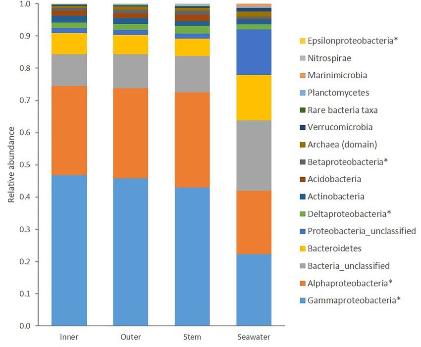
Ho et al.                                                                                                               Microbial Diversity of Cliona patera

to over a meter in height and diameter. C. patera was common                time of processing. Samples for culture-dependent methods were
and abundant in Singapore waters in the early 19th century.                 transported to the laboratory for immediate processing within
However, due to its impressive size, it was much sought after               2.5 h of sample collection.
by natural history museums and private collectors in the past.                 In addition to sponge samples, seawater (two and three
Since the early 20th century, this sponge has not been recorded             samples in January 2019 and February 2019, respectively) was
from this region, and was thought to be extinct (Lim et al.,                collected in 1 L-polypropylene Nalgene bottles (previously
                                                                                                                               R

2012). In early 2011, during a routine survey dive, this sponge             sterilized by autoclaving at 121◦ C for 15 min) and placed on
was fortuitously rediscovered off Singapore’s southern islands              ice in a cooler for transportation to the Singapore Centre for
(Lim et al., 2012). Another population of C. patera was recently            Environmental Life Sciences Engineering (SCELSE), Nanyang
discovered near the islands of Koh Rong Sanloem and Koh                     Technological University (NTU). In the laboratory, each seawater
Koun, Cambodia in 2018. The reappearance of C. patera in                    sample was filtered through a 0.2 µM cellulose acetate filter
Singapore waters presents a unique opportunity to study the                 paper (Sartorius Stedim), which was stored at −80◦ C until
sponge in its natural habitat, including the diverse associated             time of processing.
microbes. This study aimed to evaluate the diversity of the
C. patera associated prokaryotic community found on the top                 Microbial DNA Extraction and 16S rRNA
oval concave disk and stalk of the gamma stage (massive, free-              Analysis
living) of the sponge using 16S rRNA amplicon sequencing (Lim               DNA was extracted from each sponge sample and seawater
et al., 2012; Zhang et al., 2016). In addition, sponge-associated           filter using the DNeasy PowerSoil Kit (QIAGEN) according
                                                                                                          R

bacteria obtained from two sponges were cultured and their                  to the manufacturer’s instructions. Concentration and purity
extracts assessed for quorum sensing inhibitory (QSI) activity              of the DNA were determined using a NanodropTM 2000
based on a Pseudomonas aeruginosa PAO1 lasB-gfp biosensor                   spectrophotometer (Thermo Fisher Scientific) and QubitTM 2.0
strain. The disruption of bacterial quorum sensing systems in               fluorometer (Thermo Fisher Scientific). To study the microbial
pathogenic bacteria using small molecules represents a novel                communities in the sponge samples, PCR was used to amplify
chemotherapy against infectious diseases (Saurav et al., 2016).             the V4 region of the 16S rRNA gene using the 515F and 806R
Furthermore, mass spectroscopic-based metabolomics method,                  primers (Caporaso et al., 2011). PCR reactions required 12.5 µL
using the Global Natural Product Social Molecular Networking                of KAPA HiFi HotStart ReadyMix 2X, 5 µL of primers (1 µM)
platform, was used to annotate the metabolomes of selected QSI              and 2.5 µL of 5 ng/µL of DNA template. The mixture was
active marine bacterial strains (Nguyen et al., 2013).                      adjusted to a final volume of 25 µL using sterile water. The
                                                                            reaction was cycled in a thermocycler for an initial denaturation
                                                                            at 95◦ C for 3 min, denaturation at 98◦ C, annealing at 52◦ C
MATERIALS AND METHODS                                                       and extension at 72◦ C (30 s, 30 cycles) and a final extension at
                                                                            72◦ C for 5 min. Gel extraction was employed to excise the band
Collection of Cliona patera Samples                                         of interest from the pooled PCR products using the Invitrogen
Sponge samples from Cliona patera (Class Demospongiae; Family               PureLinkTM Quick Gel Extraction Kit (Thermo Fisher Scientific)
Clionaidae; gamma stage) were collected at two time points,                 according to manufacturer’s instructions. The quality of the
approximately 1.5 months apart on January 14, 2019 and                      purified PCR products was checked using the Agilent 2200 Tape
February 27, 2019, in the waters surrounding the southern coast             station before they were sent to sequencing facility at SCELSE,
of Singapore along the Singapore Strait at depth of about 12 m              NTU for amplicon sequencing on the Illumina MiSeq Platform.
(National Parks Board Permit Number: NP/RP19-036) (Lim                          Raw sequencing data were processed in Mothur v 1.31.1
et al., 2012). In the first collection, a total of 12 sponge samples,       (Kozich et al., 2013) using the MiSeq SOP pipeline2 . Sequence
each measuring approximately 3 cm1 , from two C. patera                     reads of low quality were removed and the remaining sequences
colonies, designated as NP1 and NP6, were obtained. A second                trimmed and aligned (maxambig = 0, minlength = 309,
collection of 36 sponge samples from six colonies of C. patera,             maxlength = 379, maxhomop = 8). Pre-clustering was done on
including NP1 to NP6, was carried out. The height of the sponges            sequences using the Deblur method (Amir et al., 2017) and
ranged from 47 to 61 cm (Supplementary Figure 1). From each                 chimeras were removed with VSEARCH. Taxonomy was assigned
sponge, a total of six samples were excised: two samples from the           with the SILVA reference database version 132 to identify the sub-
inner surface of the sponge cup (Inner), two samples from outer             Operational Taxonomic Units (OTUs) following the application
surface of the sponge cup (Outer), and two samples from                     of Deblur. Non-bacterial and non-archaeal (i.e., mitochondria,
stem of sponge (Stem). Upon collection, sponge samples were                 chloroplast, eukaryotes, and unclassified) reads were filtered out.
placed individually in NascoTM Whirl-Pak bags containing
                                                  R
                                                                            All sequencing data were uploaded to the NCBI Sequence Read
seawater. Samples were divided equally for microbial analysis               Archive under BioProject accession number PRJNA623254.
using culture-dependent and culture-independent methods (see
below sections). Samples reserved for microbial diversity study,            Statistical Analysis
using culture-independent method, were drained of seawater and              For each sample group, mean relative abundance of the
immediately frozen in a dry shipper, then stored at −80◦ C until            representative taxa was calculated. Using Mothur, the data
1                                                                           2
    https://jgi.doe.gov/data-and-tools/bbtools/                                 https://www.mothur.org/wiki/MiSeq_SOP

Frontiers in Microbiology | www.frontiersin.org                         3                                           June 2021 | Volume 12 | Article 631445
Ho et al.                                                                                                        Microbial Diversity of Cliona patera

were rarefied to 55,140 sequence reads per sample (Weiss                  salt and potassium dichromate and incubated at 25◦ C with
et al., 2017). Alpha diversity was assessed in terms of Sobs              150 rpm shaking for 14 days and stored at 5◦ C while awaiting
(observed richness; number of OTUs), Chao (estimated richness),           further processing.
Shannon evenness and Inverse Simpson Diversity. To test
for difference in means amongst sample groups, Analysis of                Small-Scale Fermentation of Marine
variance (ANOVA) was conducted in R v 3.6.1 with post hoc                 Bacterial Strains and Preparation of
Tukey tests used to identify significantly different groups. Beta
diversity, or prokaryotic community structure, among groups
                                                                          Organic Extracts
was examined in PRIMER v 7 using permutational multivariate               Ten milliliters of each isolated marine bacterial strain, maintained
analysis of variance (PERMANOVA) with Bray-Curtis similarity              in marine broth, was transferred into a 250 mL-Erlenmeyer flask
matrices of square root transformed data and visualized in non-           containing 100 mL of marine broth supplemented with nalidixic
metric multidimensional scaling (nMDS) plots. Additionally,               acid sodium salt and potassium dichromate and incubated at
permutational multivariate analysis of dispersion (PERMDISP)              25◦ C with 150 rpm shaking for 14 days. After the incubation
was used to test for homogeneity of dispersion among samples              period, an equal volume of ethyl acetate (EtOAc) was added
within groups. The specific OTUs contributing to the differences          to the liquid cultures and the mixture were transferred into
observed in the PERMANOVA analysis were determined by                     separatory funnel to collect the EtOAc phase. Solvent partitioning
running multivariate generalized linear models (GLMs) with a              using EtOAc was repeated twice for each liquid culture. EtOAc
negative binomial distribution. The GLMs were performed in R              was removed in vacuo using a rotary evaporator, and the dried
v 3.6.1 using the mvabund package (Wang et al., 2012) with data           extracts were reconstituted with equal parts absolute ethanol and
subsampled to the top 500 OTUs to focus the analysis on the most          isopropanol before transferring to scintillation vials, stored at
abundant OTUs present in the sponge samples. OTUs of interest             −20◦ C, for subsequent biological evaluation in the QSI assay and
were screened using the nucleotide-nucleotide BLAST search to             metabolomics analysis.
determine closely related sequences (Altschul et al., 1990).
                                                                          Pseudomonas aeruginosa Quorum
                                                                          Sensing Inhibitory Assay
Cultivable Marine Bacterial Isolation                                     The bacterial quorum sensing inhibitory (QSI) assay was
From Sponge Samples                                                       conducted based on the biosensor strain, Pseudomonas
Sponge samples from the inner cup, outer cup and stem                     aeruginosa PAO1 lasB-gfp(ASV) (Hentzer et al., 2002). The
were washed with sterile artificial sterile seawater to remove            monitor strain has their promoter fused to an unstable GFP
loosely attached microorganisms. They were then homogenized               (green fluorescent protein) that has a C-terminal oligopeptide
thoroughly with a mortar and pestle with 10 mL of sterile                 extension containing the amino acids ASV [gfp(ASV)]. This
artificial seawater for the purpose of bacterial isolation. Fifteen       causes the GFP to be more susceptible to degradation by
different marine media, including A1 to A5, marine agar, starch           housekeeping proteases, resulting in a short half-life. As such,
casein agar, actinomycete isolation agar, YEME, YPG, A1 + C,              unstable gfp(ASV) allows for monitoring of temporal QS-
YMP, YMP + C, TCG, and SIM were used for the isolation                    regulated gene expression (Andersen et al., 1998). The GFP,
of cultivable marine bacteria from the first sponge collection            composed of 238 amino acid residues (26.9 kDa), exhibits
(Supplementary Table 1). All media, containing DifcoTM                    bright green fluorescence when exposed to light in the blue to
Marine agar, were sterilized by autoclaving before supplemented           ultraviolet range. The lasB gene is responsible for the production
with concentration of potassium dichromate/cycloheximide and              of the virulence factor, elastase, which can cause degradation
nalidixic acid sodium salt solution to minimize fungal growth and         of elastin, collagen as well as necrosis of macrophages in host
fast growing Gram-negative bacteria, respectively. The antibiotic         during infections (Lee and Zhang, 2015). Marine bacterial-
solution was filtered (0.2 µm pore size) before being added to            derived extracts were tested in triplicates and prepared in
the autoclaved media in petri dishes as isolation plates. The             a 96-well microtiter plates. Stock solutions of extracts were
homogenized supernatants of sponge samples were heat-shocked              prepared in 100% DMSO at concentration of 1 mg/mL. Each
at 65◦ C for 10 min to reduce the growth of fast growing                  bacterial stock solution was then mixed with ABTGC medium
Gram negative bacteria. Twenty microliters of the heat-shocked            (Supplementary Table 1) and serial diluted to give a final
supernatant was then inoculated in duplicates onto isolation agar         concentration of 200 µg/mL in each well of the microtiter plate.
plates and incubated at 25◦ C for 6–8 weeks.                              A culture of the PAO1 lasB-gfp strain was grown overnight
   All isolation agar plates were checked every day and the               in Luria-Bertani medium at 37◦ C with 200 rpm shaking. The
bacterial isolates were picked or selected based on the criteria          culture was subsequently diluted with ABTGC medium to a final
of colonies having unique or interesting morphology, including            optical density at 600 nm (OD600 ) of 0.02. An equal amount of
matte textures, irregular colony shapes, and bright colors. They          the bacterial suspension was added to the wells to reach final
were then re-streaked onto MBA medium (DifcoTM Marine                     inhibitor concentration of 100 µg/mL. All bacterial extracts
Agar 2216) and incubated at 25◦ C for approximately 3–7 days.             were initially screened at a concentration of 100 µg/mL before
The isolated marine bacterial strains were then inoculated into           proceeding with dose-dependent assay (ranging from 100 to
50 mL falcon tubes containing 20 mL of marine broth (DifcoTM              1.563 µg/mL using ABTGC medium for dilution) for selected
Marine Broth 2216), supplemented with nalidixic acid sodium               extracts that showed significant QSI activity. DMSO control

Frontiers in Microbiology | www.frontiersin.org                       4                                      June 2021 | Volume 12 | Article 631445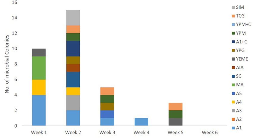
Ho et al.                                                                                                      Microbial Diversity of Cliona patera

(0.1% final concentration) and blank control were added into                The network spectra and the library reference spectra were
the microtiter plates in triplicates. The microtiter plates were         required to have a minimum cosine score threshold of 0.7 and
then incubated at 37◦ C for 17 h in a Tecan Infinite 200 Pro plate       a minimum of six matched peaks in order to be considered
reader (Tecan Group Ltd., Maännedorf, Switzerland) to measure            for spectral library annotation and a minimum of six matched
fluorescence and cell density. Bacterial growth was measured at          fragment ions. The precursor ion mass tolerance was set at 2.0 Da
an optical density at 450 nm while fluorescence was measured             and a MS/MS fragment ion tolerance of 0.5 Da to create a
as excitation and emission wavelength at 485 nm and 535 nm,              consensus spectra. Further edges between two nodes were kept
respectively. Expression of GFP was normalized by dividing its           in the network if each of the nodes appeared in each other’s
value with the growth measured at the respective time points             top 10 most similar nodes. The input data were searched against
over a period of 10–16 h. Bacterial extracts were further tested         annotated reference spectra of the MS/MS library within GNPS.
using PAO1-gfp strain to ensure that they were targeting the             For the visualization of compounds from the dereplication hits,
QS genes and not the GFP (Yang et al., 2007). Bacterial extracts         the results were exported and viewed directly with the pie-
that did not interfere with fluorescent output signals in the            chart creating tool (nodeCharts plugin for Cytoscape) within
PAO1-gfp strain were then subjected to dose-dependent QS                 Cytoscape 3.7.1.
inhibition study.
                                                                         Genome Sequencing and de novo
Mass Spectrometry-Based Molecular                                        Assembly Identification of Selected QSI
Network of QSI Active Marine Bacterial                                   Active Marine Bacterial Strains
Extracts                                                                 Selected marine bacterial strains, isolated from week 3 onward,
Quorum sensing inhibitory active marine bacterial extracts were          from the first collection of sponge samples that showed significant
filtered over C18 SPE cartridges by application of 1.0 mL sample         QSI activity were subjected to de novo assembly identification
(1 mg/mL) and elution with 3 mL acetonitrile (CH3 CN). Solvent           based on bacterial genomes by 1st Base, Singapore. Bacterial
was removed in vacuo using a rotary evaporator before being              isolates were inoculated into a 250 mL Erlenmeyer flask
redissolved in 1 mL CH3 CN, then vortex mixing over 5 min                containing 100 mL of liquid media (DifcoTM Marine Broth 2216
and transferred into separate Eppendorf tubes. Tubes were then           supplemented with 0.015 g/L nalidixic acid sodium salt, 0.05 g/L
centrifuged at 10,000 rpm at 4◦ C over 10 min and the supernatant        potassium dichromate) and incubated at 25◦ C with 150 rpm
was aliquoted and diluted with CH3 CN to 10,000 × dilution.              shaking for 14 days. The bacterial cells were pelleted down using
One-and-a-half microliters of each diluted sample was subjected          centrifuge and sent to 1st Base, Singapore for genomic workup.
to LC-HRMS/MS [Q Exactive Plus Hybrid Quadrupole-Orbitrap                Briefly, the bacterial genomic DNA was extracted from each
Mass Spectrometer (Thermo Fisher Scientific) equipped with a             cell pellet using the Quick-DNATM fungal/bacterial microprep
heated electrospray ionization (H-ESI) probe] performed with             kit (Zymo Research) protocol. The quality and quantity of the
a Thermo Scientific Hypersil GOLD (C18 50 mm × 2.1 mm,                   genomic DNA were measured using PicoGreen, Nanodrop and
1.9 µm) column and maintained at a column temperature of                 gel electrophoresis. The samples passed the QC measurement
40◦ C and sample temperature of 4◦ C using a gradient elution            and hence proceed for library preparation using QIAseq FX
program of 0.1% aq. HCOOH (mobile phase A) and 98%                       DNA library kit from QIAGEN. The quality of the libraries
CH3 CN in 0.1% aq. HCOOH (mobile phase B) at a flow rate of              was measured using TapeStation 4200, PicoGreen and qPCR.
0.5 mL/min. The gradient program began at 10% and increased              The libraries were then pooled according to the protocol
to 50% of mobile phase B within 2 min and was held at 50%                recommended by Illumina and proceeded straight to sequencing
mobile phase B for 2 min. It was then increased to 100% of               using the MiSeq platform at 2 × 251 PE format with QC
mobile phase B within 6 min and was held at 100% B for 0.5 min           reads assembled de novo using SPAdes 3.11.1 (Bankevich et al.,
before reconditioning back to the starting composition in 0.5 min        2012). Paired end Illumina sequences were first removed of
and was held at the starting composition of 10% B for 3 min              sequence adaptors and reads with low quality scores using bbduk
bringing the total run time to 14 min. All the mass spectra              of the BBTools Packages3 . The resulting contigs of >1000 bp
were collected in the positive ion and data-dependent acquisition        were subjected to MEGABLAST against the NCBI Nucleotide
mode, where the first five most intense ions of each full-scan           database. All genomes were subsequently annotated using the
mass spectrum (mass range: m/z 100–1500) were subjected to               RAST pipeline (Overbeek et al., 2014).
tandem mass spectrometry (MS/MS) analysis. MS scan time
of 0.25 s over 14 min, MS/MS scan time of 0.06 s, and a 3-
step normalized collision energy of 25, 35, and 55. The MS/MS            RESULTS
data files were converted from .RAW to .mzXML files using
MSConvert software and uploaded to the Global Natural Product            Marine Microbes Associated With Cliona
Social Molecular Networking (GNPS) server3 and the molecular             patera: Culture-Independent Method
networking performed using the GNPS data analysis workflow               A total of 16,381 unique OTUs were identified from the four
employing a special spectral clustering algorithm.                       sample groups, Stem, Inner cup, Outer cup, and Seawater. The
                                                                         Stem group had the highest mean number of unique OTUs
3
    http://gnps.ucsd.edu                                                 (3268 ± 203.2 standard deviation), including 25 bacterial phyla,

Frontiers in Microbiology | www.frontiersin.org                      5                                     June 2021 | Volume 12 | Article 631445
Ho et al. Microbial Diversity of Cliona patera
66 bacterial classes, five archaeal phyla, and 11 archaeal classes. were only present in the sponge group (Supplementary Table 3).
This was followed by the Inner cup with 3000 (±431.9) unique The genus Ruegeria was not found in the seawater group but
OTUs (25 bacterial phyla, 62 bacterial classes, six archaeal phyla, was present in all sponge samples. Within the sponge samples,
and 12 archaeal classes) and Outer cup with 2901 (±349.6) the GLM identified 21.2% of the 500 most abundant OTUs as
unique OTUs (24 bacterial phyla, 59 bacterial classes, four contributing significantly to the differences detected between the
archaeal phyla, and 12 archaeal classes). Taken together, C. patera cup and stem (Table 3 and Supplementary Table 4).
had an average of 5522 (±613.1) unique OTUs comprising of
26 bacterial phyla, 74 bacterial classes, six archaeal phyla, and Isolation of Marine Microbes Associated
12 archaeal classes. The seawater group had the lowest number With Cliona patera: Culture-Dependent
of OTUs with 1998 (±233.8), consisting of 26 bacterial phyla,
66 bacterial classes, six archaeal phyla, and 12 archaeal classes
Method
(Supplementary Table 2). Culture-dependent methods were used to isolate bacteria
Within C. patera, Proteobacteria, particularly the classes associated with C. patera from different parts of the sponge
Gammaproteobacteria and Alphaproteobacteria, dominated the colonies in order to assess their ability to interfere with bacterial
sponge microbiome making up more than 70% of the quorum sensing system in the Pseudomonas aeruginosa PAO1
prokaryotic community (Figure 1). The seawater group was lasB-gfp biosensor. The first collection of C. patera samples,
also dominated by more than 40% Proteobacteria of the namely from NP1 and NP6, yielded a total of 110 marine
classes Gammaproteobacteria and Alphaproteobacteria, but also bacterial strains isolated over a 6-week incubation period, using
contained a larger proportion of Bacteroidetes compared 15 different marine culture media (Figure 3). Sixty-four and
to the sponge samples. Overall, the sponge microbiomes 46 bacterial strains were isolated from the two sponges, NP1
were significantly different from the seawater (PERMANOVA: and NP6, respectively. In addition, the majority of the marine
Pseudo-F = 35.249, P = 0.001; Supplementary Figure 2). bacterial colonies were isolated from sponge samples obtained
There were significant differences among some of the sponge from the cup region as compared to the stem, with 85 and
and seawater groups for the alpha diversity metrics: Richness 25 bacterial strains, respectively. In addition, colonies that were
(Sobs ; F = 5.4, p = 0.00527), Chao (F = 1.911, p = 0.154), not considered unique were selected to prevent bias. Of the
Inverse Simpson (F = 4.45, p = 0.0123), and Shannon (F = 18.51, 15 different marine culture media used for the isolation of
p < 0.0001). Tukey’s HSD tests revealed differences among all bacterial strains, A1, TCG, YPG, MA, and YEME provided the
groups – the seawater and sponge (Inner, Outer, and Stem) group, highest yield of isolated colonies, with 16, 13, 12, 11, and 10
as well as within the sponge when comparing the Inner and strains, respectively (Supplementary Figure 3). The number
Outer cup to the Stem (Table 1). The sponge groups exhibited of isolated bacterial strains peaked at week 2 from 13 marine
higher richness (Sobs ) and evenness compared to the seawater. media (Figure 3). However, the weekly number of new bacterial
Within C. patera, the diversity and evenness of the prokaryotic strains appearing on marine media started to tail off from week
community was significantly lower in the cup as compared to 3 onward. For instance, on week 5 and 6, of the 15 different
the Stem. The Inner cup and Outer cup had similarly even media, only seven (i.e., A3, MA, SC, AIA, YEME, YPM, and
prokaryotic communities. For both richness metrics, Sobs and TCG) and four (e.g., A2, A4, AIA, and YPM + C) marine
Chao, the Inner cup group had the highest standard deviation in media types, respectively, showed the appearance of new bacterial
contrast to the other groups which indicated greater variance in colonies (Figure 3).
the number of OTUs per sample.
PERMANOVA analysis did not reveal any significant Anti-quorum Sensing Activity of Marine
differences between the microbial communities of the six sponges Bacterial Extracts
sampled in this study when each sponge was considered as All 110 marine bacterial strains isolated from samples derived
a whole (Pseudo-F = 1.1118, P = 0.192), or between sponges from sponge colonies NP1 and NP6 underwent small-scale
collected in January and February (Pseudo-F = 0.8920; P = 0.608). fermentation in order to obtain sufficient organic extracts for
However, within C. patera, PERMANOVA analysis revealed evaluation of their QSI activity using the biomonitor strain,
significant differences among the regions of the sponge (Pseudo- Pseudomonas aeruginosa PAO1 lasB-gfp. The organic extracts
F = 1.6642, P = 0.009), specifically between the Inner cup from cultures of 34 (30.9%) marine bacterial strains exhibited
and Stem, as well as the Outer cup and Stem (Table 2). moderate (24 bacterial strains with 60–80% inhibition) to
When visualized in the nMDS plot the Inner and Outer cup significant (10 bacterial strains with >80% inhibition) florescence
samples appear grouped more closely than the Stem samples reduction on the biosensor PAO1 lasB-gfp strain when tested at
(Figure 2), but there was no differences in dispersion among 100 µg/mL (Figure 4). A majority of the QSI active extracts,
groups (PERMDISP: F = 0.4539, P = 0.719). about 44% (15 bacterial extracts), were prepared from marine
Generalized linear models supported the PERMANOVA bacterial strains picked solely from week 2 of the incubation
analysis, confirming the distinction between the sponge and period (Figure 4). In addition, the marine media, including
seawater prokaryotic communities, as well as between the sponge A1, A4, MA, TCG, and YPM, gave the highest number of QSI
cup and stem prokaryotic communities. Between the sponge active marine bacterial strains. In spite of the lower number of
and seawater groups, 98.8% of the 500 OTUs tested contributed marine bacterial strains isolated from the stem region of C. patera,
significantly (P < 0.01) to the dissimilarity, of which 52 OTUs the proportion of QSI active strains, showing more than 60%
Frontiers in Microbiology | www.frontiersin.org 6 June 2021 | Volume 12 | Article 631445Ho et al. Microbial Diversity of Cliona patera
FIGURE 1 | Taxonomic composition of prokaryotic communities in the Inner cup, Outer cup, and Stem of Cliona patera and ambient seawater. Asterisks (∗ ) denote
classes within the phylum Proteobacteria, and all representatives of the Domain Archaea have been grouped.
TABLE 1 | Alpha diversity metrics for prokaryotic communities in Cliona patera and ambient seawater.
Group Richness (Sobs ) Richness (Chao) Evenness (Shannon) Diversity (inv. Simpson)
Inner 3897.25 ± 1177.21a 7999.51 ± 3473.79 0.65 ± 0.02ac 26.43 ± 4.60a
Outer 3560.13 ± 586.83b 7318.48 ± 1986.14 0.65 ± 0.02bc 27.61 ± 5.74b
Stem 3964.63 ± 383.50c 7676.98 ± 1370.09 0.68 ± 0.02c 39.96 ± 13.58ab
Seawater 2410.80 ± 343.93abc 5096.26 ± 692.7 0.60 ± 0.01abc 32.29 ± 2.26
Different letters represent significant pairwise comparisons, where groups with the same letters indicated significant difference between them.
florescence inhibition, was comparable to the overall cup region TABLE 2 | Pairwise comparisons of prokaryotic community structure
(both inner and outer cup), with 36% and 29.4%, respectively. (PERMANOVA) in Cliona patera Inner cup, Outer cup, and Stem.
With the emphasis on slow growing marine bacterial strains,
18 QSI active organic extracts of bacterial strains, isolated from C. patera PERMANOVA
week 3 onward, were further subjected to QSI assay in a dose
t P
dependent manner ranging from 1.563 to 100 µg/mL. Twelve
crude extracts showed some degree of anti-quorum sensing Inner cup, Outer cup 0.927 0.695
activity in a dose-dependent manner (Supplementary Figure 4). Inner cup, Stem 1.551 0.005
Outer cup, Stem 1.323 0.014
Identification and MS-Based Molecular Bold represent significant pairwise comparisons.
Networking of Selected QSI Active
Marine Bacterial Colonies 55%. The criterion used for the identification was based on
De novo assembly of bacterial genome for bacterial identification genome annotation using RAST. It was revealed that these QSI
was performed on selected marine bacterial strains with extracts active bacterial strains belonged to either class Bacilli (phylum
that showed QSI florescence inhibitory activity of more than Firmicutes) or class Alphaproteobacteria (phylum Proteobacteria)
Frontiers in Microbiology | www.frontiersin.org 7 June 2021 | Volume 12 | Article 631445Ho et al. Microbial Diversity of Cliona patera
FIGURE 2 | nMDS plot of prokaryotic community structure in the Inner cup, Outer cup, and Stem of Cliona patera.
(Table 4). The bacterial strains were found to be affiliated with having different degrees of QSI activity (Supplementary
Labrenzia alba (strains #91 and #93), Bacillus stratosphericus Table 5). From the organic extracts of 11 QSI active bacterial
(strain #41), Ruegeria arenilitoris (strain #48), and Staphylococcus strains, a total of 973 parent ions were detected (Figure 6).
haemolyticus (strain #53) (Table 4). Additional new structural classes were also observed, including
In order to gain insight into the natural products chemistry pyrenocine B, 2,6-dihydroxyanthraquinone and their derivatives
of these QSI active bacterial extracts, the mass spectrometry- (Figure 6). The molecular networkings comparison between
based metabolomic approach based on molecular networking two classes of bacteria, belonging to either class Bacilli or
platform (GNPS) was performed. A total of 591 parent ions class Alphaproteobacteria, revealed the presence of a number of
(shown as molecular networking clusters in Figure 5) were molecular families that are unique to each bacterial class. For
detected in QSI active extracts derived from five marine bacterial instance, two molecular families, designated in boxes A and B in
strains, including #41, #48, #53, #91, and #93 (Figure 5). Of the Figure 6, are only detected in bacterial strains belonging to class
591 parent ions, compound dereplication based on the GNPS Bacilli. These two molecular families did not yield any hits from
mass spectral library was performed and revealed a total of the GNPS library and are potential new molecules. Furthermore,
120 compound hits. A number of noteworthy natural products, the diversity of surfactin-related molecules, including potential
namely diketopiperazines (e.g., compound 1), harmaline (2), new surfactin derivatives, appeared to be detected predominantly
surfactin D (3), anisomycin (4), and dehydroxynocardamine (5), in bacterial strains belonging to class Bacilli (box C in Figure 6).
were detected in the molecular networking clusters (Figure 5). Taken together, the molecular networking clusters of QSI active
In addition, a majority of identical/similar ionizable molecules bacterial extracts revealed the present of diverse classes of
were observed to be present in the extracts of all five marine natural products.
bacterial strains. For instance, molecular families related to
dehydroxynocardamine and anisomycin were found in all
bacterial extracts. One molecular networking cluster, containing
16 nodes with m/z ranging from 394.505 to 819.508, was unique
DISCUSSION
to strains #53 (class Bacilli) and #48 (class Alphaproteobacteria)
(refer to box A in Figure 5). Moreover, this cluster showed
Microbial Communities Associated With
no library hits in the GNPS database. As both marine bacterial Cliona patera: Culture-Independent
strains #91 and #93 are related to Labrenzia alba, their molecular Method
signatures were almost identical with only a few ionizable The Neptune’s Cup sponge, Cliona patera, was characterized
molecules unique to either strains (Supplementary Figure 5). by a complex and diverse prokaryotic microbiome with more
The MS-based molecular networking analyses were further than double the number of distinct OTUs detected in the
expanded to include extracts from 11 marine bacterial strains surrounding seawater. The prokaryotic community structure
Frontiers in Microbiology | www.frontiersin.org 8 June 2021 | Volume 12 | Article 631445Ho et al. Microbial Diversity of Cliona patera
TABLE 3 | Highly abundant (>0.1 mean relative abundance of total community ± SD) OTUs identified as contributing significantly (P < 0.01) to the differences between
the cup and stem of Cliona patera.
OTU Phylum Lowest taxonomic classification Mean relative abundance
Cup Stem
4 Proteobacteria Order Chromatiales 1.690 ± 0.226 0.632 ± 0.107
6 Proteobacteria Class Gammaproteobacteria 1.715 ± 0.186 0.575 ± 0.266
8 Proteobacteria Order Rhodobacteraceae 1.393 ± 0.188 0.453 ± 0.141
15 Proteobacteria Class Gammaproteobacteria 1.165 ± 0.339 0.341 ± 0.144
20 Proteobacteria Order Rhodospirillales 0.568 ± 0.091 0.422 ± 0.132
27 Proteobacteria Family Erythrobacteraceae 0.706 ± 0.195 0.145 ± 0.067
28 Proteobacteria Class Gammaproteobacteria 0.683 ± 0.096 0.162 ± 0.081
30 Proteobacteria Class Betaproteobacteria 0.401 ± 0.102 0.291 ± 0.045
33 Proteobacteria Order Rhodospirillales 0.337 ± 0.068 0.240 ± 0.071
34 Proteobacteria Class Gammaproteobacteria 0.304 ± 0.077 0.237 ± 0.067
36 Proteobacteria Class Gammaproteobacteria 0.397 ± 0.068 0.141 ± 0.077
37 Proteobacteria Family Rhodospirillaceae 0.288 ± 0.051 0.247 ± 0.116
43 Proteobacteria Class Gammaproteobacteria 0.328 ± 0.048 0.112 ± 0.029
57 Proteobacteria Class Alphaproteobacteria 0.210 ± 0.048 0.153 ± 0.038
61 Bacteroidetes Family Flavobacteriaceae 0.252 ± 0.054 0.093 ± 0.022
67 Actinobacteria Genus Ilumatobacter 0.181 ± 0.047 0.038 ± 0.020
72 Proteobacteria Genus Porphyrobacter 0.244 ± 0.060 0.053 ± 0.013
74 Proteobacteria Class Gammaproteobacteria 0.179 ± 0.040 0.126 ± 0.024
89 Proteobacteria Order Rhodospirillales 0.125 ± 0.020 0.103 ± 0.032
91 Bacteroidetes Family Flavobacteriaceae 0.179 ± 0.071 0.028 ± 0.027
93 Proteobacteria Class Gammaproteobacteria 0.109 ± 0.025 0.090 ± 0.034
94 Proteobacteria Order Myxococcales 0.082 ± 0.033 0.104 ± 0.068
95 Proteobacteria Class Gammaproteobacteria 0.143 ± 0.025 0.052 ± 0.013
98 Actinobacteria Class Actinobacteria 0.140 ± 0.036 0.045 ± 0.017
100 Proteobacteria Class Alphaproteobacteria 0.104 ± 0.022 0.086 ± 0.027
102 Nitrospirae Genus Nitrospira 0.037 ± 0.027 0.149 ± 0.119
103 Actinobacteria Class Actinobacteria 0.133 ± 0.033 0.043 ± 0.008
106 Actinobacteria Order Acidimicrobiales 0.132 ± 0.031 0.035 ± 0.013
107 Proteobacteria Genus Haliea 0.107 ± 0.017 0.074 ± 0.014
110 Bacteroidetes Genus Robiginitalea 0.140 ± 0.059 0.024 ± 0.010
113 Proteobacteria Genus Thiohalomonas 0.117 ± 0.038 0.025 ± 0.019
135 Bacteroidetes Family Flavobacteriaceae 0.106 ± 0.021 0.030 ± 0.006
139 Proteobacteria Family Rhodospirillaceae 0.112 ± 0.050 0.022 ± 0.010
147 Unclassified Bacteria 0.107 ± 0.037 0.018 ± 0.009
of the six C. patera sponges examined were all similar and Ramsby et al., 2018; Easson et al., 2020; Meenatchi et al., 2020;
distinct from the seawater, suggesting a host-specific microbiome, Mote et al., 2020; Sacristán-Soriano et al., 2020). This is,
consistent with previous sponge microbiome research (Schmitt however, the first study of a Clionid sponge to identify distinct
et al., 2007; Sharp et al., 2007; Turon et al., 2018). Moreover, microbiomes separated into different sections of an individual
there was no difference in the prokaryotic community structures sponge. The difference in prokaryotic community structure
between the two sponges sampled in January and 1 month later found in the cup and stem of the sponges is likely related to
in February. However, additional sampling would be required the differences in structural composition of the tough fibrous
to assess long-term temporal stability in the microbiomes of stem compared to the cup, and the physiological demands on
C. patera. the cup tissue, as it is regularly regrown from turtle predation.
Interestingly, C. patera supported distinct symbiont Additional research should be conducted to examine the role of
communities within the cup and stem portions of the sponge. the microbiome in sponge function for both the cup and stem of
While this is the first study examining the microbiome of C. patera.
C. patera, the microbiomes of several Cliona species have been Of the OTUs contributing to the differences in microbiomes
studied, namely Cliona celata, Cliona delitrix, Cliona lobata, of the sponge cups and stems, the majority of OTUs (78.3%)
Cliona orientalis, Cliona thomasi, Cliona tumula, Cliona varians, were present in higher relative abundance in the cup of C. patera.
and Cliona viridis (Jeong et al., 2015; Thomas et al., 2016; OTU 6 was the most abundant, significant OTU in the cup
Frontiers in Microbiology | www.frontiersin.org 9 June 2021 | Volume 12 | Article 631445Ho et al. Microbial Diversity of Cliona patera FIGURE 3 | Number of marine bacterial colonies isolated from samples of Cliona patera, NP1 and NP6, over a 6-week incubation period in various marine media. FIGURE 4 | Number of QSI active marine bacterial colonies, with more than 60% florescence inhibition, isolated from samples of Cliona patera, NP1 and NP6, over a 6-week incubation period in various marine media. TABLE 4 | Overview of best BLAST hits of 16S rRNA gene from genomes of selected colonies of interest showing QSI activity. Bacterial Description Phylum/Class % Identical % Pairwise % GC NCBI % QS Strain sites identity Accession # Inhibition #41 Bacillus stratosphericus Firmicutes/Bacilli 99.9% 99.9% 55.2% MZ328876 58.9% #48 Ruegeria arenilitoris Proteobacteria/Alphaproteobacteria 99.1% 99.1% 55.5% MZ328874 68.2% #53 Staphylococcus haemolyticus Firmicutes/ Bacilli 99.4% 99.4% 51.1% MZ328872 84.8% #91 Labrenzia alba Proteobacteria/Alphaproteobacteria 98.2% 98.2% 55.9% MZ328867 78.8% #93 Labrenzia alba Proteobacteria/Alphaproteobacteria 98.2% 98.2% 55.9% MZ328866 67.5% Frontiers in Microbiology | www.frontiersin.org 10 June 2021 | Volume 12 | Article 631445
Ho et al.                                                                                                                                Microbial Diversity of Cliona patera

  FIGURE 5 | Molecular network of 591 parent ions detected in extracts of five QSI active marine bacterial strains. Blue: strain #53; Pink: strain #91; Yellow: strain #48;
  Green: strain #93; Purple: strain #41; Gray: MeOH blank. Within each box, the nodes represent the number of ions detected in the bacterial extracts while the node
  size represents the relative abundance of the different parent ions. Within each node, the relative abundance of a particular ion detected in the different bacterial
  strains is depicted by the sizes of the colored wedge. The edge thickness reflects the similarity between each parent ions with thicker edge showing higher similarity.
  Nodes with associated numbers 1 to 5 correspond to the detected compounds and their chemical structures are shown in the top row of boxes.

of C. patera, with a relative abundance of 1.715 ± 0.186                                  specifically, Gammaproteobacteria and Alphaproteobacteria
in the cup compared to 0.575 ± 0.266 in the stem. OTU                                     (Webster and Thomas, 2016; Pita et al., 2018; Sacristán-Soriano
6 was a Gammaproteobacteria that matched 99% identity in                                  et al., 2020). This high abundance of Proteobacteria support the
BLAST to an uncultured bacterium isolated from the coral                                  placement of C. patera within the LMA sponges, as predicted by
Galaxea fascicularis (Accession KU351051) and 99% identity to                             Moitinho-Silva et al. (2017). The other dominant taxa included
an uncultured bacterium retrieved from the Caribbean coral                                Bacteroidetes (6.05%), Actinobacteria (1.82%), Acidobacteria
Montastraea faveolata (Sunagawa et al., 2009). On the other                               (1.66%), the Archaea domain (0.77%), Planctomycetes (0.22%),
hand, OTU 4, which belonged to the order Chromatiales (also                               Verrucomicrobia (0.2%), Nitrospirae (0.2%), Marinimicrobia
within class Gammaproteobacteria), was the most abundant OTU                              (0.02%), and a large chunk of bacteria remained unclassified
found in the stem of C. patera and matched 100% identity                                  (10.5%). Although present in the sponge, the phylum Chloroflexi
in BLAST to an uncultured bacterium isolated from surface                                 was not a dominant group, unlike reports for a majority
seawaters in the East China Sea (Accession KU173719), as well                             of marine sponges (Thomas et al., 2016). In addition, the
as another uncultured bacterium found in marine sediments in                              commonly described exclusively sponge-specific phylum
the Philippines (Garren et al., 2009).                                                    Poribacteria was not evident in C. patera (Jeong et al., 2015).
   Similar to previous studies on the microbiome of marine                                Only one individual sequence read of Poribacteria was detected
sponges and within the sponge family Clionaidae, the                                      in one of the seawater samples.
microbial communities of C. patera were predominantly                                        Cliona patera did not harbor any Cyanobacteria, although it is
composed of Proteobacteria (relative abundance: 70%);                                     a bacterial group commonly reported in sponges (Mariani et al.,

Frontiers in Microbiology | www.frontiersin.org                                      11                                             June 2021 | Volume 12 | Article 631445
Ho et al.                                                                                                                               Microbial Diversity of Cliona patera

  FIGURE 6 | Molecular network of 973 parent ions detected in 11 marine bacterial strains belonging to class Bacilli (phylum Firmicutes) (Blue) and class
  Alphaproteobacteria (phylum Proteobacteria) (Pink). Nodes due to MeOH blank are in gray. Within each box, the different nodes represent the different ions detected
  in the bacterial extracts while the node sizes represent the relative abundance of the different parent ions. Within each node, the relative abundance of a particular
  ion detected in the different bacterial strains is depicted by the sizes of the colored wedge. The edge thickness reflects the similarity between each parent ions with
  thicker edge showing higher similarity. Nodes with associated numbers 1 to 3 correspond to the detected compounds and their chemical structures are shown in
  the top row of boxes.

2001). This absence is consistent with previous research which                           This genus belongs to the Roseobacter clade, which is an
states that while most phototrophic species are affiliated with                          ecologically relevant and major marine group equipped with a
Cyanobacteria, sponges in the Clionaidae family are a notable                            diverse range of metabolic activities, including the production
exception (Schönberg et al., 2006). Instead, the dinoflagellate,                         of novel bioactive compounds (Martens et al., 2007; Luo
Symbiodinium has been reported to form symbiotic partnerships                            and Moran, 2014). For instance, a Ruegeria strain associated
with 13 Clionaidae species (Schönberg et al., 2005). For instance,                       with the sponge Suberites domuncula has been reported to
vertical transmission of Symbiodinium in Cliona viridis has                              produce exocellular cyclic dipeptides (cell signaling compounds)
been observed (Ramsby et al., 2017) and it appears that the                              aiding in symbiosis with their hosts (Mitova et al., 2004a,b).
metabolism of C. viridis is related to the photosynthetic activities                     Research has also shown that Ruegeria species are able to
of Symbiodinium (Bourne and Webster, 2013). However, the                                 derive sulfur from dimethylsulfoniopropionate, an abundant
presence of eukaryotes was not evaluated in this study.                                  compound available in marine environments (Wirth et al., 2020).
   The genus Ruegeria, phylum Proteobacteria, which was                                  A strain of Ruegeria isolated from the marine sponge Suberites
present in all sponge samples, was not detected in the seawater                          domucula was also shown to exhibit anti-bacterial activity toward
group. Ruegeria (class Alphaproteobacteria) is a genus mainly                            Bacillus subtilis (Mitova et al., 2004a,b). Ruegeria cultured from
comprised of marine bacteria and has been repeatedly isolated                            Irciniidae sponges showed mild antimicrobial activity against
and cultured from diverse sponge species (Karimi et al., 2019).                          Staphylococcus aureus in another study (Esteves et al., 2013).

Frontiers in Microbiology | www.frontiersin.org                                     12                                            June 2021 | Volume 12 | Article 631445
Ho et al.                                                                                                          Microbial Diversity of Cliona patera

Incidentally, from the culture dependent method used in this                 samples. In fact, a recent survey of the literature reported
study, a Ruegeria related bacterial strain #48 was isolated and              that QS-inhibitory marine bacteria belonged to several classes,
its crude organic extracts showed quorum sensing inhibitory                  including Alphaproteobacteria (20.5%), Gammaproteobacteria
activity. In addition to unknown molecular families, various                 (26.6%), Actinobacteria (6.1%), Bacilli (37.7%), and Flavobacteria
classes of known natural products, such as surfactin and                     (8.8%) (Zhao et al., 2019). Furthermore, a range of QS inhibitors
anisomycin related molecules were detected in the molecular                  and antimicrobial agents, including diketopiperazines, aromatic
networking of the bacterial extract (Figure 5).                              polyketides, lipopeptides, furanones and cyclic depsipeptides
                                                                             and peptides, have been reported from marine bacterial strains
                                                                             isolated from a variety of marine samples, such as sediments and
Quorum Sensing Inhibitory Activity of                                        marine macroorganisms (Indraningrat et al., 2016; Ma et al.,
Marine Bacteria-Associated With Cliona                                       2018; Borges and Simões, 2019; Chen et al., 2019; Zhao et al.,
patera: Culture-Dependent Method                                             2019).
Studies have shown that only a fraction of microbial diversity is
accurately represented in culture-dependent methods (Connon
and Giovannoni, 2002). In order to recover and assess the                    MS/MS Molecular Networking of QSI
biomedical potential of marine bacterial strains from sponge                 Active Marine Bacterial Extracts
samples of Cliona patera, a range of marine media, including                 MS-based molecular networking of organic extracts prepared
low nutrient agar, as well as extended incubation period (up                 from selected QSI active marine bacterial strains, belonging
to 6 weeks) were used in this study. These methods resulted                  to class Bacilli and class Alphaproteobacteria, revealed diverse
in the isolation of 110 bacterial colonies from sponge samples.              chemistry (Figures 5, 6). This metabolomic approach based on
Moreover, the number of media used in the study played an                    MS/MS molecular networking has been used for compound
important role in the isolation since no single medium yielded               dereplication, as well as for effective screening and detection
majority of bacterial isolates (Figure 3). These isolation strategies        of potential novel bioactive molecules in extracts derived
have been used successfully for the cultivation of unique slow-              from natural sources (Yang et al., 2013; Fox Ramos et al.,
growing bacterial strains with pharmaceutical importance (Olson              2019). From a total of 591 precursor ions, derived from
et al., 2000; Cho and Giovannoni, 2004; Jensen et al., 2005;                 initial analysis of five QSI active marine bacterial extracts
Gontang et al., 2007; Hameş-Kocabaş and Uzel, 2012; Choi et al.,           that showed more than 50% QS inhibitory activity, a range
2015).                                                                       of natural products molecular family clusters, ranging from
   A significant number of bacterial extracts, about 30.9%,                  siderophores (e.g., dehydroxynocardamine), pyrrolidine
initially tested at 100 µg/mL showed moderate (60–80%) to                    derivatives (e.g., anisomycin), biosurfactants (e.g., surfactin
significant (>80%) fluorescence reduction in the biosensor PAO1              D), diketopiperazines [e.g., cyclo(L-Val-L-Pro)], and indole
lasB-gfp strain (Figure 4). A majority of the QSI active marine              alkaloids (e.g., harmaline), were detected (Figure 5). This
bacterial strains were observed in marine microbes isolated from             chemical space is extended to include molecular families of
week 2 of the incubation. This is probably due to the higher                 other structural classes, such as aromatic polyketides (e.g.,
number of marine bacterial strains being isolated at that time               2,6-dihydroxyanthraquinone) and pyrone-derivatives (e.g.,
point. QSI active bacterial strains continued to be detected from            pyrenocine B), as well as addition surfactin-derivatives when
bacterial strains isolated after more than 2 weeks of incubation.            the organic extracts of 11 QSI active strains were analyzed
The percentage of QSI active strains from each sponge colony of              (Figure 6). Despite the biotechnological applications and
NP1 and NP6 was 21.8% and 43.5%, respectively. This relatively               ecological functions, such as biosurfactants, antibiotics and metal
high percentage of marine bacteria exhibiting QS inhibitory                  chelators, of some of these molecules, their quorum sensing
activity is not unexpected as sponge-associated microbes are                 inhibitory activities have not been reported (Stubbendieck
known to produce quorum sensing quenching compounds                          et al., 2019; Sajid et al., 2020). The only exception are the
(Saurav et al., 2016; Borges and Simões, 2019; Ong et al., 2019;             diketopiperazine class of compounds where certain members
Singh et al., 2020). This relatively high percentage of QSI active           were reported to modulate bacterial quorum sensing system by
bacterial strains was also detected from both the cup and stem               binding to the receptors of LuxR family (Holden et al., 1999;
regions of the sponge colonies. To our knowledge, this study is              Abbamondi et al., 2014). Moreover, diketopiperazines display
the first of its kind on the occurrence of marine bacterial strains          diverse biological properties, including antibacterial, antitumor,
having QS inhibitory activity isolated from sponge samples taken             antifungal and antiviral activity, making them attractive sources
from different parts of the sponge host.                                     of therapeutic agents (Ortiz and Sansinenea, 2017).
   It is not surprising that the QSI active bacterial strains                    A majority of identical or similar ionized molecules were
uncovered in this study belonged to either phylum                            found to be present in the extracts of all bacterial strains.
Firmicutes (class Bacilli) or phylum Proteobacteria (class                   However, there are specific molecular family clusters or nodes
Alphaproteobacteria) since these phyla are known to harbor                   that are restricted to one or two bacterial strains. For instance,
diverse bioactive compounds (Aleti et al., 2015; Timmermans                  molecular clusters containing 16 nodes with m/z ions ranging
et al., 2017; Buijs et al., 2019). Phylum Proteobacteria, in                 from 394.505 to 819.508 were only detected in marine bacterial
particular, was also one of the major taxonomic bacterial groups             strains #53 and #48, related to Staphylococcus haemolyticus and
revealed from the 16S rRNA amplicon sequencing of the sponge                 Ruegeria arenilitoris, respectively (box A in Figure 5). Moreover,

Frontiers in Microbiology | www.frontiersin.org                         13                                     June 2021 | Volume 12 | Article 631445
You can also read