Review Article Overview of Candida albicans and Human Papillomavirus (HPV) Infection Agents and their Biomolecular Mechanisms in Promoting Oral ...
←
→
Page content transcription
If your browser does not render page correctly, please read the page content below
Hindawi
BioMed Research International
Volume 2021, Article ID 7312611, 11 pages
https://doi.org/10.1155/2021/7312611

Review Article
Overview of Candida albicans and Human Papillomavirus (HPV)
Infection Agents and their Biomolecular Mechanisms in
Promoting Oral Cancer in Pediatric Patients

 Lorenzo Lo Muzio ,1 Andrea Ballini ,2,3 Stefania Cantore ,4,5 Lucrezia Bottalico ,6
 Ioannis Alexandros Charitos ,7 Mariateresa Ambrosino ,1 Riccardo Nocini ,8
 Annarita Malcangi ,9 Mario Dioguardi ,1 Angela Pia Cazzolla ,1 Edoardo Brauner ,10
 Luigi Santacroce ,11 and Michele Di Cosola 1
 1
 Department of Clinical and Experimental Medicine, Università Degli Studi di Foggia, Foggia 71122, Italy
 2
 School of Medicine, University of Bari “Aldo Moro”, Bari 70124, Italy
 3
 Department of Precision Medicine, University of Campania “Luigi Vanvitelli”, Naples 80138, Italy
 4
 Department of Interdisciplinary Medicine, University of Bari “Aldo Moro”, Bari 70124, Italy
 5
 Polypheno Academic Spin Off, University of Bari “Aldo Moro”, Bari 70124, Italy
 6
 Interdepartmental Research Center for Pre-Latin, Latin and Oriental Rights and Culture Studies (CEDICLO), University of Bari
 “A. Moro”, Bari 70124, Italy
 7
 Department of Emergency and Urgency, National Poisoning Centre, Riuniti University Hospital of Foggia, Foggia 71122, Italy
 8
 Section of Ear Nose and Throat (ENT), Department of Surgical Sciences, Dentistry, Gynecology and Pediatric, University of Verona,
 Verona 37126, Italy
 9
 Azienda Sanitaria Locale BAT, Trani 76123, Italy
 10
 Department of Oral Sciences, Maxillofacial Surgery Unit, Roma 00161, Italy
 11
 Department of Interdisciplinary Medicine, Microbiology and Virology Unit, School of Medicine, University of Bari “Aldo Moro”,
 Bari 70124, Italy

 Correspondence should be addressed to Andrea Ballini; andrea.ballini@me.com
 and Stefania Cantore; stefaniacantore@pec.omceo.bari.it

 Received 14 September 2021; Accepted 16 October 2021; Published 2 November 2021

 Academic Editor: Iole Vozza

 Copyright © 2021 Lorenzo Lo Muzio et al. This is an open access article distributed under the Creative Commons Attribution
 License, which permits unrestricted use, distribution, and reproduction in any medium, provided the original work is
 properly cited.

 Oral carcinoma represents one of the most common malignancies worldwide. Oral squamous cell carcinomas (OSCCs) account
 over 90% of all oral malignant tumors and are characterized by high mortality in the advanced stages. Early diagnosis is often a
 challenge for its ambiguous appearance in early stages. Mucosal infection by the human papillomavirus (HPV) is responsible
 for a growing number of malignancies, particularly cervical cancer and oropharyngeal carcinomas. In addition, Candida
 albicans (C. albicans), which is the principal fungi involved in the oral cancer development, may induce carcinogenesis
 through several mechanisms, mainly promoting inflammation. Medical knowledge and research on adolescent/pediatric
 patients’ management and prevention are in continuous evolution. Besides, microbiota can play an important role in
 maintaining oral health and therefore all human health. The aim of this review is to evaluate epidemiological and
 pathophysiological characteristics of the several biochemical pathways involved during HPV and C. albicans infections in
 pediatric dentistry.
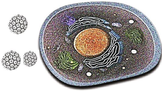
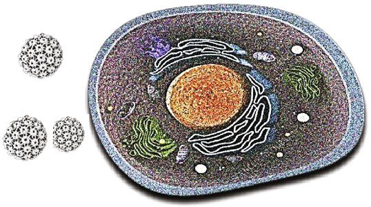
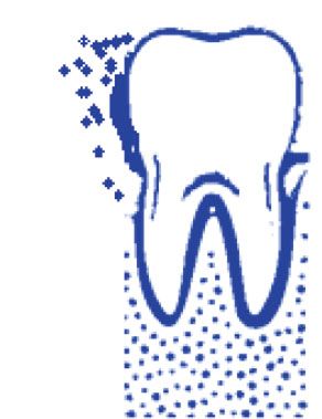
2 BioMed Research International

1. Introduction nant transformations remain unknown. Lifestyle factors,
 PMODs, modification of the microbiome, systemic sclerosis,
Oral carcinoma is the fifth most common malignant tumor genetic disease with dysregulation of DNA metabolism
worldwide and accounts for the majority of head and neck (Zinsser–Engman–Cole syndrome, Fanconi anemia, and
tumors [1, 2]. During the last decades, there has been a Xeroderma pigmentosum), mucosal inflammation and oral
progressive increase in proportion of incidence of oral mucosa chronic trauma, and hematinic and micronutrient
cancer not related to a known etiologic factor, such as the deficiency are the most important causes associated with
so-called “oral cancer in young,” a relevant disease in non- oral carcinoma [8].
smoker nondrinker (NSND) patients [3]. The topic is matter In several Asian countries, the combination of tobacco
of long standing debate, and adequate study models to ana- and/or betel nuts increases the risk for PMODs and oral
lyze this entity are lacking [4]. cancer. In addition, the synergistic effect in consuming large
 Squamous cell carcinomas (OSCCs) are the most com- quantities of alcohol and/or tobacco has a high risk associ-
mon type of oral tumor and can occur in larynx and pharynx ated with consumption and is proportional to the amount
too, with over 90% of oral cancers and represents 2-3% of all of alcohol consumed [9, 10]. Cigarette smoke from combus-
cancers [5]. Besides, OSCCs are more common in people tion releases several carcinogenic chemicals substances such
over the age of 50, while some studies report that 1%-6% as benzopyrene, dimethylbenzanthracene, nitrosazine, and
occur under the age of 40. There is a special preference for free radicals [11, 12]. On the other hand, large amounts of
men: in fact, the ratio of men to women in Western societies alcohol can cause the mitochondrial salivary suppression of
is 2 : 1 [6]. On the other hand, the incidence of injury in aldehyde dehydrogenase ALDH 2 allele gene which leads
young adults has been noted to be increasing worldwide to high levels of serum acetaldehyde (derived from the
[4, 7]. In recent years, despite advances in diagnosis and ethanol metabolism and turn to acetic acid by ALDH),
oncologic therapy, the 5-year survival rate of oral carci- increasing the risk of carcinogenesis [13, 14].
noma is still 50–60%, with a slight increase in the United Virus infections such as HPV (mainly type 16) have been
States (US) during the last decade (66%) [6]. linked to oral carcinomas [15, 16]. Chronic Candida spp.
 The current model of oral and pharyngeal carcinoma infections also appear to be a major risk factor [17, 18].
development suggests a progressive multistep transition The risk by HPV strain infections is involved in a growing
from normal mucosa to OSCCs through a series of progres- percentage of oral cancer, but other infectious agents such
sive histological changes (oral epithelial dysplasia) reflecting as the Candida spp. (more specifically C. albicans) could be
the accumulation of genetic and epigenetic abnormalities involved for their production of endogenous nitrosamines
and genetic susceptibility [5, 6]. starting from dietary nitrites present in the mouth, particu-
 The evaluation of the literature and surveillance data larly in saliva [19, 20].
concerning oral carcinoma is difficult because this neoplasia
often is reported associated with other head and neck malig- 2.1. The Role of Microbiota Dysbiosis in Candidiasis. The
nancies, and report of anatomic subsites is often unclear or terms of oral microbiome, oral microbiota, or oral micro-
can create confusion between its localization between oral flora are used for the microorganisms present in the human
cavity and oropharynx. mouth [21]. The mouth is the second largest and diverse
 Candida albicans is a commensal fungal species com- microbiota niche after the gut, harboring over 700 species
monly colonizing the human mucosal surfaces [7]. Carriage of bacteria. Every person has his or her own microbiome sig-
rates, corresponding to 18.5–40.9% in healthy individuals, nature [22]. The human microbiota consists of archaeal
are usually higher in individuals with compromised immu- cells, bacteria, fungi, viruses, and protozoa. Thus, various
nity, such as human immunodeficiency virus-positive bacteria of the oral microbiota have a cross talking with C.
individuals, diabetes patients, and infants and elder popula- albicans and HPV by modifying its pathogenicity or the
tions [7]. Besides, C. albicans can strongly interact with behavior of bacterial population [23].
additional oral microorganisms and enforce noteworthy The over presenting relative phyla, according to the
impact on the virulence of polymicrobial biofilms [7]. Fur- Human Oral Microbiome Database (eHOMD), as reported
thermore, it is recognized that coinfection of C. albicans is in Figure 2, are the Streptococcus spp. [24].
strongly associated with increasing diseases in pediatric den- The core microbiome is shared to all the persons, while
tistry, such as severe caries in children (S-ECC) [7]. variable microbiome is distinctive for single subject and is
 Thus, the purpose of the present paper is to review the cur- due to the lifestyle and to the physiological differences
rent evidence on the role of C. albicans and human papilloma- [22]. The mouth has two different types of surfaces on which
virus (HPV), as well their biomolecular mechanisms, in the bacteria can colonize: the hard and the soft tissues, respec-
pathogenesis of potentially malignant oral disorders (PMODs) tively, of the teeth and the oral mucosa [22]. Moreover, the
and oral carcinoma (Figure 1) in pediatric dentistry. model environment for the development of microorganisms
 is presented by the oral cavity and related nasopharyngeal
2. Risk Factors for Oral Cancer in regions. In fact, the usual temperature of mouth is about
 Pediatric Dentistry 37°C without significant changes, lead in this way to a stable
 environment to bacterial survive [23]. In addition, in normal
Several risk factors have been involved in the pathogenesis of conditions, saliva too has a pH between 6.5 and 7, favorable
these lesions, but the mechanisms and the causes of malig- for most bacteria species [8]. The Streptococcus mitis is one
BioMed Research International 3

 Candida albicans
 Proteins E6 and E7 integrate into DNA

 Oral dysbiosis microbiota -> bacterial synergism ->
 immunosuppression-> aid fungal colonization -> deep infection

 Overexpression
 HPV
 (I)production of the cytolytic toxin candidalysin
 oncogenetic strains risk groups
 (II)production of nitrosamines
 (III)production of acid aspartyl-proteinase
 (IV)production of acetaldehyde (carcinogen due to mutagenic qualities in
 DNA)
 (V)over-expression of Ki-67 labeling index, Prostaglandin-endoperoxide
 synthase 2 (COX-2) and P53
 (VI) , IL-1 ,
 IL-6, IL-8, IL-18, tumor necrosis factor (TNF)- , IFN- , and GM-CSF)
 (VII)reduction of -defensins which helps with candida superinfections

 ORAL CARCINOGENESIS

Figure 1: The pathways of carcinogenesis in the oral cavity through the oncogenetic HPV virus strain and Candida albicans. Several
hypothetical mechanisms have been proposed for the fungal infection that can induce PMODs and malignant lesions in oral epithelium.
The dysbiosis of the microbiota can play an important role with the subsequent alteration of the microbiome. There follows a synergism
between “bad” bacteria with Candida albicans and HPV.

 Actinobaculum 0.06%
 Carymebacterium 0.60%
 Haemophilus 0.70%
 Rothia 0.70%
 Propionibacterium 1.00%
 Bacteroides 1.10%
 Atopobium 1.10%
 Eubacterium 1.30%
 Enterococcus 1.30%
 Treponema 1.50%
 Campylobacter 1.50%
 Granulicatella 1.90%
 Capnocytophaga 1.90%
 Actinomyces 1.90%
 Dialister 2.00%
 Neiseria 2.30%
 Lactobacillus 2.30%
 Prevotella 2.70%
 Fusobacterium 3.00%
 Gemella 3.30%
 Selenomonas 3.50%
 Veillonella 9.80%
 Streptococcus 26.90%

 Figure 2: The main relative percentage (%) phyla of oral microbiome reported in eHOMD (source eHOMD, http://www.homd.org/).

of the first bacteria to colonize the oral cavity, establish den- que [27]. Vestibular streptococcus produces hydrogen perox-
tal plaque and, under certain conditions (such as the tooth ide and urease from which ammonia is produced, with a
extraction), may cause systemic infection such as endocardi- consequent increases of local pH [28]. On the other hand,
tis [25]. Streptococcus salivarius electively colonizes the oral Streptococcus anginosus, usually isolated from the mucous
mucosa and is over presenting in saliva, acting as an oppor- membranes, gingival fissure, and dental plaque, do not
tunistic pathogen that produce polysaccharides in the secrete polysaccharides playing a protective role in caries
presence of fructose, increasing in this way salivary pH development [28]. Staphylococci spp. are isolated to a small
[26]. Streptococcus mutans (based on the antigens, there extent from saliva, gingival cleft of the mucous membranes,
are 8 different serotypes), producing bacteriokines and and dental plaque (mainly from immunocompromised sub-
extracellular and intracellular polysaccharides (thus acidify- jects), with over represented the S. aureus [26]. Lactobacillus
ing the oral environment), seems to have a role in the genesis species are members of the oral flora in a small percentage
of caries, as well influencing the composition of dental pla- (less than 1%) but increase in tooth areas showing caries,
4 BioMed Research International

also for the characteristic that it could grow in a low pH in C. albicans infection because it can inhibit adhesion and
environment [23, 27]. Several species of potential opportu- fungal filamentation. The S. aureus may be helped by C.
nistic pathogens, such as Propionibacterium and Corynebac- albicans in an infection, such as R. dentocariosa, and S. mitis
terium, have been isolated from the oral environment [28]. aids fungal colonization [34, 35]. It has been noted that
Actinomyces spp. are also common members of dental Fusobacterium nucleatum, S. salivarius, A. actinomycetem-
plaque flora [29]. Other pathogens, such as the Porphyromo- comitans, and E. faecalis can have a protective role in a C.
nas gingivalis, Fusobacterium nucleatum, and Treponema albicans infections, because it can inhibit the overall viru-
denticola, which are anaerobic bacterium Gram-negative lence and fungal biofilm formation (Figure 3) [36, 37].
(-), can cause periodontitis and are linked to systemic dis- Finally, some studies reported several interactions
eases [30, 31]. Archaeal or archaic cells are only an exceed- between C. albicans and P. gingivalis that could be used
ingly small percentage of the oral microbiome with a to increase infectivity [38, 39]. In some patients, the
limited diversity (very few species and phytotypes) that can inflammatory reaction in candidiasis can be self-limiting,
be adapted as a small minority of organisms in this environ- but in other patients, multiple genetic, epigenetic, or exter-
ment. Methanes have been isolated from the oral cavity and, nal factors (tobacco, alcohol, diet, diabetes, etc.), as well in
in fact, in 36% of patients with periodontitis archaeal were synergetic crosstalk with oral bacterial, can cause excessive
detected by in situ fluorescent hybridization [27]. It was and chronic inflammation, leading to PMODs and alveolar
found that the archaeal were confined to a subset of human bone injure [40–42].
beings and consisted of two different Rinna fililipi within the Moreover recent evidence points out a causal relation-
genus Methanobrevibacter. The archaeal community in ship between specific bacterial infections, such as by P.
periodontal disease was dominated by a Methanobrevibacter gingivalis, and the development of oral and esophageal
oralis type phytologist and a separate subspecies of Methano- carcinoma [43].
brevibacter such as Methanobrevibacter cuticularis, Methano-
brevibacter filiformi, Methanobrevibacter ruminantium, and 2.2. Candidiasis Can Generate Oral Precancerous Conditions?
Methanobrevibacter arboriphilius [21, 22]. Fungi are a small Major predisposing factors to oral candidiasis could be
part of the oral microbiome. The predominant species is diabetes, mobile prosthesis, antibiotics, antiblastic or
Candida albicans. Other fungal species present in the mouth inhaled corticosteroid therapy, xerostomia by radiations,
are Cladosporium, Saccharomycetes, Aspergillus spp. (such or Sjógren’s syndrome and HIV infection, presenting clini-
as A. Penicillium), Gibberella, Cryptococcus, Fusarium, cal forms as angular cheilitis, mucocutaneous (rare), pseu-
Rhodotorula, and Schizophyllum [21, 25]. Two species of domembranous, hyperplastic, and erythematous (atrophic)
protozoa were found in physiological flora of the mouth: [44, 45]. Moreover, current evidence revealed correlation
the Entamoeba gingivalis amoeba and the Trichomonas between Candida infection and malignant transformation
tenax [21, 25]. The number of these organisms is high of the oral cavity [46, 47]. In fact, several hypothetical
in people with poor oral hygiene and gingivitis and was once mechanisms have been proposed for C. albicans interac-
considered potential pathogens [28]. At present, saprophytes tions with oral epithelium, leading to PMODs and malig-
are considered harmless, and the apparent association with nant lesions [48–50]:
the disease is linked to diet, because a poor oral hygiene can
allow an increase in the quantities of food intake and bacte- (a) Production of the cytolitic toxin candidalysin
rial residues, which are the main nutrients for the protozoa (b) Production of nitrosamines
[28]. The conditions of dysbiosis can lead to the imbalance
of growth and reduction of some microorganisms present (c) Production of acid aspartyl-proteinase
in the oral microbiota. Bacteria are not the only microorgan-
 (d) Production of acetaldehyde (carcinogen inducing
isms present in the periodontal area, and there are also
 mutations in genes)
several fungi members. Recent studies in patients with
periodontal infections found at least 150 species of fungi (e) Overexpression of Ki-67 labeling index, prostaglandin-
belonging to the generate Candida albicans, Ascomycota, endoperoxide synthase 2 (COX-2), and p53
Basidiomycota, Glomeromycota, and Chytridiomycota [29, 30].
This subsequently damages the periodontal tissue, in turn (f) Upregulation of proinflammatory cytokines, such as
creating nutrients for the bacteria of the dysbiotic micro- interleukin- (IL-) 1α, IL-1β, IL-6, IL-8, IL-18, tumor
biome [31]. It has been noted that Mitis Group Streptococci, necrosis factor- (TNF-) α, interferon gamma (IFN-γ),
Salivarius Group Streptococci, S. gordonii, S. mutans, S. ora- and granulocyte-macrophage colony-stimulating fac-
lis, S. sanguinis, and S. parasanguini can interact with C. tor (GM-CSF)
albicans. The S. mutans adhering to C. albicans interact (g) Reduction of β-defensins which facilitate Candida
through three mechanisms. Firstly, C. ablicans metabolizes superinfections
carbohydrates and improves the sugar metabolism of S.
mutans and S. mitis; secondly, S. mutans can influence the In a prospective cohort study (between 2007 and 2009)
coding of adhesins in C. albicans; finally, the synergism of on 103 patients, Candida spp. was isolated from 31 (30%)
S. mutans and C. albicans can promote an increase of patients with carcinoma and from 33 (32%) patients with
growth and invasion of mucous tissues in coinfection PMODs [51]. In another prospective cohort study, it was
[32, 33]. The S. salivarius K19 may play a protective role noted that the presence of Candida infections was related
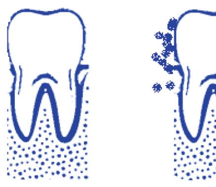
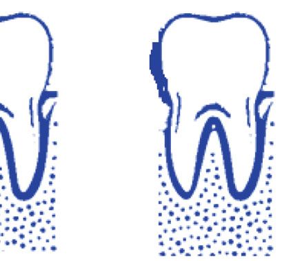
BioMed Research International 5

 Biofilm Biofilm
 Host factors Start adhesion
 maturation Dispersal

Figure 3: Example stages of biofilm formation process on dental surface (this mechanism occurs throughout the oral cavity). Biofilms are an
organized community of microorganisms. Microbes with weak and reversible forces attach first, dental surface. All bacteria that are not
immediately removed are rigidly attached to special structures, such as fibrils. Bacteria multiply and offer additional attack sites for other
microorganisms. The ability to maintain the consistency of microbes in dental plaque is based on the balance between cooperative and
competitive relationships between microorganisms and their host. This microbial homeostasis in combination with host defense prevents
the formation of pathogenic microorganism colonization. However, when this community is unbalanced quantitatively and qualitatively,
dysbiosis occurs.

to an increased risk of cancer [52]. In addition, some bacte- including autoinoculation from skin lesion HPV in adoles-
ria such as S. viridans are able to convert ethanol into acet- cent and pediatric patients [69, 70]. The HPV infection is
aldehyde for the presence of the alcohol-dehydrogenase commonly is commomly associated to benign lesions (vul-
enzyme in them [53–55]. The ability to switch among differ- gar warts, warts, focal epithelial hyperplasia, squamous cell
ent phenotypic forms has been thought to contribute to C. papilloma, Bowen’s papillomatosis), or to cancerous lesions
albicans virulence, and phenotypic switching events in C. such as squamous cell carcinoma (SCC) [64]. There must
albicans can be induced by hydroxyurea [55, 56]. Besides, be chafing or small lesions for the virus to enter the epithe-
some chemotherapeutic agents, such as 5-fluorouracil, could lium, and direct contact with the skin or mucosa is required
reduce the susceptibility of C. albicans to the antifungal for virus transmission. The strain of virus determines the
drugs [57]. The genotype “A” of C. albicans is more repre- different types of lesion that will be developed, as well as
sented in the OSCCs, while the genotype “C” of C. albicans the location of the infection. Hence, HPV can be transmitted
is more represented in the leucoplakia [58, 59]. Candida leu- in many ways through different abrasions, with sexual inter-
koplakia (CL) lesions are complex to discriminate from non- course, when the newborn passes through an infected genital
Candida leukoplakias (NCLs) clinically, but the presence of tract, and from a variety of self-inoculation positions, for
invading Candida hyphae in the superficial layer of epithe- example, by scratching the skin [68].
lium accompanied by infiltratration of polymorphic neutro- Since HPV does not have a protein or ribosomal synthe-
phils histologically discriminates CL lesions [60, 61]. sis, for its proliferation, it employs the genetic mechanism of
Moreover, Candida was isolated by exfoliative cytology and the host cell [69, 70]. The virus uses, to direct the metabolic
periodic acid–Schiff (PAS) staining of biopsies in a study functions of the host cell in its favor, the production of viral
that analyzed 44 cases with 59.1% presenting oral leucopla- messenger RNA, which is produced by transcription of viral
kia, showing Candida detection in 62.5% of the cases [62]. genetic material. The genome is divided into three parts. The
Furthermore, in the observed cases with Candida, the “E” (early region) which codes for proteins necessary for the
DNA alterations were higher [62]. In a retrospective study viral DNA genome duplication. It includes 7-9 open reading
on 136 patients with oral leukoplakia divided into two frames (ORFs), coding regions for proteins E1, E2, E4, E5,
groups, it was found that lesions had higher degrees of cell E6, E7, and E8 (although E5 and E8 ORF are not present
abnormalities (epithelial dysplasia) with Candida coinfec- in the genomes of all HPV types). The “L” (late region)
tion. The first group presenting multiple oral leucoplakia which encodes the structural proteins of the HPV capsid
lesions, Candida infection was detected in the 47.9% of virus (L1 and L2). Finally, the URR or NCR (upstream reg-
the sample (with 28.6% of dysplasia), while in the second ulatory region or noncoding region) comprised between “E”
group with single oral leucoplakia lesion, Candida infec- and “L” regions and regulates the function of the viral
tion was detected in the 19.0% of the sample (with genome, in addition to four binding sites to E2 proteins
20.0% of dysplasia) [63]. and multiple binding sites of the transcription [69–71].
 The differences between different strains of the virus are
2.3. The Role of Human Papillomavirus (HPV). HPVs are evident when comparing lesions of the same epithelial area.
members of the Papillomaviridae (PV) family, presenting a In fact, cell proliferation obtained from the expression of E6
circular, with supercoiled and double-stranded DNA, and and E7 proteins, due to infection with oncogenic HPV virus
some are considered as oncogenic promoters [64–66]. The strains, facilitates expansion of lesion, with a higher risk of
virus has a hexahedral symmetry capsid (consisting of pen- metastasis [71, 72].
tameric capsomeres) and has two structural proteins, L1
and L2 [67, 68]. Oral HPV is often sexually transmitted, 2.4. Which Are the Pathways of Oncogenesis in HPV
but nonsexual modes of transmission should be considered, Infection? The receptor that HPV uses to enter cells is

6 BioMed Research International

Table 1: There are over 450 types of HPV. The HPV’s genotypes are divided into 4 groups according to the associate oncogenic risk by
IARC/WHO (source from https://monographs.iarc.who.int/agents-classified-by-the-iarc/).

Group HPV virus
 Carcinogenic to humans: human papillomavirus types 16, 18, 31, 33, 35, 39, 45, 51, 52, 56, 58, and 59 (the HPV types that have
1
 been classified as carcinogenic to humans can differ by an order of magnitude in risk for cervical cancer)
2A Probably carcinogenic to humans: human papillomavirus type 68
 Possibly carcinogenic to humans: human papillomavirus types 26, 53, 66, 67, 70, 73, and 82; human papillomavirus types 30, 34,
2B 69, 85, and 97 (classified by phylogenetic analogy to the HPV genus alpha types classified in group 1); human papillomavirus types
 5 and 8 (in patients with epidermodysplasia verruciformis)
 Not classifiable as to its carcinogenicity to humans: human papillomavirus genus beta (except types 5 and 8) and genus gamma;
3
 human papillomavirus types 6 and 11

 Decomposition of Nuclear
 transcription factor, X
 box-binding protein 1-91
 (NFX1-91) -> Increase Binding and cleavage of Post- Decomposition (Bax, Bak,
 Telomerase reverse synaptic densityprotein(PDZ) FADD, FLICE) -> Increase in
 transcriptasehTERT -> Cell protein strands -> Host cell IAP-2Inhibition of apoptosis ->
 immortalization metaplasia Cell proliferation

 HPV E6 protein CANCER

 Inhibition of IRF3, CBP/300, Binding and cleavage of p53
 TADA3 and Tyk2 -> Inhibition of (suppression of p53 target
 the immune response in cancer genes)-> altered DNA
 cells replication -> increases of
 mutations

 Figure 4: Integration of HPV DNA into the host genome leading to the E6 protein overexpression and to carcinogenic processes.

integrin A6 [71]. On one hand, there are many types of HPV process [81]. Briefly, the Rb protein pathway could induce car-
such as 16, 18, 31, and 52, and on the other hand, they are cinogenesis by inactivating cell cycle regulators [82]. Activation
characterized by a high potential for malignancy and also of p53 turns on the production of the CD-kinase inhibitor pro-
have a high risk of metastasis (Table 1) [73, 74]. tein, p21WAF1 which contributes to cell cycle regulation by act-
 The role of the virus on carcinogenesis is due to proteins ing on different CD-kinase/cyclin complexes [83]. Thus, failure
E6 and E7 that integrate into host DNA. These two proteins of p53 has been observed to cause carcinogenesis. This is attrib-
could induce the neoplastic growth, since they block the two uted to the mutation of the genetic locus 17p13. Furthermore,
tumor suppressor proteins, the retinoblastoma protein E6 sequentially causes the cleavage of p53 [84]. Oncoprotein
(pRb) and p53, which regulate the transition from the G1 E7 causes p16 overexpression. In fact, the gene that codes for
phase of the cell cycle to S. E7 binds to p53 blocking its abil- the regulatory protein p16 is found in chromosome 9p21, and
ity to interact with the transcription factor E2F [75, 76]. its mutation is correlated with OSCCs [85, 86] (Figure 4).
Therefore, cells characterized by E7 overexpression lose the Besides, E6 inhibits the activity of p73, a type 16 homol-
control of the transition from G1 to S phase, which causes ogous p53. HPV blocks cell apoptosis by inhibiting Bax gene
continuous cell cycles and therefore cell proliferation with- expression in keratinocytes. This results in increased muta-
out suppression, as well, degradation of the pRb [76]. tions in the DNA of the cells. The apoptosis can be inhibited
 Levels of p53 in normal cells are exceptionally low. During by E7 binding to the tumor necrosis factor 1 receptor (TNF-
the E7 overexpression, an increase in p53 was shown for the R1) [87], originating a variety of bimolecular effects that
inhibition of its breakdown in normal cells which is in turn promote carcinogenesis (Figure 5).
regulated by MDM2 (mediator of DNA damage 2) [77, 78]. According to carcinogenesis model proposed by Califano
E7 also interacts with many other factors; among these, the et al., healthy tissues near the lesions show the same pattern
CDK (cyclin-dependent kinase) inhibits p27 and p21, which of mutations (loss of heterozygosity) of neoplastic cells,
induce the dysregulation of cell cycle [78]. E7 forms protein since all population come from a single mutant progenitor
complexes with pRb, p107, and p130 and participates in E6 cell [88]. Another model reports that the progenitor cell of
cleavage of p53 via the E6 complex, E6AP (associated protein the basal layer acquires a genetic mutation, which it trans-
E6), and p53 [79, 80]. Dysregulation of pRb phosphorylation- fers to its daughter cells. The result is the creation of a mass
dephosphorylation is an early event. There is a gradual reduc- of cells that grows, strains, and also affects nearby tissues, in
tion of pRb expression in dysplastic lesions and in neoplastic the development of lesions (clinically, they appear as
BioMed Research International 7

 Inactivation or Activation of Protein
 Retinoblastoma protein Immunological kinase of the Protein
 (Rb), Cyclin-Dependent non-recognition kinase B or Akt
 Kinase Inhibitors (p21 and HPV-positive (PKB/Akt) Pathway ->
 p27)-> immortal Cell tumors Inhibition of apoptosis

 HPV E7 protein
 CANCER

 Increased the Levels of Inhibition of Interferon
 interleukin 6 and Mcl-1 -> regulatory factor 1(IRF-1) and
 Accelerated cell cycle and IRF-9 transcription factors
 cell division

 Figure 5: Integration of HPV DNA into the host genome leading to E7 overexpression and to carcinogenic processes.

leukoplakia or erythroplakia). In the final stage, specific catenin release and subsequently cytoplasmic aggregation
clone cells after further mutations acquire a tumoral geno- and its transport to the nucleus, through the interaction with
type. In this context, loss of heterozygosity at loci 9p21 certain genes (with inhibition of apoptosis and increase of
and 3p21 appears to increase the risk of malignant recur- cell proliferation) such as COX-2, cyclin D1, and cMyc
rence [89, 90]. A mutation in the type of gene duplication [105, 111]. Furthermore, this pathway increases the expres-
has been shown to be involved in oral carcinogenesis [91]. sion of metalloproteinases, which catalyze the basement
 Replication of genetic material in the TERT (telomerase membrane and the dense structure of the epithelium, favor-
reverse transcriptase) protein coding gene results in overex- ing infiltration [111]. Lastly, HPV proteins E6 and E7 can
pression of the TERT human protein (hTERT). Studies regulate the epigenetic mechanisms, as DNA methylation,
conducted on OSCCs have shown an increase in both telome- histone modification, chromatin remodeling, and miRNA
rase activity and hTERT expression [92, 93]. This hTERT production in cell host or viral genes. Therefore, on one
expression occurs not only in cancerous cells but also around hand, E6/E7 induced DNA methylation shut out normal epi-
in the normal epithelium in the early course of carcinogenesis genetic processes, and on the other hand, E7 binds and mod-
[94]. The spindle assembly checkpoint (SAC) describes the ulates methyltransferase activity [112, 113].
way point to control mitosis, and Cdc20 is a SAC protein able
to activate the anaphase promoting complex (APC) which is a
key pathway factor and is also responsible for the formation of 3. Conclusions
aneuploidy cells [95–97].
 It has been noted that in 70% of oral tumors there is an Nowadays, several suggestive and consistent correlations
overexpression of the Cdc20 mRNA [98, 99]. Besides, as fur- between C. albicans and HPV infection in oral cancer pro-
ther bimolecular mechanisms in the etiology of oral cancer, gression in adolescent/pediatric patients have been reported
several studies reported the role of the epidermal growth fac- worldwide. Candida spp. and in particular C. albicans can
tor (EGF), the mitogen-activated protein kinase (MAPK) produce carcinogens such as nitrosamine or promote the
pathway, the PI3K/AKT/mTOR pathway, the signal trans- development of oral carcinoma. HPV are involved in the
ducer and activator of transcription (STAT) pathway, TGF pathogenesis of different types of cancer. In particular, from
(transforming growth factor), NF-κB (nuclear factor κB), literature data, it emerges that HPV infection plays an
and Wnt/β-catenin [100–102]. The ERbB1 gene, responsible important role in the risk of PMODs of the oral mucosa
for EGF encoding, is found on chromosome 7p12 and and consequently in the dysplastic and malignant transfor-
has been associated with increased activity in oral cancer mation of these lesions. Based on existing evidence, we can
[103, 104]. ERbB1 dysregulation in head and neck squa- also conclude that, in the composition of the microbiome
mous epithelial neoplasms is attributed more to its over- associated with OSCCs, there are no specific species to
expression than to the presence of mutations [105, 106]. implicate in its etiology, of course excluding oncoviruses
TGF promotes tumor progression by increasing angiogenesis (i.e., HPV) or fungi (i.e., C. albicans) that are associated with
and reducing sensitivity to the immune system. This action is oral cancer. Large multicenter trials are required in order to
caused by the tolerance of tumor cells to TGF-induced apo- study the biological behavior and formulate treatment strat-
ptosis [107, 108]. The TGFβR-II mutation is important, egies in the management of the same. Perhaps, these find-
and thus, a reduction of the TGFβR-II/TGFβR-I ratio (sus- ings will produce interest in the possible association among
pension/development) is determined, and thus, the protec- oral microbiota dysbiosis, C. albicans, HPV, and oral cancer
tive role of TGF is canceled [109]. A gradual decrease in and spur controlled prospective clinical studies in this field.
expression of TGF-β, TGFβR-I, and TGFβR-II was observed
in the different stages of carcinogenesis. In cancer cells, there
is a reduction in TGFβR-II levels, and this can promote the Data Availability
development and growth of oral cancer, acting as an indica-
tor of differentiation and therefore of aggressive behavior The data used to support the findings of this study are
[110]. Activation of the Wnt/β-catenin pathway induces β- included within the article.
8 BioMed Research International

Conflicts of Interest [11] H. Rubin, “Synergistic mechanisms in carcinogenesis by
 polycyclic aromatic hydrocarbons and by tobacco smoke: a
The authors declares that there is no conflict of interest bio-historical perspective with updates,” Carcinogenesis,
regarding the publication of this paper. vol. 22, no. 12, pp. 1903–1930, 2001.
 [12] A. Wyss, M. Hashibe, S. C. Chuang et al., “Cigarette, cigar,
Authors’ Contributions and pipe smoking and the risk of head and neck cancers:
 pooled analysis in the International Head and Neck Cancer
A.B., I.A.C., and L.S. contributed to the concept and design. Epidemiology Consortium,” American Journal of Epidemiol-
M.A., L.B., I.A.C., and A.B. contributed to the acquisition, ogy, vol. 178, no. 5, pp. 679–690, 2013.
analysis, and interpretation of data. I.A.C., E.B., and A.B. [13] H. K. Seitz and F. Stickel, “Molecular mechanisms of alcohol-
contributed to the drafting of the manuscript. S.C., E.B., mediated carcinogenesis,” Nature Reviews Cancer, vol. 7,
A.P.C., and A.B. contributed to the bibliographic research. no. 8, pp. 599–612, 2007.
M.D.C., S.C., and L.B. contributed to the critical revision of [14] J. Ferlay, M. Ervik, F. Lam et al., Global Cancer Observatory:
the manuscript for important intellectual content. A.M., Cancer Tomorrow, International Agency for Research on
R.N., and M.D. contributed to the data interpretation, tech- Cancer, Lyon, France, 2018, https://gco.iarc.fr/tomorrow.
nical, and material support. L.S., A.B., M.D.C., and L.L.M [15] J. M. García-Martín, P. Varela-Centelles, M. González, J. M.
contributed to the supervision and final approval. All Seoane-Romero, J. Seoane, and M. J. García-Pola, “Epidemi-
authors have read and agreed to the published version of ology of oral cancer,” in Oral Cancer Detection, P. Panta, Ed.,
the manuscript. Lorenzo Lo Muzio, Andrea Ballini, and Springer, Cham, 2019.
Stefania Cantore contributed equally as co-first authors. [16] X. Chen and Y. Zhao, “Human papillomavirus infection in
Michele Di Cosola, Luigi Santacroce, and Edoardo Brauner oral potentially malignant disorders and cancer,” Archives
contributed equally as co-last authors. of Oral Biology, vol. 83, pp. 334–339, 2017.
 [17] H. Mehanna, T. Beech, T. Nicholson et al., “Prevalence of
 human papillomavirus in oropharyngeal and nonoropharyn-
References geal head and neck cancer-systematic review and meta-
 [1] Q. B. Rahman, O. Iocca, K. Kufta, and R. M. Shanti, “Global analysis of trends by time and region,” Head Neck, vol. 35,
 burden of head and neck cancer,” Oral and Maxillofacial Sur- no. 5, pp. 747–755, 2013.
 gery Clinics of North America, vol. 32, no. 3, pp. 367–375, [18] F. Javed, A. A. Al-Kheraif, S. V. Kellesarian, F. Vohra, and G. E.
 2020. Romanos, “Oral Candida carriage and species prevalence in
 [2] World Health Organization, Oral cancer, 2021, https://www denture stomatitis patients with and without diabetes,” Journal
 .who.int/cancer/prevention/diagnosis-screening/oral-cancer/ of Biological Regulators and Homeostatic Agents, vol. 31, no. 2,
 en. pp. 343–346, 2017.
 [3] R. Grigolato, R. Accorona, G. Lombardo et al., “Oral cancer in [19] M. Hashibe, “Risk Factors for Cancer of the Mouth: Tobacco,
 non-smoker non-drinker patients. Could comparative pet Betel Quid, and Alcohol,” in Textbook of Oral Cancer. Text-
 oncology help to understand risk factors and pathogenesis?,” books in Contemporary Dentistry, S. Warnakulasuriya and J.
 Critical Reviews in Oncology/Hematology, vol. 166, p. 103458, Greenspan, Eds., Springer, Cham, 2020.
 2021. [20] A. Ballini, M. D. Cosola, R. Saini et al., “Comparison of
 [4] International Agency for Research on Cancer, GLOBOCAN, manual nylon bristled versus thermoplastic elastomer tooth-
 2020, http://gco.iarc.fr/today/data/factsheets/cancers/1-Lip- brushes in terms of cleaning efficacy and biological potential
 oral-cavity-fact-sheet.pdf. role on gingival health,” Applied Sciences, vol. 11, no. 16,
 [5] World Health Organization, WHO Classification Head and p. 7180, 2021.
 Neck Tumours (IARC)http://screening.iarc.fr/doc/BB9.pdf. [21] P. J. Turnbaugh, R. E. Ley, M. Hamady, C. M. Fraser-
 [6] B. S. Siriwardena, A. Tilakaratne, E. A. Amaratunga, and Liggett, R. Knight, and J. I. Gordon, “The human micro-
 W. M. Tilakaratne, “Demographic, aetiological and survival biome project,” Nature, vol. 449, no. 7164, pp. 804–810,
 differences of oral squamous cell carcinoma in the young 2007.
 and the old in Sri Lanka,” Oral Oncology, vol. 42, no. 8, [22] P. Nimish Deo and R. Deshmukh, “Oral microbiome:
 pp. 831–836, 2006. unveiling the fundamentals,” Journal of oral and maxillo-
 [7] Q. Du, B. Ren, J. He et al., “Candida albicans promotes tooth facial pathology: JOMFP, vol. 23, no. 1, pp. 122–128,
 decay by inducing oral microbial dysbiosis,” The ISME Jour- 2019.
 nal, vol. 15, no. 3, pp. 894–908, 2021. [23] M. A. Ghannoum, R. J. Jurevic, P. K. Mukherjee et al., “Char-
 [8] L. Lorini, C. Bescós Atín, S. Thavaraj et al., “Overview of oral acterization of the oral fungal microbiome (mycobiome) in
 potentially malignant disorders: from risk factors to specific healthy individuals,” PLoS Pathogens, vol. 6, no. 1, article
 therapies,” Cancers, vol. 13, no. 15, p. 3696, 2021. e1000713, 2010.
 [9] World Health Organization, WHO global report on trends in [24] expanded Human Oral Microbiome Database (eHOMD), All
 prevalence of tobacco smoking 2000-2025-third edition Oral genomes, 2021, http://homd.org/index.php?name=
 2015file:///C:/Users/Operatore3/Down- GenomeList&link=GenomeList&type=all_oral.
 loads/9789240000032-eng.pdf. [25] T. Chen, W. H. Yu, J. Izard, O. V. Baranova, A. Lakshmanan,
 [10] Y. Wang, X. Jia, P. Lin, M. Geng, R. Wang, and S. Li, “Cancer and F. E. Dewhirst, “The Human Oral Microbiome Database:
 mortality attributable to cigarette smoking in 2005, 2010 and a web accessible resource for investigating oral microbe
 2015 in Qingdao, China,” PLoS One, vol. 13, no. 9, article taxonomic and genomic information,” Database, vol. 2010,
 e0204221, 2018. p. baq013, 2010.
BioMed Research International 9

[26] K. Krishnan, T. Chen, and B. J. Paster, “A practical guide to carcinogenesis. a brief review,” Journal of Fungi, vol. 7, no. 6,
 the oral microbiome and its relation to health and disease,” p. 476, 2021.
 Oral Diseases, vol. 23, no. 3, pp. 276–286, 2017. [44] A. D. Alnuaimi, A. N. Ramdzan, D. Wiesenfeld et al., “Can-
[27] L. Gao, T. Xu, G. Huang, S. Jiang, Y. Gu, and F. Chen, “Oral dida virulence and ethanol-derived acetaldehyde production
 microbiomes: more and more importance in oral cavity and in oral cancer and non-cancer subjects,” Oral Diseases,
 whole body,” Protein & Cell, vol. 9, no. 5, pp. 488–500, 2018. vol. 22, no. 8, pp. 805–814, 2016.
[28] Y. Zhang, X. Wang, H. Li, C. Ni, Z. Du, and F. Yan, “Human [45] E. Chimenos-Küstner, I. Font-Costa, and J. López-López,
 oral microbiota and its modulation for oral health,” Biomed- “Oral cancer risk and molecular markers,” Medicina Oral,
 icine & Pharmacotherapy, vol. 99, pp. 883–893, 2018. Patología Oral y Cirugía Bucal, vol. 9, no. 5, pp. 381–384,
[29] P. Le Bars, S. Matamoros, E. Montassier et al., “The oral cav- 2004.
 ity microbiota: between health, oral disease, and cancers of [46] P. Pärnänen, J. H. Meurman, L. Samaranayake, and
 the aerodigestive tract,” Canadian Journal of Microbiology, I. Virtanen, “Human oral keratinocyte E-cadherin degrada-
 vol. 63, no. 6, pp. 475–492, 2017. tion by Candida albicans and Candida glabrata.,” Journal
[30] J. Chen, J. C. Domingue, and C. L. Sears, “Microbiota dysbio- of Oral Pathology & Medicine, vol. 39, no. 3, pp. 275–
 sis in select human cancers: evidence of association and cau- 278, 2010.
 sality,” Seminars in Immunology, vol. 32, pp. 25–34, 2017. [47] P. Lovreglio, N. Bukvic, S. Fustinoni et al., “Lack of genotoxic
[31] G. Mori, G. Brunetti, S. Colucci et al., “Alteration of activity effect in workers exposed to very low doses of 1, 3-butadiene,”
 and survival of osteoblasts obtained from human periodonti- Archives of Toxicology, vol. 80, no. 6, pp. 378–381, 2006.
 tis patients: role of TRAIL,” Journal of Biological Regulators [48] M. L. Gainza-Cirauqui, M. T. Nieminen, L. Novak Frazer,
 and Homeostatic Agents, vol. 21, no. 3-4, pp. 105–114, 2007. J. M. Aguirre-Urizar, M. D. Moragues, and R. Rautemaa,
[32] D. Montelongo-Jauregui and J. L. Lopez-Ribot, “Candida “Production of carcinogenic acetaldehyde by Candida albi-
 interactions with the oral bacterial microbiota,” Journal of cans from patients with potentially malignant oral mucosal
 Fungi, vol. 4, no. 4, p. 122, 2018. disorders,” Journal of Oral Pathology & Medicine, vol. 42,
[33] S. Patil, R. S. Rao, B. Majumdar, and S. Anil, “Clinical appear- no. 3, pp. 243–249, 2013.
 ance of oral Candida infection and therapeutic strategies,” [49] E. Cicinelli, A. Ballini, M. Marinaccio et al., “Microbiological
 Frontiers in Microbiology, vol. 6, p. 1391, 2015. findings in endometrial specimen: our experience,” Archives
[34] C. A. Fux, P. Stoodley, L. Hall-Stoodley, and J. W. Costerton, of Gynecology and Obstetrics, vol. 285, no. 5, pp. 1325–1329,
 “Bacterial biofilms: a diagnostic and therapeutic challenge,” 2012.
 Expert Review of Anti-infective Therapy, vol. 1, no. 4, [50] A. R. Casaroto, R. A. da Silva, S. Salmeron et al., “Candida
 pp. 667–683, 2003. albicans-cell interactions activate innate immune defense in
[35] S. F. Yang, H. D. Huang, W. L. Fan et al., “Compositional and human palate epithelial primary cells via nitric oxide (NO)
 functional variations of oral microbiota associated with the and β-defensin 2 (hBD-2),” Cell, vol. 8, no. 7, p. 707, 2019.
 mutational changes in oral cancer,” Oral Oncology, vol. 77, [51] F. Galli, G. Colella, V. Di Onofrio, R. Rossiello, I. F. Angelillo,
 pp. 1–8, 2018. and G. Liguori, “Candida spp. in oral cancer and oral precan-
[36] R. Guerrero-Preston, F. Godoy-Vitorino, A. Jedlicka et al., cerous lesions,” The New Microbiologica, vol. 36, no. 3,
 “16S rRNA amplicon sequencing identifies microbiota pp. 283–288, 2013.
 associated with oral cancer, human papilloma virus infection [52] M. Nørgaard, R. W. Thomsen, D. K. Farkas, M. F. Mogensen,
 and surgical treatment,” Oncotarget, vol. 7, no. 32, pp. 51320– and H. T. Sørensen, “Candida infection and cancer risk: a
 51334, 2016. Danish nationwide cohort study,” European Journal of Inter-
[37] B. A. Peters, J. Wu, R. B. Hayes, and J. Ahn, “The oral fungal nal Medicine, vol. 24, no. 5, pp. 451–455, 2013.
 mycobiome: characteristics and relation to periodontitis in a [53] J. H. Meurman and J. Uittamo, “Oral micro-organisms in the
 pilot study,” BMC Microbiology, vol. 17, no. 1, p. 157, 2017. etiology of cancer,” Acta Odontologica Scandinavica, vol. 66,
[38] J. Chandra, M. Retuerto, P. K. Mukherjee, and no. 6, pp. 321–326, 2008.
 M. Ghannoum, “The fungal biome of the oral cavity,” [54] E. Marttila, J. Uittamo, P. Rusanen, C. Lindqvist,
 Methods in Molecular Biology, vol. 1356, pp. 107–135, 2016. M. Salaspuro, and R. Rautemaa, “Acetaldehyde production
[39] D. Azzolino, P. C. Passarelli, P. de Angelis, G. B. Piccirillo, and microbial colonization in oral squamous cell carcinoma
 A. D’Addona, and M. Cesari, “Poor oral health as a determi- and oral lichenoid disease,” Oral Surgery, Oral Medicine, Oral
 nant of malnutrition and sarcopenia,” Nutrients, vol. 11, Pathology and Oral Radiology, vol. 116, no. 1, pp. 61–68,
 no. 12, p. 2898, 2019. 2013.
[40] G. Mori, G. Brunetti, S. Colucci et al., “Osteoblast apoptosis [55] K. Pakshir, K. Zomorodian, M. Karamitalab, M. Jafari,
 in periodontal disease: role of TNF-related apoptosis- H. Taraz, and H. Ebrahimi, “Phospholipase, esterase and
 inducing ligand,” International Journal of Immunopathology hemolytic activities of Candida spp. isolated from onychomy-
 and Pharmacology, vol. 22, no. 1, pp. 95–103, 2009. cosis and oral lichen planus lesions,” Journal de Mycologie
[41] F. Posa, G. Colaianni, M. Di Cosola et al., “The myokine irisin Médicale, vol. 23, no. 2, pp. 113–118, 2013.
 promotes osteogenic differentiation of dental bud-derived [56] J. Morschhäuser, R. Virkola, T. K. Korhonen, and J. Hacker,
 MSCs,” Biology (Basel), vol. 10, no. 4, p. 295, 2021. “Degradation of human subendothelial extracellular matrix
[42] A. Ballini, S. Capodiferro, M. Toia et al., “Evidence-based by proteinase-secreting Candida albicans,” FEMS microbiol-
 dentistry: what’s new?,” International Journal of Medical Sci- ogy letters, vol. 153, no. 2, pp. 349–355, 1997.
 ences, vol. 4, no. 3, pp. 174–178, 2007. [57] P. Krogh, B. Hald, and P. Holmstrup, “Possible mycological
[43] M. Di Cosola, A. P. Cazzolla, I. A. Charitos, A. Ballini, etiology of oral mucosal cancer: catalytic potential of
 F. Inchingolo, and L. Santacroce, “Candida albicans and oral infecting Candida albicans and other yeasts in production
10 BioMed Research International

 of N-nitrosobenzylmethylamine,” Carcinogenesis, vol. 8, [73] L. Santacroce, P. Luperto, M. L. Fiorella, and T. Losacco,
 no. 10, pp. 1543–1548, 1987. “Carcinoma of unknown origin++ with latero-cervical
[58] R. S. Kumar, S. Ganvir, and V. Hazarey, “Candida and calco- metastasis. Diagnostic problems. Retrospective analysis of
 fluor white: study in precancer and cancer,” Journal of Oral 110 cases of latero-cervical tumefaction,” Clinical Therapeu-
 and Maxillofacial Pathology, vol. 13, no. 1, pp. 2–8, 2009. tics, vol. 151, no. 3, pp. 199–201, 2000.
[59] M. H. Abdulrahim, B. A. McManus, S. R. Flint, and D. C. [74] M. F. Coscia, R. Monno, A. Ballini et al., “Human papilloma
 Coleman, “Genotyping Candida albicans from Candida leu- virus (HPV) genotypes prevalence in a region of South Italy
 koplakia and non-Candida leukoplakia shows no enrichment (Apulia),” Annali dell'Istituto Superiore di Sanità, vol. 51,
 of multilocus sequence typing clades but enrichment of ABC no. 3, pp. 248–251, 2015.
 genotype C in Candida leukoplakia,” PLoS One, vol. 8, no. 9, [75] J. Cao and Z. Y. Zhang, “Human papillomavirus infection and
 article e73738, 2013. p53 alteration in oral squamous cell carcinoma,” The Chinese
[60] A. Ballini, S. Cantore, L. Fatone et al., “Transmission of non- Journal of Dental Research, vol. 3, no. 3, pp. 44–49, 2000.
 viral sexually transmitted infections and oral sex,” The Jour- [76] I. A. Charitos, A. Ballini, S. Cantore et al., “Stem cells: a his-
 nal of Sexual Medicine, vol. 9, no. 2, pp. 372–384, 2012. torical review about biological, religious, and ethical issues,”
[61] G. P. Bombeccaria, F. Spadaria, M. Rossia, M. Porrinia, Stem Cells International, vol. 2021, Article ID 9978837, 11
 M. Bosottia, and A. B. Giannì, “Biology of Candida spp. in pages, 2021.
 potentially malignant disorders and carcinoma of the oral [77] WHO/IARC Agents Classified by the IARC Monographs, Vol-
 cavity,” Dental Cadmos, vol. 84, no. 10, pp. 624–634, 2016. umes 1–128https://monographs.iarc.who.int/agents-
[62] K. Nakazawa, S. F. Fifita, and K. Kuyama, “The cytological classified-by-the-iarc/.
 findings of oral inflammatory lesions, lichen planus and leu- [78] N. Ganguly and S. P. Parihar, “Human papillomavirus E6 and
 koplakia coexisted with and without Candida: with special E7 oncoproteins as risk factors for tumorigenesis,” Journal of
 reference to clinical, histopathological, immunohistochemi- Biosciences, vol. 34, no. 1, pp. 113–123, 2009.
 cal and flow cytometrical analyses,” International Journal of [79] G. Pannone, P. F. Nocini, L. Lo Muzio, M. Procaccini,
 Oral-Medical Sciences, vol. 6, no. 2, pp. 81–90, 2007. G. Pannone, and L. Santacroce, “Instability of micro-
[63] C. T. Chiu, C. F. Li, J. R. Li et al., “Candida invasion and influ- satellite sequences of DNA associated with genetic alterations
 ences in smoking patients with multiple oral leucoplakias–a in head and neck neoplasms. Review of the literature and pre-
 retrospective study,” Mycoses, vol. 54, no. 5, pp. e377–e383, liminary results of a research plan,” Minerva Stomatologica,
 2011. vol. 47, no. 11, pp. 589–596, 1998.
[64] “ICTV 9th report,” Papillomaviridaehttps://talk.ictvonline [80] N. Dyson, P. M. Howley, K. Münger, and E. Harlow, “The
 .org/ictv-reports/ictv_9th_report/dsdna-viruses-2011/w/ human papilloma virus-16 E7 oncoprotein is able to bind
 dsdna_viruses/121/papillomaviridae. to the retinoblastoma gene product,” Science, vol. 243,
[65] L. Santacroce, M. Di Cosola, L. Bottalico et al., “Focus on no. 4893, pp. 934–937, 1989.
 HPV infection and the molecular mechanisms of oral carci- [81] S. Soni, J. Kaur, A. Kumar et al., “Alterations of rb pathway
 nogenesis,” Viruses, vol. 13, no. 4, p. 559, 2021. components are frequent events in patients with oral epithe-
[66] H. Dong, X. Shu, Q. Xu et al., “Current Status of Human lial dysplasia and predict clinical outcome in patients with
 Papillomavirus-Related Head and Neck Cancer: From Viral squamous cell carcinoma,” Oncology, vol. 68, no. 4-6,
 Genome to Patient Care,” Virologica Sinica, 2021. pp. 314–325, 2005.
[67] K. Devaraja, S. Aggarwal, S. S. Verma, and S. C. Gupta, [82] S. Choi and J. N. Myers, “Molecular pathogenesis of oral
 “Clinico-pathological peculiarities of human papilloma virus squamous cell carcinoma: implications for therapy,” Journal
 driven head and neck squamous cell carcinoma: a compre- of Dental Research, vol. 87, no. 1, pp. 14–32, 2008.
 hensive update,” Life Sciences, vol. 245, p. 117383, 2020. [83] R. Ralhan, S. Agarwal, M. Mathur, B. Wasylyk, and
[68] B. K. Erickson, R. D. Alvarez, and W. K. Huh, “Human pap- A. Srivastava, “Association between polymorphism in p 21
 illomavirus: what every provider should know,” American (Waf 1/Cip1) cyclin-dependent kinase inhibitor gene and
 Journal of Obstetrics and Gynecology, vol. 208, no. 3, human oral cancer,” Clinical Cancer Research, vol. 6, no. 6,
 pp. 169–175, 2013. pp. 2440–2447, 2000.
[69] Z. M. Zheng and C. C. Baker, “Papillomavirus genome struc- [84] W. H. Westra, J. M. Taube, M. L. Poeta, S. Begum,
 ture, expression, and post-transcriptional regulation,” Fron- D. Sidransky, and W. M. Koch, “Inverse relationship between
 tiers in Bioscience, vol. 11, pp. 2286–2302, 2006. human papillomavirus-16 infection and disruptive p53 gene
[70] S. Benyo, A. Keane, J. Warrick, and K. Y. Choi, “HPV-posi- mutations in squamous cell carcinoma of the head and neck,”
 tive oral papillomas in an adolescent—a diagnostic dilemma,” Clinical Cancer Research, vol. 14, no. 2, pp. 366–369, 2008.
 Clinical Case Reports, vol. 9, no. 8, article e04546, 2021. [85] C. T. Allen, J. S. Lewis Jr., S. K. El-Mofty, B. H. Haughey, and
[71] IARC Working group on the evaluation of carcinogenic B. Nussenbaum, “Human papillomavirus and oropharynx
 risks to humans and Human papillomaviruses, “(IARC cancer: biology, detection and clinical implications,” Laryn-
 Monographs on the Evaluation of Carcinogenic Risks to goscope, vol. 120, no. 9, pp. 1756–1772, 2010.
 Humans, No. 90.) 1,” in Human Papillomavirus (HPV) [86] S. Ohta, H. Uemura, Y. Matsui et al., “Alterations of p16 and
 Infection, International Agency for Research on Cancer, p14ARF genes and their 9p21 locus in oral squamous cell car-
 Lyon (FR), 2007, https://www.ncbi.nlm.nih.gov/books/ cinoma,” Oral Surgery, Oral Medicine, Oral Pathology, Oral
 NBK321770/. Radiology, and Endodontology, vol. 107, no. 1, pp. 81–91,
[72] S. Nair and M. R. Pillai, “Human papillomavirus and disease 2009.
 mechanisms: relevance to oral and cervical cancers,” Oral [87] M. Filippova, H. Song, J. L. Connolly, T. S. Dermody, and
 Diseases, vol. 11, no. 6, pp. 350–359, 2005. P. J. Duerksen-Hughes, “The human papillomavirus 16 E6
BioMed Research International 11

 protein binds to tumor necrosis factor (TNF) R1 and pro- [103] G. Sethi, B. Sung, and B. B. Aggarwal, “Nuclear factor-kappa
 tects cells from TNF-induced apoptosis,” Journal of Biolog- B activation: from bench to bedside,” Experimental Biology
 ical Chemistry, vol. 277, no. 24, pp. 21730–21739, 2002. and Medicine, vol. 233, no. 1, pp. 21–31, 2008.
 [88] J. Califano, P. van der Riet, W. Westra et al., “Genetic [104] C. H. Squarize, R. M. Castilho, V. Sriuranpong, D. S. Pinto Jr.,
 progression model for head and neck cancer: implications and J. S. Gutkind, “Molecular cross-talk between the NFkap-
 for field cancerization,” Cancer Research, vol. 56, no. 11, paB and STAT3 signaling pathways in head and neck squa-
 pp. 2488–2492, 1996. mous cell carcinoma,” Neoplasia, vol. 8, no. 9, pp. 733–746,
 [89] B. J. Braakhuis, C. R. Leemans, and R. H. Brakenhoff, “A 2006.
 genetic progression model of oral cancer: current evidence [105] C. Sakanaka, T. Q. Sun, and L. T. Williams, “New steps in the
 and clinical implications,” Journal of Oral Pathology & Med- Wnt/beta-catenin signal transduction pathway,” Recent Prog-
 icine, vol. 33, no. 6, pp. 317–322, 2004. ress in Hormone Research, vol. 55, pp. 225–236, 2000.
 [90] K. Uchida, A. Oga, M. Okafuji et al., “Molecular cytogenetic [106] H. S. Patmore, L. Cawkwell, N. D. Stafford, and J. Greenman,
 analysis of oral squamous cell carcinomas by comparative “Unraveling the chromosomal aberrations of head and neck
 genomic hybridization, spectral karyotyping, and fluores- squamous cell carcinoma: a review,” Annals of Surgical
 cence in situ hybridization,” Cancer Genetics and Cytogenet- Oncology, vol. 12, no. 10, pp. 831–842, 2005.
 ics, vol. 167, no. 2, pp. 109–116, 2006. [107] M. A. Sheikh Ali, M. Gunduz, H. Nagatsuka et al., “Expres-
 [91] K. Freier, S. Pungs, C. Flechtenmacher et al., “Frequent high sion and mutation analysis of epidermal growth factor recep-
 telomerase reverse transcriptase expression in primary oral tor in head and neck squamous cell carcinoma,” Cancer
 squamous cell carcinoma,” Journal of Oral Pathology & Med- Science, vol. 99, no. 8, pp. 1589–1594, 2008.
 icine, vol. 36, no. 5, pp. 267–272, 2007. [108] M. Pring, S. Prime, E. K. Parkinson, and I. Paterson, “Dysreg-
 [92] R. Christensen, N. H. Park, P. Sapp, M. K. Kang, and N. H. ulated TGF-beta1-induced Smad signalling occurs as a result
 Park, “Elevated expression of hTERT is associated with dys- of defects in multiple components of the TGF-beta signalling
 plastic cell transformation during human oral carcinogenesis pathway in human head and neck carcinoma cell lines,”
 in situ,” Kim HR, Clinical Cancer Research, vol. 7, no. 10, International Journal of Oncology, vol. 28, no. 5, pp. 1279–
 pp. 3079–3086, 2001. 1285, 2006.
 [93] A. Musacchio and E. D. Salmon, “The spindle-assembly [109] I. C. Paterson, J. B. Matthews, S. Huntley et al., “Decreased
 checkpoint in space and time,” Nature Reviews Molecular Cell expression of TGF-beta cell surface receptors during progres-
 Biology, vol. 8, no. 5, pp. 379–393, 2007. sion of human oral squamous cell carcinoma,” The Journal of
 [94] G. Mondal, S. Sengupta, C. K. Panda, S. M. Gollin, W. S. Pathology, vol. 193, no. 4, pp. 458–467, 2001.
 Saunders, and S. Roychoudhury, “Overexpression of Cdc20 [110] G. Tian, Y. Fu, D. Zhang, J. Li, Z. Zhang, and X. Yang, “Iden-
 leads to impairment of the spindle assembly checkpoint and tification of four key prognostic genes and three potential
 aneuploidization in oral cancer,” Carcinogenesis, vol. 28, drugs in human papillomavirus negative head and neck squa-
 no. 1, pp. 81–92, 2007. mous cell carcinoma,” Cancer Cell International, vol. 21,
 [95] P. J. Roberts and C. J. Der, “Targeting the Raf-MEK-ERK no. 1, p. 167, 2021.
 mitogen-activated protein kinase cascade for the treatment [111] T. Shimada, H. Nakamura, K. Yamashita et al., “Enhanced
 of cancer,” Oncogene, vol. 26, no. 22, pp. 3291–3310, 2007. production and activation of progelatinase A mediated by
 [96] P. Richter, F. D. Böhmer, W. Hindermann et al., “Analysis membrane-type 1 matrix metalloproteinase in human oral
 of activated EGFR signalling pathways and their relation to squamous cell carcinomas: implications for lymph node
 laminin-5 gamma2 chain expression in oral squamous cell metastasis,” Clinical & Experimental Metastasis, vol. 18,
 carcinoma (OSCC),” Histochemistry and Cell Biology, no. 2, pp. 179–188, 2000.
 vol. 124, no. 2, pp. 151–160, 2005. [112] M. Burley, S. Roberts, and J. L. Parish, “Epigenetic regulation
 [97] C. Segrelles, M. Moral, M. F. Lara et al., “Molecular determi- of human papillomavirus transcription in the productive
 nants of Akt-induced keratinocyte transformation,” Onco- virus life cycle,” Seminars in Immunopathology, vol. 42,
 gene, vol. 25, no. 8, pp. 1174–1185, 2006. no. 2, pp. 159–171, 2020.
 [98] M. W. Lingen, W. Xiao, A. Schmitt et al., “Low etiologic [113] O. O. Orenuga, A. Oluwo, R. T. Oluwakuyide, and A. B.
 fraction for high-risk human papillomavirus in oral cavity Olawuyi, “Recurrent oral squamous papilloma in a pediatric
 squamous cell carcinomas,” Oral Oncology, vol. 49, no. 1, patient: case report and review of the literature,” Nigerian
 pp. 1–8, 2013. journal of clinical practice, vol. 21, no. 12, pp. 1674–1677,
 [99] A. A. Molinolo, P. Amornphimoltham, C. H. Squarize, R. M. 2018.
 Castilho, V. Patel, and J. S. Gutkind, “Dysregulated molecular
 networks in head and neck carcinogenesis,” Oral Oncology,
 vol. 45, no. 4-5, pp. 324–334, 2009.
[100] J. L. Roh, K. J. Cho, G. Y. Kwon et al., “The prognostic value
 of hypoxia markers in T2-staged oral tongue cancer,” Oral
 Oncology, vol. 45, no. 1, pp. 63–68, 2009.
[101] A. Onishi, Q. Chen, J. O. Humtsoe, and R. H. Kramer,
 “STAT3 signaling is induced by intercellular adhesion in
 squamous cell carcinoma cells,” Experimental Cell Research,
 vol. 314, no. 2, pp. 377–386, 2008.
[102] J. I. Song and J. R. Grandis, “STAT signaling in head and neck
 cancer,” Oncogene, vol. 19, no. 21, pp. 2489–2495, 2000.
You can also read