Demonstration of brain region-specific neuronal vulnerability in human iPSC-based model of familial Parkinson's disease
←
→
Page content transcription
If your browser does not render page correctly, please read the page content below
Human Molecular Genetics, 2020, Vol. 29, No. 7           1180–1191

                                                                               doi: 10.1093/hmg/ddaa039
                                                                               Advance Access Publication Date: 11 March 2020
                                                                               General Article

GENERAL ARTICLE

Demonstration of brain region-specific neuronal

                                                                                                                                                        Downloaded from https://academic.oup.com/hmg/article/29/7/1180/5803130 by guest on 12 October 2020
vulnerability in human iPSC-based model of familial
Parkinson’s disease
Razvan-Marius Brazdis1,2 , Julian E. Alecu1 , Daniel Marsch3 , Annika Dahms3 ,
Katrin Simmnacher1 , Sandra Lörentz1 , Anna Brendler1 , Yanni Schneider4 ,
Franz Marxreiter4 , Laurent Roybon5 , Beate Winner1 , Wei Xiang4,*,† and
Iryna Prots1,*,†
1 Department  of Stem Cell Biology, Friedrich-Alexander-Universität Erlangen-Nürnberg, Erlangen 91054,
Germany, 2 Department of Psychiatry and Psychotherapy, University Hospital Erlangen,
Friedrich-Alexander-Universität Erlangen-Nürnberg, Erlangen 91054, Germany, 3 Institute of Biochemistry
(Emil-Fischer-Center), Friedrich-Alexander-Universität Erlangen-Nürnberg, Erlangen 91054, Germany,
4 Department of Molecular Neurology, University Hospital Erlangen, Friedrich-Alexander-Universität

Erlangen-Nürnberg, Erlangen 91054, Germany and 5 Stem Cell Laboratory for CNS Disease Modeling,
Department of Experimental Medical Science, Lund University, Lund 22184, Sweden

*To whom correspondence should be addressed. Tel: +49 91318539303; Fax: +49 91318539311; Email: iryna.prots@uk-erlangen.de (I.P.);
Tel: +49 91318535881; Fax: +49 91318534672; Email: wei.xiang@fau.de (W.X.)

Abstract
Parkinson’s disease (PD) is a neurodegenerative disorder characterized by protein inclusions mostly composed of aggregated
forms of α-synuclein (α-Syn) and by the progressive degeneration of midbrain dopaminergic neurons (mDANs), resulting in
motor symptoms. While other brain regions also undergo pathologic changes in PD, the relevance of α-Syn aggregation for
the preferential loss of mDANs in PD pathology is not completely understood yet. To elucidate the mechanisms of the brain
region-specific neuronal vulnerability in PD, we modeled human PD using human-induced pluripotent stem cells (iPSCs)
from familial PD cases with a duplication (Dupl) of the α-Syn gene (SNCA) locus. Human iPSCs from PD Dupl patients and a
control individual were differentiated into mDANs and cortical projection neurons (CPNs). SNCA dosage increase did not
inf luence the differentiation efficiency of mDANs and CPNs. However, elevated α-Syn pathology, as revealed by enhanced
α-Syn insolubility and phosphorylation, was determined in PD-derived mDANs compared with PD CPNs. PD-derived mDANs
exhibited higher levels of reactive oxygen species and protein nitration levels compared with CPNs, which might underlie
elevated α-Syn pathology observed in mDANs. Finally, increased neuronal death was observed in PD-derived mDANs
compared to PD CPNs and to control mDANs and CPNs. Our results reveal, for the first time, a higher α-Syn pathology,
oxidative stress level, and neuronal death rate in human PD mDANs compared with PD CPNs from the same patient. The
finding implies the contribution of pathogenic α-Syn, probably induced by oxidative stress, to selective vulnerability of
substantia nigra dopaminergic neurons in human PD.

† These   authors contributed equally to this work.
Received: July 16, 2019. Revised: February 29, 2020. Accepted: March 3, 2020
© The Author(s) 2020. Published by Oxford University Press.
This is an Open Access article distributed under the terms of the Creative Commons Attribution License (http://creativecommons.org/licenses/by/4.0/),
which permits unrestricted reuse, distribution, and reproduction in any medium, provided the original work is properly cited.
                                                                                                                                               1180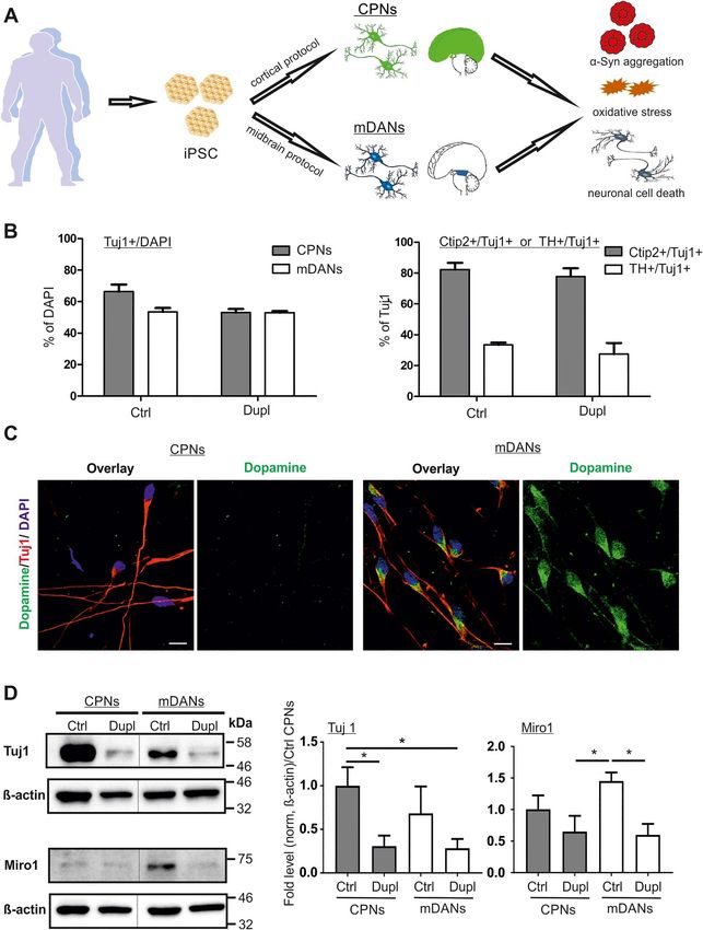
Human Molecular Genetics, 2020, Vol. 29, No. 7       1181

Introduction                                                          levels of ROS and protein nitration, when compared with CPNs,
                                                                      indicating an increased oxidative stress level in mDANs. Conse-
Parkinson’s disease (PD) is the most common neurodegenerative
                                                                      quently, PD Dupl mDANs were characterized by a higher basal
movement disorder affecting around 1% of the population aged
                                                                      neuronal death, resembling a selective loss of mDANs in PD
over 65 years (1). Neuropathologically, PD is characterized by
                                                                      brain. Our results demonstrate, for the first time, a stronger
the progressive degeneration of dopaminergic neurons in the
                                                                      vulnerability and a more pronounced α-Syn pathology of mDANs
substantia nigra (SN) of the midbrain (2). The resulting progres-
                                                                      compared with CPNs from the same PD patients with SNCA Dupl.
sive loss of nigrostriatal dopamine signaling is the main cause
                                                                      These results support the role of α-Syn aggregation, probably
of the cardinal motor symptoms of PD, such as bradykinesia,
                                                                      potentiated by SNCA dosage increase and oxidative stress, in
rigidity and resting tremor (3). Besides, emotional and cognitive
                                                                      promoting the selective vulnerability of dopaminergic neurons
disturbances, contributing to the non-motor symptoms of PD,
                                                                      in the SN in human PD.
are based on the dysregulation of cortical circuits, which are
partly dependent on the midbrain dopamine system (4–7).
    Another neuropathological hallmark of PD is the deposition        Results
of intracellular inclusions, mainly consisting of pathologically

                                                                                                                                           Downloaded from https://academic.oup.com/hmg/article/29/7/1180/5803130 by guest on 12 October 2020
                                                                      Cortical and midbrain neuronal differentiation is not
aggregated α-synuclein (α-Syn) in neuronal cell bodies and neu-
                                                                      altered by the SNCA dosage increase
rites, called Lewy bodies (LBs) and Lewy neurites (LNs), respec-
tively (8). α-Syn is a small, physiologically soluble protein, pre-   To address the selective vulnerability of mDANs and detect
dominantly located at the presynaptic site, which aggregates in       whether they exhibit a PD-related pathology, we generated
brains of PD patients. The aggregation of α-Syn proceeds through      mDANs and CPNs from iPSCs of a heathy donor (Ctrl) and a
monomers and aggregation intermediates (such as oligomers             PD patient carrying a Dupl of the SNCA locus (PD Dupl). To avoid
and protofibrils) to insoluble fibrils, which finally deposit along   genetic variabilities (21), we used identical iPSC lines of the PD
with other components, forming LBs and LNs (9). Among aggre-          Dupl patient and of the healthy donor for the differentiation of
gated α-Syn species, oligomers were suggested to be the most          both neuronal subtypes. We previously demonstrated that CPNs
toxic species (10,11), causing neurite pathology and dysregula-       differentiated from the same iPSC line of the PD Dupl patient
tion of axonal transport, whose disruption is strongly connected      exhibit impaired axonal transport, accompanied by an increased
to neuronal cell death (12,13). In addition to α-Syn deposition in    formation of α-Syn aggregation intermediates as compared with
diseased brains, α-Syn gene (SNCA) mutations and multiplica-          Ctrl CPNs (12).
tions of the SNCA locus cause early onset genetic forms of PD             IPSCs from a healthy control and the PD Dupl patient were
(14), further strengthening the pathological importance of α-Syn      differentiated into mDANs and CPNs (Fig. 1A). Due to the involve-
for PD. Thus, PD patients carrying the SNCA locus duplication         ment of fibroblast growth factor (FGF) signaling pathways in the
(Dupl) are characterized by the presence of a widespread Lewy         forebrain and midbrain patterning (22), CPNs were differentiated
pathology from the brainstem to the neocortex and neuronal            by using FGF2 as a modulator of proliferation and differenti-
loss in the SN (15).                                                  ation of the neural precursor cells (NPCs) (23), according to a
    Analysis of human post-mortem brain tissues suggests a pre-       previously published protocol (24). mDANs were generated by a
dictable PD-specific spreading pattern of α-Syn pathology during      combination of FGF8, known to promote dopaminergic differen-
disease progression, initiating in the brainstem and progressing      tiation (25), and small molecules to inhibit signaling pathways of
toward the midbrain and, later, to the neocortex (16). Despite of     transforming growth factor-β and bone morphogenetic protein
the widespread of Lewy pathology in various neuronal types in         (26).
PD brains, preferential degeneration of midbrain dopaminergic             Neuronal differentiation was assessed by immunocytochem-
neurons (mDANs) in PD suggests their particular vulnerability.        istry (ICC), based on the expression of β3-tubulin (Tuj1), a pan
Although the underlying mechanisms are yet unclear, several           neuronal marker, and was similarly effective in Ctrl and PD Dupl
interconnected pathways may contribute to the selective vul-          cultures, resulting in 53–66% of Tuj1-positive neurons generated
nerability of mDANs: (1) mDANs produce the neurotransmitter           by both cortical and midbrain protocols (Fig. 1B, left panel; rep-
dopamine that is highly prone to self-oxidation, leading to the       resentative ICC images used for the quantification are shown
generation of reactive oxygen species (ROS), implying a high          in Fig. 4A). Human iPSC differentiation into CPNs and mDANs
basal oxidative stress level in mDANs (17); (2) oxidative stress is   was evaluated using antibodies (Abs) against the CPN marker—
known as a risk factor of α-Syn oligomerization (18), suggesting      COUP-TF-interacting protein 2 (Ctip2) and the mDAN marker—
that the intracellular milieu of mDANs facilitates the aggregation    tyrosine hydroxylase (TH). Cortical and midbrain differentiation
of α-Syn; and (3) since α-Syn is involved in dopamine synthe-         efficiencies were comparable between Ctrl and PD Dupl iPSC
sis, release, and reuptake (19), α-Syn aggregation may impact         lines, reaching about 80% Ctip2-positive CPNs and about 30% TH-
dopamine homeostasis, by e.g. stimulating dopamine reuptake,          positive mDANs, respectively (Fig. 1B, right panel; representative
thereby exacerbating oxidative stress and leading to cellular         ICC images used for the quantification are shown in Fig. 4A).
damage (20).                                                          These data indicate that SNCA dosage increase has no major
    In this study, we aimed to address the vulnerability of mDANs     impact on either neuronal or neurotransmitter-specific differ-
and the correlation to α-Syn pathology in human PD by employ-         entiation efficiency. Importantly, dopamine staining revealed
ing human-induced pluripotent stem cell (iPSC)-derived neu-           dopamine-positive cells among Tuj1-positive neurons, differen-
rons. To study the selective vulnerability of mDANs in PD, we         tiated by the midbrain protocol, while Tuj1-positive neurons,
compared mDANs and cortical projection neurons (CPNs) dif-            developed by the cortical protocol, showed only the marginal sig-
ferentiated from iPSCs of PD patients with SNCA Dupl and of           nals of dopamine, confirming a neurotransmitter-specific phe-
a healthy donor. We observed an elevated α-Syn aggregation            notype of iPSC-derived neurons, in particular mDANs (Fig. 1C).
propensity in PD mDANs compared with both, control mDANs                  Although Ctrl and PD Dupl iPSC lines differentiated equally
and CPNs, but even more importantly compared with PD CPNs             well into CPNs and mDANs, the total β3-tubulin level was
from the same patient. Coincidently, mDANs showed higher              reduced in PD Dupl-derived neurons, either in CPNs or mDANs,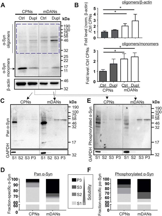
1182        Human Molecular Genetics, 2020, Vol. 29, No. 7

                                                                                                                                                                           Downloaded from https://academic.oup.com/hmg/article/29/7/1180/5803130 by guest on 12 October 2020

Figure 1. α-Syn gene (SNCA) locus duplication (Dupl) does not alter neuronal differentiation efficiency. (A) Schematic summary of the study. CPNs and mDANs were
differentiated from human iPSCs of a control individual (Ctrl) and PD patients carrying SNCA Dupl (Dupl) using cortical and small-molecule-based midbrain protocols,
respectively. α-Syn aggregation was analyzed biochemically. Oxidative stress was evaluated by the level of ROS and protein nitration. Neuronal cell death was determined
by ICC analysis of cleaved Caspase-3 (C-Casp3). (B) Neuronal differentiation was equally effective in Ctrl and Dupl as assessed by ICC of neuronal marker β3-tubulin
(Tuj1), cortical projection marker Ctip2 and midbrain dopaminergic marker TH (representative images used for the quantification are shown in Fig. 4A). Amounts of
neurons were determined as the percentage (%) of Tuj1-positive cells over DAPI-positive cells, whereas cortical and midbrain neurons were evaluated as the proportion
(%) of Ctip2- or TH-positive neurons over total Tuj1-positive neurons in three independent differentiation rounds. Values are shown as mean ± SD. Two-tailed Student’s
t-test was used. (C) ICC staining of dopamine in iPSC neuronal cultures revealed positive dopamine signals in Tuj1-positive neurons, differentiated from Ctrl iPSCs by
the midbrain protocol, while dopamine was barely detectable in Ctrl CPNs, confirming the neurotransmitter-specific phenotype of mDANs. Scale bars 10 μm. (D) Tuj1
and Miro1 protein expression was assessed in Ctrl and PD Dupl CPNs and mDANs by WB. Quantification was conducted by the normalization of Tuj1 or Miro1 signals
to β-actin levels followed by setting Ctrl CPN levels to 1. Reduced Tuj1 and Miro1 expression was determined in both PD Dupl CPNs and mDANs compared with Ctrl
neurons. ∗ P ≤ 0.05; values are shown as mean ± SD (three independent experiments). One-way ANOVA with multiple comparisons test was used. Blots for Tuj1 and
Miro1 within one black frame are derived from the same membrane. β-Actin was used as a loading control and was probed on the same membrane as the respective
target protein. Lanes from different parts of the same membrane are separated by black dashed lines.
Human Molecular Genetics, 2020, Vol. 29, No. 7 1183
when compared with respective neurons derived from Ctrl In summary, complementary aggregation analyses revealed
iPSCs (Fig. 1D, Tuj1 panel). Given the comparable differentiation an increased aggregation propensity of α-Syn in PD mDANs
efficiency of neurons in both Ctrl and PD Dupl cultures (as compared with PD CPNs, characterized by a higher level of α-Syn
analyzed by the percentages of Tuj1-positive cells, Fig. 1B), oligomers, enhanced insolubility and a proportional increase of
these results suggest reduced β3-tubulin level per individual insoluble phosphorylated α-Syn.
PD Dupl neurons compared with Ctrl neurons. Consistently, the
levels of mitochondrial Rho GTPase 1 (Miro1), required for a
proper development, morphology and intracellular distribution Increased levels of ROS and protein nitration in PD Dupl
of mitochondria and associated with PD pathology (27), were mDANs
decreased in PD Dupl-derived neurons (Fig. 1D, Miro1 panel). The comparison between mDANs and CPNs demonstrates a
Taken together, in accordance with neuronal impairments in PD higher α-Syn aggregation tendency in PD Dupl mDANs with a
Dupl CPNs described previously in our study (12), mDANs from particularly strong effect when compared with PD Dupl CPNs.
the same PD Dupl iPSC also showed neuronal deficits. As mDANs are assumed to have high oxidative stress levels
due to active dopamine metabolism, we next asked whether
Downloaded from https://academic.oup.com/hmg/article/29/7/1180/5803130 by guest on 12 October 2020
the degree of oxidative stress in different neuronal subtypes
Increased high molecular weight α-Syn oligomers in PD
correlates with distinct α-Syn aggregation patterns. Oxidative
Dupl mDANs
stress is characterized by the overproduction of ROS and reactive
We next investigated whether the level of α-Syn aggregation dif- nitrogen species (RNS). Therefore, we first measured the levels of
fers in CPN and mDAN populations and whether α-Syn pathology ROS in CPNs and mDANs, respectively, derived from Ctrl and PD
is especially evident in neurons from the PD Dupl patient. To Dupl. We applied CellRox and MitoSox fluorescent reagents to
these aims, whole cell lysates of CPNs and mDANs derived from assess cytosolic ROS and mitochondrial superoxide, respectively.
Ctrl and PD Dupl were analyzed by denaturing sodium dodecyl CellRox and MitoSox fluorescence intensities were measured in
sulfate-polyacrylamide gel electrophoresis (SDS-PAGE) followed living neurons using a multimode microplate reader in order
by western blot (WB) for determining the detergent-stable higher to assure fast and simultaneous detection in multiple neuronal
molecular weight α-Syn oligomers that appear between 32 kDa lines. Moreover, to exclude any possible effects of components
(the molecular weight of dimers on SDS-PAGE) and 190 kDa in different neuronal differentiation media used for mDANs and
(Fig. 2A, blue dashed box). To evaluate the level of SDS-stable α- CPNs on ROS levels, all subtypes of neurons were cultured in
Syn oligomers, α-Syn oligomer immunosignals were normalized the same minimal essential neuronal media for the last 48 h
either to β-actin for determining the fold changes of oligomers prior to fluorescence detection. Significantly higher cytosolic
(Fig. 2B, upper diagram) or to α-Syn monomers for determin- ROS and mitochondrial superoxide levels were determined in PD
ing the ratio changes associated with monomer-to-oligomers Dupl mDANs compared with PD Dupl CPNs (P < 0.01 for CellRox
transition (Fig. 2B, lower diagram). We observed that both total and P < 0.001 for MitoSox, Fig. 3A and B, respectively). Of note,
and proportional α-Syn SDS-stable oligomer levels are generally MitoSox, but not CellRox, levels were significantly higher in PD
higher in mDANs than in CPNs (Fig. 2B). Whereas a tendency of Dupl mDANs when compared with Ctrl mDANs, suggesting a
increased α-Syn oligomers in PD Dupl mDANs compared with PD more prominent mitochondrial pathology in PD Dupl mDANs
Dupl CPNs was found, a significant increase in α-Syn oligomer (P < 0.0001, Fig. 3B). Altogether, these results indicate neuronal
levels was detected in PD Dupl and Ctrl mDANs compared with subtype-specific, higher ROS levels in PD Dupl mDANs, which
Ctrl CPNs (P ≤ 0.05, Fig. 2B). An increase of α-Syn oligomers in are independent on the differentiation protocol.
mDANs compared with CPNs suggests a particular predisposi- ROS/RNS can react with susceptible amino acid residues of
tion of mDANs to form neurotoxic α-Syn species. proteins, thereby triggering post-translational modification of
To further verify an enhanced oligomerization propensity, proteins, such as tyrosine nitration (30). We additionally exam-
we performed sequential extraction of proteins, which allows to ined protein nitration levels biochemically by using an antibody
separate insoluble α-Syn (in S3 and P3 fractions) from soluble against nitrotyrosine and could show that mDANs, derived either
(in S1 fraction) and less soluble or vesicle-bound α-Syn (in S2 from Ctrl or from PD Dupl iPSCs, exhibit markedly higher protein
fraction) under native conditions prior to denaturing SDS-PAGE. nitration levels (Fig. 3C). Together with our data from ROS anal-
In order to determine differences between mDNAs and CPNs yses (Fig. 3A and Fig. 3B), protein nitration results demonstrate
related to PD pathology, we focused on comparing α-Syn solu- a higher oxidative stress level in mDANs compared with CPNs,
bility in PD Dupl neurons. We detected a proportional increase which was especially pronounced in PD Dupl mDANs.
of insoluble α-Syn species in PD Dupl mDANs compared with
PD Dupl CPNs using a pan α-Syn antibody (Fig. 2C and D). In
agreement with a reduced solubility of total α-Syn, the propor- Increased cell death rate in mDANs compared with
tional levels of α-Syn phosphorylated at the amino acid residue CPNs in PD Dupl cases
S129 were also increased in insoluble fractions generated by Since PD Dupl mDANs were characterized by a particularly high
sequential extraction of proteins (Fig. 2E and F). Phosphorylation levels of α-Syn pathology and oligomers compared with CPNs,
of α-Syn at S129 is closely related to PD and was found in LBs and we asked whether different neuronal types (mDANs versus
LNs (28). Therefore, phosphorylation of α-Syn is widely used as CPNs) derived from Ctrl and PD Dupl iPSCs exhibit different
an indicator for α-Syn aggregation, especially for oligomerization basal neuronal death rate. For this, cell death was evaluated
(29). Notably, the levels of oligomeric species with a molecular by ICC of the cleaved Caspase-3 (C-Casp3), a marker for early
weight of 46–58 kDa showed a remarkable increase in insoluble stages of apoptosis (31), in neurons double-positive for Tuj1
S3 and P3 fractions in PD Dupl mDANs (Fig. 2C). Interestingly, and a respective neuronal marker (Ctip2 or TH; Fig. 4A and B).
these insoluble oligomeric species were also detected in the IPSC-derived CPNs and mDANs from two different PD Dupl
same samples using an antibody against phosphorylated S129 α- patients (Dupl and Dupl#1A) were examined for neuronal cell
Syn (Fig. 2E), indicating that prominent oligomeric α-Syn bands death. While no significant differences were found between
correspond to insoluble phosphorylated species. Ctrl and PD Dupl CPNs (3.1 versus 2.9% [Dupl] and 3.2%1184 Human Molecular Genetics, 2020, Vol. 29, No. 7 Figure 2. Higher α-Syn aggregation levels in mDANs compared with CPNs in PD Dupl case. (A) High molecular weight SDS-stable α-Syn oligomers were determined Downloaded from https://academic.oup.com/hmg/article/29/7/1180/5803130 by guest on 12 October 2020 by WB using a pan α-Syn antibody (Syn1). β-Actin was used as a loading control and was probed on the same membrane. (B) α-Syn oligomer level in each sample was quantified by measuring the signal intensity in the region ranging from 32 kDa (corresponding to the molecular weight of α-Syn dimers) to 190 kDa (blue dashed box in A). Quantifications were performed by the normalization of signals either to β-actin levels (in order to determine the levels of α-Syn oligomers) or to α-Syn monomer (to analyze relative ratios of α-Syn oligomers to monomers in each sample) followed by setting Ctrl CPN levels to 1 (upper and lower diagrams, respectively). PD Dupl mDANs exhibit the highest α-Syn oligomer levels as shown in the representative WB (A) and by quantifications (B). Numbers on the right of the WB panel represent molecular weights of a protein ladder in kDa. Values are shown as mean ± SD of three independent experiments. ∗ P ≤ 0.05 by one-way ANOVA. (C–F) The solubility of α-Syn was determined by sequential extraction of proteins, followed by WB analysis of α-Syn distribution in fractions carrying proteins with decreasing solubility (solubility S1 > S2 > S3 > P3 fractions). GAPDH, a soluble cytosolic protein, was probed on the same membranes for α-Syn to control the sequential extraction. α-Syn and phosphorylated α-Syn (S129) in different fractions were probed using (C and D) a pan α-Syn antibody and (E and F) a phosphorylated α-Syn antibody, respectively. Solubility analysis reveals a decreased α-Syn solubility and increased formation of insoluble phosphorylated α-Syn species. A prominent band between 46 and 58 kDa found in S3 and P3 fractions derived from PD Dupl mDANs represents phosphorylated oligomeric α-Syn. Quantification shown in (D) and (F) was done by calculating the proportion of α-Syn positive signals (from monomeric and oligomeric α-Syn) in each fraction. Blots for α-Syn within one black frame (in C and E) are derived from the same membrane. GAPDH was probed on the same membrane as α-Syn. Lanes from different parts of the same membrane are separated by black dashed lines.
Human Molecular Genetics, 2020, Vol. 29, No. 7                  1185

                                                                                                                                                                           Downloaded from https://academic.oup.com/hmg/article/29/7/1180/5803130 by guest on 12 October 2020
Figure 3. Higher oxidative stress levels in mDANs compared with CPNs. (A) Cytosolic ROS and (B) mitochondrial superoxide were measured in Ctrl and PD Dupl CPNs and
mDANs by the CellRox reagent and MitoSox, respectively, using a CLARIOStar Plus plate reader. CellRox and MitoSox signals were normalized to respective DAPI signals
followed by the normalization to an external CPN and mDAN control line. Significantly higher ROS levels both (A) cytosolic and (B) mitochondrial were determined in
PD Dupl mDANs compared with PD Dupl CPNs. (C) Total protein nitration level was determined in CPNs and mDNAs generated from Ctrl and PD Dupl iPSCs by WB
using an antibody against nitrotyrosine. The blots were stained by Ponceau for protein loading control prior to immunodetection. Protein nitration levels are remarkably
higher in mDANs compared with CPNs. Blots with samples from two independent differentiation experiments (#1 and #2) are shown. Blots for nitration within one
black frame are derived from the same membrane. Ponceau staining was performed on the same membrane prior to the nitration detection. Lanes from different parts
of the same membrane are separated by black dashed lines.

[Dupl#1A] of C-Casp3/Ctip2-double positive neurons, respec-                            from pathological PD Dupl iPSCs. The same holds true for α-
tively, P > 0.05), significantly elevated proportion of apoptotic                      Syn aggregation potential, which is higher in PD Dupl mDANs
PD Dupl mDANs compared with Ctrl was detected (9.5% [Dupl]                             compared with PD Dupl CPNs and might underlie a particular
and 9.9% [Dupl#1A] versus 3.5% of C-Casp3/TH-double positive                           vulnerability of mDANs in PD.
neurons, respectively, P ≤ 0.05, Fig. 4C). Moreover, significantly
higher neuronal death was determined in mDAN cultures                                  Discussion
compared with CPN cultures of both PD Dupl patients (P ≤ 0.05
for Dupl neurons and P ≤ 0.01 for Dupl#1A neurons, Fig. 4C).                           mDANs are a heavily affected neuronal population in PD.
Altogether, these results demonstrate higher vulnerability of                          Although the selective loss of midbrain TH-positive neurons
mDANs compared with CPNs, in particularly those derived                                has been shown in post-mortem PD brains and in various
1186 Human Molecular Genetics, 2020, Vol. 29, No. 7
Downloaded from https://academic.oup.com/hmg/article/29/7/1180/5803130 by guest on 12 October 2020
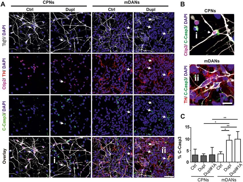
Figure 4. Increased apoptosis in mDANs compared with CPNs in PD Dupl cases. (A) iPSC-derived CPNs (Tuj1+/Ctip2+) and mDANs (Tuj1+/TH+) from the control
individual (Ctrl) and PD Dupl patients (Dupl) were stained for cleaved Caspase-3 (C-Casp3) to determine neuronal death rate in respective neurons. Representative
images used for neuronal subtype differentiation efficiency (Fig. 1B) and for the C-Casp3 (Fig. 4C) quantifications are shown. Arrows indicate the examples of C-Casp3+
neurons. (B) Enlarged views of selected cells marked by white frames in (A), representing a Tuj1+/Ctip2+/C-Casp3+ cell (i) and a Tuj1+/TH+/C-Casp3+ cells (ii). (C)
Quantification of ICC analysis. iPSC-derived CPNs and mDANs from two PD Dupl patients (Dupl and Dupl#1A) were analyzed for neuronal cell death. Control (Ctrl)
combines data from CPNs or mDANs, independently differentiated from two different iPSC clones of the same healthy individual. Significantly higher apoptosis rates
were detected in mDANs from both PD Dupl patients (Dupl and Dupl#1A; % of TH+/C-Casp3+ over Tuj1+) compared with Ctrl, as well as to CPNs (% of Ctip2+/C-Casp3+
over Tuj1+) of PD Dupl cases. DAPI visualized cell nuclei. Values are shown as mean ± SD of three independent differentiations. ∗ P ≤ 0.05, ∗∗ P ≤ 0.01 by one-way ANOVA.
Scale bar 50 μm in (A) and 12.5 μm in (B).
animal PD models, preferential loss of mDANs in PD compared dosage increase and oxidative stress, both are risk factors for
with other neuronal subtypes and the underlying mechanisms α-Syn aggregation. Our results might provide an explanation of
are less investigated, in particular in human disease models. a selective vulnerability of SN neurons in human PD.
In this study, using human iPSC-based disease modeling, we The neuronal differentiation from human iPSCs in our study
detected higher levels of α-Syn aggregation in mDANs compared yielded in total 53–66% Tuj1-positive neurons. The neuronal
with CPNs in familial PD. α-Syn pathology in PD Dupl mDANs differentiation efficiency in our study is in accordance with
was characterized by decreased α-Syn solubility, a proportional previously published data using human iPSC-based disease
increase of detergent-stable oligomers and the formation of modeling (24,26). Moreover, neuronal generation from iPSCs was
insoluble, phosphorylated oligomeric α-Syn species. Moreover, comparable between Ctrl and PD Dupl iPSCs, indicating that gene
we clearly demonstrated in our model system that ROS and dosage increase of SNCA does not impair neuronal differentia-
protein nitration levels, both indicators of oxidative stress, tion. This finding suggests that there might be no major defects
are higher in mDANs, particularly in PD Dupl mDANs, than of neuronal development in PD Dupl cases. Similar to the total
in CPNs. Importantly, pronounced α-Syn pathology and high neuronal numbers, Ctrl and PD Dupl iPSCs were equally well dif-
oxidative stress levels in PD Dupl mDANs were coincident with ferentiated to CPNs and mDANs, indicating that SNCA Dupl does
a higher neuronal death when compared with PD Dupl CPNs not compromise neurotransmitter-specific development. In line
and Ctrl neurons. Taken together, by directly comparing human- with our results, previous studies reported the similar efficiency
derived neurons, we demonstrate that mDANs are particularly of cortical glutamatergic differentiation (32) and midbrain
vulnerable in human familial form of PD with SNCA Dupl, dopaminergic neuronal differentiation of iPSCs from healthy
probably due to the susceptibility of this neuronal type to SNCA individuals (33), idiopathic (34) or genetic PD patients (26,35,36).Human Molecular Genetics, 2020, Vol. 29, No. 7 1187
A widespread cortical Lewy pathology and the development remarkable decline of α-Syn solubility and increased formation
of dementia are characteristic for the majority of PD patients of insoluble, phosphorylated α-Syn oligomers. In contrast,
carrying SNCA Dupl (15). Cortical Lewy pathology is known to neurons with a lower death rate (Ctrl CPNs, Ctrl mDANs and
be associated with cognitive impairment in PD Dupl patients PD Dupl CPNs) showed lower α-Syn aggregation levels. Using
(37). Despite the presence of Lewy pathology in the cortex of PD various biochemical procedures, we have previously shown an
patients, no PD-specific neuronal loss in the cortex was found increased formation of α-Syn aggregation intermediates in PD
based on the investigations of post-mortem brain tissues (38). Dupl CPNs compared with Ctrl CPNs (12). Combining the results
In our study, the survival rate of CPNs from healthy control and of the current study, α-Syn aggregation propensity in PD Dupl
PD Dupl patients was not significantly different. Thus, although mDANs appears to be even higher than in PD Dupl CPNs.
α-Syn pathology may be responsible for neuronal dysfunction Our analysis of oxidative stress by directly assessing ROS lev-
in the cortical regions of PD patients resulting in dementia els in living neurons revealed significantly higher ROS levels in
symptoms, our results suggest a higher resistance of CPNs to PD- PD Dupl mDANs compared with PD Dupl CPNs. Moreover, signif-
driving factors such as α-Syn pathology. Due to the fact that PD icant differences in mitochondrial superoxide levels between PD
is characterized by a broad Lewy pathology localization and at Dupl mDANs and Ctrl mDANs suggest a stronger mitochondrial
Downloaded from https://academic.oup.com/hmg/article/29/7/1180/5803130 by guest on 12 October 2020
the same time by a neuronal loss evident only in limited brain pathology in PD Dupl mDANs. Evaluation of protein nitration
regions, including subsets of brain nuclei and the SN, it is indeed level showed a generally higher degree in mDNAs in comparison
supposed that there might be common features rendering PD with CPNs, regardless of their origin (Ctrl or PD Dupl), which
pathology-related neurons more vulnerable (reviewed in 39). was coincident with a more pronounced α-Syn pathology in
In contrast to CPNs and in agreement with neuronal loss mDANs. Our study suggests that both the distinct intracellular
in the SN in PD, a significantly enhanced cell death rate was milieu of mDANs (oxidative stress) and abnormal α-Syn levels
found in PD Dupl mDANs compared with Ctrl mDANs and PD are important for the selective vulnerability of mDANs. One
Dupl CPNs, indicating a particular vulnerability of mDANs in intriguing contributor to oxidative stress in mDANs could be
human PD pathology. Thus, our results from the iPSC-based dopamine metabolism in this specific neuronal subtype, which
model of familial PD recapitulate a prominent and selective is supported by the clear difference in intracellular dopamine
loss of dopaminergic neurons in the SN of PD patients. Higher between mDANs and CPNs from the control iPSCs in our model
death rates of mDANs from PD Dupl patients compared with system. Dopamine is an unstable neurotransmitter and highly
Ctrl mDANs are in accordance with previous data: iPSC-based prone to oxidation. During its self-oxidation, free radicals are
studies, comparing mDNAs generated from Ctrl and familial produced, contributing to the high basal oxidative stress level.
PD patients with either SNCA triplication or point mutations Indeed, dopamine treatment was shown to result in the dark-
in the genes LRRK2, PARKIN or PINK1, also pointed out an ening of midbrain spheroids containing mDANs generated from
increased neuronal death and elevated susceptibility of mDANs a PD patient carrying the variation p.Q811R in POLG1 gene, but
to dopamine-induced oxidative stress in PD mDNAs (40– not control (52). This phenotype suggests dopamine oxidation
44). Moreover, increased extracellular-signal-regulated kinase and neuromelanin deposition in mDANs and was associated
phosphorylation (26), mitochondrial dysfunctions and decreased with metabolic dysfunction and occurrence of high molecular
dopaminergic differentiation were reported in mDANs from weight α-Syn oligomers in PD mDANs (52). Furthermore, a pro-
familial PD patients (45,46). Studies analyzing human post- gressive accumulation of oxidized dopamine was reported in
mortem brain tissue from idiopathic PD patients (47) or from iPSC-derived mDANs from sporadic and a number of different
familial PD patients with SNCA Dupl (15) showed an elevated genetic PD cases, including SNCA triplication, suggesting that
mDAN death level. All these previous findings suggest that this phenotype might be a common pathology in both sporadic
despite widespread pathological changes of PD in different areas and familial forms of PD (53).
of the brain, the most heavily affected neurons in PD are mDANs Interestingly, mitochondrial oxidative stress and oxidized
in pars compacta of the SN. dopamine were previously shown to start a pathological cascade
The vulnerability of mDANs may be attributed to their resulting in α-Syn accumulation in PD iPSC-derived mDANs (53).
intrinsic milieu, characterized by a high basal oxidative stress Our findings of a pronounced α-Syn pathology coincident with
level and the related high α-Syn aggregation propensity. a higher oxidative stress level in mDANs further strengthen the
Oxidative stress is an important player of α-Syn aggregation. role of α-Syn aggregation in human PD pathology. Beside several
One underlying mechanism involves the increased formation of known toxic cellular mechanisms, aggregated α-Syn species,
α-Syn oligomers under oxidative stress via α-Syn modification such as oligomers, may specifically trigger mDAN death. They
by ROS/RNS or oxidative stress metabolites (18,48,49). The might mediate this effect either via modulating dopamine reup-
accumulation of oxidative stress-induced modified oligomeric take and homeostasis, thereby increasing oxidative stress (19)
α-Syn species has been shown to be neurotoxic in a variety of cell and α-Syn own aggregation. Alternatively, α-Syn oligomers may
models. For example, our previous study revealed that specific destroy the membrane integrity, resulting in either inducing
oxidative stress-mediated α-Syn oligomers are selectively toxic mitochondrial pathology (12) or increasing abnormal Ca2+ cur-
to human mDNAs (differentiated LUHMES cells) (18). All these rents (10) that would modulate the pacemaker firing pattern of
converging findings suggest a causative link between α-Syn neurons in the SN (54). Further studies using strategies to inhibit
oligomerization and the death of mDANs in PD. An increased α-Syn oligomer formation (55) applied to PD mDANs could clarify
α-Syn expression or accumulation has been previously shown this question.
in iPSC-derived mDANs from PD patients carrying SNCA In conclusion, our study shows a higher α-Syn aggregation
triplication (40,45,50,51) or LRRK2 mutation (41,42). Here, by level preferentially in mDANs, derived from a familial PD patient
comparing PD Dupl mDANs and CPNs directly, we could show with SNCA locus Dupl, when compared with CPNs of the same
that an increase of SNCA dosage preferentially promotes α- patient and mDANs and CPNs from a control donor. We proved
Syn aggregation in PD Dupl mDANs. The α-Syn aggregation higher oxidative stress levels in mDANs compared with CPNs.
pattern in this particular neuronal subtype is characterized Moreover, PD Dupl mDANs were characterized by the reduced
by a proportional increase of SDS-stable α-Syn oligomers, a survival rate. Thus, by directly comparing mDANs and CPNs1188 Human Molecular Genetics, 2020, Vol. 29, No. 7
from the identical individuals, our study suggests that α-Syn were incubated in the ICC blocking solution 1 h at RT. Cell
pathology, induced by an increased SNCA dosage and oxidative nuclei were stained with 1 μg/ml 4 ,6-diamidino-2-phenylindole
stress, renders vulnerability to mDANs in human PD pathology. (DAPI, Sigma-Aldrich). Coverslips were mounted on glass micro-
scope slides (Thermo Fisher Scientific) in Aqua Polymount (Poly-
sciences). Images were acquired using a ZEISS LSM 780 confocal
Materials and methods laser scanning microscope and Zen Software. Images were quan-
tified using the cell counter plugin and the ImageJ software1.51j
Cells and cell culture
(NIH). In total, three independent differentiation rounds were
Human iPSC lines from two PD patients carrying an SNCA Dupl performed for each NPC line and three independent images
were kindly provided by Professor Galasko (line SDi1-R-C3 pre- were evaluated in each differentiation round to assess neuronal
viously described (12), referred here as Dupl) and by Dr Roy- differentiation efficiency.
bon (line CSC-1A, previously described in (56), referred here as
Dupl#1A). Both PD Dupl patients were female and had a disease
onset at the age of 58 (Dupl) and 53 years (Dupl#1A), respec- Denaturing SDS-PAGE and WB analysis of neuronal
cells
Downloaded from https://academic.oup.com/hmg/article/29/7/1180/5803130 by guest on 12 October 2020
tively. Both PD patients had a progressive disease course char-
acterized by tremor, muscle cramps and dementia. The dupli- Neuronal cells were homogenized in detergent-free Tris-
cation of SNCA was confirmed in iPSCs from both PD patients buffered saline (TBS, 50 mm Tris pH 7.4, 150 mm NaCl) containing
using Multiplex Ligation-Dependent Probe Amplification analy- 2 mm Ethylenediaminetetraacetic acid (EDTA) und protease
sis (P051/P052 probe mixes, MRC-Holland), which revealed SNCA inhibitor cocktail (Roche) in a Potter dounce homogenizer on
dosage increase of three copies corresponding to a heterozygous ice. Protein content was determined using a bicinchoninic acid
duplication. IPSCs from an age- and sex-matched healthy Cau- assay (Thermo Fisher Scientific). A total protein of 30 μg of the
casian individual with no history of neurologic disease served homogenate were mixed with the equal volume of 2xSDS sample
as a control (lines UKERi33Q-R1–06 and UKERi33Q-R1–002). PD buffer (0.125 M Tris/HCl pH 6.8, 4% SDS, 20% glycerol, 20 mm
Dupl and Ctrl line UKERi33Q-R1–06 were used for all exper- Dithiothreitol [DTT] and 0.01% bromophenol blue) and separated
iments, while neuronal death levels were additionally deter- in a 12% SDS-PAGE gel. In this study, samples were processed for
mined in neurons derived from PD Dupl, PD Dupl#1A and both the denaturing SDS-PAGE under reducing conditions (10 mm
Ctrl UKERi33Q-R1–06 and UKERi33Q-R1–002 lines. Human iPSCs DTT), which allowed to detect α-Syn and other protein markers
were reprogrammed from fibroblasts by retroviral transduction in the same samples and in the same WB membranes, when
with SOX2, KLF4, c-MYC and OCT3/4, differentiated through needed. Since α-Syn does not contain any cysteine residues
NPCs into CPNs or mDANs by either applying a FGF2-based cor- and α-Syn oligomerization is not based on the disulfide bonds
tical protocol (24) or a FGF8- and small molecule-based midbrain between cysteine residues, the use of DTT does not influence the
protocol, respectively, as previously described (26,57). detection of SDS-stable α-Syn species and meanwhile improves
All experiments with human iPSC-derived cells were carried the separation and detection of other proteins in WB. For
out in accordance with the local Institutional Review Board WB, the gel was blotted on a nitrocellulose membrane (Merk
approval (No. 259_17B, University Hospital Erlangen, Friedrich- Millipore, Darmstadt, Germany) by a semi-dry transfer apparatus
Alexander-Universität Erlangen-Nürnberg, Erlangen, Germany) (Biometra, Analytik Jena, Jena Germany).
and national and European Union directives. Written informed For immunodetection, blotted membranes were fixed for
consents were received from the participants prior to inclusion 30 min in 4% PFA to increase the sensitivity of protein detection
into the study at the movement disorder clinic at the Depart- (58) and washed twice with TBS. To control protein loading,
ment of Molecular Neurology, University Hospital Erlangen a total protein staining with 0.2% ponceau in 3% acetic acid
(Erlangen, Germany) and at the Department of Neurosciences, was performed. The membranes were next blocked with TBS
UCSD (San Diego, CA, USA). The iPSC line CSC-1A was generated containing 3% bovine serum albumin and probed with primary
from fibroblasts obtained with informed consent and after and horseradish peroxidase-conjugated secondary Abs. For
ethical committee approval at the Parkinson institute in Milan, visualization, chemiluminescent substrates (SuperSignal West
Italy. The permit for reprogramming of CSC-1A was delivered by Chemiluminescent Substrate kits, Thermo Fisher Scientific)
the Swedish work environment authority and registered under were applied to the membranes, and chemiluminescent signals
the number 20200-3211. were detected by Gel Doc XR system (Bio-Rad Laboratories,
Munich, Germany) and quantified by Image Lab Software (5.2.1,
Bio-Rad).
Immunocytochemistry
The following primary and secondary Abs were used for
Cells were fixed with 4% paraformaldehyde (PFA) for 30 min WB: monoclonal anti-β-actin (AC-15, #A5441 Sigma-Aldrich,
at 37◦ C, permeabilized by treatment with a mixture of ethanol: 1:5000), monoclonal mouse anti-Miro1 (CL1083, #ab188029,
acetic acid (2:1) for 5 min at −20◦ C (for staining of all tar- Abcam, 1:800), polyclonal rabbit anti-nitrotyrosine (#06-284,
get proteins except dopamine, where this step was omitted), Upstate, 1:1000), monoclonal mouse anti-α-Syn (Syn1, #610787,
washed three times with phosphate-buffered saline (PBS, Invit- BD Biosciences, 1:2000), monoclonal rabbit anti-phosphorylated
rogen) and subsequently permeabilized/blocked by incubating α-Syn (S129, clone EP1536Y, #ab51253, Abcam, 1:500) and
in ICC blocking solution (0.3% Triton X100, 3% donkey serum monoclonal mouse anti-β3-tubulin (Tuj1, MMS-435P, Covance,
[both from Sigma-Aldrich] in PBS) for 45 min at room tem- 1:1000).
perature (RT). Primary Abs: anti-ß3-tubulin (Tuj1; 1:500; BioLe-
gend), anti-TH (C-20, 1:500; Santa Cruz Biotechnology), anti-Ctip2
Sequential extraction of proteins
(25B6; 1:500; Abcam), anti-C-Casp3 (Asp175, 1:500; Cell Signal-
ing) and anti-dopamine (ab6427; 1:1000; Abcam) were incubated α-Syn aggregation was assessed by sequential extraction of
in the ICC blocking solution overnight at 4◦ C. Fluorescently proteins, which determined the proportion of insoluble α-Syn,
labeled secondary Abs (1:500, all from Thermo Fisher Scientific) indicative for aggregation. The assay was performed as describedHuman Molecular Genetics, 2020, Vol. 29, No. 7 1189
previously (12). Briefly, the whole cell homogenate (prepared Abbreviations
as described in WB section) was centrifuged at 100 000g for
Abs, antibodies; ANOVA, analysis of variance; BCA, bicinchoninic
1 h at 4◦ C and the resulting supernatant was collected as
acid; BMP, bone morphogenetic protein; C-Casp3, cleaved
soluble fraction (S1). After re-suspending the pellet in TBS
Caspase 3; CPNs, cortical projection neurons; Ctip2, chicken
containing 1% Triton X100, followed by a second centrifugation
ovalbumin upstream promoter transcription factor (COUP-
step (100 000g, 1 h, 4◦ C), the Triton X100-soluble fraction (S2) was
TF)-interacting protein 2; Ctrl, control; DA, dopaminergic;
collected. The Triton X100-insoluble pellet was re-suspended
DAPI, 4 ,6-diamidino-2-phenylindole; DTT, dithiothreitol; Dupl,
with radioimmunoprecipitation assay (RIPA) buffer (50 mm Tris-
SNCA duplication; EDTA, ethylenediaminetetraacetic acid; ERK,
HCl, pH 7.4, 175 mm NaCl, 5 mm EDTA, 1% NP-40 and 0.5% sodium
extracellular-signal-regulated kinase; FGF, fibroblast growth
deoxycholate containing 1% SDS). RIPA-soluble S3 fraction was
factor; ICC, immunocytochemistry; IF, immunofluorescence;
collected after a third centrifugation step (100 000g, 1 h, 4◦ C).
iPSCs, induced pluripotent stem cells; LBs, Lewy bodies; LNs,
The remaining RIPA-insoluble pellet (P3) was solubilized in 8 M
Lewy neurites; mDANs, midbrain dopaminergic neurons;
Urea/5% SDS (P3). α-Syn content within different fractions was
Miro1, Mitochondrial Rho GTPase 1; MLPA, Multiplex Ligation-
analyzed by WB using an anti-α-Syn Ab (Syn1). Intensity of bands
Dependent Probe Amplification; NPCs, neural precursor cells;
Downloaded from https://academic.oup.com/hmg/article/29/7/1180/5803130 by guest on 12 October 2020
corresponding to α-Syn monomers and high molecular weight
PBS, phosphate-buffered saline; PD, Parkinson’s disease; PFA,
oligomers were generated by the Image Lab 5.0 software (Biorad).
paraformaldehyde; RNS, reactive nitrogen species; ROS, reactive
oxygen species; RT, room temperature; SD, standard deviation;
ROS measurements SDS, sodium dodecyl sulfate; SN, substantia nigra; SNCA, α-
synuclein gene; TBS, tris-buffered saline; TGF-β, transforming
Direct ROS detection was performed in living neurons. To this
growth factor β; TH, tyrosine hydroxylase; Tuj1, β3-tubulin; WB,
aim, NPCs from Ctrl and PD Dupl iPSC lines were differentiated
western blot; α-Syn, α-synuclein.
to either CPNs or mDANs as described above in 96-well plates
with flat-bottom and black walls at a density of 50 × 103 cells per
well. CPNs and mDANs were cultured under the same conditions Acknowledgements
in a minimal essential neuronal media (20 ng/ml brain-derived
neurotrophic factor, 20 ng/ml glial cell-derived neurotrophic We would like to acknowledge Annika Sommer and Steven
factor [both from Peprotech], 200 nm ascorbic acid and 1 mm Havlicek for providing iPSC-derived NPCs and Douglas Galasko
cAMP [both from Sigma-Aldrich]) for the last 48 h to eliminate for providing iPSC of the PD Dupl patient (line SDi1-R-C3A). We
possible differences in ROS induced by different neuronal differ- also thank Daniela Graef, Holger Wend and Petra Wenzeler for
entiation protocols. On the final differentiation day, CPNs and excellent technical support. We are also thankful to the ‘Cell Line
mDANs were stained with 5 μM CellRox Deep Red fluorescence and DNA Biobank from Patients affected by Genetic Diseases’
reagent (Thermo Fisher Scientific), which was added directly (Istituto G. Gaslini, Genova, Italy), the ‘Parkinson Institute
to the culture medium, for 30 min at 37◦ C in order to detect Biobank’ and members of the Telethon Network of Genetic
cytosolic ROS. To detect the mitochondrial superoxide, cells were Biobanks funded by Telethon Italy (project no. GTB12001, http://
pre-washed with warm PBS containing Ca2+ and Mg2+ (Thermo biobanknetwork.telethon.it) for providing fibroblast samples
Fisher Scientific) followed by an incubation with 5 μM MitoSox used to generate the Dupl#1A iPSC line. The present work was
reagent (Thermo Fisher Scientific) diluted in warm PBS contain- performed in fulfillment of the requirements for obtaining the
ing Ca2+ and Mg2+ for 10 min at 37◦ C. After CellRox and MitoSox degree “Dr med.” for Razvan-Marius Brazdis.
treatment, cells were washed once with PBS containing 1 μg/ml
Conflict of Interest statement: The authors declare that they have
DAPI for cell nuclei staining for 5 min at RT and finally washed
no competing interests.
twice with PBS prior to measurement. CellRox, MitoSox and DAPI
fluorescence intensities were measured using a CLARIOstar Plus
plate reader (BMG Labtech) at the following Excitation/Emis- Funding
sion wavelengths: CellRox - 625-30/680-30 nm; MitoSox - 510-
15/580-20 nm; DAPI - 360-20/460-30 nm, respectively. CellRox and Deutsche Forschungsgemeinschaft (DFG, German Research
MitoSox values in each well were normalized to DAPI to account Foundation) (270949263/GRK2162 to R.-M.B., K.S., J.E.A., and
for cell numbers following by the normalization to the respective B.W.); Johannes and Frieda Marohn Foundation (to W.X. and I.P.);
values of an external neuronal line, differentiated either with the Fritz Thyssen Foundation (to I.P.); Swedish research council and
cortical or with the midbrain protocol, to account for cortical the Crafoord Foundation (to L.R.); Interdisciplinary Center for
and midbrain cell culture conditions. Each neuronal line was Clinical Research of the University Hospital Erlangen, Germany
differentiated in quadruplicates for each CellRox and MitoSox (E11 to W.X., E25 to B.W., J51 to F.M.); Bavarian Ministry of
detection. Education and Culture, Science and the Arts in the framework
of the BioSysNet and the ForIPS network and by the German
Federal Ministry of Education and Research (BMBF: 01EK1609B).
Statistical methods Microscopy/Image analysis was performed with support from
Differences among groups were evaluated by one-way analysis DFG grant INST 410/45-1 FUGG.
of variance (ANOVA) followed by Bonferroni’s or Holm-Sidak
multiple comparison tests, while comparisons between two
groups were done by two-tailed, paired Student’s t-test.
References
P-values ≤ 0.05 were considered significant. Graphs are pre- 1. Pringsheim, T., Jette, N., Frolkis, A. and Steeves, T.D. (2014)
sented as mean of three independent experiments (n = 3) ± The prevalence of Parkinson’s disease: a systematic review
standard deviation (SD). Statistical analyses were performed and meta-analysis. Mov. Disord., 29, 1583–1590.
using GraphPad Prism version 5.03 (GraphPad Software, 2. Kalia, L.V. and Lang, A.E. (2015) Parkinson’s disease. Lancet,
Inc.). 386, 896–912.1190 Human Molecular Genetics, 2020, Vol. 29, No. 7
3. Fahn, S. (2003) Description of Parkinson’s disease as a clinical 20. Emamzadeh, F.N. (2016) Alpha-synuclein structure, func-
syndrome. Ann. N. Y. Acad. Sci., 991, 1–14. tions, and interactions. J. Res. Med. Sci., 21, 29.
4. Rutledge, R.B., Skandali, N., Dayan, P. and Dolan, R.J. (2015) 21. Popp, B., Krumbiegel, M., Grosch, J., Sommer, A., Uebe, S., Kohl,
Dopaminergic modulation of decision making and subjec- Z., Plotz, S., Farrell, M., Trautmann, U., Kraus, C. et al. (2018)
tive well-being. J. Neurosci., 35, 9811–9822. Need for high-resolution genetic analysis in iPSC: results
5. Sawaguchi, T. and Goldman-Rakic, P.S. (1994) The role of D1- and lessons from the for IPS consortium. Sci. Rep., 8, 17201.
dopamine receptor in working memory: local injections of 22. Maric, D., Fiorio Pla, A., Chang, Y.H. and Barker, J.L. (2007)
dopamine antagonists into the prefrontal cortex of rhesus Self-renewing and differentiating properties of cortical neu-
monkeys performing an oculomotor delayed-response task. ral stem cells are selectively regulated by basic fibroblast
J. Neurophysiol., 71, 515–528. growth factor (FGF) signaling via specific FGF receptors. J.
6. Salgado-Pineda, P., Delaveau, P., Blin, O. and Nieoullon, A. Neurosci., 27, 1836–1852.
(2005) Dopaminergic contribution to the regulation of emo- 23. Raballo, R., Rhee, J., Lyn-Cook, R., Leckman, J.F., Schwartz,
tional perception. Clin. Neuropharmacol., 28, 228–237. M.L. and Vaccarino, F.M. (2000) Basic fibroblast growth fac-
7. Chaudhuri, K.R. and Schapira, A.H. (2009) Non-motor symp- tor (Fgf2) is necessary for cell proliferation and neuro-
Downloaded from https://academic.oup.com/hmg/article/29/7/1180/5803130 by guest on 12 October 2020
toms of Parkinson’s disease: dopaminergic pathophysiology genesis in the developing cerebral cortex. J. Neurosci., 20,
and treatment. Lancet Neurol., 8, 464–474. 5012–5023.
8. Spillantini, M.G., Schmidt, M.L., Lee, V.M., Trojanowski, J.Q., 24. Havlicek, S., Kohl, Z., Mishra, H.K., Prots, I., Eberhardt, E.,
Jakes, R. and Goedert, M. (1997) Alpha-synuclein in Lewy Denguir, N., Wend, H., Plotz, S., Boyer, L., Marchetto, M.C. et al.
bodies. Nature, 388, 839–840. (2014) Gene dosage-dependent rescue of HSP neurite defects
9. Lashuel, H.A., Overk, C.R., Oueslati, A. and Masliah, E. (2013) in SPG4 patients’ neurons. Hum. Mol. Genet., 23, 2527–2541.
The many faces of alpha-synuclein: from structure and 25. Xi, J., Liu, Y., Liu, H., Chen, H., Emborg, M.E. and Zhang, S.C.
toxicity to therapeutic target. Nat. Rev. Neurosci., 14, 38–48. (2012) Specification of midbrain dopamine neurons from
10. Danzer, K.M., Haasen, D., Karow, A.R., Moussaud, S., Habeck, primate pluripotent stem cells. Stem Cells, 30, 1655–1663.
M., Giese, A., Kretzschmar, H., Hengerer, B. and Kostka, M. 26. Reinhardt, P., Glatza, M., Hemmer, K., Tsytsyura, Y., Thiel, C.S.,
(2007) Different species of alpha-synuclein oligomers induce Hoing, S., Moritz, S., Parga, J.A., Wagner, L., Bruder, J.M. et al.
calcium influx and seeding. J. Neurosci., 27, 9220–9232. (2013) Derivation and expansion using only small molecules
11. Winner, B., Jappelli, R., Maji, S.K., Desplats, P.A., Boyer, L., of human neural progenitors for neurodegenerative disease
Aigner, S., Hetzer, C., Loher, T., Vilar, M., Campioni, S. et al. modeling. PLoS One, 8, e59252.
(2011) In vivo demonstration that alpha-synuclein oligomers 27. Hsieh, C.H., Shaltouki, A., Gonzalez, A.E., Bettencourt da
are toxic. Proc. Natl. Acad. Sci. USA, 108, 4194–4199. Cruz, A., Burbulla, L.F., St Lawrence, E., Schule, B., Krainc,
12. Prots, I., Grosch, J., Brazdis, R.M., Simmnacher, K., Veber, D., Palmer, T.D. and Wang, X. (2016) Functional impairment
V., Havlicek, S., Hannappel, C., Krach, F., Krumbiegel, M., in miro degradation and mitophagy is a shared feature in
Schutz, O. et al. (2018) Alpha-Synuclein oligomers induce familial and sporadic Parkinson’s disease. Cell Stem Cell, 19,
early axonal dysfunction in human iPSC-based models of 709–724.
synucleinopathies. Proc. Natl. Acad. Sci. USA., 115, 7813–7818. 28. Oueslati, A. (2016) Implication of alpha-synuclein phospho-
13. Prots, I., Veber, V., Brey, S., Campioni, S., Buder, K., Riek, R., rylation at S129 in synucleinopathies: what have we learned
Bohm, K.J. and Winner, B. (2013) Alpha-Synuclein oligomers in the last decade? J. Parkinsons Dis., 6, 39–51.
impair neuronal microtubule-kinesin interplay. J. Biol. Chem., 29. Oueslati, A., Fournier, M. and Lashuel, H.A. (2010) Role of
288, 21742–21754. post-translational modifications in modulating the struc-
14. Ross, O.A., Braithwaite, A.T., Skipper, L.M., Kachergus, J., Huli- ture, function and toxicity of alpha-synuclein: implications
han, M.M., Middleton, F.A., Nishioka, K., Fuchs, J., Gasser, for Parkinson’s disease pathogenesis and therapies. Prog.
T., Maraganore, D.M. et al. (2008) Genomic investigation of Brain Res., 183, 115–145.
alpha-synuclein multiplication and parkinsonism. Ann. Neu- 30. Giasson, B.I., Duda, J.E., Murray, I.V., Chen, Q., Souza, J.M.,
rol., 63, 743–750. Hurtig, H.I., Ischiropoulos, H., Trojanowski, J.Q. and Lee,
15. Konno, T., Ross, O.A., Puschmann, A., Dickson, D.W. and V.M. (2000) Oxidative damage linked to neurodegeneration
Wszolek, Z.K. (2016) Autosomal dominant Parkinson’s dis- by selective alpha-synuclein nitration in synucleinopathy
ease caused by SNCA duplications. Parkinsonism Relat. Disord., lesions. Science, 290, 985–989.
22, S1–S6. 31. Yakovlev, A.G. and Faden, A.I. (2004) Mechanisms of neural
16. Braak, H., Del Tredici, K., Rub, U., de Vos, R.A., Jansen Steur, cell death: implications for development of neuroprotective
E.N. and Braak, E. (2003) Staging of brain pathology related to treatment strategies. NeuroRx, 1, 5–16.
sporadic Parkinson’s disease. Neurobiol. Aging, 24, 197–211. 32. Cao, S.Y., Hu, Y., Chen, C., Yuan, F., Xu, M., Li, Q., Fang,
17. Gonzalez-Hernandez, T., Cruz-Muros, I., Afonso-Oramas, D., K.H., Chen, Y. and Liu, Y. (2017) Enhanced derivation of
Salas-Hernandez, J. and Castro-Hernandez, J. (2010) Vulner- human pluripotent stem cell-derived cortical glutamatergic
ability of mesostriatal dopaminergic neurons in Parkinson’s neurons by a small molecule. Sci. Rep., 7, 3282.
disease. Front Neuroanat, 4, 140. 33. Doi, D., Samata, B., Katsukawa, M., Kikuchi, T., Morizane,
18. Xiang, W., Schlachetzki, J.C., Helling, S., Bussmann, J.C., A., Ono, Y., Sekiguchi, K., Nakagawa, M., Parmar, M. and
Berlinghof, M., Schaffer, T.E., Marcus, K., Winkler, J., Klucken, Takahashi, J. (2014) Isolation of human induced pluripotent
J. and Becker, C.M. (2013) Oxidative stress-induced post- stem cell-derived dopaminergic progenitors by cell sorting
translational modifications of alpha-synuclein: specific for successful transplantation. Stem Cell Reports, 2, 337–350.
modification of alpha-synuclein by 4-hydroxy-2-nonenal 34. Kikuchi, T., Morizane, A., Doi, D., Magotani, H., Onoe, H.,
increases dopaminergic toxicity. Mol. Cell. Neurosci., 54, 71–83. Hayashi, T., Mizuma, H., Takara, S., Takahashi, R., Inoue, H.
19. Venda, L.L., Cragg, S.J., Buchman, V.L. and Wade-Martins, R. et al. (2017) Human iPS cell-derived dopaminergic neurons
(2010) Alpha-Synuclein and dopamine at the crossroads of function in a primate Parkinson’s disease model. Nature, 548,
Parkinson’s disease. Trends Neurosci., 33, 559–568. 592–596.You can also read