Insulin Resistance and Alzheimer s Disease: Molecular Links & Clinical Implications
←
→
Page content transcription
If your browser does not render page correctly, please read the page content below
Current Alzheimer Research, 2008, 5, 000-000                                              1

Insulin Resistance and Alzheimer´s Disease: Molecular Links & Clinical
Implications

Karen F. Neumann1,2, Leonel Rojo1,3, Leonardo P. Navarrete1,2, Gonzalo Farías1, Paula Reyes1 and
Ricardo B. Maccioni1,2,*

1
 Laboratory of Cellular and Molecular Neurosciences, University of Chile, 2International Center for Biomedicine (ICC),
Santiago, Chile and 3Arturo Prat University, Iquique, Chile

            Abstract: Hyperinsulinemia as well as type II diabetes mellitus are among the risk factors for Alzheimer´s disease (AD).
            However, the molecular and cellular basis that link insulin resistance disorders and diabetes with AD are far from clear.
            Here, we discuss the potential molecular mechanisms that may explain the participation of these metabolic disorders in
            the pathogenesis of AD. The human brain uses glucose as a primary fuel; insulin secreted by the pancreas cross the blood-
            brain barrier (BBB), reaching neurons and glial cells, and exerts a region-specific effect on glucose metabolism. Glucose
            homeostasis is critical for energy generation, neuronal maintenance, neurogenesis, neurotransmitter regulation, cell sur-
            vival and synaptic plasticity. It also plays a key role in cognitive function. In an insulin resistance condition, there is a re-
            duced sensitivity to insulin resulting in hyperinsulinemia; this condition persists for several years before becoming full-
            blown diabetes. Toxic levels of insulin negatively influence neuronal function and survival, and elevation of peripheral in-
            sulin concentration acutely increases its cerebrospinal fluid (CSF) concentration. Peripheral hyperinsulinemia correlates
            with an abnormal removal of the amyloid beta peptide (A) and an increase of tau hyperphosphorylation as a result of
            augmented cdk5 and GSK3 activities. This leads to cellular cascades that trigger a neurodegenerative phenotype and de-
            cline in cognitive function. Chronic peripheral hyperinsulinemia results in a reduction of insulin transport across the BBB
            and a reduced insulin signaling in brain, altering all of insulin’s actions, including its anti-apoptotic effect. However, the
            increase in brain insulin levels resulting from its peripheral administration at optimal doses has shown a cognition-
            enhancing effect in patient with AD. Some drugs utilized in type II diabetes mellitus reduce cognitive impairment associ-
            ated with AD. The link between insulin resistance and neurodegeneration and AD, and the possible therapeutic targets in
            preventing the insulin-resistance disorders are analyzed.
Keywords: Insulin, Insulin Resistance, Hyperinsulinemia, Diabetes, Cognition, Neurodegeneration, Alzheimer´s Disease.

A) GLUCOSE & INSULIN SIGNALING                                                 kinase phosphatidylinositol 3-kinase (PI3K) and the mitogen
                                                                               activation protein kinase (MAPK) cascade [4,5]. PI3K is
1. Insulin and Insulin Receptor (IR)
                                                                               associated with almost all of the metabolic actions of insulin
    Insulin is a small polypeptide, with a molecular weight of                 (refer to Fig. 1).
about 6,000 kDa, synthesized in significant quantities in -                       The effects of insulin on the central nervous system
cells of the pancreas. An increased level of glucose in blood                  (CNS) are affected by its availability to this separate physio-
stimulates -cells in pancreas to secret insulin by exocytosis,                logical compartment. The term BBB in its most restrictive
which diffuses into the islets capillary blood [1]. Insulin pro-               sense refers to the vascular bed of the CNS, which is spe-
vides both short-term and long-term homeostatic signals,                       cially modified to prevent the unrestricted transfer of mole-
since it is secreted acutely in response to an increase in blood               cules between the blood and the extracellular fluid of the
glucose and because plasma insulin levels are directly corre-                  CNS [6]. The BBB plays a critical role in the transduction of
lated with the degree of long term increase in body adiposity                  signals between the CNS and peripheral tissues. It does so
[2,3].                                                                         through several mechanisms, including the direct transport of
    Insulin exerts its effect on glucose uptake in peripheral                  peptides and regulatory proteins such as insulin [7].
tissue by binding to the Insulin Receptor (IRs), a cell surface                    There is solid evidence that insulin can cross the BBB by
protein, which belongs to the family of tyrosine kinase re-                    a saturable transport process mediated by the insulin receptor
ceptors. Binding of insulin leads to a rapid autophosphoryla-                  protein [6, 8–14]. This transporter is not uniformly distrib-
tion on several tyrosine residues, which provide docking                       uted throughout the BBB [7]. There is also evidence of local
sites for adaptor proteins, such as the insulin receptor sub-                  insulin synthesis in brain [12, 14–16], although its function
strate (IRS) proteins. Docking of adaptor proteins induces                     has not been elucidated. Therefore, the origin of brain insulin
the activation of downstream pathways such as the lipid                        is a subject under investigation. IRs are present in the CNS,
                                                                               and were localized for the first time by ligand autoradiogra-
*Address correspondence to this author at the International Center for Bio-    phy and confirmed by immunohistochemistry and autoradio-
medicine, Providencia 455, Dept. 303, Providencia, Santiago, Chile;            graphy [17–19]. CNS insulin receptors differ from their pe-
E-mail: rmaccion@manquehue.net                                                 ripheral counterparts both in structure, function, and molecu-

                                                 1567-2050/08 $55.00+.00       ©2008 Bentham Science Publishers Ltd.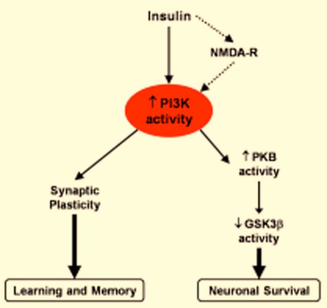
2 Current Alzheimer Research, 2008, Vol. 5, No. 5 Neumann et al.
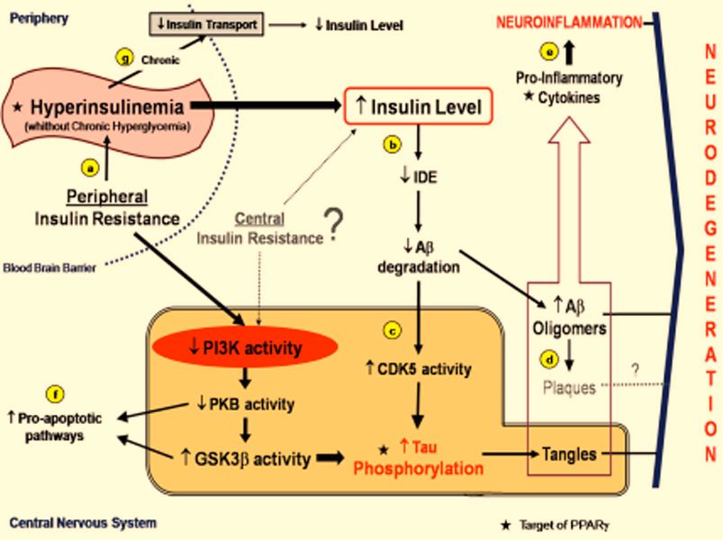
Fig. (1). Schematic representation of molecular pathways linking insulin resistance and neurodegeneration, and therapeutic target of
PPAR ligands.
(a) Peripheral insulin resistance results in periods of hyperinsulinemia to overcome the hyperglycemia. (b) Hyperinsulinemia, with the possi-
ble contribution of central insulin resistance, leads to increased insulin levels in the CNS, that kidnaps IDE resulting in a decrease of A
degradation, which upregulates cdk5 with consequent tau hyperphosphorylation, in the meantime that increases A oligomers. (c) Peripheral
insulin resistance is associated with decreased insulin signaling in neuron, that also can be a result of central insulin resistance, with de-
creased PI3K and PKB activity, leading an increased GSK3 activity. Cdk5 and GSK3 activities increase tau phosphorylation and tangles
formation. (d) A oligomers are more toxic than Senile Plaques, they are one of the responsible elements for activation of the innate immune
response in the glia. (e) These oligomers are among factors that lead to Neuroinflammation. (f) Downregulation of PKB activity and a high
GSK3 activity are also related with pro-apoptotic pathways that result in neurodegeneration. (g) Thus, chronic hyperinsulinemia (without
hyperglicemia) results in down-regulation of insulin transporters in the blood brain barrier and decreases the levels of insulin in the CNS.
lar weight [20]. IRs are widely distributed in the brain with functions as supported by neuronal activity, and in the con-
the highest concentration in the olfactory bulb, hypothala- trol of aging-related processes [26].
mus, cerebral cortex, cerebellum and hippocampus. They are
expressed in all regions of brain in both neurons and glial 2. Glucose, Insulin and Cognition
cells. IRs are distributed throughout the neuronal processes
The brain consumes metabolic energy disproportionate to
and the cell body, but are concentrated in the synaptic end-
its size. It uses glucose (primary fuel), largely through oxida-
ings [4, 21–24].
tive metabolism. Glucose deprivation leads to coma, sei-
The signaling and biological effects of the insulin have zures, and produces potentially permanent brain damage.
been widely studied mainly in the “classical” insulin target Glucose is continually supplied from cerebral blood flow and
tissues, e.g. liver, fat and skeletal muscle, with respect to must be transported into the brain through the endothelial
glucose uptake, regulation of cell proliferation, gene expres- cells that form the BBB to reach neurons and glial cells by
sion and the suppression of hepatic glucose production [4]. facilitated diffusion. Carriers of glucose are the glucose
However, insulin plays many roles within the CNS. It has transport proteins (GLUT), which allow glucose entry into
been shown that some of the CNS effects of insulin are the individual cells (neurons and glial cells) [27]. Animal mod-
opposite to those exerted in peripheral tissues. In particular, els show that the cerebral blood flux depends on glucose
CNS insulin increases glucose and inhibits feeding, whereas levels in the blood stream; there is a compensatory mecha-
serum insulin decreases glucose and increases feeding nism to maintain adequate delivery of glucose fuel to the
[14,25]. Thus, to some extent, insulin acts as its own coun- brain [28,29].
terregulatory hormone, with CNS insulin producing features
Evidence suggests regionally specific effects of insulin
of insulin resistance [6]. Thus, in the CNS, insulin partici-
on brain glucose metabolism. Insulin does not seem to influ-
pates in the regulation of feeding behavior and energy ho-
meostasis, neuronal maintenance, neurogenesis, and neuro- ence basal cerebral glucose metabolism or transport of glu-
cose into the brain [30–35]. Insulin affects the use of glucose
transmitter regulation. In addition, it has a role on cognitiveInsulin Resistance and Alzheimer´s Disease Current Alzheimer Research, 2008, Vol. 5, No. 5 3
in specific regions of the brain, most likely by selective dis- 2. Hyperinsulinemia/Hyperglicemia in the Brain
tribution of insulin-sensitive GLUT isoforms, which overlap
Alterations in circulating glucose levels can negatively
with the distribution of insulin and IRs in the brain [36–39].
affect the CNS because neurons have a consistently high
Insulin, through the IRs localized in the hypothalamus, con-
glucose demand. Neuronal glucose uptake depends on ex-
tributes to the regulation of food intake and energy homeo-
stasis and leads to changes in body weight by anorexigenic tracellular glucose concentration, but chronic hyperglycemia
results in cellular damage (glucose neurotoxicity). Therefore,
or orexigenic effects produced by increasing or decreasing
a tight metabolic control is very important [67–69]. Studies
levels of insulin in brain respectively. Moreover, insulin in
the CNS modifies peripheral glucose metabolism by increas-
on hyperglycemic rodents have shown cognitive im-
ing insulin sensitivity in peripheral tissue [10,40,41].
pairment in addition to functional and structural altera-
tions in the brain [68].
In brain, insulin is not a major regulator of glucose me-
A growing body of evidence suggests that peripheral
tabolism. In vitro studies showed that insulin regulates glu-
insulin abnormalities increase the risk for memory loss and
cose uptake of glial cells, but did not influence neuronal glu-
neurodegenerative disorders such as AD, but acute and
cose uptake [42,43]. Insulin can influence neurons directly
chronic hyperinsulinemia have opposing effects on memory
by mechanisms unrelated to modulation of glucose uptake.
performance (Fig. 2). A possible explanation for that is dis-
Neurotransmitter release, neuronal-outgrowth, tubulin activ-
ity, neuronal survival and synaptic plasticity are all directly cussed below. Chronic hyperinsulinemia has a negative in-
fluence on memory, since type II Diabetes Mellitus has been
modulated by insulin [5, 44–46]
associated with long-term impairment in cognitive function
Studies in human and animal models have shown that an in humans and animal model studies. On the other hand,
increase in brain insulin has a cognition-enhancing effect, acute increases of peripheral or brain insulin have an en-
independently of changes in peripheral glucose [47–51]. hanced memory performance effect [47,49, 63, 70–73]. Pre-
Moreover, there is cumulative evidence to support the effects diabetic conditions with hyperinsulinemia but not chronic
of insulin and IRs on cognition, mediated by a modulatory hyperglycemia may persist for many years before progres-
role in learning and memory processes [1,4,5,26,52]. Insulin sion to type II Diabetes Mellitus. Hyperinsulinemia exposes
also modulates CNS concentration of neurotransmitters as- the cells (including neurons) to unphysiologically high levels
sociated with important roles in cognition such as acetylcho- of insulin for a long period of time. Studies have shown that
line, norepinephine and dopamine [53,54]. It has been shown high concentrations of insulin affect the function and sur-
that, in an early stage of memory formation, an alteration of vival of neurons in culture by sensitizing them to toxin and
gene expression of IRs in the rat hippocampus in response to stress-induced insults [74].
learning experiences occurs [55]. Taken together, all these
findings suggest that insulin may influence normal memory
function (Figs. 1 and 2).
The biological basis of learning and memory processes
resides in synaptic strength, where insulin signaling plays a
modulator role on synaptic long-term potentiation (LTP) and
long-term depression (LTD), two opposite forms of activity-
dependent synaptic modifications. Insulin signaling modu-
lates synaptic plasticity by: 1) Promoting the recruitment of
GABA receptors on post-synaptic membranes; 2) Influenc-
ing NMDA receptor conductance (neuronal Ca2+ influx); and
3) Regulating AMPA receptor cycling [56–62].
B) INSULIN RESISTANCE
1. Insulin Resistance
Reduced sensitivity to insulin by the main target organs
(liver, fat, muscle) is known as Insulin Resistance, where
there is an elevated level of insulin in the bloodstream (hy-
perinsulinemia). This reduced sensitivity also results in im-
paired response to oral glucose (referred to as Impaired Glu-
cose Tolerance), where it takes longer to restore normal glu- Fig. (2). Schematic representation of the relationships between
cose levels after eating. Peripheral Insulin Resistance is insulin signaling, learning, memory and neuronal survival.
known to be the major contributor to the progression to hy- Acute high levels of insulin in the CNS have been related with the
perglycemia and Type II Diabetes Mellitus, since the pan- improvement of cognitive function and Neuronal Survival by a
creas can not secrete enough insulin to overcome the insulin pathway that it involves the NMDA-R, PI3K and PKB activity and
resistance and prevent these events. Hyperinsulinemia with- inactivation of GSK3.
out hyperglycemia is an indication of Insulin Resistance, a
pre-diabetic condition. There is evidence of an increased risk
of cognitive decline and neurodegeneration in populations Moreover, a relationship between glycosylation of tau
with peripheral insulin resistance (hyperinsulinemia without proteins, its aggregation ability and the neurodegenerative
hyperglycemia) [63–66]. phenotype has been revealed [75]. Peripheral injection of4 Current Alzheimer Research, 2008, Vol. 5, No. 5 Neumann et al.
high doses of insulin in mice caused a rapid and dose- the first generation of AD drugs, the cholinesterase inhibitors
dependent increase in tau phosphorylation in the CNS [76]. donepecil, galantamine and rivastigmine. The reduced cellu-
In vitro studies indicate that insulin modulates the levels of lar availability of acetyl-CoA may also cause decreased for-
A peptide by promoting the release of intracellular A. mation of both intracellular cholesterol and neurosteroids,
Physiological insulin levels promote A clearance by peptide which are the main lipid components of cell membranes.
degradation, a mechanism that involves insulin degrading Membrane abnormalities have been observed in AD brains.
enzyme (IDE) activity (detailed below in section III.1) [77–
The A peptide (or A oligomers) has been shown to
79]. A low level of insulin in brain reduces A release from
cause decreased glucose uptake/metabolism [97]. Further-
intracellular to extracellular compartments and high levels
more, apolipoprotein E4 (apoE4) and diabetes mellitus are
reduce A degradation in the extracellular compartment [80–
known to be major risk factors for AD [98]. Individuals with
84]. apoE4 alleles have lower brain glucose metabolism than
Impaired verbal memory has been reported in individuals those carrying apoE2 or apoE3 alleles. Diabetes patients are
with hyperinsulinemia and not chronic hyperglycemia [85]. characterized by deficient glucose uptake/metabolism.
Epidemiological studies have indicated that hyperinsuline- Hence, it can be speculated that apoE4 genotype together
mia, independent of glucose levels, constitutes one of the with diabetes may increase the risk for AD by impairment in
risk factors for dementia in the type II diabetes mellitus brain glucose uptake/metabolism.
population as well as in the non-diabetic population, since
the raising of peripheral insulin concentration acutely in- C) LINK BETWEEN INSULIN RESISTANCE AND
creases its concentration in the CSF and brain [63,65,70,86]. ALZHEIMER DISEASE
Prolonged peripheral hyperinsulinemia downregulates BBB
1. Relationships between Insulin Resistance and AD
functions and the IR activity and reduces insulin transport
Pathogenesis
into the brain [86, 87]. Thus, hyperinsulinemia during the
development of type II diabetes mellitus is neurotoxic. De- Abnormalities in insulin metabolism, characteristic of
velopment of these complications depend on the duration of type II diabetes, are among the major factors thought to
a diabetic condition, upregulation of circulating glucose, mechanistically influence the onset of AD. These abnormali-
glycosylated proteins, etc., and the quality of metabolic con- ties are thought to play a role in AD via their influence on
trol [67]. the synthesis and degradation of A and as a consequence of
Insulin resistance, hyperinsulinemia and type II Diabetes the cascade of neuronal alterations resulting from the effects
Mellitus are associated with elevated inflammatory markers of danger/alarm signals from oligomeric amyloid species
and increased risk for AD [63,65,88–90] (Fig. 1). In adults (Fig. 1) [99]. Additionally, recent studies have indicated that
with type II Diabetes Mellitus and impaired glucose toler- certain signal transduction pathways downstream of the insu-
ance, an abnormal level of soluble TNF-R1 (death receptor lin receptor may also promote the generation of A peptides
domain) and TNF-R2 (cell survival) has been reported. In- by modulating the cleavage of the parent A precursor pro-
creased levels of TNF-R1 and decreased levels of TNF-R2 tein (APP) at the -secretase site, a cleavage site necessary
have also been observed in AD brain [91–93]. for A amyloidogenicity [100].
Although this evidence tentatively suggests that type II
3. Impaired Glucose Uptake and Metabolism in the AD Diabetes Mellitus might play an important role in AD
Brain through mechanisms that involve A peptide generation,
alternative studies suggest that insulin may also provoke
Studies using positron emission tomography (PET) have
demonstrated that glucose metabolism is reduced markedly amyloid accumulation by limiting A degradation via direct
competition for the IDE. Another major substrate of IDE is
in the cerebral cortex in early stage AD and in the mild cog-
the A peptide. The IDE degrades monomeric but not the
nitive impairment (MCI) that is believed to be a precursor of
oligomeric A peptide [77,78]. IDE is a zinc-metallo-
AD, and that the reduction parallels the worsening of demen-
peptidase that preferentially cleaves proteins with a propen-
tia symptoms, establishing that glucose uptake and metabo-
sity to form -pleated sheet-rich amyloid fibrils, such as A
lism are impaired in AD brain [94]. However, the causes of
the impairment of glucose uptake/metabolism in AD brain peptides. This relationship of IDE with A is supported by
recent evidence indicating that IDE activity in the brain is
are not well understood. Several findings suggest that this
negatively correlated with A content, and that IDE expres-
impairment is a cause, rather than a consequence, of neu-
sion is decreased in the AD brain [83]. Insulin regulates the
rodegeneration. The impaired cerebral glucose consumption
levels of IDE. Hyperinsulinemia reduces A degradation by
is more severe than the impaired oxygen consumption in
reduction of IDE levels and binding competition (Fig. 1)
AD, suggesting that the former is not the result of the latter.
[80–84].
The brain is highly dependent upon glucose as a source
It has been reported that A40 and A42 reduce insulin
of energy. The impaired glucose uptake/metabolism might
binding and insulin receptor autophosphorylation. The re-
lead to deficient synaptic activity and cellular homeostasis,
duction in this binding seems to be caused by a decrease in
as these are very sensitive to energy deficiency. Impaired
the affinity of insulin to the insulin receptor. This suggests
glucose uptake/metabolism also causes reduced formation of
acetyl-CoA and, consequently, affects the synthesis of ace- that A is a direct competitive inhibitor of insulin binding
and action [101], an aspect that demands further investiga-
tylcholine [95]. Deficient activity of the cholinergic system
tion.
is one of the most significant biochemical deficiencies in AD
pathology, discovered decades ago [96], and is the basis ofInsulin Resistance and Alzheimer´s Disease Current Alzheimer Research, 2008, Vol. 5, No. 5 5
The strikingly reduced CNS expression of genes encod- resistance when genetic background predisposes to AD.
ing insulin, IGF-I, and IGF-II, as well as the insulin and IGF- However, further studies are necessary to clarify this.
I receptors in AD led to some authors to suggest that AD
Insulin binding to the IRs leads to autophosphorylation of
may represent a neuro-endocrine disorder that resembles
the IRs which initiates several signaling cascades. One of
diabetes mellitus [102]. In addition, the same researchers these is the lipid kinase phosphatidylinositol 3-kinase (PI3K)
demonstrated that alterations insulin levels and insulin-like
cascade, which is associated with almost all of the metabolic
growth factor expression and deterioration of insulin func-
action of insulin. PI3K activation leads to the activation of
tion with the course of AD progression, were linked with an
Protein Kinase B (PKB or Akt) and Glycogen Synthase
acetylcholine decrease in the brain [103]. There is evidence
Kinase 3 (GSK3). Activation of PKB inactives GSK3
supporting the notion that high plasma insulin levels and
by phosphorylation [110,111]. The neuroprotective effect of
peripheral insulin resistance affect A42 levels, inflamma- IGF-1 results from activation of PKB [112]. Expression of
tion in the CNS and cognitive performance of individuals
PKB protects neurons against toxin-induced death and pro-
[104]. From such evidence, a model can be constructed de-
tects PC12 cells against A peptide-induced death [113–
scribing how this metabolic profile contributes to the patho-
115].
genesis of AD. There are several etiological factors leading
to the final common expression in the AD pathology [103]. Cole and coworkers have reviewed the molecular link
between insulin action, diabetes and AD [5]. Insulin resis-
Insulin plays an important role in memory and brain
tance in the periphery produces hyperinsulinemia, while in
function in general. Peripheral hyperinsulinemia and insulin
the brain it decreases IDE activity. The effects of IDE in-
resistance induce a number of deleterious effects in the CNS
clude abnormal A removal and plaque formation, increased
that interfere with these functions, in a manner that is exac-
cdk5 activity, and increased activity of GSK3 (Fig. 1). Dys-
erbated by obesity and aging. In particular, effects on A
regulation of cdk5 is a major molecular event in the pathway
regulation and neuroinflammation are potential culprits in to neurodegeneration [116–118]. GSK3 activity has been
promoting aging-related memory impairment in some cases
implicated in the pathology of AD in different ways. AD
of AD (see Fig. 1). This possibility has obvious relevance for
brain exhibits a dysregulated expression of this kinase as
adults with type II Diabetes Mellitus. However, it is worth
well as changes in its activity [119,120], leading to hyper-
noting that hyperinsulinemia and insulin resistance afflict
phosphorylation of tau and tangle formation. Several studies
many non-diabetic adults with conditions such as obesity,
have shown that GSK3 activity is required for induction of
impaired glucose tolerance, cardiovascular disease, and hy- neuronal apoptosis (refer to Fig. 1), while inhibition of
pertension [105–107].
GSK3 promotes neuronal survival (illustrated schematically
Indeed, in recent years, cumulative evidence has been in Fig. 2) (reviewed by Cole et al, 2007 [5]). Insulin signal-
gained on the involvement of alteration in neuronal lipopro- ing induces the phosphorylation and inhibition of GSK3
teins activity, as well as a role of cholesterol and other lipids [110].
in the pathogenesis of this neurodegenerative disorder. In All of the negative effects of hyperinsulinemia in the
relation to hypercholesterolemia, several reports have shown
CNS and their associated functions could be exacerbated or
that elevated serum cholesterol levels and elevated levels of
similarly produced by a central insulin resistance in conjunc-
A are linked with AD risk. Cholesterol influences the activ-
tion with the peripheral insulin resistance or secondary to it.
ity of the enzymes involved in the metabolism of the amy-
This is supported by the evidence of central molecular insu-
loid precursor protein and in the production of A, but the
lin resistance in a mouse model of AD with induced periph-
mechanism by which cholesterol affects A production and eral insulin resistance, where a reduced basal signaling (re-
metabolism is not fully understood [108].
duced phosphorylation of IR, PKB and GSK3, as well as
decreased PI3K activity) was shown in the cerebral cortex
2. Metabolomics of Insulin in the Context of Neuronal
(Fig. 1) [84,109].
Survival and AD Onset
Studies of insulin action in brain are focused on the basic
To understand the link between insulin, insulin resis- effects of insulin signaling. Insulin acts like a neurotrophic
tance, neuronal survival and AD onset, it is important iden- factor, since it promotes neuronal survival [46,121]. Studies
tify the link between the key molecules involved in the intra- in vitro of intracellular pathways utilized by insulin for syn-
cellular pathways utilized by insulin to exert its effect. Insu- aptic plasticity have identified a link to neuronal protection
lin resistance in the periphery produces acute episodes of against cell death [46,121]. Intracellular pathways utilized by
hyperinsulinemia without chronic hyperglycemia. High lev- insulin to influence synaptic plasticity and neuronal survival
els of insulin in plasma are correlated with high levels of converge on the PI3K pathway (Fig. 2) (reviewed by van der
insulin in brain (Fig. 1) [86], leading to neurotoxic effects Heide et al. 2006 [1]). The increased catalytic activity of
[74]. PI3K results in the phosphorylation and activation of anti-
Studies in transgenic mouse models of AD have shown apoptotic substrates [1]. PKB is of major importance in me-
that diet-induced peripheral insulin resistance promotes amy- diating the effects of PI3K in neuronal survival, and in vitro
loidosis suggesting that peripheral insulin resistance can in- and in vivo studies have shown that activated PKB protects
fluence A production in the brain [84,109]. These findings against apoptosis. Dominant negative PKB does not [122–
in association of a reduced basal signaling of insulin in cor- 124]. Studies on insulin-facilitated LTP and LTD induction
tex with an increased degree of AD neuropathology argue show that this process is completely NMDA receptor-
that peripheral insulin resistance promotes neuronal insulin dependent [62]. PI3K activity is required for LTP mainte-
nance in the hippocampal CA1 region [125]. An increased6 Current Alzheimer Research, 2008, Vol. 5, No. 5 Neumann et al.
gene expression of IR in the rat hippocampus in response to ated glucose uptake. In addition to these functions, they also
learning experiences has been shown [55]. All together, the have been demonstrated to diminish the levels of multiple
effect of insulin on neuronal survival and its effects on inflammatory mediators (Fig. 1) (reviewed by Jiang, 2008,
NMDA-dependent synaptic plasticity, require similar intra- Rojo et al., 2008 [90,133]). Studies in cellular models have
cellular signaling routes (schematically illustrated in Fig. 2). shown that PPAR agonists down-regulate A peptide depo-
sition that occurs in AD, although the mechanisms of this
Clinical Studies on Insulin Resistance and AD phenomenon demand further investigation [133–135]. Tro-
glitazone, a thiazolidinedione, significantly reduced phos-
In the last five years a growing body of evidence has
phorylation of tau protein at Ser202 and Ser396/404, resi-
been developed suggesting that glucose metabolism is asso-
dues phosphorylated in early and later stages of neurofibril-
ciated with the pathogenesis of AD, age-related cognitive
lary tangle accumulation in AD and other neurodegenerative
decline and neuroinflamation [90,126–128]. Intravenous disorders (Fig. 1, PPAR ligands) [136].
insulin infusion in healthy, eugylcemic older subjects (a pro-
cedure known as hyperinsulinemic-euglycemic clamp) in- Interestingly, the PPAR Pro12Ala polymorphism, which
duces a facilitation of memory while it increases cerebrospi- is associated with an increased risk of type II diabetes [137],
nal fluid levels of A1-42 [126]. Insulin mediated memory has been found to influence plasma 24S-hydroxycholesterol/
facilitation is less marked at supraoptimal levels of insulin cholesterol ratio in AD patients. This fact is important con-
and in subjects with AD the hyperinsulinemic facilitation of sidering that elevated cholesterol in blood is a risk factor for
memory is obtained at higher concentrations of insulin when AD and 24S-hydroxycholesterol is the major product of
compared with normal subjects. This phenomenon is more brain cholesterol metabolism and is released into the blood
clear in patients who are not carriers of the APOE-4 gene stream [108]. At present pioglitazone and rosiglitazone are
[51]. the two thiazolidinediones that are available for clinical use.
Both share the adverse side-effect of generating edema and
The clinical effects of insulin and/or glucose administra- potentially heart failure [138]. A recent meta-analysis [139]
tion on memory in patients with AD have been extensively
revealed a possible increased risk of myocardial infarction
reviewed [128,129]. As a result, it has been suggested that
and a borderline increase in the risk of death from cardiovas-
moderate administration of both glucose and insulin could
cular causes in patients receiving rosiglitazone. However, the
improve memory in patients. These observations appear to
significance of the data has been debated [140]. Thus,
be in disagreement with Gispen and Biessels [68] who de-
rosiglitazone as a possible therapy for AD is an active field
scribed that both chronic hyperglycemia and chronic hyper- of clinical investigation.
insulinemia are associated with accelerated cognitive decline
in the elderly. This apparent discrepancy may be explained Rosiglitazone has been shown to preserve memory func-
in part by the mechanisms underlying spontaneous chronic tion in AD patients when compared with a placebo-assigned
hyperglycemia and chronic hyperinsulinaemia during insulin group. In a double blind trial, 20 mild AD or MCI patients
resistance. Hoyer [130] has comprehensively addressed this who received 4 mg of rosiglitazone for 6 months showed
issue in his recent review. He classified AD as an ‘‘insulin improvements in memory and selective attention, associated
resistant brain state’’. This interesting hypothesis is sup- with less reduction in plasma A-40 and A-42 when com-
ported by a recent clinical trial performed by Reger et al. pared to 10 control patients [129]. This stabilization of
[73] where the administration of intranasal insulin improved plasma A could be of importance, considering that plasma
cognitive performance in elderly patients. In this pilot study, A1-42 decreases with AD progression, as previously de-
the authors administered 20 IU BID intranasal insulin to 13 scribed [82]. Another study on 511 AD patients treated with
patients and compared them with 12 controls receiving pla- 2 mg, 4 mg or 8 mg of rosiglitazone or placebo found that
cebo. They found no changes in plasma levels of glucose and there is an improvement in cognitive function when evalu-
insulin with the treatment. However the insulin-treated group ated by the Alzheimer’s Disease Assesment Scale-cognitive
retained more verbal information after a delay and showed (ADAS-cog) for patients treated with 8 mg of rosiglitazone,
improved attention and functional status. Insulin treatment and that this result is restricted to those patients who are not
also increased the A1-40/A1-42 ratio in plasma. Previous carriers of APOE-4 [141]. This modulation of the effect of
studies by the same author [131] demonstrated that this ef- rosiglitazone by the APOE genotype strongly resembles
fect was restricted to APOE-4 negative subjects. what has been observed for insulin and support the recom-
mendation for APOE genotyping of patients at risk of cogni-
Modulation of the response to insulin by the APOE geno-
tive decline [142]. Even though this evidence is still prelimi-
type has led to speculation regarding whether APOE-4 is an
nary, it supports the role of PPAR ligands with their dual
independent risk factor for AD, so that people carrying this
action over insulin resistance and on neuroinflammation as a
allele do not have a greater risk of being insulin resistant novel strategy for the treatment of cognitive decline associ-
than the general population. In the other hand, people that
ated with AD and stress the importance of further studies in
are insulin resistant are at risk of developing AD regardless
this perspective.
of their APOE-4 status [128]. The evidence that favors the
role of insulin resistance in AD has opened new perspectives
ACKNOWLEDGMENTS
for treatment options. The family of antidiabetic Thia-
zolidinedione drugs function as agonists of the nuclear re- Research has been supported by a grant from Fondecyt
ceptor peroxisome proliferator-activated receptor (PPAR)- 1080254 and a grant from the Alzheimer´s Association,
and have been shown to improve sensitivity to insulin by U.S.A. Authors are grateful to Sarah Murray MD. for care-
decreasing circulating insulin and increasing insulin medi- fully revising the text of this paper.Insulin Resistance and Alzheimer´s Disease Current Alzheimer Research, 2008, Vol. 5, No. 5 7
REFERENCES weight of marmots during the summer feeding period. Physiol Be-
hav 49: 335–338 (1991).
[1] Van der Heide LP, Ramakers GM and Smidt MP. Insulin signaling [26] Craft S and Watson GS. Insulin and neurodegenerative disease:
in the central nervous system: Learning to survive. Prog Neurobiol shared and specific mechanisms. Lancet Neurol 3: 169–178 (2004).
79: 205–221 (2006). [27] Clarke DD and Sokoloff L. Circulation and energy metabolism of
[2] Bagdade JD, Bierman EL and Porte Jr D. The Significance of Basal the brain. In: ‘Basic neurochemistry’. (Eds: Siegel G J, Agranoff
Insulin Levels in the Evaluation of the Insulin Response to Glucose BW, Albers RW, Molinoff SK, Fisher PB, Uhler MD), Lippincott-
in Diabetic and Nondiabetic Subjects. J Clin Invest 46: 1549-1557 Ratven, Philadelphia, p. 637–669 (1999).
(1967). [28] Duckrow RB, Beard DC and Brennan RW. Regional cerebral blood
[3] Polonsky KS, Given BD, Hirsch L, Shapiro ET, Tillil H, Beebe C, flow decreases during chronic and acute hyperglycemia. Stroke 18:
et al. Quantitative Study of Insulin Secretion and Clearance in 52–58 (1987).
Normal and Obese Subjects. J Clin Invest 81:435–441 (1987). [29] Pelligrino DA, Segil LJ and Albrecht RF. Brain glucose utilization
[4] Plum L, Schubert M and Brüning JC. The role of insulin receptor and transport and cortical function in chronic vs. acute hypoglyce-
signaling in the brain. Trends Endocrino Metab 16: 59–65 (2005). mia. Am J Physiol. 259: E729–E735 (1990).
[5] Cole AR, Astell A, Green C and Sutherland C. Molecular connex- [30] Lucignani G, Namba H, Nehlig A, Porrino LJ, Kennedy C and
ions between dementia and diabetes. Neurosci Biobehav Rev 31(7): Sokoloff L. Effects of insulin on local cerebral glucose utilization
1046–1063 (2007). in the rat. J Cereb Blood Flow Metab 7: 309–314 (1987).
[6] Banks WA. The source of cerebral insulin. Eur J Pharmacol 490: [31] Marfaing P, Penicaud L, Broer Y, Mraovitch S, Calando Y and
5–12 (2004). Picon L. Effects of hyperinsulinemia on local cerebral insulin bind-
[7] Banks WA. Blood-brain barrier and energy balance. Obesity (Sil- ing and glucose utilization in normoglycemic awake rats. Neurosci
ver Spring) 14 Suppl 5: 234S-237S (2006). Lett 115: 279–285 (1990).
[8] Margolis RU and Altszuler N. Insulin in the cerebrospinal fluid. [32] Doyle P, Cusin I, Rohner-Jeanrenaud F and Jeanrenaud B. Four-
Nature 215: 1375–1376 (1967). day hyperinsulinemia in euglycemic conditions alters local cerebral
[9] Steffens AB, Scheurink AJ, Porte D Jr and Woods SC. Penetration glucose utilization in specific brain nuclei of freely moving rats.
of peripheral glucose and insulin into cerebrospinal fluid in rats. Brain Res 684(1): 47–55 (1995).
Am J Physiol 255: R200–R204 (1988). [33] Fanelli C, Dence C and Markham J. Blood-to-brain glucose trans-
[10] Schwartz MW, Figlewicz DP, Baskin DG, Woods SC and Porte D port and cerebral glucose metabolism are not reduced in poorly
Jr. Insulin in the brain: a hormonal regulator of energy balance. controlled type 1 diabetes. Diabetes 47: 1444–1450 (1998).
Endocr Rev 13: 387–414 (1992). [34] Hasselbalch S, Knudsen G and Videback C. No effect of insulin on
[11] Baura GD, Foster DM, Porte D Jr, Kahn SE, Bergman RN, Cobelli glucose blood-brain barrier transport and cerebral metabolism in
C, et al. Saturable transport of insulin from plasma into the central humans. Diabetes 48: 1915–1921 (1999).
nervous system of dogs in vivo. A mechanism for regulated insulin [35] Bingham EM, Hopkins D and Smith D. The role of insulin in hu-
delivery to the brain. J Clin Invest 92: 1824–1830 (1993). man brain glucose metabolism: an 18-fluoro-deoxyglucose positron
[12] Banks WA, Jaspan JB and Kastin AJ. Selective, physiological emission tomography study. Diabetes 51: 3384–3390 (2002).
transport of insulin across the blood-brain barrier: novel demonstra- [36] Brant AM, Jess TJ, Milligan G, Brown CM and Gould GW. Immu-
tion by species-specific radioimmunoassay. Peptides 18:1257–1262 nological analysis of glucose transporters expressed in different re-
(1997). gions of the rat brain and central nervous system. Biochem Biophys
[13] Banks WA, Jaspan JB, Huang W and Kastin AJ. Transport of insu- Res Commun 192: 1297–1302 (1993).
lin across the blood-brain barrier: saturability at euglycemic doses [37] El Messari S, Leloup C, Quignon M, Brisorgueil MJ, Penicaud L
of insulin. Peptides 18 (9):1423–1429 (1997). and Arluison M. Immunocytochemical localization of the insulin-
[14] Woods SC, Seeley RJ, Baskin DG and Schwartz MW. Insulin and responsive glucose transporter 4 (Glut4) in the rat central nervous
blood-brain barrier. Curr Pharm Des 9(10): 795–800 (2003). system. J Comp Neurol 399: 492–512 (1998).
[15] Devaskar SU, Giddings SJ, Rajakumar PA, Carnaghi LR, Menon [38] Ibberson M, Uldry M and Thorens B. GLUTX1, a novel mammal-
RK and Zahm DS. Insulin gene expression and insulin synthesis in ian glucose transporter expressed in the central nervous system and
mammalian neuronal cells. J Biol Chem 269: 8445–8454 (1994). insulin-sensitive tissues. J Biol Chem 275: 4607–4612 (2000).
[16] Rulifson EJ, Kim SK and Nusse R.. Ablation of insulin-producing [39] Reagan L, Gorovits N and Hoskin E. Localization and regulation of
neurons in flies: growth and diabetic phenotypes. Science 296: GLUTx1 glucose transporter in the hippocampus of streptozotocin
1118–1120 (2002). diabetic rats. Proc Natl Acad Sci USA 98:2820–2825 (2001).
[17] Havrankova J and Roth J. Insulin receptors are widely distributed [40] Obici S, Zhang BB, Karkanias G and Rossetti L. Hypothalamic
in central nervous-system of rat. Nature 212: 827–829 (1978). insulin signaling is required for inhibition of glucose production.
[18] van Houten M, Posner BI, Kopriwa BM and Brawer JR. Insulin- Nat Med 8:1376–82 (2002).
binding sites in the rat brain: in vivo localization to the circumven- [41] Niswender KD and Schwartz MW. Insulin and leptin revisited:
tricular organs by quantitative radioautography. Endocrinology adiposity signals with overlapping physiological and intracellular
105(3): 666–673 (1979). signaling capabilities. Front Neuroendocrinol 24: 1–10 (2003).
[19] Baskin DG, Woods SC, West DB, van Houten M, Posner BI, Dorsa [42] Clarke DW, Boyd FT Jr, Kappy MS and Raizada MK. Insulin
DM, et al. Immunocytochemical detection of insulin in rat hypo- binds to specific receptors and stimulates 2-deoxy-D-glucose up-
thalamus and its possible uptake from cerebrospinal fluid. Endocri- take in cultured glial cells from rat brain. J Bio Chem 259: 11672–
nology 113: 1818–1825 (1983). 11675 (1984).
[20] Heidenreich KA, Zahniser NR, Berhanu P, Brandenburg D and [43] Schubert M, Gautam D, Surjo D, Ueki K, Baudler S, Schubert D, et
Olefsky JM. Structural differences between insulin receptors in the al. Role for neuronal insulin resistance in neurodegenerative dis-
brain and peripheral target tissues. J Biol Chem 258: 8527–8530 ease. Proc Natl Acad Sci USA 101(9): 3100–3105 (2004).
(1983). [44] Mill JF, Chao MV and Ishii DN. Insulin, insulin-like growth factor
[21] Havrankova J, Brownstein M and Roth J. Insulin and insulin- II, and nerve growth factor effects on tubulin mRNA levels and
receptors in rodent brain. Diabetologia 20: 26873 (1981). neurite formation. Proc Natl Acad Sci USA 82: 7126–7130 (1985).
[22] Wozniak M, Rydzewski B, Baker SP and Raizadai M. The cellular [45] Wang C, Li Y, Wible B, Angelides KJ and Ishii DN. Effects of
and physiological actions of insulin in the central nervous system. insulin and insulin-like growth factors on neurofilament mRNA
Neurochem Int 22: 1–10 (1993). and tubulin mRNA content in human neuroblastoma SH-SY5Y
[23] Unger JW and Betz M. Insulin receptors and signal transduction cells. Brain Res Mol Brain Res 13: 289–300 (1992).
proteins in the hypothalamo-hypophyseal system: a review on mor- [46] Tanaka M, Sawada M, Yoshida S, Hanaoka F and Marunouchi T.
phological findings and functional implications. Histol Histopathol Insulin prevents apoptosis of external granular layer neurons in rat
13: 1215–1224 (1998). cerebellar slice cultures. Neurosci Lett 199: 37–40 (1995).
[24] Abbott MA, Wells DG and Fallon JR. The insulin receptor tyrosine [47] Craft S, Asthana S, Newcomer JW, Wilkinson CW, Matos IT,
kinase substrate p58/53 and the insulin receptor are components of Baker LD, et al. Enhancement of memory in Alzheimer's disease
CNS synapses. J Neurosci 19: 7300–7308 (1999). with insulin and somatostatin, but not glucose. Arch Gen Psychia-
[25] Florant GL, Singer L, Scheurink AJ, Park CR, Richardson RD and try 56: 1135–1140 (1999).
Woods SC. Intraventicular insulin reduces food intake and body8 Current Alzheimer Research, 2008, Vol. 5, No. 5 Neumann et al.
[48] Kern W, Born J, Schreiber H and Fehm HL. Central nervous sys- population-based cohort study. Am J Epidemiol 145: 301–308
tem effects of intranasally administered insulin during euglycemia (1997).
in men. Diabetes 48: 557–563 (1999). [72] Fehm HL, Perras B, Smolnik R, Kern W and Born J. Manipulating
[49] Park CR, Seeley RJ, Craft S and Woods SC. Intracerebroventricu- neuropeptidergic pathways in humans: a novel approach to neuro-
lar insulin enhances memory in a passive-avoidance task. Physiol pharmacology?. Eur J Pharmacol 405: 43–54 (2000).
Behav 68: 509–514 (2000). [73] Reger MA, Watson GS, Green PS, Wilkinson CW, Baker LD,
[50] Kern W, Peters A, Fruehwald-Schultes B, Deininger E, Born J and Cholerton B, et al. Intranasal insulin improves cognition and modu-
Fehm HL. Improving influence of insulin on cognitive functions in lates -amyloid in early AD. Neurology 70(6): 440–448 (2008).
humans. Neuroendocrinology 74: 270–280 (2001). [74] Schafer M and Erdo SL. Development of glutamate neurotoxicity
[51] Craft S, Asthana S, Cook DG, Baker LD, Cherrier M, Purganan K, in cortical cultures: induction of vulnerability by insulin. Brain Res
et al. Insulin dose–response effects on memory and plasma amyloid Dev Brain Res 62(2): 293-296 (1991).
precursor protein in Alzheimer's disease: interactions with apolipo- [75] Gonzalez C, Farías G and Maccioni RB. Modification of tau to an
protein E genotype. Psychoneuroendocrinology 28: 809–822 Alzheimer's type protein interferes with its interaction with micro-
(2003). tubules. Cell Mol Biol (Noisy-le-grand) 44(7): 1117-1127 (1998).
[52] Dou JT, Chen M, Dufour F, Alkon DL and Zhao WQ. Insulin re- [76] Freude S, Plum L, Schnitker J, Leeser U, Udelhoven M, Krone W,
ceptor signaling in long-term memory consolidation following spa- et al. Peripheral hyperinsulinemia promotes tau phosphorylation in
tial learning. Learn Mem 12: 646–655 (2005). vivo. Diabetes 54: 3343–3348 (2005).
[53] Figlewicz DP, Szot P, Israel PA, Payne C and Dorsa DM. Insulin [77] Qiu WQ, Ye Z, Kholodenko D, Seubert P and Selkoe DJ. Degrada-
reduces norepinephrine transporter mRNA in vivo in rat locus co- tion of amyloid beta-protein by a metalloprotease secreted by mi-
eruleus. Brain Res 602: 161–164 (1993). croglia and other neural and non-neural cells. J Biol Chem 272:
[54] Kopf S and Baratti C. Effects of posttraining administration of 6641–6646 (1997).
insulin on retention of a habituation response in mice: participation [78] Qiu WQ, Walsh DM, Ye Z, Vekrellis K, Zhang J, Podlisny MB, et
of a central cholinergic mechanism. Neurobiol Learn Mem 71: 50– al.. Insulin-degrading enzyme regulates extracellular levels of amy-
61 (1999). loid beta-protein by degradation. J Biol Chem 273: 32730–32738
[55] Zhao W, Chen H, Xu H, Moore E, Meiri N, Quon MJ, et al. Brain (1998).
insulin receptors and spatial memory. correlated changes in gene [79] Gasparini L, Gouras GK, Wang R, Gross RS, Beal MF, Greengard
expression, tyrosine phosphorylation, and signaling molecules in P, et al. Stimulation of beta-amyloid precursor protein trafficking
the hippocampus of water maze trained rats. J Biol Chem 274: by insulin reduces intraneuronal beta-amyloid and requires mito-
34893–34902 (1999). gen-activated protein kinase signaling. J Neurosci 21: 2561–2570
[56] Wang YT and Salter MW. Regulation of NMDA receptors by (2001).
tyrosine kinases and phosphatases. Nature 369: 233–5 (1994). [80] Perez A, Morelli L, Cresto JC and Castano EM. Degradation of
[57] Wan Q, Xiong ZG, Man HY, Ackerley CA, Braunton J, Lu WY, et soluble amyloid beta peptides 1 – 40, 1 – 42, and the Dutch variant
al. Recruitment of functional GABA(A) receptors to postsynaptic 1 –40Q by insulin degrading enzyme from Alzheimer disease and
domains by insulin. Nature 388: 686–690 (1997). control brains. Neurochem Res 25: 247– 255 (2000).
[58] Man HY, Lin JW, Ju WH, Ahmadian G, Liu L, Becker LE, et al. [81] Farris W, Mansourian S, Chang Y, Lindsley L, Eckman EA, Frosch
Regulation of AMPA receptor-mediated synaptic transmission by MP, et al. Insulin-degrading enzyme regulates the levels of insulin,
clathrin-dependent receptor internalization. Neuron 25: 649–662 amyloid beta-protein, and the beta-amyloid precursor protein intra-
(2000). cellular domain in vivo. Proc Natl Acad Sci USA 100: 4162–4167
[59] Kneussel M. Dynamic regulation of GABA(A) receptors at synap- (2003).
tic sites. Brain Res Brain Res Rev 39: 74–83 (2002). [82] Mayeux R, Honig LS, Tang MX, Manly J, Stern Y, Schupf N, et al.
[60] Malenka RC. Synaptic plasticity and AMPA receptor trafficking. Plasma A[beta]40 and A[beta]42 and Alzheimer’s disease: relation
Ann NY Acad Sci 1003: 1–11 (2003). to age, mortality, and risk. Neurology 61: 1185–1190 (2003).
[61] Huang CC, Lee CC and Hsu KS.. An investigation into signal [83] Cook DG, Leverenz JB, McMillan PJ, Kulstad JJ, Ericksen S, Roth
transduction mechanisms involved in insulin-induced long-term RA, et al. Reduced hippocampal insulin-degrading enzyme in
depression in the CA1 region of the hippocampus. J Neurochem lateonset Alzheimer’s disease is associated with the apolipoprotein
89: 217–231 (2004). Eepsilon4 allele. Am J Pathol 162: 313–319 (2003).
[62] van der Heide LP, Kamal A, Artola A, Gispen WH and Ramakers [84] Ho L, Qin W, Pompl PN, Xiang Z, Wang J, Zhao Z, et al. Dietin-
GM. Insulin modulates hippocampal activity-dependent synaptic duced insulin resistance promotes amyloidosis in a transgenic
plasticity in a N-methyl-d-aspartate receptor and phosphatidyl- mouse model of Alzheimer’s disease. FASEB J 18: 902– 904
inositol-3-kinase-dependent manner. J Neurochem 94: 1158–1166 (2004).
(2005). [85] Vanhanen M, Koivisto K, Kuusisto J, Mykkänen L, Helkala EL,
[63] Ott A, Stolk RP, van Harskamp F, Pols HA, Hofman A and Bre- Hänninen T, et al. Cognitive function in an elderly population with
teler MM. Diabetes mellitus and the risk of dementia—The Rotter- persistent impaired glucose tolerance. Diabetes Care 21: 398–402
dam Study. Neurology 53: 1937–1942 (1999). (1998).
[64] Greenwood C and Winocur G. Glucose treatment reduces memory [86] Wallum BJ, Taborsky GJ Jr, Porte D Jr, Figlewicz DP, Jacobson L,
deficits in young rats fed high-fat diets. Neurobiol Learn Mem 75: Beard JC, et al. Cerebrospinal fluid insulin levels increase during
179–189 (2001). intravenous insulin infusions in man. J Clin Endocrinol Metab 64:
[65] Luchsinger JA, Tang MX, Shea S and Mayeux R. Hyperinsuline- 190–194 (1987).
mia and risk of Alzheimer disease. Neurology 63: 1187–1192 [87] Kaiyala KJ, Prigeon RL, Kahn SE, Woods SC and Schwartz MW.
(2004). Obesity induced by a high-fat diet is associated with reduced brain
[66] Yaffe K, Blackwell T, Kanaya AM, Davidowitz N, Barrett-Connor insulin transport in dogs. Diabetes 49: 1525–1533 (2000).
E and Krueger K.. Diabetes, impaired fasting glucose, and devel- [88] Strachan MW, Deary IJ, Ewing FM and Frier BM. Is type II diabe-
opment of cognitive impairment in older women. Neurology 63: tes associated with an increased risk of cognitive dysfunction? A
658–663 (2004). critical review of published studies. Diabetes Care 20: 438– 445
[67] Pirart J.. Diabetes mellitus and its degenerative complications— (1997).
prospective- study of 4400 patients observed between 1947 and [89] Caballero AE. Endothelial dysfunction, inflammation, and insulin
1973. Diab Metab 3: 245–256 (1977). resistance: a focus on subjects at risk for type 2 diabetes. Curr Diab
[68] Gispen WH and Biessels GJ. Cognition and synaptic plasticity in Rep 4: 237–246 (2004).
diabetes mellitus. Trends Neurosci 23: 542–549 (2000). [90] Rojo LE, Fernández JA, Maccioni AA, et al. Neuroinflammation:
[69] Tomlinson DR and Gardiner NJ. Glucose neurotoxicity. Nat Rev implications for the pathogenesis and molecular diagnosis of Alz-
Neurosci 9(1): 36–45 (2008). heimer's disease. Arch Med Res 39(1): 1–16 (2008).
[70] Ott A, Stolk RP, Hofman A, van Harskamp F, Grobbee DE and [91] Aggarwal S, Gollapudi S and Gupta S. Increased TNF-alpha-
Breteler MM. Association of diabetes mellitus and dementia: The induced apoptosis in lymphocytes from aged humans: changes in
Rotterdam study. Diabetologia 39: 1392–1397 (1996). TNF-alpha receptor expression and activation of caspases. J
[71] Leibson CL, Rocca WA, Hanson VA, Cha R, Kokmen E, Obrien Immunol 162: 2154– 2161(1999).
PC, et al. Risk of dementia among persons with diabetes mellitus: aInsulin Resistance and Alzheimer´s Disease Current Alzheimer Research, 2008, Vol. 5, No. 5 9
[92] Dzienis-Straczkowska S, Straczkowski M, Szelachowska M, Abeta(25-35) on Akt/PKB kinase and survival of PC12 cells. J
Stepien A, Kowalska I and Kinalska I. Soluble tumor necrosis fac- Neurochem 78: 1000–1008 (2001).
tor-alpha receptors in young obese subjects with normal and im- [116] Maccioni RB, Otth C, Concha II and Muñoz JP. The protein kinase
paired glucose tolerance. Diabetes Care 26: 875–880 (2003). Cdk5. Structural aspects, roles in neurogenesis and involvement in
[93] Zhao M, Cribbs DH, Anderson AJ, Cummings BJ, Su JH, Wasser- Alzheimer's pathology. Eur J Biochem 268(6): 1518–1527 (2001).
man AJ, et al. The induction of the TNFalpha death domain signal- [117] Alvarez A, Toro R, Caceres A, Maccioni RB. Inhibition of tau
ing pathway in Alzheimer’s disease brain. Neurochem Res 28: phosphorylating protein kinase cdk5 prevents beta-amyloid-
307–318 (2003). induced neuronal death. FEBS Lett 459(3): 421-426 (1999).
[94] Jagust WJ, Seab JP, Huesman RH, Valk PE, Mathis CA, Reed BR, [118] Alvarez A, Muñoz JP and Maccioni RB. A Cdk5–p35 Stable Com-
et al. Diminished glucose transport in Alzheimer’s disease : dy- plex Is Involved in the -Amyloid-Induced Deregulation of Cdk5
namic PET studies. J Cerebr Blood Flow Metab 11: 323-330 Activity in Hippocampal Neurons. Exp Cell Res 264(2): 266-274
(1991). (2001).
[95] Gibson GE, Petersen C and Jenden DJ. Brain acetylcholine synthe- [119] Shiurba RA, Ishiguro K, Takahashi M, Sato K, Spooner ET,
sis decline with senescence. Science 213: 674-676 (1981). Mercken M, et al. Immunocytochemistry of tau phosphoserine 413
[96] Bigl V, Arendt T, Fischer S, Werner M and Arendt A. The cho- and tau protein kinase I in Alzheimer pathology. Brain Res 737(1-
linergic system in aging, Gerontology 33: 172–180 (1987). 2): 119– 132 (1996).
[97] Prapong TJ, Buss WH, Hsu P and Heine H.. West Greenlee and E. [120] Leroy K, Boutajangout A, Authelet M, Woodgett JR, Anderton BH
Uemura. Amyloid beta-peptide decreases neuronal glucose uptake and Brion JP. The active form of glycogen synthase kinase- 3beta
despite causing increase in GLUT3 mRNA transcription and is associated with granulovacuolar degeneration in neurons in Alz-
GLUT3 translocation to the plasma membrane. Exp Neurol 174: heimer’s disease. Acta Neuropathol (Berl) 103: 91–99 (2002).
253–258 (2002). [121] Ryu BR, Ko HW, Jou I, Noh JS and Gwag BJ. Phosphatidylinositol
[98] Zhu X, Perry G and Smith MA. Insulin signaling, diabetes mellitus 3-kinase-mediated regulation of neuronal apoptosis and necrosis by
and risk of Alzheimer disease. J Alzheimers Dis 7: 81–84 (2005). insulin and IGF-I. J Neurobiol 39: 536–546 (1999).
[99] Fernandez J, Rojo L, Kuljis RO and Maccioni RB. The danger [122] Philpott KL, McCarthy MJ, Klippel A and Rubin LL.. Activated
signals hypothesis of Alzheimer s disease. J Alz Dis (2008). In phosphatidylinositol 3-kinase and Akt kinase promote survival of
press. superior cervical neurons. J Cell Biol 139: 809–815 (1997).
[100] Moreira PI, Santos MS, Seica R and Oliveira CR. Brain mitochon- [123] Matsuzaki H, Tamatani M, Mitsuda N, Namikawa K, Kiyama H,
drial dysfunction as a link between Alzheimer's disease and diabe- Miyake S, et al. Activation of Akt kinase inhibits apoptosis and
tes. J Neurol Sci 257: 206–214 (2007). changes in Bcl-2 and Bax expression induced by nitric oxide in
[101] Xie L, Helmerhorst E, Taddei K, Plewright B, Van Bronswijk W primary hippocampal neurons. J Neurochem 73: 2037–2046
and Martins R. Alzheimer's beta-amyloid peptides compete for in- (1999).
sulin binding to the insulin receptor. J Neurosci 22: RC221 (2002). [124] Yamaguchi A, Tamatani M, Matsuzaki H, Namikawa K, Kiyama
[102] Steen EJ, Terry BJ, Rivera EJ, Cannon JL, Neely TR, Tavares R, et H, Vitek MP, et al. Akt activation protects hippocampal neurons
al. Impaired insulin and insulin-like growth factor expression and from apoptosis by inhibiting transcriptional activity of p53. J Biol
signalling mechanisms in Alzheimer’s disease – Is this type 3 dia- Chem 276: 5256–5264 (2001).
betes?. J Alz Dis 7: 63–80 (2005). [125] Sanna PP, Cammalleri M, Berton F, Simpson C, Lutjens R, Bloom
[103] Rivera EJ, Goldin A, Fulmer N, Tavares R, Wands JR and de la FE, et al. Phosphatidylinositol 3-kinase is required for the expres-
Monte SM.. Insulin and insulin-like growth factor expression and sion but not for the induction or the maintenance of long-term po-
function deteriorate with progression of Alzheimer's disease: link to tentiation in the hippocampal CA1 region. J Neurosci 22: 3359–
brain reductions in acetylcholine. J Alzheimers Dis 8: 247–68 3365 (2002).
(2005). [126] Watson GS and Craft S.. The role of insulin resistance in the
[104] Steven ES, Jongsoon L and Allison BG. Inflammation and insulin pathogenesis of Alzheimer's disease: implications for treatment.
resistance. J Clin Invest 116(7): 1793–1801 (2006). CNS Drugs 17(1): 27–45 (2003).
[105] Röder HM and Ingram VM. Two novel kinases phosphorylate tau [127] Craft S. Insulin resistance syndrome and Alzheimer’s disease: Age-
and the KSP site of heavy neurofilament subunits in high and obesity-related effects on memory, amyloid, and inflammation.
stoichiometric ratios. J Neurosci 11: 3325–3342 (1991). Neurobiol Aging 26S: S65–S69 (2005).
[106] Bush ML, Miyashiro JS and Ingram VM. Activation of a neuro- [128] Craft S. Insulin resistance syndrome and Alzheimer disease: patho-
filament kinase, a tau kinase, and a tau phosphatase by decreased physiologic mechanisms and therapeutic implications. Alzheimer
ATP levels in nerve growth factor-differentiated PC12 cells. Proc Dis Assoc Disord 20: 298–301 (2006).
Natl Acad Sci USA 92: 1962–1965 (1995). [129] Watson GS, Cholerton BA, Reger MA, Baker LD, Plymate SR,
[107] Hong M and Lee VM. Insulin and insulin-like growth factor-1 Asthana S, et al. Preserved cognition in patients with early Alz-
regulate tau phosphorylation in cultured human neurons. J Biol heimer disease and amnestic mild cognitive impairment during
Chem 272: 19547–19553 (1997). treatment with rosiglitazone: a preliminary study. Am J Geriatr
[108] Rojo L, Sjöberg MK, Hernández P, Zambrano C and Maccioni RB. Psychiatry 13: 950–958 (2005).
Roles of cholesterol and lipids in the etiopathogenesis of Alz- [130] Hoyer S. Glucose metabolism and insulin receptor signal transduc-
heimer’s disease. J Biomed Biotechnol 2006(3): 73976 (2006). tion in Alzheimer disease. Eur J Pharmacol 490(1-3): 115–125
[109] Gasparini L, Netzer WJ, Greengard P and Xu H. Does insulin dys- (2004).
function play a role in Alzheimer’s disease?. Trends Pharmacol Sci [131] Reger MA, Watson GS, Frey II WH, Baker L, Cholerton B, Keel-
23: 288–293 (2002). ing M, et al. Effects of intranasal insulin on cognition in memory-
[110] Cross DA, Alessi DR, Cohen P, Andjelkovich M, Hemmings BA. impaired older adults: Modulation by APOE genotype. Neurobiol
Inhibition of glycogen synthase kinase-3 by insulin mediated by Aging 27: 451–458 (2006).
protein kinase B. Nature 378: 785–789 (1995). [132] Jiang Q, Heneka M and Landreth GE.. The Role of Peroxisome
[111] Fisher TL and White MF. Signaling pathways: the benefits of good Proliferator-Activated Receptor-gamma (PPARgamma) in Alz-
communication. Current Biology 14: R1005–R1007 (2004). heimer's Disease: Therapeutic Implications. CNS Drugs 22(1): 1–
[112] Dudek H, Datta SR, Franke TF, Birnbaum MJ, Yao R, Cooper GM, 14 (2008).
et al. Regulation of neuronal survival by the serine-threonine pro- [133] Sastre M, Dewachter I, Landreth GE, Willson TM, Klockgether T,
tein kinase Akt. Science 275: 661–665 (1997). van Leuven F and Heneka MT. Nonsteroidal anti-inflammatory
[113] Zhou H, Summers SA, Birnbaum MJ and Pittman RN. Inhibition of drugs and peroxisome proliferator-activated receptorgamma ago-
Akt kinase by cell-permeable ceramide and its implications for ce- nists modulate immunostimulated processing of amyloid precursor
ramide-induced apoptosis. J Biol Chem 273: 16568–16575 (1998). protein through regulation of beta-secretase. J Neurosci 23: 9796–
[114] Zhou H, Li XM, Meinkoth J and Pittman RN. Akt regulates cell 9804 (2003).
survival and apoptosis at a postmitochondrial level. J Cell Biol 151: [134] Camacho IE, Serneels I, Spittaels K, Merchiers P, Dominguez D
483–494 (2000). and De Strooper B. Peroxisome-proliferator-activated receptor
[115] Martin D, Salinas M, Lopez-Valdaliso R, Serrano E, Recuero M gamma induces a clearance mechanism for the amyloid-beta pep-
and Cuadrado A. Effect of the Alzheimer amyloid fragment tide. J Neurosci 24: 10908–10917 (2004).You can also read