PEGANUM HARMALA ENHANCED GLP 1 AND RESTORED INSULIN SIGNALING TO ALLEVIATE ALCL3 INDUCED ALZHEIMER LIKE PATHOLOGY MODEL - NATURE
←
→
Page content transcription
If your browser does not render page correctly, please read the page content below
www.nature.com/scientificreports
OPEN Peganum harmala enhanced
GLP‑1 and restored insulin
signaling to alleviate AlCl3‑induced
Alzheimer‑like pathology model
Rofida A. Saleh1, Tarek F. Eissa2, Dalaal M. Abdallah1*, Muhammed A. Saad1,3 &
Hanan S. El‑Abhar1,4
Peganum harmala (P. harmala) is a folk medicinal herb used in the Sinai Peninsula (Egypt) as a remedy
for central disorders. The main constituents, harmine and harmaline, have displayed therapeutic
efficacy against Alzheimer’s disease (AD); however, the P. harmala potential on sensitizing central
insulin to combat AD remains to be clarified. An AD-like rat model was induced by aluminum chloride
(AlCl3; 50 mg/kg/day for six consecutive weeks; i.p), whereas a methanolic standardized P. harmala
seed extract (187.5 mg/kg; p.o) was given to AD rats starting 2 weeks post AlCl3 exposure. Two
additional groups of rats were administered either the vehicle to serve as the normal control or the
vehicle + P. harmala seed extract to serve as the P. harmala control group. P. harmala enhanced
cognition appraised by Y-maze and Morris water maze tests and improved histopathological structures
altered by AlCl3. Additionally, it heightened the hippocampal contents of glucagon-like peptide
(GLP)-1 and insulin, but abated insulin receptor substrate-1 phosphorylation at serine 307 (pS307-
IRS-1). Besides, P. harmala increased phosphorylated Akt at serine 473 (pS473-Akt) and glucose
transporter type (GLUT)4. The extract also curtailed the hippocampal content of beta amyloid (Aβ)42,
glycogen synthase (GSK)-3β and phosphorylated tau. It also enhanced Nrf2, while reduced lipid
peroxides and replenished glutathione. In conclusion, combating insulin resistance by P. harmala is a
novel machinery in attenuating the insidious progression of AD by enhancing both insulin and GLP-1
trajectories in the hippocampus favoring GLUT4 production.
Abbreviations
Aβ42 Amyloid beta 42
AD Alzheimer’s disease
Akt Protein kinase B
AlCl3 Aluminum chloride
ANOVA Analysis of variance
BBB Blood–brain barrier
CA1 Cornu ammonis 1
DG Dentate gyrus
DPP-4 Dipeptidyl peptidase IV
DYRK1A Dual-specificity tyrosine phosphorylation-regulated kinase 1A
ELISA Enzyme-linked immunosorbent assay
GLP-1 Glucagon-like peptide-1
GLUT4 Glucose transporter type 4
GSH Glutathione
GSK-3β Glycogen synthase kinase 3β
H&E Hematoxylin and eosin
1
Department of Pharmacology and Toxicology, Faculty of Pharmacy, Cairo University, Cairo, Egypt. 2Faculty of
Biotechnology, October University for Modern Sciences and Arts (MSA), Giza, Egypt. 3Department of Pharmacology
and Toxicology, School of Pharmacy, Newgiza University, Cairo, Egypt. 4Department of Pharmacology, Toxicology
& Biochemistry, Faculty of Pharmaceutical Sciences and Pharmaceutical Industries, Future University in Egypt,
Cairo, Egypt. *email: dalaal.abdallah@pharma.cu.edu.eg
Scientific Reports | (2021) 11:12040 | https://doi.org/10.1038/s41598-021-90545-4 1
Vol.:(0123456789)
www.nature.com/scientificreports/
HPLC High-performance liquid chromatography
IGF Insulin growth factor
IR Insulin receptor
IRS-1 Insulin receptor substrate-1
MDA Malondialdehyde
MWM Morris water maze
NFT Neurofibrillary tangles
Nrf2 Nuclear factor erythroid 2-related factor 2
P. harmala Peganum harmala
p-tau Phosphorylated tau
PI3K Phosphoinositide 3-kinase
pS307-IRS-1 Insulin receptor substrate-1 phosphorylation at serine 307
ROS Reactive oxygen species
SAP Spontaneous alternation percentage
SD Standard deviation
Insulin is conveyed from the periphery through a blood–brain barrier (BBB) transporter system to different
brain regions1. Insulin receptor (IR) is densely expressed in the hippocampus, hypothalamus, and cerebral
cortex2, where the de novo synthesis of insulin takes p lace1,3. In the brain, insulin signaling plays a crucial role
in cognition, where insulin/IR interaction activates signal transduction cascades that augment synaptic plasticity
mechanisms4. Besides the involvement of insulin and its trajectory in memory consolidation, the glucagon-like
peptide (GLP)-1, another peptide hormone, plays a crucial role in maintaining the physiology of the central
nervous system5. This hormone can also pass BBB and interacts with its widely expressed receptor in differ-
ent brain regions6. GLP-1 has documented its capacity to enhance learning and memory, as well as to mediate
neuroprotective effects among its pleiotropic pharmacological p otentials7. Previous studies revealed that GLP-1
prompts the insulin signaling cascade to be one of its neuroprotective m echanisms7,8.
Over the past decade, hippocampal insulin resistance has been considered a major incentive for Alzheimer’s
disease (AD) p athogenesis9,10. Currently, it has widely been recognized that AD may be a brain type of diabetes
mellitus (DM) or “type 3 diabetes”, combining insulin deficiency with insulin resistance11. An early investiga-
tion probed by Steen et al.12 demonstrated an irregular neuronal insulin and insulin growth factor (IGF)-I/-II
signaling mechanisms in AD brains. In augmentation, the amyloid beta (Aβ) protein aberrantly interferes with
the insulin signaling cascade and acts as a nexus between insulin resistance and cognitive impairment in A D13,14.
This link, hence, highlights the mutual causality between insulin resistance and Aβ o ligomerization15 that con-
sequently fosters mitochondrial dysfunction to induce oxidative damage16 and initiates membrane-dependent
Aβ oligomerization mediated lipid p eroxidation17. Oxidative stress promotes serine phosphorylation of insulin
receptor substrate-1 (IRS-1) at 307, thereby progressing insulin resistance metabolic dysfunction in the b rain18.
On the other hand, GLP-1 receptor agonists reduced AD pathologic markers and improved memory in the
absence of diabetes e xperimentally19; recently, a randomized double-blind placebo-controlled trial reported that
long-term treatment with dulaglutide, a long-acting GLP-1 receptor agonist, reduced the hazard of substantive
cognitive impairment in type 2 diabetic p atients20.
Peganum harmala (P. harmala) L. is a traditional plant that belongs to the family of Zygophyllaceae and it is
popularly known as Harmal or Haramlaan in North Africa21,22, but it is also well-adapted to other dry zones,
including the Middle East, India, Iran, and Mongolia23,24. In Egypt, it has been used for centuries in folk medicine
predominantly in the Sinai Peninsula, where the plant seeds were consumed as an analgesic and the smoke emit-
ted from its burning leaves was inhaled for the relief of headaches and CNS d isorders22. Phytochemical studies
have established that P. harmala has an abundance of β-carboline alkaloids, mainly greater in the seeds23,25. These
include harmine, harmaline, harmalol, norharmane, and h armane25; allowing P. harmala to have a wide array
24
of therapeutic activities . Studies have confirmed that harmine and harmaline retain an acetylcholinesterase
inhibitory effect and an antioxidant activity, to afford a potential in AD t reatment25,26. Moreover, harmine and
harmaline exhibited an antidiabetic action, when tested in a streptozotocin rat model, an effect that was attributed
to enhancing insulin s ensitivity24,27.
In view of the aforementioned data, this study investigated the potential modulatory effect of a standardized
methanolic P. harmala seed extract against a rat model of AD underscoring its impact on alleviating insulin
resistance with relation to Aβ, tau and oxidative stress.
Methods
Preparation of the extract. The seeds of P. harmala were collected in September 2017 from the mountain
of Saint Katherine (Sinai, Egypt). The herb was identified by Ibrahim El Gamal [Nature Conservation Sector
(NCS), Egyptian Environmental Affairs Agency (EEAA), Egypt] and voucher specimens of P. harmala were
deposited without identification number at Saint Katherine Protectorate, NCS, EEAA, Egypt.
The crushed seeds were successively extracted with methanol 99.8% at room temperature for 96 h, using a Sox-
hlet extractor. The combined methanolic extract was filtered (Whatman No.1 filter paper, CAT# WHA1001325,
Sigma-Aldrich Co, MO, USA), then concentrated to dryness in a rotary evaporator at 55οC and preserved at 4ο
C till use.
Chemical reagents for the HPLC analysis. Standard harmine (98%) and harmaline were purchased
from Sigma-Aldrich Co, (MO, USA), CAT#286,044, CAT#51,330; respectively. All used chemicals and reagents
for the standardization process were of HPLC grade, procured from Merck Millipore, (Darmstadt, Germany).
Scientific Reports | (2021) 11:12040 | https://doi.org/10.1038/s41598-021-90545-4 2
Vol:.(1234567890)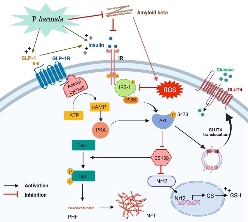
www.nature.com/scientificreports/
Figure 1. Schematic presentation of the experimental timeline. AlCl3 was injected on day 1 and continued till
day 42 and P. harmala administration started on day 15. Behavioral tests were commenced with Y-maze on day
37 followed by MWM with training sessions carried on days 38–41, probe test on day 42 and animal euthanasia
on day 43. AlCl3 aluminum chloride, MWM Morris Water Maze, P. harmala Peganum harmala.
Standardization of P. harmala extract by HPLC. The triplet analysis of the P. harmala extract was car-
ried out using the μBondapak C18 Column (125A, 10 μm, 3.9 × 300 mm; WAT027324; Waters, Dublin, Ireland)
and the Shimadzu (Kyoto, Japan) LC-10AD vp pump and SPD-10A vp UV/VIS detector. The mobile phase
consisted of isopropanol: acetonitrile: water: formic acid (100:100:325:0.3) at a flow rate of 1 ml/min. Standard
solutions of harmine (100µ/ml) and harmaline (100µ/ml) were prepared in the mobile phase and 10 µl of the
prepared standard solutions and the P. harmala extract were injected and detected at the UV wavelength 330 nm.
Animals. Adult male Wistar rats, weighing 180–250 g, were obtained from the Animal House Unit (AHU)
of the Faculty of Pharmacy, Cairo University (Cairo, Egypt). The animals were kept under standard housing
conditions of constant temperature (25 ± 2 °C) and humidity (60 ± 10%) with 12:12 h light/dark cycles. They
were allowed standard rat chow diet and water ad libitum. This study abided by ARRIVE guideline28 and was
evaluated and approved by the Research Ethics Committee (REC) at Faculty of Pharmacy, Cairo University [PT:
2071]. All experiments were carried out in accordance with relevant guidelines and regulations.
Experimental design. Fifty-two rats were randomly allocated into 4 groups (n = 13 each). Rats in group I
received saline (i.p) and served as the normal control group, whereas those in group II provided the AD model
in which rats were injected with AlCl3 dissolved in saline (50 mg/kg/day; i.p) for 6 consecutive weeks29. In group
III, normal rats received daily i.p saline for 2 weeks followed by daily doses of P. harmala per se (187.5 mg/kg;
lCl3-exposed rats were treated with the oral doses of P. harmala 30 min
p.o) for 4 weeks25, while in group IV, A
after each neurotoxin injection for a period of 4 weeks, starting from the 15th day of AlCl3 injection in rats that
showed declined cognition29.
Behavioral studies. Six days prior to the end of the trial, rats were subjected to behavioral tests starting
with the least stressful Y-maze test on day 37, followed by Morris Water Maze (MWM) test that was carried out
for 5 successive days as follows; training on days 38 to 41 and on the 42nd day, the probe trial was performed
(Fig. 1).
Y‑maze task. The test is a simple and quick measure of spatial memory that depends upon the natural pro-
pensity of rats to exhibit “spontaneous alternation behavior”, in which all three arms in the Y-maze are entered
successively without repeated entries30. The Y-maze apparatus consisted of 3 identical painted wooden arms (40
cm long, 35 cm high, 12 cm wide) designated as A, B, and C, with an angle of 120° between them. Each rat was
placed in the center of the maze and was allowed to navigate freely through the maze for 5 min without cues
or reinforcements, such as food or water. The number of entries/arm was recorded manually and the arm entry
was considered valid when the hind paws of the animal are completely inside the arm. The total number of
arm entries and the number of spontaneous alternations were counted to calculate the spontaneous alternation
percentage (SAP) according to the following Eq. 31: SAP (%) = [(number of alternations)/ (total number of arm
entries − 2)] × 100.
MWM. This test is particularly sensitive to hippocampal-dependent spatial memory in r ats32. The test was
carried out following the Morris 1984 protocol33 using a circular pool (150 cm diameter, 60 cm high), filled to a
depth of 40 cm with water (26 ± 1οC). The pool was divided into 4 virtual quadrants of equal sizes and a circular
Scientific Reports | (2021) 11:12040 | https://doi.org/10.1038/s41598-021-90545-4 3
Vol.:(0123456789)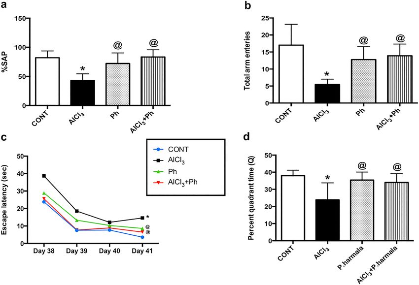
www.nature.com/scientificreports/
Figure 2. HPLC chromatogram. Graph represents (a) P. harmala extract against standard (b) harmine and (c)
harmaline at 330 nm.
transparent movable escape platform (10 cm diameter) that was submerged 1 cm below the water surface in the
middle of one quadrant (i.e. the target quadrant). For the 4 subsequent days of the training session (acquisi-
tion phase), rats were exposed to 4 daily trials to find the hidden platform within 120 s. The time taken to find
the platform was recorded for each trial (i.e. escape latency) and the animal was left on the platform for 10 s;
however, rats that failed to allocate the platform were guided and placed on the platform for 30 s with a given
latency of 120 s. On the 5th day of the MWM paradigm (day 42 of experimentation), the probe test was carried
out, where the platform was removed and rats were allowed to explore the maze for 120 s, in which the percent
quadrant time (Q)34 was recorded using the following equation: Q = (time spent in target quadrant (s)/120) × 100.
Twenty-four hours after the completion of behavioral testing, animals were euthanized using a high dose
of thiopental (100 mg/kg) and brains were carefully harvested. The hippocampi were gently dissected out to be
used for the determination of the biochemical parameters.
Western blot analysis. The hippocampi from 4 rats per group were homogenized in radioimmuniprecipi-
tation assay (RIPA) buffer (150 mM NaCl, 0.1% sodium dodecyl sulfate, 1% Triton X-100, 50 mM Tris–HCl,
1% sodium deoxycholate, pH 7.8) with protease inhibitor cocktail for Western blot analysis. The total protein
content of hippocampal homogenates was assessed using the Bicinchoninic Acid Assay (BCA) assay kit (Abcam,
Cambridge, UK) and then 20 µg protein aliquots of each sample were separated by SDS-PAGE then transferred
into a nitrocellulose membrane using a semi-dry transfer apparatus (Bio-Rad, CA, USA). The membranes were
placed in 5% bovine serum albumin in Tris buffered saline containing 0.1% Tween 20 (TBST) to block the bind-
ing sites on the membrane and to reduce background interference. After that, the membranes were incubated
with primary antibodies including phospho-Akt1 (Ser473) polyclonal antibody (1:500, CAT# PA5-104,445),
phospho-IRS1 (Ser307) polyclonal antibody (1:1000, CAT# 44-813G), phospho-tau (Ser262) polyclonal anti-
body (1:1000, CAT# 44-750G), and a β-actin monoclonal antibody as a reference protein (1:1000, CAT# MA5-
15,739) overnight on a roller shaker at 4 °C. All the primary antibodies used were purchased from ThermoFisher
Scientific (MA, USA). On the next day, the membranes were washed in TBST and incubated with horseradish
peroxidase-conjugated secondary antibody (1:1000, CAT# ab6734, Abcam) for 1 h at room temperature. Signals
were developed following enhanced chemiluminescent (ECL) reagent (CAT# 32,209, ThermoFisher Scientific).
Eventually, the optical densities of the expressed protein bands were quantified via densitometry using a laser
scanning densitometer (CAT# GS800, Bio-Rad). The results were normalized to β-actin and expressed as arbi-
trary units (AU).
Scientific Reports | (2021) 11:12040 | https://doi.org/10.1038/s41598-021-90545-4 4
Vol:.(1234567890)
www.nature.com/scientificreports/
Figure 3. Effect of P. harmala on AlCl3-induced: (a) % SAP and (b) total arm entries in Y-maze test and (c)
learning and (d) memory deficits in MWM paradigm. Values represent mean ± SD (n = 13). Statistical analysis
was carried out using one-way ANOVA for %SAP and total arm entries in Y-maze and percent quadrant time in
the probe MWM test, while two-way ANOVA was used for the escape latency in the training session of MWM
test followed by Tukey’s Multiple Comparisons test. Significantly different from (*) CONT and (@) A lCl3 groups
at p < 0.05. AlCl3aluminum chloride, CONT control, MWM Morris Water Maze, Ph Peganum harmala, %SAP
spontaneous alternation percentage.
ELISA assessment. The hippocampi of 6 rats/group were homogenized in phosphate buffer and aliquoted
for ELISA assay; then kept at -80οC. The following hippocampal parameters were quantified using the corre-
sponding rat-specific ELISA kits: Aβ1-42 and GLUT4 (Cusabio technology LLC, TX, USA, CAT# CSB-E10786r,
CAT# CSB-E13908r; respectively); insulin, GSH, MDA and Nrf-2 (MyBioSource, CA, USA, CAT# MBS724709,
CAT# MBS265966, CAT# MBS268427, CAT# MBS752046; respectively); glucagon like peptide-1 (GLP-1) and
pS9-GSK3β (RayBiotech, CA, USA, CAT# EIAR-GLP1-1, CAT# PEL-GSK3b-S9-; respectively). The manufac-
turers’ instructions were precisely followed for each ELISA.
Histopathological examination. Moreover, the whole brains of the last 3 rats/group were fixed in 10%
neutral buffered formalin for 72 h followed by paraffin embedding for histopathological examination. The for-
malin fixed brains of each group were trimmed, dehydrated in serial grades of ethanol, cleared in xylene, and
embedded in Paraplast tissue embedding media. Sagittal brain Sects. (4 µm thickness) were obtained using a
rotatory microtome and were stained with Harris Hematoxylin and Eosin (H&E) staining as a general tissue
structure examination. Likewise, other sections were subjected to Nissl staining using Toluidine blue to assess
neuronal survival in hippocampal Cornu Ammonis 1 (CA1) and Dentate gyrus (DG) regions. Six random non-
overlapping fields were analyzed blindly for counting intact neurons by using full HD microscopic camera oper-
ated by Leica application Suite for tissue sections analysis (Leica Microsystems GmbH, Wetzlar, Germany). All
histopathological procedures and evaluation were performed by an external investigator in a blinded manner.
Statistical analysis. All data sets were represented as mean ± standard deviation (SD). GraphPad Prism
software 6.0 (GraphPad Software, CA, USA) was used to perform statistical analysis. Comparisons between
means were carried out using one-way Analysis of Variance (ANOVA) followed by Tukey’s Multiple Compari-
sons test. Escape latency results from the MWM training sessions were analyzed using two-way ANOVA and
followed by Tukey’s Multiple Comparisons test. Statistical significance was achieved when p < 0.05.
Results
The administration of P. harmala to normal rats showed no substantial changes compared with their control
counterparts for all measured parameters, with the exception of hippocampal GLUT4 content. Therefore, com-
parisons were only made with reference to the control group.
Scientific Reports | (2021) 11:12040 | https://doi.org/10.1038/s41598-021-90545-4 5
Vol.:(0123456789)www.nature.com/scientificreports/
Figure 4. Effect of P. harmala on (a–d) hippocampal CA1 and (e–h) DG histopathological changes-induced
by AlCl3 in rats. Section of (a) normal hippocampal CA1 area, shows intact pyramidal neurons (black arrow),
while section of (b) AlCl3 group displays marked neuronal loss with varied areas of darkly pyknotic, shrunken
degenerated neurons (blue arrow) with mild edema and vacuolation of neutrophils (dashed arrow). Section of
(c) Ph group presents intact histological structures similar to normal rats and photomicrograph of (d) AlCl3 + Ph
group reveals fewer degenerated shrunken pyknotic pyramidal neurons (blue arrow) combined with many
intact cells and milder records of glial cell infiltrates (arrow head). In the hippocampal DG area, section of
(e) the control group receiving saline or (g) P. harmala shows normal morphological features of hippocampal
layers, including granule cells with intact nuclear details (black arrow) along with an unaltered hilar region. The
section of (f) AlCl3-treated rats reveals conspicuous degenerative aberrations and many pyknotic granule cells,
as well as hilar cells (blue arrow) with moderate records of edema and vacuolation of neuropil (dashed arrow).
However, section of (h) AlCl3 + Ph group exhibits few scattered degenerated granule cells besides hilar cells (blue
arrow) and various apparent intact granule cells (black arrow) with lower records of intracellular edema (H&E,
scale bar: 50 μm). AlCl3 aluminum chloride, CA1 cornu Ammonis 1, CONT control, DG dentate gyrus, H&E
Hematoxylin and Eosin, Ph Peganum harmala.
Standardization of P. harmala extract. As illustrated in Fig. 2, the (a) harmine and harmaline in P. har‑
mala extract, quantified through HPLC analysis, showed a retention time of 4.667 and 9.108 min, respectively,
which was similar to those of the two standards; viz., (b) harmine and (c) harmaline. The amount of harmine and
harmaline present in P. harmala extract was estimated to be about 14 and 21% (w/w), respectively.
P. harmala alleviates the AlCl3‑induced learning and memory deficits. The results of the Y-maze
test (Fig. 3) confirmed the spatial memory deficit and the decreased general activity in the AlCl3-treated rats,
as evidenced by the marked inhibition in the (a) SAP and (b) total arm entries by 48 and 68%, respectively, as
compared to the control group. On the contrary, post-treatment with P. harmala in AlCl3-exposed rats improved
spatial memory and restored both measured parameters. Moreover, using the MWM test, the insulted rats
showed a learning shortfall indicated by the prolongation of the (c) escape latency across the 4 training days in
the acquisition phase compared to the control. Conversely, rats administered P. harmala 2 weeks after initiating
AlCl3 injection, showed shortness in the escape latency, as compared with the A lCl3 group signifying improved
learning. Additionally, the MWM probe test confirmed the cognitive deficit in A lCl3-treated rats to show com-
promised spatial memory that was expressed as a salient decline in (d) Q (63%) compared to the control group.
However, P. harmala effectively ameliorated the AlCl3-induced memory impairment, restoring the time spent in
the target quadrant to its value in normal control rats.
P. harmala mitigates AlCl3‑induced histopathological changes and intact neuron count in CA1
and DG regions of the hippocampus. Compared to the normal architecture of hippocampal (a) CA1 and
(e) DG areas (Fig. 4), the sections of AlCl3 rats show (b) variable alternated areas of darkly pyknotic, shrunken
degenerated pyramidal neurons (CA1), and (f) granule/hilar cells (DG), besides apparent intact neurons with
mild edema and vacuolation of neutrophils. However, sections of P. harmala post-treatment reveal fewer degen-
erated shrunken pyknotic cells in the (d) CA1pyramidal neurons with mild glial cells infiltrate, besides apparent
intact (h) granule/hilar cells in DG with diminished records of intercellular edema. Figure 5 reveals a remarkable
reduction in the number of intact neurons in hippocampal CA1 (72%) and DG (50%) regions of A lCl3-treated
rats compared with the control. Contrarily, treatment with P. harmala prevented the loss of viable neurons in
both regions (CA1: 1.3 folds; DG: 1.9 folds) in comparison to the AlCl3 group.
Scientific Reports | (2021) 11:12040 | https://doi.org/10.1038/s41598-021-90545-4 6
Vol:.(1234567890)www.nature.com/scientificreports/
Figure 5. Effect of P. harmala on AlCl3-induced neuronal loss in hippocampal CA1 and DG regions. (a)
Representative images of Nissl staining across hippocampal CA1 and DG sections (scale bar 50 μm). The lower
panel shows the effect of P. harmala treatment on the number of surviving neurons in hippocampal (b) CA1
and (c) DG regions. Values represent mean ± SD (n = 3). Statistical analysis was carried out by one-way ANOVA
followed by Tukey’s Multiple Comparisons test. (*) Significantly different from CONT and (@) A lCl3 groups
at p < 0.05; AlCl3 aluminum chloride, CONT control, CA1 cornu Ammonis 1, DG dentate gyrus, Ph Peganum
harmala.
P. harmala activates hippocampal GLP‑1 and insulin signaling in AlCl3‑induced AD‑like pathol-
ogy in rats. As presented in Fig. 6, exposure to A
lCl3 provoked a prominent decrease in hippocampal con-
tent of (a) GLP-1 by 74% versus control that was greatly raised by P. harmala (2.5 folds) compared to the insult.
Additionally, AlCl3-treated rats halved (b) insulin content, effect that was associated with a marked elevation in
(c) pS307-IRS-1 (6.1 folds) and the reduction in both (d) pS473-Akt (34%) and (e) GLUT4 (45%), as compared
to the control group. In a sharp contrast, P. harmala extract notably boosted insulin content (1.5 folds), while
reduced pS307-IRS-1 (51%) and enhanced pS473-Akt (2.1 folds), as well as GLUT4 (2 folds) when compared to
the AlCl3 group.
P. harmala ameliorates AlCl3‑induced alterations in hippocampal Aβ42, p‑tau, and pS9‑GSK‑3β
in AD rat model. In Fig. 7, AlCl3 showed a substantial leap in the hippocampal contents of (a) Aβ42 and (b)
p-tau to reach 9.8 and 5.3 folds, respectively, while it notably diminished (c) pS9-GSK-3β by 55%, as compared
with the control group. However, these effects were opposed by treatment with P. harmala, which suppressed
Aβ42 (33%) and p-tau (41%) and augmented pS9-GSK-3β (165%), as compared with AlCl3 rats.
P. harmala abates AlCl3‑induced oxidative stress by enhancing hippocampal Nrf2 signal-
ing. As shown in Fig. 8, AlCl3-injected rats provoked oxidative stress in the hippocampus as accentuated by
the elevated content of (a) MDA (2.5 folds) in accordance with lowered contents of (b) Nrf2 (44%) and (c) GSH
(32%) versus their counterpart. Conversely, P. harmala halved MDA, but boosted Nrf2 (1.8 folds) and GSH (2
folds), as compared with AlCl3-treated rats to signify its antioxidant capacity.
Scientific Reports | (2021) 11:12040 | https://doi.org/10.1038/s41598-021-90545-4 7
Vol.:(0123456789)www.nature.com/scientificreports/
Figure 6. Effect of P. harmala on hippocampal GLP-1 and insulin signaling in AlCl3-induced AD-like
pathology in rats. Values represent mean ± SD (n = 6/4). The blots of pS307-IRS-1, pS473-Akt and β-actin were
cropped and the full-length blots are shown in the Supplementary file (Fig. S1). Statistical analysis was carried
out by one-way ANOVA followed by Tukey’s Multiple Comparisons test. Significantly different from (*) CONT
and (@) A lCl3 groups at p < 0.05; pS473-Akt phosphorylated Akt at serine 473, AlCl3 aluminum chloride, AU
arbitrary units, CONT control, GLP-1 glucagon-like peptide-1, GLUT4 glucose transporter type 4, pS307-IRS-1
phosphorylated insulin receptor substrate-1 at serine 307, Ph Peganum harmala.
Discussion
The current report provides novel insights into the anti-amnestic effects of the methanolic extract of P. harmala
seed in a sporadic AD model. The enhanced cognition with reduced Aβ42/tau pathology in A lCl3-injected rats
coincided with an attenuated hippocampal insulin resistance. P. harmala activated the Akt trajectory to inhibit
GSK3β and enhance GLUT4 via two influential molecules, namely, GLP-1 and insulin that mediated pS307-IRS-1
reduction (Fig. 9). The standardized extract also activated Nrf2 to enhance the brain antioxidant capacity that
receded oxidative stress to additionally improve insulin sensitivity.
Intriguingly, our findings firstly validated that the insulin-signaling dysregulation in the hippocampi of rats
exposed to AlCl3 could be ameliorated after the administration of P. harmala seed extract for 4 weeks. P. harmala
commenced its central insulin sensitizing effect by enhancing the hippocampal content of insulin, results that
further support a similar effect in the periphery. In a previous work, P. harmala seeds disclosed their hypoglyce-
mic effect via triggering insulin secretion from pancreatic β-cells in a streptozotocin diabetic m odel24.
The enhancing effect of P. harmala on central insulin, recorded in our work, can be owed to the elevated
hippocampal content of GLP-1, a hormone that is known to improve insulin signaling35. Our finding underpins
the results of Gu et al.36, who reported that upon screening of plants for their possible dipeptidyl peptidase IV
(DPP-4) enzyme inhibitory activity, harmine, a main active constituent of P. harmala, showed an antidiabetic
DPP-4 inhibitory effect that in turn increased the endogenous levels of GLP-136,37. It is worth mentioning that
both GLP-1 and its receptor are decreased in AD human brain and in experimental AD models, a fact that
coincide with the present findings in the current m odel38.
Following the increased hippocampal insulin and GLP-1 contents, P. harmala thrusted the insulin trajectory
forward and extended its insulin sensitizing effect by downregulating the protein expression of pS307IRS-1, a
leading instigator of brain insulin resistance39. Previously, it was reported that the decrement in IRS-1 serine
phosphorylation allows the activation of the PI3K cascade9; this verity is confirmed herein, where the inhibition
of pS307IRS-1 by P. harmala was allied by an increase in the hippocampal protein expression of the phosphoryl-
ated/activated pS473Akt, a downstream of the PI3K molecule. Although the central insulin sensitizing effect of P.
harmala has not been reported before, a study conducted in 2 01627 has shown that P. harmala extract mitigated
palmitic acid-induced insulin resistance in muscle cells by modulating the p-IRS/p-Akt hub. Moreover, our
results are compatible with an earlier study conducted by Naresh et al.40, which recounted that treatment of L6
skeletal muscle cells with 4-hydroxypipecolic acid, isolated from P. harmala seeds, stimulated GLUT4 transloca-
tion through the PI3K-dependent insulin signaling pathway.
In further support of our current results, the aptitude of P. harmala to increase GLP-1 adds to the activation of
insulin cascade, since GLP-1 agonists, such as liraglutide and exendin-4, have activated the IRS-1/Akt trajectory
Scientific Reports | (2021) 11:12040 | https://doi.org/10.1038/s41598-021-90545-4 8
Vol:.(1234567890)www.nature.com/scientificreports/
Figure 7. Effect of P. harmala on AlCl3-induced alterations in hippocampal (a) Aβ42, (b) p-tau, and (c) pS9-
GSK-3β in AD rat model. Values represent mean ± SD (n = 6/4). The blots of p-tau and β-actin were cropped
and the full-length blots are shown in the Supplementary file (Fig. S1). Statistical analysis was carried out by
one-way ANOVA followed by Tukey’s Multiple Comparisons test. Significantly different from (*) CONT and
(@) AlCl3 groups at p < 0.05; AlCl3 aluminum chloride, CONT control, Aβ42 amyloid beta 42, AU arbitrary
units, pS9-GSK-3β phosphorylated glycogen synthase kinase-3 beta at serine 9, Ph Peganum harmala, p-tau
phosphorylated tau.
in mouse models of A D41,42. Notably, GLP-1 mediates its effect by stimulating adenylyl cyclase to consequently
trigger the PI3K/Akt signaling p athway43. This fact was further verified in other studies; Gupta et al.44 in their
in vitro study revealed that treatment with exendin-4 has phosphorylated elements of the insulin-signaling hub,
including Akt1. Moreover, another study showed that GLP-1 improves glucose metabolism through activating
Akt45.
The modulation of p-IRS-1/p-Akt signaling by insulin and GLP-1 was coupled by an increase in the hip-
pocampal content of GLUT4 to form the last step of the pathway and to highlight the beneficial role of P. har‑
mala in improving cerebral insulin sensitivity that was reflected also on the refining of memory function. This
notion was documented in a recent study stating that GLUT4 is one of the foremost molecules responsible for
hippocampal memory and insulin s ensitivity46. In addition to its effect on GLUT4, increased levels of GLP-1 play
a critical role in improving cognition. An earlier study on GLP-1 receptor knockout mice has revealed memory
impairment and cognitive dysfunction in the MWM t est47 to coincide with the present data in AD rats. These
findings, hence, emphasize the role of the active insulin cascade along with GLP-1 in the P. harmala-induced
upturn in memory and cognition.
A crosstalk between insulin cascade and AD manifestations has been highlighted earlier, where it was identi-
fied that Aβ, a surrogate marker of AD pathology, competitively inhibits the binding of insulin to its neuronal
receptors48,49 and interferes with tyrosine phosphorylation/activation of IRS-150, while permitting its serine
phosphorylation/inhibition to hinder GLUT 4 translocation15. Moreover, post-mortem studies51,52 have displayed
decreased levels of insulin in different AD brain regions; besides, brain insulin deficiency was reported to impede
memory, synaptic transmission, IRS-1 tyrosine phosphorylation/activation, and the activation of Akt53, events
that contribute to AD pathology54. In the present work, the treatment with P. harmala for 4 weeks, not only acti-
vated the insulin trajectory, but it curbed also the bolstered hippocampal content of Aβ42, as well as the protein
expression of the phosphorylated/activated tau, the two main molecules related to AD pathology. Hence, these
effects support the folk notion about the effectiveness of P. harmala in easing CNS disorders and offer another
mechanism to alleviate AD-like pathology besides augmenting a cetylcholine25,26. The aptitude of P. harmala to
turn on the insulin cascade can explain in part the decreased levels of Aβ42 and p-tau, where increased insulin
was reported to foster Aβ clearance and prevent Aβ plaque f ormation15,55. Furthermore, the elevated insulin level
triggers the tau phosphatase to deter tau p athology56.
Not only the increased insulin, but also GLP-1 plays a central role in lowering both Aβ42 and p-tau as
mentioned before57,58. These authors displayed that different GLP-1 receptor agonists have inhibited Aβ, tau
Scientific Reports | (2021) 11:12040 | https://doi.org/10.1038/s41598-021-90545-4 9
Vol.:(0123456789)www.nature.com/scientificreports/
Figure 8. Effect of P. harmala on AlCl3-induced oxidative stress related markers (a) MDA, (b) Nrf2, and (c)
GSH in AD rat model. Values represent mean ± SD (n = 4). Statistical analysis was carried out by one-way
ANOVA followed by Tukey’s Multiple Comparisons test. Significantly different from (*) CONT and (@) A lCl3
groups at p < 0.05; CONT control, AlCl3 aluminum chloride, GSH glutathione, MDA malondialdehyde, Nrf2
Nuclear factor erythroid 2-related factor 2, Ph Peganum harmala.
hyperphosphorylation and neuronal damage. Besides, using DPP-4 inhibitors59,60 or the GLP-1 analogue Liraglu-
tide in AD animal models succeeded to attenuate the amyloid load and improve cognitive abilities and m emory61.
Additionally, the P. harmala-induced pS473-Akt plays a role too, since earlier investigations confirmed that
pS473-Akt bestows neuronal protection against A D62,63. A fundamental effect of the pS473-Akt is to phosphoryl-
ate one of its arsenal downstream molecules glycogen synthase kinase (GSK)-3β at its serine 9 r esidue64, a fact
that was recorded here in the P. harmala treated group. The aberrant insulin signaling augments Aβ neuropa-
thology via activating GSK3β65, which interferes with the two main steps responsible for a myloidogenesis65,66
and is considered the main kinase responsible for tau phosphorylation, a step that precedes the emergence of
neurofibrillary tangles (NFT)67. Mutually, Aβ upregulates GSK3β activity by inhibiting its phosphorylation as
evidenced from an earlier in vitro study67 and here. Hence, the inhibition of the hippocampal GSK-3β adds to the
mechanism of the current extract in dwindling the AD pathomolecules. The activation of Akt/ GSK-3β pathway
is partly attributed to the increased GLP-1, where its analogs have restored this trajectory and attenuated memory
deficits in amyloid precursor peptide/presenilin 1 m ice58,68.
Although harmine has been proven to effectively enhance spatial learning and memory, as shown here and
hitherto69, Aβ was not lowered by harmine, but rather by quinazoline derivatives70. On the contrary, harmine
is known as a potent inhibitor of dual-specificity tyrosine phosphorylation-regulated kinase 1A (DYRK1A),
which plays a central role in tau h yperphosphorylation71,72 to explain partially the current inhibition of p-tau
by P. harmala.
From another prospective, Aβ is tied to insulin resistance via inciting oxidative stress that serves as a midpoint
mechanism in this trajectory. In this context, high binding protein metals tend to interact with Aβ a ggregates73
to produce reactive oxygen species (ROS)74, where hydrogen peroxide (H2O2) promotes IRS-1 phosphorylation
at serine 30718, hence adversely afflicting insulin signaling and aggravating insulin resistance. Meanwhile, as
depicted from the well-recognized oxidative stress theory of AD, the neuronal lipid membrane is penetrated
by Aβ oligomers to initiate and propagate lipid peroxidation by acting as sulfur free radicals, evoking thus
neurotoxicity and synapse d eterioration17. This fact is considered as an important factor in initiating neurode-
generation in both AD patients75 and AlCl3 exposed animals76 to validate the present Aβ-mediated cognitive
decline associated by insulin resistance and oxidative stress. The latter was documented by the increased lipid
Scientific Reports | (2021) 11:12040 | https://doi.org/10.1038/s41598-021-90545-4 10
Vol:.(1234567890)www.nature.com/scientificreports/
Figure 9. A proposed novel mechanism of P. harmala against insulin resistance in an AlCl3 sporadic model
of AD. P. harmala restored insulin signaling via a favorable stimulation of GLP-1 and insulin/p-IRS-1/Akt/
GLUT-4 axes versus inhibition of Aβ, GSK3β along with sequential induction of Nrf2 signaling while abating
p-tau; hence combating AD. Akt protein kinase B, ATP adenosine triphosphate, cAMP cyclic adenosine
monophosphate, GLP-1/GLP-1R glucagon-like peptide-1/receptor, GLUT4 glucose transporter type 4, GS
glutathione synthetase, GSH glutathione, IR insulin receptor, IRS-1 insulin receptor substrate-1, Nrf2 nuclear
factor erythroid 2-related factor 2, P harmala Peganum harmala, PI3K phosphoinositide 3-kinase, PKA protein
kinase A, ROS reactive oxygen species. “Created with BioRender.com".
peroxidation with the depletion of the defense molecule GSH and the suppression of the antioxidant transcrip-
tion factor Nrf2. In our study, P. harmala displayed an antioxidant capacity through bolstering GSH and Nrf2 to
attenuate MDA formation. Such results are supported by Li et al. (2018) who stated that harmine and harmaline
reversed scopolamine-induced oxidative damage in C57BL/6 mice77. Of note, activating Nrf2 has been previously
reported to abrogate Aβ production through blunting β-secretase e xpression78 and control GSH synthesis being
an upstream regulator of glutathione synthetase79. Since active GSK3β causes the proteasomal degradation of
Nrf280, hence, P. harmala-mediated Akt activation/phosphorylation could be one culprit for GSK3β deactivation
to enhance Nrf2, improve cognition, and reduce insulin resistance.
To this end, the therapeutic benefit of P. harmala extract against AD-like pathology resides in its central abil-
ity to ameliorate hippocampal insulin resistance and oxidative stress through the crossed interaction of insulin/
PI3K/Akt/GLUT4, GLP-1/Akt/GLUT4 and Akt/pGSk3β/Nrf2 cues. The activated pathways acted on tackling
Aβ aggregation, tau phosphorylation mainly by inactivating/phosphorylating GSK3β. Accordingly, the novel
findings of this study propose a compelling therapeutic mechanism for P. harmala, beyond its traditional use,
in the treatment of AD, which needs further clinical proof.
Data availability
All data generated or analyzed during this study are included in this article and its Supplementary information
files.
Scientific Reports | (2021) 11:12040 | https://doi.org/10.1038/s41598-021-90545-4 11
Vol.:(0123456789)www.nature.com/scientificreports/
Received: 2 January 2021; Accepted: 22 April 2021
References
1. Blázquez, E., Velázquez, E., Hurtado-carneiro, V. & Ruiz-Albusac, J. M. Insulin in the brain: Its pathophysiological implications
for states related with central insulin resistance, type 2 diabetes and alzheimer ’ s disease. Front. Endocrinol. 5, 1–21 (2014).
2. Kandimalla, R., Thirumala, V. & Reddy, P. H. Is Alzheimer’s disease a type 3 diabetes? A critical appraisal. Biochim. Biophys. Acta.
Mol. Basis Dis. 1863, 1078–1089 (2017).
3. Kuwabara, T. et al. Insulin biosynthesis in neuronal progenitors derived from adult hippocampus and the olfactory bulb. EMBO
Mol. Med. 3, 742–754 (2011).
4. Ferrario, C. R. & Reagan, L. P. Insulin-mediated synaptic plasticity in the CNS: Anatomical, functional and temporal contexts.
Neuropharmacology 136, 182–191 (2018).
5. Satoh, F. et al. Characterization of human and rat glucagon-like peptide-1 receptors in the neurointermediate lobe: Lack of coupling
to either stimulation or inhibition of adenylyl cyclase. Endocrinology 141, 1301–1309 (2000).
6. Li, Y. et al. GLP-1 receptor stimulation preserves primary cortical and dopaminergic neurons in cellular and rodent models of
stroke and Parkinsonism. Proc. Natl. Acad. Sci. USA 106, 1285–1290 (2009).
7. Müller, T. D. et al. Glucagon-like peptide 1 (GLP-1). Mol. Metab. 30, 72–130 (2019).
8. Chang, C., Lin, T., Ho, H., Kuo, C. & Li, H. GLP-1 analogue liraglutide attenuates mutant huntingtin-induced neurotoxicity by
restoration of neuronal insulin signaling. Int. J. Mol. Sci. 19, 2505 (2018).
9. Biessels, G. J. & Reagan, L. P. Hippocampal insulin resistance and cognitive dysfunction. Nat. Rev. Neurosci. 16, 660–671 (2015).
10. De la Monte, S. M. & Wands, J. R. Alzheimer’s disease is type 3 diabetes: Evidence reviewed. J. Diabetes Sci. Technol. 2, 1101–1113
(2008).
11. de la Monte, S. M. Type 3 diabetes is sporadic Alzheimer׳s disease: Mini-review. Eur. Neuropsychopharmacol. 24, 1954–1960 (2014).
12. Steen, E. et al. Impaired insulin and insulin-like growth factor expression and signaling mechanisms in Alzheimer’s disease: Is this
type 3 diabetes ?. J. Alzheimers Dis. 7, 63–80 (2005).
13. Han, X. et al. Insulin attenuates beta-amyloid-associated Insulin/Akt/EAAT signaling perturbations in human astrocytes. Cell.
Mol. Neurobiol. 36, 851–864 (2016).
14. Lee, J. H., Jahrling, J. B., Denner, L. & Dineley, K. T. Targeting insulin for Alzheimer’s disease: Mechanisms, status and potential
directions. J. Alzheimers Dis. 64, 427–453 (2018).
15. Mullins, R. J., Diehl, T. C., Chia, C. W. & Kapogiannis, D. Insulin resistance as a link between amyloid-beta and tau pathologies
in Alzheimer’s disease. Front. Aging Neurosci. 9, 1–16 (2017).
16. Butterfield, D. A., Di Domenico, F. & Barone, E. Elevated risk of type 2 diabetes for development of Alzheimer disease: A key role
for oxidative stress in brain. Biochim. Biophys. Acta 1842, 1693–1706 (2014).
17. Butterfield, D. A., Swomley, A. M. & Sultana, R. Amyloid β-peptide (1–42)-induced oxidative stress in Alzheimer disease: Impor-
tance in disease pathogenesis and progression. Antioxid. Redox Signal 19, 823–835 (2013).
18. Verdile, G. et al. Inflammation and oxidative stress: The molecular connectivity between insulin resistance, obesity, and Alzheimer’s
disease. Mediat. Inflamm. 2015, 105828 (2015).
19. YildirimSimsir, I., Soyaltin, U. E. & Cetinkalp, S. Glucagon like peptide-1 (GLP-1) likes Alzheimer’s disease. Diabetes Metab. Syndr.
12, 469–475 (2018).
20. Cukierman-Yaffe, T. et al. Effect of dulaglutide on cognitive impairment in type 2 diabetes: An exploratory analysis of the REWIND
trial. Lancet Neurol. 19, 582–590 (2020).
21. Asgarpana, J. & Ramezanloo, F. Chemistry, pharmacology and medicinal properties of Peganum harmala L. Afr. J. Pharm. Phar‑
macol. 6, 1573–1580 (2012).
22. Eissa, T. A. F., Palomino, O. M., Carretero, M. E. & Gómez-serranillos, M. P. Ethnopharmacological study of medicinal plants used
in the treatment of CNS disorders in Sinai Peninsula, Egypt. J. Ethnopharmacol. 151, 317–332 (2014).
23. Araujo, I. et al. Chemical study of Peganum harmala seeds. Afr. J. Biotechnol. 18, 462–471 (2019).
24. Komeili, G., Hashemi, M. & Bameri-niafar, M. Evaluation of antidiabetic and antihyperlipidemic effects of Peganum harmala seeds
in diabetic rats. Cholesterol 2016, 1–6 (2016).
25. Osman, N. N., Alanbari, K. H. & Al-Shreef, H. A. Evaluation of the possible antioxidant effects of Peganum harmala and Ginkgo
biloba in ameliorating Alzheimer’s disease in rat model. IJPSR 9, 3189–3198 (2018).
26. Ali, S. K. et al. In-vitro evaluation of selected Egyptian traditional herbal medicines for treatment of Alzheimer disease. BMC
Complement. Altern. Med. 13, 1–10 (2013).
27. Ghaffar, S. et al. Attenuation of palmitate induced insulin resistance in muscle cells by harmala, clove and river red gum. Pak. J.
Pharm. Sci 29, 1795–1800 (2016).
28. Kilkenny, C., Browne, W. J., Cuthill, I. C., Emerson, M. & Altman, D. G. Improving bioscience research reporting: The ARRIVE
guidelines for reporting animal research. PLOS Biol. 8, e1000412 (2010).
29. Bazzari, F. H., Abdallah, D. M. & El-abhar, H. S. Chenodeoxycholic acid ameliorates AlCl3-induced Alzheimer’s disease neuro-
toxicity and cognitive deterioration via enhanced insulin signaling in rats. Molecules 24, 1–17 (2019).
30. Hughes, R. N. The value of spontaneous alternation behavior (SAB) as a test of retention in pharmacological investigations of
memory. Neurosci. Biobehav. Rev. 28, 497–505 (2004).
31. Justin-thenmozhi, A. et al. Attenuation of aluminum chloride-induced neuroinflammation and caspase activation through the
AKT/GSK-3β pathway by hesperidin in wistar rats. Neurotox. Res. 34, 463–476 (2018).
32. Snow, W. M. et al. Morris water maze training in mice elevates hippocampal levels of transcription factors nuclear factor (erythroid-
derived 2)-like 2 and nuclear factor kappa B p65. Front. Mol. Neurosci. 8, 1–12 (2015).
33. Morris, R. Developments of a water-maze procedure for studying spatial learning in the rat. J. Neurosci. Methods 11, 47–60 (1984).
34. Vorhees, C. V. & Williams, M. T. Morris water maze: Procedures for assessing spatial and related forms of learning and memory.
Nat. Protoc. 1, 848–858 (2006).
35. Cai, H.-Y. et al. Lixisenatide reduces amyloid plaques, neurofibrillary tangles and neuroinflammation in an APP/PS1/tau mouse
model of Alzheimer’s disease. Biochem. Biophys. Res. Commun. 495, 1034–1040 (2018).
36. Gu, L. H. et al. A thin-layer chromatography-bioautographic method for detecting dipeptidyl peptidase IV inhibitors in plants. J.
Chromatogr. A 11, 116–122 (2015).
37. Kosaraju, J. et al. Saxagliptin: A dipeptidyl peptidase-4 inhibitor ameliorates streptozotocin induced Alzheimer’s disease. Neurop‑
harmacology 72, 291–300 (2013).
38. Chen, S. et al. DPP-4 inhibitor improves learning and memory deficits and AD-like neurodegeneration by modulating the GLP-1
signaling. Neuropharmacology 157, 107668 (2019).
39. Talbot, K. Brain insulin resistance in Alzheimer’s disease and its potential treatment with GLP-1 analogs. Neurodegener. Dis. Manag.
4, 31–40 (2014).
40. Naresh, G. et al. Glucose uptake stimulatory effect of 4-hydroxypipecolic acid by increased GLUT 4 translocation in skeletal muscle
cells. Bioorg. Med. Chem. Lett. 22, 5648–5651 (2012).
Scientific Reports | (2021) 11:12040 | https://doi.org/10.1038/s41598-021-90545-4 12
Vol:.(1234567890)www.nature.com/scientificreports/
41. Talbot, K. & Wang, H.-Y. The nature, significance, and glucagon-like peptide-1 analog treatment of brain insulin resistance in
Alzheimer’s disease. Alzheimers Demen. 10, 1–25 (2014).
42. Long-Smith, C. M. et al. The diabetes drug liraglutide ameliorates aberrant insulin receptor localisation and signalling in parallel
with decreasing both amyloid-β Plaque and glial pathology in a mouse model of Alzheimer’ s disease. Neuromol. Med. 15, 102–114
(2013).
43. Yarchaon, M. & Arnold, S. E. Repurposing diabetes drugs for brain insulin resistance in Alzheimer disease. Diabetes 63, 2253–2261
(2014).
44. Gupta, N. A. et al. Glucagon-like peptide-1 receptor is present on human hepatocytes and has a direct role in decreasing hepatic
steatosis in vitro by modulating elements of the insulin signaling pathway. Hepatology 51, 1584–1592 (2010).
45. Vyas, A. K. et al. Exenatide improves glucose homeostasis and prolongs survival in a murine model of dilated cardiomyopathy.
PLoS ONE 6, e17178 (2011).
46. Mcnay, E. C. & Pearson-leary, J. GluT4: A central player in hippocampal memory and brain insulin resistance. Exp. Neurol. 323,
1–9 (2020).
47. Bae, C. S. & Song, J. The role of glucagon-like peptide 1 (GLP1) in type 3 diabetes: GLP-1 controls insulin resistance, neuroinflam-
mation and neurogenesis in the brain. Int. J. Mol. Sci. 18, 1–7 (2017).
48. Aulston, B. D., Shapansky, J., Huang, Y., Odero, G. L. & Glazner, G. W. Secreted amyloid precursor protein alpha activates neuronal
insulin receptors and prevents diabetes-induced encephalopathy. Exp. Neurol. 303, 29–37 (2018).
49. Huang, C., Wang, C., Id, C. L., Yen, A. & Li, H. Abelmoschus esculentus subfractions attenuate beta amyloid-induced neuron
apoptosis by regulating DPP-4 with improving insulin resistance signals. PLoS ONE 14, 1–13 (2019).
50. Xie, L. et al. Alzheimer’s beta-amyloid peptides compete for insulin binding to the insulin receptor. J. Neurosci. 22, 1–5 (2002).
51. Gabbouj, S. et al. Altered insulin signaling in Alzheimer’s disease brain: Special emphasis on PI3K-Akt pathway. Front. Neurosci.
13, 1–8 (2019).
52. Liu, Y., Liu, F., Grundke-iqbal, I., Iqbal, K. & Gong, C. Deficient brain insulin signalling pathway in Alzheimer’s disease. J. Pathol.
225, 54–62 (2011).
53. Ferreira, L. S. S., Fernandes, C. S., Vieira, M. N. N. & De Felice, F. G. Insulin resistance in Alzheimer’s disease. Front. Neurosci. 12,
1–11 (2018).
54. Neth, B. J. & Craft, S. Insulin resistance and Alzheimer’s disease: Bioenergetic linkages. Front. Neurosci. 12, 1–20 (2017).
55. Rad, S. K. et al. Mechanism involved in insulin resistance via accumulation of β-amyloid and neurofibrillary tangles: Link between
type 2 diabetes and Alzheimer’s disease. Drug. Des. Dev. Ther. 12, 3999–4021 (2018).
56. Gratuze, M., Julien, J., Petry, F. R., Morin, F. & Planel, E. Insulin deprivation induces PP2A inhibition and tau hyperphosphoryla-
tion in hTau mice, a model of Alzheimer’s disease-like tau pathology. Sci. Rep. 12, 1–13 (2017).
57. Perry, T. et al. Glucagon-like peptide-1 decreases endogenous amyloid-beta peptide (Abeta) levels and protects hippocampal
neurons from death induced by Abeta and iron. J. Neurosci. Res. 72, 603–612 (2003).
58. McClean, P. L. & Hölscher, C. Lixisenatide, a drug developed to treat type 2 diabetes, shows neuroprotective effects in a mouse
model of Alzheimer’s disease. Neuropharmacology 86, 241–258 (2014).
59. Tumminia, A., Vinciguerra, F., Parisi, M. & Frittitta, L. Type 2 diabetes mellitus and Alzheimer’s disease: Role of insulin signalling
and therapeutic implications. Int. J. Mol. Sci. 19, 1–17 (2018).
60. Angelopoulou, E. & Piperi, C. DPP-4 inhibitors: A promising therapeutic approach against Alzheimer’s disease. Ann. Transl. Med.
6, 255 (2018).
61. Batista, A. F. et al. The diabetes drug liraglutide reverses cognitive impairment in mice and attenuates insulin receptor and synaptic
pathology in a non-human primate model of Alzheimer’s disease. J. Pathol. 245, 85–100 (2018).
62. Lee, H., Kumar, P., Fu, Q., Rosen, K. M. & Querfurth, H. W. The insulin/akt signaling pathway is targeted by intracellular beta-
amyloid. Mol. Biol. Cell 20, 1533–1544 (2009).
63. Magrané J, Rosen KM, Smith RC, Walsh K, Gouras GK, Querfurth HW. Intraneuronal beta-amyloid expression downregulates
the Akt survival pathway and blunts the stress response. J. Neurosci. 25, 10960–10969 (2005)
64. Shieh, J. C., Huang, P. & Lin, Y. Alzheimer’s disease and diabetes: Insulin signaling as the bridge linking two pathologies. Mol.
Neurobiol. 57, 1966–1977 (2020).
65. Qu, Z.-S. et al. Glycogen synthase kinase-3 regulates production of amyloid-β peptides and tau phosphorylation in diabetic rat
brain. Sci. World J. 2014, 1–8 (2014).
66. Ly, P. T. T. et al. Inhibition of GSK3β-mediated BACE1 expression reduces Alzheimer-associated phenotypes. J. Clin. Invest. 123,
224–235 (2013).
67. Llorens-Marítin, M., Jurado, J., Hernández, F. & Ávila, J. GSK-3 β, a pivotal kinase in Alzheimer disease. Front. Mol. Neurosci. 7,
1–11 (2014).
68. McClean, P. L., Parthsarathy, V., Faivre, E. & Hölscher, C. The diabetes drug liraglutide prevents degenerative processes in a mouse
model of Alzheimer’s disease. J. Neurosci. 31, 6587–6594 (2011).
69. He, D. et al. Effects of harmine, an acetylcholinesterase inhibitor, on spatial learning and memory of APP/PS1 transgenic mice
and scopolamine-induced memory impairment mice. Eur. J. Pharmacol. 5, 96–107 (2015).
70. Brunhofer, G. et al. Bioorganic & Medicinal Chemistry Exploration of natural compounds as sources of new bifunctional scaffolds
targeting cholinesterases and beta amyloid aggregation: The case of chelerythrine. Bioorg. Med. Chem. 20, 6669–6679 (2012).
71. Ryoo, S.-R. et al. DYRK1A-mediated hyperphosphorylation of Tau A functional link between Down syndrome and Alzheimer
disease. J. Biol. Chem. 282, 34850–34857 (2007).
72. Frost, D. et al. β-Carboline compounds, including harmine, inhibit DYRK1A and tau phosphorylation at multiple Alzheimer’s
disease-related sites. PLoS ONE. 6, e19264 (2011).
73. Maynard, C. J., Bush, A. I., Masters, C. L., Cappai, R. & Li, Q.-X. Metals and amyloid-beta in Alzheimer’s disease. Int. J. Exp. Pathol.
86, 147–159 (2005).
74. Cheignon, C. et al. Oxidative stress and the amyloid beta peptide in Alzheimer’s disease. Redox. Biol. 14, 450–464 (2018).
75. Manoharan, S. et al. The role of reactive oxygen species in the pathogenesis of Alzheimer’s disease, parkinson’s disease, and hun-
tington’s disease: A mini review. Oxid. Med. Cell. Longev. 2016, 8590578 (2016).
76. Rui, D. & Yongjian, Y. Aluminum chloride induced oxidative damage on cells derived from hippocampus and cortex of ICR mice.
Brain Res. 1324, 96–102 (2010).
77. Li, S.-P. et al. Analogous β-carboline alkaloids harmaline and harmine ameliorate scopolamine-induced cognition dysfunction by
attenuating acetylcholinesterase activity, oxidative stress, and inflammation in mice. Front. Pharmacol. 9, 346 (2018).
78. Osama, A., Zhang, J., Yao, J., Yao, X. & Fang, J. Nrf2: A dark horse in Alzheimer’s disease treatment. Ageing Res. Rev. 64, 1–46
(2020).
79. Steele, M. L. et al. Effect of Nrf2 activators on release of glutathione, cysteinylglycine and homocysteine by human U373 astroglial
cells. Redox Biol. 1, 441–445 (2013).
80. Sotolongo, K., Ghiso, J. & Rostagno, A. Nrf2 activation through the PI3K/GSK-3 axis protects neuronal cells from Aβ-mediated
oxidative and metabolic damage. Alzheimer’s Res. Ther. 12, 1–22 (2020).
Scientific Reports | (2021) 11:12040 | https://doi.org/10.1038/s41598-021-90545-4 13
Vol.:(0123456789)You can also read