Diseases and Causes of Death in European Bats: Dynamics in Disease Susceptibility and Infection Rates
←
→
Page content transcription
If your browser does not render page correctly, please read the page content below
Diseases and Causes of Death in European Bats:
Dynamics in Disease Susceptibility and Infection Rates
Kristin Mühldorfer1*, Stephanie Speck2, Andreas Kurth3, René Lesnik3, Conrad Freuling4, Thomas
Müller4, Stephanie Kramer-Schadt1, Gudrun Wibbelt1
1 Leibniz Institute for Zoo and Wildlife Research, Berlin, Germany, 2 Bundeswehr Institute of Microbiology, Munich, Germany, 3 Robert Koch Institute, Berlin, Germany,
4 Friedrich-Loeffler-Institute, Wusterhausen, Germany

     Abstract
     Background: Bats receive increasing attention in infectious disease studies, because of their well recognized status as
     reservoir species for various infectious agents. This is even more important, as bats with their capability of long distance
     dispersal and complex social structures are unique in the way microbes could be spread by these mammalian species.
     Nevertheless, infection studies in bats are predominantly limited to the identification of specific pathogens presenting a
     potential health threat to humans. But the impact of infectious agents on the individual host and their importance on bat
     mortality is largely unknown and has been neglected in most studies published to date.

     Methodology/Principal Findings: Between 2002 and 2009, 486 deceased bats of 19 European species (family
     Vespertilionidae) were collected in different geographic regions in Germany. Most animals represented individual cases
     that have been incidentally found close to roosting sites or near human habitation in urban and urban-like environments.
     The bat carcasses were subjected to a post-mortem examination and investigated histo-pathologically, bacteriologically and
     virologically. Trauma and disease represented the most important causes of death in these bats. Comparative analysis of
     pathological findings and microbiological results show that microbial agents indeed have an impact on bats succumbing to
     infectious diseases, with fatal bacterial, viral and parasitic infections found in at least 12% of the bats investigated.

     Conclusions/Significance: Our data demonstrate the importance of diseases and infectious agents as cause of death in
     European bat species. The clear seasonal and individual variations in disease prevalence and infection rates indicate that
     maternity colonies are more susceptible to infectious agents, underlining the possible important role of host physiology,
     immunity and roosting behavior as risk factors for infection of bats.

  Citation: Mühldorfer K, Speck S, Kurth A, Lesnik R, Freuling C, et al. (2011) Diseases and Causes of Death in European Bats: Dynamics in Disease Susceptibility and
  Infection Rates. PLoS ONE 6(12): e29773. doi:10.1371/journal.pone.0029773
  Editor: Anthony R. Fooks, Veterinary Laboratories Agency, United Kingdom
  Received August 11, 2011; Accepted December 4, 2011; Published December 28, 2011
  Copyright: ß 2011 Mühldorfer et al. This is an open-access article distributed under the terms of the Creative Commons Attribution License, which permits
  unrestricted use, distribution, and reproduction in any medium, provided the original author and source are credited.
  Funding: This study was supported by the Adolf and Hildegard Isler-Stiftung, the FAZIT-Stiftung (fellowship to KM) and the Klara Samariter-Stiftung. The funders
  had no role in study design, data collection and analysis, decision to publish, or preparation of the manuscript.
  Competing Interests: The authors have declared that no competing interests exist.
  * E-mail: muehldorfer@izw-berlin.de

Introduction                                                                            world [6–12]. However, most studies are limited to the
                                                                                        identification of microorganisms detected and investigations
   Bats are among the most successful and diverse mammals on                            regarding infectious diseases and causes of death in bats are
earth. Approximately 1230 chiropteran species are found on every                        sparse [13–16].
continent except Antarctica and inhabit a multitude of diverse                             In Europe, research is predominantly focused on European bat
ecological niches [1]. Bats play essential roles in maintaining                         lyssaviruses [17,18] and coronaviruses [19,20], but first indications
healthy ecosystems, as they act as plant pollinators, seed dispersers,                  of bat-pathogenic bacteria [13,14,21–23] and novel viruses [24,25]
and predators of populations of insects including harmful forest                        isolated from deceased bats in Germany and Great Britain were
and agricultural pests [2]. Most bat species are listed in the IUCN                     found. In this study, we provide new data on infectious diseases in
Red list of endangered species and almost half of these are                             European bat species, considering factors likely to affect the
considered threatened or near-threatened [3]. To estimate and                           susceptibility of bats to infectious agents including effects of
prevent further population declines, research has been primarily                        seasonality, individual and species-specific heterogeneities, and
focused on bat biology, ecology and behavior, while disease                             possible intra- and inter-species transmission dynamics.
aspects were largely neglected [4].
   In the last two decades, the importance of chiropteran species as                    Materials and Methods
potential vectors of significant viral diseases especially in regard to
zoonoses has received growing attention. Besides bat rabies that                           All bat species in Europe are strictly protected under the Flora-
has been studied for more than half a century, extensive research                       Fauna-Habitat Guidelines of the European Union (http://ec.europa.
efforts identified a large number of microbial agents [5] including                     eu/environment/nature/legislation/habitatsdirective/index_en.htm)
important emerging zoonotic viruses detected in bats across the                         (92/43/EEC) and the Agreement on the Conservation of Populations

        PLoS ONE | www.plosone.org                                                  1                           December 2011 | Volume 6 | Issue 12 | e29773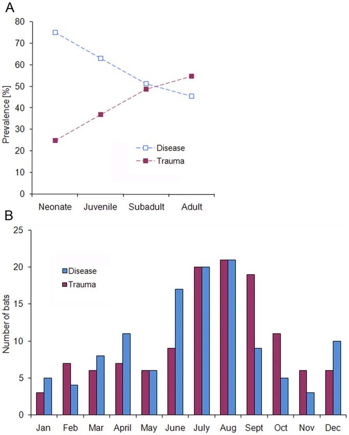
Diseases and Causes of Death in European Bats

of European Bats (www.eurobats.org) that prohibit invasive sampling           (Chocolate agar 5% CO2) for 24–48 h. Specific culture media and
of bats for research purposes. For the animals investigated in this           conditions for the isolation of Yersinia, Salmonella and anaerobic
study, carcasses of deceased bats found in Germany were kindly                bacteria were used if appropriate. Primary identification of
provided by bat researchers and bat rehabilitation centers of different       bacterial strains was based on colony morphology, hemolysis,
federal states.                                                               Gram-staining, indol production, catalase and oxidase reaction.
                                                                              Bacterial species identification was carried out using the relevant
Study material                                                                commercial Api test system (bioMérieux, Germany). Additional
   Between 2002 and 2009, a total of 486 deceased bats of 19                  conventional biochemical tests [29,30] were applied to confirm
European vespertilionid species (i.e., Family Vespertilionidae) were          Api test results where necessary. In case of ambiguous biochemical
investigated (Fig. 1A, [26]). The bat carcasses originated from 6             test results, 16S rDNA gene analysis was performed for final
different geographic regions in Germany, i.e. Berlin greater                  identification [23]. Salmonella isolates were characterized at the
metropolitan area (n = 223), Bavaria (n = 165), Brandenburg                   National Reference Laboratory for the Analysis and Testing of
(n = 38), Lower Saxony (n = 36), Thuringia (n = 21), and Baden-               Zoonoses (Salmonella) at the Federal Institute for Risk Assessment,
Wuerttemberg (n = 3), and were collected by bat researchers and               Berlin, Germany. Identification and characterization of Yersinia
bat rehabilitation centers. Most animals represented individual               and Pasteurella species have been reported earlier [22,23].
cases that were found dead, injured or moribund near human
habitation. Thus, the species composition in this study predom-               Virological investigation
inately reflected the urban and suburban bat fauna, which is                     Homogenized organ tissue of lung, liver, heart, kidney, spleen,
characterized by a disproportionate abundance of a few bat                    brain and salivary gland of 210 bats were pooled for each
species (Fig. 1A, [27,28]). Two groups of 2 and 21 adult noctules             individual and used for RNA/DNA extraction and further
(Nyctalus noctula), respectively, were collected from tree hibernacula        molecular analysis by generic PCR assays detecting flavi- [31],
destroyed during wood logging. A further group of 25 deceased                 hanta- [32], corona- [33], and influenza A-viruses [34]. Also, PCR
adult N. noctula originated from a colony that was trapped in a rain          assays specific for 8 previously described herpesviruses [24] from
pipe in December. Nine dead juvenile Pipistrellus pipistrellus were           European vespertilionid bats were used. For this purpose, RNA/
collected from a nursery roost.                                               DNA was isolated using the NucleoSpinH RNA II Kit (Macherey-
   If bats died in care or had to be euthanized for animal welfare            Nagel, Germany) and the NucleoSpinH Tissue Kit (Macherey-
reasons, the carcasses were immediately stored at 220uC and were              Nagel), respectively, according to the manufacturer’s instructions.
shipped to the Leibniz Institute for Zoo and Wildlife Research,               Because of limitations in sample volume, for 180 out of the 210
Berlin, Germany, for diagnostic investigations. Of all carcasses              bats PCR assays could only be applied for 4 different bat
examined histo-pathologically, about 90% were suitable for                    herpesviruses. Internal controls were used for all PCR assays to
bacteriological investigation. A lesser extend (43%) was also                 test for inhibition. For confirmation, all retrieved fragments of bat
examined for selected viral agents at the Robert Koch Institute,              herpesvirus-specific PCR assays were checked for sequence
Berlin, Germany. In addition, a brain sample of each animal was               identity to previously published isolates [24].
submitted to the Friedrich-Loeffler-Institute, Wusterhausen, Ger-                For detection of lyssavirus antigen in brain tissue the fluorescent
many, for rabies diagnosis.                                                   antibody test (FAT) using a polyclonal antirabies conjugate (Sifin,
                                                                              Germany) was used [35]. FAT-positive brain tissues were subject
Pathological investigation                                                    of virus isolation in murine neuroblastoma cell culture (Na 42/13)
   A full necropsy was performed on each bat and all macroscopic              using the Rabies Tissue Culture Infection Test (RTCIT) as
findings including ectoparasite infestation were recorded. For                described elsewhere [36]. Lyssaviruses isolated in cell culture were
histo-pathological examination, small slices of multiple organ                characterized using both a panel of 10 anti-nucleocapsid
tissues (i.e., lung, liver, heart, kidney, adrenal gland, spleen,             monoclonal antibodies (MAb) [37] and partial sequencing of a
intestine, pancreas, brain, tongue, larynx, salivary gland and                fragment of the nucleoprotein gene after RNA extraction using
pectoral muscle) and tissues conspicuous for pathological changes             Trizol (Invitrogen, Germany) essentially as described [18].
were fixed in buffered 4% formalin, processed using standard
methods and embedded in liquid paraffin. Sections were cut at 2–              Genetic identification of bat species
5 mm and routinely stained with hematoxylin-eosin (HE). In                       Genomic DNA was extracted from organ homogenates using
addition, special histological staining methods were used depend-             the NucleoSpinH Tissue Kit (Macherey-Nagel) according to
ing on microscopic findings, i.e. for the detection of bacteria               manufacturer’s recommendations. Genetic identification of the
(Gram or Giemsa staining), fungi (periodic acid Schiff or Grocott’s           bat species was performed by amplification and sequencing of a
Gomori methenamine silver nitrate staining), iron (Prussian blue              241 bp fragment of the cytochrome B (cytB) gene [38] using
stain), mineralization (von Kossa staining), connective and                   primers FM up (59- CCC CHC CHC AYA TYA ARC CMG
collagen tissue (trichrome staining). Details on pathological results         ART GAT A -39) and FM down (59- TCR ACD GGN TGY CCT
are published elsewhere [26].                                                 CCD ATT CAT GTT A -39). In addition, for differentiation of
   The causes of mortality were rigorously standardized with the              the 2 distinct Pipistrellus species, P. pipistrellus and P. pygmaeus, a
primary cause of death identified for each bat as the most serious            rapid multiplex PCR assay was performed as described by Kaňuch
injury, disease or event subsequently fatal to the animal. To ensure          et al. [39] using primers PIP-F (59- CTC ATT CAT TGA YCT
independence of primary and contributing causes of death, the                 ACC AGC -39), PIP-R (59- CAG CRA ATA GTA AAA TAA
categorization was based on the severity of pathological findings.            CTC C -39) and Ppip-F (59- CAT CTG TTT GGG ACT ACA
                                                                              GAT CC -39).
Bacteriological investigation
   Samples of lung, liver, heart and kidney, and tissues conspicuous          Statistical analysis
for pathological changes (e.g. enlarged spleen) of 430 bats were                The bat data were categorized in regard to different explanatory
plated onto Columbia (5% sheep blood), Chocolate, Gassner, and                numeric and factor variables, e.g. bat species, sex and age class.
MacConkey agar (Oxoid, Germany) and were incubated at 37uC                    The variable ‘age class’ ranked between 1 and 4 with increasing

       PLoS ONE | www.plosone.org                                         2                       December 2011 | Volume 6 | Issue 12 | e29773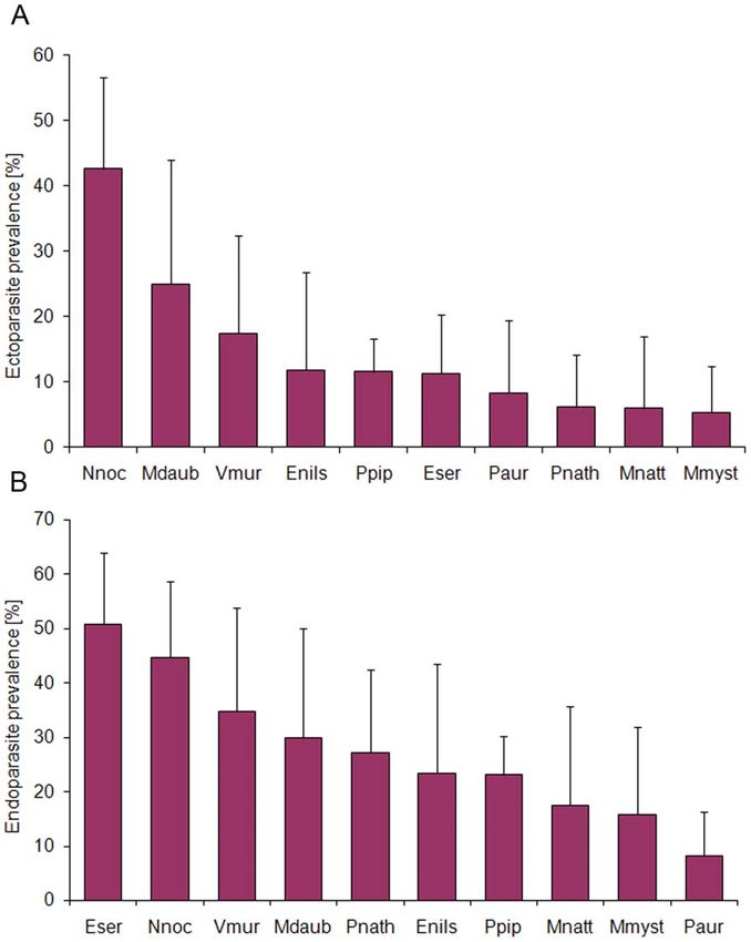
Diseases and Causes of Death in European Bats

PLoS ONE | www.plosone.org   3   December 2011 | Volume 6 | Issue 12 | e29773
Diseases and Causes of Death in European Bats
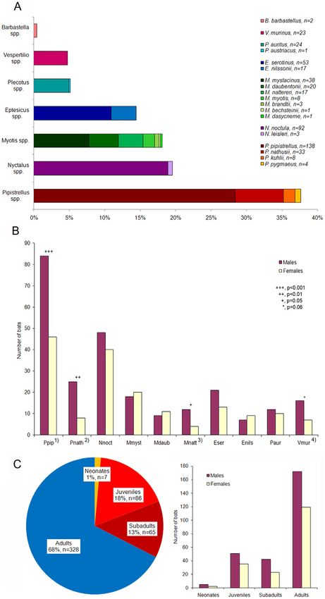
Figure 1. Details on bats from Germany. (A) Bat species distribution among the study sample (n = 486). (B) Male-to-female ratio (bat species .10
individuals). Footnotes: 1) Chi-square test, x2 = 11.1, df = 1, p = 0.0009, 2) x2 = 8.8, df = 1, p = 0.003, 3) x2 = 4.0, df = 1, p = 0.05, 4) x2 = 3.5, df = 1, p = 0.06.
Abbreviations: Ppip, Pipistrellus pipistrellus; Pnath, Pipistrellus nathusii; Nnoc, Nyctalus noctula; Mmyst, Myotis mystacinus; Mdaub, Myotis daubentonii;
Mnatt, Myotis nattereri; Eser, Eptesicus serotinus; Enils, Eptesicus nilssonii; Paur, Plecotus auritus; Vmur, Vespertilio murinus. (C) Age-sex distribution
among the study sample (n = 486).
doi:10.1371/journal.pone.0029773.g001
age (i.e. neonates, juveniles, subadults, and adults) and was used as variable had not been significant as fixed effect (results not shown),
numeric variable. For endoparasitic analysis, we defined a 3 level but from other studies we can assume that there are species-
variable ‘bat size’ according to the body size of a certain bat specific differences in susceptibility of bats to certain infectious
species to reduce the degrees of freedom of the full model, i.e. large agents and therefore included it as random effect. We further used
species (N. noctula, Eptesicus serotinus, and Vespertilio murinus), generalized linear models (GLM with logit link and binomial error
medium-sized species (E. nilssonii, Plecotus auritus, Myotis daubentonii, structure; for datasets with bat species .10 individuals) to test for
M. nattereri, and P. nathusii) and small species (P. pipistrellus, and M. individual and species-specific differences in parasite infection
mystacinus). To detect effects of seasonality, 4 different activity rates (hypotheses C and D).
periods were specified according to the date of sampling, i.e. We created a full model for each hypothesis (A–D) to examine
hibernation period (November to March), post-hibernation period multiple and interaction effects of the specified variables. To select
(April/May), maternity period (June to August), and swarming the final model variables, we used a stepwise backward algorithm
period (September/October). As dependent binary variable for the (function stepAIC in library MASS) based on Akaike’s information
respective models we either classified the mortality cause being criterion (AIC) [40]. The DAIC of the final model was calculated
disease or not (i.e. trauma), or the presence-absence of bacterial, relative to a random intercept model to demonstrate the effect size
ecto- and endoparasitic infections. of the selected variables.
We formulated 4 different hypotheses to test for individual and
species-specific differences in disease susceptibility and infection Results
rates: (A) Disease-related mortality in bats is influenced by sex, age
and species-specific differences, and degree of endoparasitic Results of the diagnostic analyses follow the full data splitting
infection. (B) Bacterial infection in bats is influenced by sex, age into several subsets (see section ‘Statistical analysis’ in Material and
and species-specific differences, occurrence of traumatic injuries Methods; Table 1).
and cat predation. (C) Ecto- or (D) endoparasitic infection in bats
is affected by age, sex and species-specific differences. Seasonal Full dataset: Bat samples
effects were not analyzed because of too many missing data points. All sampled bats belonged to 7 different genera (i.e. Pipistrellus,
Because the long-term dataset was highly biased towards sampling Nyctalus, Myotis, Eptesicus, Plecotus, Vespertilio, and Barbastella) and 19
procedure, preservation of bat carcasses and following diagnostic European vespertilionid species (Fig. 1A). Three bat species, the
investigations, we split and filtered the full data into several subsets common pipistrelle (P. pipistrellus, n = 138), the noctule bat (N.
reflecting the different analyses (Table 1). noctula, n = 92), and the serotine bat (E. serotinus, n = 53) constituted
All statistical analyses were performed using the R software V. about 60% of all bat carcasses investigated in this study, whereas P.
2.13.1 (R Development Core Team 2011, Vienna, Austria). We pygmaeus, Nyctalus leisleri, Myotis brandtii, M. bechsteinii, M. dasycneme,
used the chi-square test for given probabilities to evaluate Plecotus austriacus and Barbastella barbastellus were represented in small
significant differences in the sex ratio among bats of different numbers of 1 to 4 animals. The overall sex ratio was 1.5 males to 1
species. For hypotheses A and B, we used a generalized linear female with significant species-specific differences (Fig. 1B). Animals
mixed modeling approach (binomial GLMM using function lmer in their first year of life (neonates, juveniles, and subadults)
in library lme4) with bat species included as random effect. This represented one third (32.5%, n = 158) of bat samples (Fig. 1C).
Table 1. Description of the data sets used for different analyses.
Data set Sex Age Bat species
Analysis (total n) (% males) (% adults) (total n)
Full dataset Bat samples 486 55.6 67.5 19
a
Subset 1 Causes of death 433 55.0 65.4 19
GLMM: disease- vs. trauma-related mortality (A) 289a 55.0 65.7 17
Subset 2 Bacteriological results 430 58.4 65.3 18
GLMM: bacterial infection vs. no infection (B) 377a 58.1 62.6 18
Subset 3 Virological results 210b 56.7 64.3 16
Subset 4 Parasitological results 433a 55.0 65.4 19
GLM: parasitic infection vs. no infection (C, D) 402a 54.7 65.2 10
GLMM, generalized linear mixed models with bat species included as random effect.
GLM, generalized linear models for datasets with bat species .10 individuals.
A–D: refers to the models analyzed on the different data sets (see chapter ‘Statistical analyses’).
a
To avoid overrepresentation of bat samples that were collected at the same time and location, a randomly selected individual of each group was included in the final
dataset.
b
For detection of lyssavirus antigen, brain tissue of all 486 bats was tested.
doi:10.1371/journal.pone.0029773.t001
PLoS ONE | www.plosone.org 4 December 2011 | Volume 6 | Issue 12 | e29773Diseases and Causes of Death in European Bats
Subset 1: Causes of death Based on the GLMM analysis, significant age- and sex-
Overall, we were able to assign a cause of death to 70% dependent differences (DAIC = 23.13) were detected between the
(n = 304) of bats investigated in this study. Two thirds of mortality general causes of mortality, disease and trauma (Table 4). The
were due to trauma (n = 145) or disease (n = 144), while almost 4% disease presence in bat samples decreased continuously with
of bats had died of other non-infectious causes like pulmonary increasing age. Neonates and juveniles of both sexes were
edema, dehydration and hypoglycemia (Table 2). In 30% (n = 129) significantly more affected by disease than older age classes
no significant pathological findings could be found. (Table 4; Fig. 2A). We also found a significant trend in disease-
Among the 145 traumatized bats, additional mild (n = 42), associated mortality between the sexes, with adult females
moderate (n = 28) and severe (n = 4) inflammatory organ changes displaying higher disease prevalence (52.5%) than males (36.4%)
were noted in one half (50.9%) of individuals, and 23% of the bats
(Table 4). No significant association was observed between a
revealed bacterial (n = 19) and/or parasitic infections (n = 15)
certain cause of mortality (i.e. disease or trauma) and severity of
(Table 3). Of the 144 bats considered as dying of disease, fatal
endoparasitic infection (DAIC = 0.75, result not shown). The
bacterial (n = 54), viral (n = 5) and parasitic infections (n = 2) were
observed in 42%. Besides, amniotic fluid aspiration was noted in a seasonal distribution of disease-related mortality cases (Fig. 2B)
neonate noctule bat (N. noctula), and a juvenile common pipistrelle described a trimodal pattern, with peaks in spring (April), summer
(P. pipistrellus) was euthanized because of severe forearm bone (June to August) and winter (December). The proportion of
deformation. The remaining 81 bats (56.3%) revealed moderate to traumatized individuals also increased obviously during the
severe pathological changes of unknown etiology or unconfirmed summer months up to and including the swarming period, but
bacterial or viral cause (Table 2). was low during the rest of the year.
Table 2. Causes of mortality of bats from Germany.
Age class Sex class
Cause of death n % Euthanasia ,1 Year Adult Female Male n.d.
Trauma 145 33.5 54 41 104 55 87 3
Unknown trauma cause 71 16.5 29 19 52 33 36 3
Cat predation 66 15.3 23 19 47 18 47 -
Roost destructiona 2 0.5 - - 2 2 - -
Trapped in rain pipea 1 0.2 - - 1 - 1 -
Trapped in window 1 0.2 - - 1 - 1 -
Trapped in lamp 1 0.2 - 1 - - 1 -
Trapped in fly strip 1 0.2 - 1 - 1 - -
Barbed wire injury 1 0.2 1 1 - 1 - -
Smoke poisoning 1 0.2 1 - 1 - 1 -
Disease 144 33.3 7 58 86 64 72 8
Unknown etiology 81 18.7 3 35 46 35 38 8
Bacterial infection 54 12.5 2 20 34 27 27 -
Viral infectionb 5 1.2 1 1 4 1 4 -
Parasitic infection 2 0.5 - - 2 - 2 -
Aspiration pneumonia 1 0.2 - 1 - 1 - -
Bone deformation 1 0.2 1 1 - - 1 -
Others 15 3.4 - 6 9 6 9 -
Pulmonary edema 9 2.1 - 3 6 1 8 -
Dehydration 2 0.5 - - 2 1 1 -
Anemiac 1 0.2 - - 1 1 - -
Hyperthermiad 1 0.2 - 1 - 1 - -
Hypothermia 1 0.2 - 1 - 1 - -
Hypoglycemia 1 0.2 - 1 - 1 - -
No significant findings 129 29.8 1 45 84 33 70 26
Total 433 100 62 150 283 158 238 37
n.d., not determined.
a
A randomly selected individual of 3 different groups of adult Nyctalus noctula.
b
Adenovirus (bat AdV-2) [25] and European bat lyssavirus (EBLV-1) infection.
c
Due to severe tick infestation.
d
A randomly selected individual of a group of juvenile Pipistrellus pipistrellus.
doi:10.1371/journal.pone.0029773.t002
PLoS ONE | www.plosone.org 5 December 2011 | Volume 6 | Issue 12 | e29773Diseases and Causes of Death in European Bats
Table 3. Pathological findings and bacterial, viral and was observed for bat gammaherpesvirus 6 (BatGHV6) in common
parasitic infections specified for the general causes of pipistrelle bats (P. pipistrellus), followed by bat gammaherpesvirus 5
mortality, trauma and disease. (BatGHV5, 42.1%) in nathusius’ pipistrelle bats (P. nathusii) and
bat gammaherpesvirus 4 (BatGHV4, 33.8%) in noctule bats (N.
noctula). Co-infection with different bat herpesviruses were
Trauma Disease recognized in 4 N. noctula (7.4%) infected with BatGHV3 and
BatGHV4, and in one N. noctula (1.5%) infected with BatGHV4
n % n %
and BatGHV5. BatGHV5 was not only detected in its initially
Total number of bats 145 33.5 144 33.3 specific host P. nathusii, but also in 3 other bat species, i.e. N. noctula,
Pathological findingsa Myotis myotis and M. mystacinus. Although the prevalence of
BatGHV3 (13.0%) and BatGHV4 (33.8%) differed significantly
Injuries 136 93.8 37 25.7
within its migrating host N. noctula, no difference was observed
Inflammatory lesions 74 51.0 124 86.1
between the sexes. Two juvenile N. noctula were found to be
Non-inflammatory lesions 1 0.7 20 13.9 infected with BatGHV4. Interestingly, for the sedentary bat
Spleen activation 81 55.9 82 56.9 species P. pipistrellus being infected with BatGHV6, a considerably
Circulatory changes 53 36.3 41 28.5 higher prevalence was observed in 22 juvenile bats (72.7%)
Metabolic disorders 10 6.8 12 8.3
resulting in an overall prevalence of 65% also without difference
between adult male and female bats.
Bacterial infection 19 13.0 54 37.5
Viral infectionb - - 5 3.5
Subset 4: Parasitological results
Parasitic infectionc 15 10.3 14 9.7 Ectoparasites (mites, fleas, and ticks) were noted in 14% (n = 62)
a
Details on pathological findings described elsewhere [26].
of bats, but a potential bias in ectoparasite numbers collected from
b
Adenovirus (bat AdV-2) [25] and European bat lyssavirus (EBLV-1) infection. dead animals in comparison to ectoparasite abundance on live
c
Severe intestinal trematode infection, disseminated nematode infection, renal animals has to be taken in account. Female bats (17.1%) were
or intestinal coccidiosis [26]. slightly more infested by ectoparasites than males (14.7%), whereas
doi:10.1371/journal.pone.0029773.t003
in different age classes ectoparasite prevalence was almost
balanced. The GLM analysis revealed significant species-specific
Subset 2: Bacteriological results differences in ectoparasite infestation (DAIC = 14.58, Table 4).
About 90% (n = 430) of bat samples were examined bacterio- Most bat species revealed low ectoparasite prevalence (range 5.3–
logically. Among these, 42 different bacterial genera with more 11.8%), while almost 43% (n = 20) of N. noctula were infested with
than 53 bacterial species were identified (Table S1). Predominant mites and/or fleas (Fig. 3A).
bacteria isolated were Enterococcus faecalis (14.7%, n = 63), Hafnia Microscopic examination of organ tissues revealed endoparasitic
alvei (11.2%, n = 48), Serratia liquefaciens (10%, n = 43), and infection in 29% (n = 124) of investigated bats, involving different
Pasteurella multocida (7.7%, n = 29). In 37% (n = 157) of bats no protozoan (families Eimeriidae and Sarcocystidae) and helminth
bacterial growth was observed at all. parasites (trematodes, cestodes, and nematodes). Helminthes were
Comparative bacteriologic and histo-pathologic analysis identi- predominantly found in the gastro-intestinal tract of the bats, while
fied 22 different bacterial species that were clearly associated with in some animals, granulomatous organ lesions were associated
pathological lesions and/or systemic infection, found in 17% with larval migration of nematode species. Based on the GLM
(n = 73) of bats investigated bacteriologically (Table 5). Members analysis, clear age- and species-specific differences (DAIC = 24.95)
of the families Pasteurellaceae (above all P. multocida) (41.1%, n = 30), were observed between infected and non-infected bats (Table 4).
Enterobacteriaceae (various bacterial species) (28.8%, n = 21), and The prevalence of endoparasitic infection in bat samples increased
Streptococcaceae (above all Enterococcus spp.) (21.9%, n = 16) were significantly with increasing age, whereas the increase in
predominant bacteria associated with disease. More than half prevalence was more rapid between juveniles and subadults
(54.8%, n = 40) of bacterial infections were observed in bats with (8.5%) compared to the older age classes (4.5%). Marginal
traumatic injuries. The GLMM analysis revealed low sex- and differences were also observed between the sexes, with female
age-dependent differences in bacterial infection (DAIC = 1.97, bats showing slightly higher (30.4%) endoparasite prevalence than
result not shown). Female bats (21.9%) and adults (21.6%) showed males (24.4%). Regarding species-specific differences, large bats
marginally higher prevalence of bacterial disease compared to like N. noctula, E. serotinus and V. murinus revealed higher
males (18.3%) and to other age classes (15.6%), respectively. endoparasite prevalence compared to individuals of medium-sized
However, we found a strong influence of cat predation or small vespertilionid species (Table 4; Fig. 3B).
(DAIC = 16) associated with bacterial infection in bats (Table 4).
Discussion
Subset 3: Virological results
Testing for human-pathogenic zoonotic viruses, no examined Causes of death and disease dynamics in bats
bat sample (0/210) was positive for influenza A virus, corona-, This study was based on a passive surveillance sampling strategy
hanta- and flaviviruses, respectively. No inhibition of the PCR that inherently influences the composition of bats sampled for
assays was notified. Out of 486 bats tested for rabies virus diagnostic investigations [27] and might also effect the data
infection, 2 serotine bats (E. serotinus) were positive for lyssavirus by presented on causes of death by ecological and anthropogenic
FAT and RTCIT. The viruses were identified as European bat impacts of urban landscapes [41]. Trauma and disease represented
lyssavirus type 1 (EBLV-1) sublineage a, both using MAbs and the most important causes of mortality in deceased bats from
sequencing. Germany, which differ from results of previous studies [13–15]
Applying bat herpesvirus-specific PCR assays, 63 out of 210 where disease-related mortality often played a subordinate role.
bats proved to be infected with 7 of the previously described 8 bat Young bats and adult females were significantly more affected by
herpesviruses (Table 6). The highest prevalence of 65% (24/37) disease, indicating that sex- and age-related disease prevalence in
PLoS ONE | www.plosone.org 6 December 2011 | Volume 6 | Issue 12 | e29773Diseases and Causes of Death in European Bats
Table 4. Result of the final model variables corresponding to 4 different analyses: (A) disease- vs. trauma-related mortality, and
presence-absence of (B) bacterial, (C) ecto- and (D) endoparasitic infection.
Analysis DAIC* Variable Factor level Estimate SE z-value p-value
(A) GLMM 23.13 Age class 20.56 0.18 23.09 0.002
Sex (male) 20.62 0.28 22.19 0.03
(B) GLMM 16.00 Cat predation 1.20 0.28 4.32 ,0.0001
(C) GLM 14.58 Bat species Nyctalus noctula 20.30 0.30 21.02 0.3
Myotis daubentonii 21.10 0.52 22.13 0.03
Vespertilio murinus 21.56 0.55 22.83 0.005
Eptesicus nilssonii 22.01 0.75 22.68 0.007
Pipistrellus pipistrellus 22.04 0.27 27.42 ,0.0001
Eptesicus serotinus 22.06 0.43 24.75 ,0.0001
Plecotus auritus 22.40 0.74 23.25 0.001
Pipistrellus nathusii 22.74 0.73 23.76 0.0002
Myotis nattereri 22.77 1.03 22.69 0.007
Myotis mystacinus 22.90 0.73 23.98 ,0.0001
(D) GLM 24.95 Age class 0.43 0.15 2.88 0.004
Bat size Large species 20.18 0.18 20.99 0.3
Medium-sized species 21.30 0.23 25.64 ,0.0001
Small species 21.29 0.19 26.86 ,0.0001
GLMM, generalized linear mixed models with bat species included as random effect.
GLM, generalized linear models for datasets with bat species .10 individuals.
AIC, Akaike’s information criterion.
*DAIC of the final model relative to a random intercept model.
doi:10.1371/journal.pone.0029773.t004
bats are strongly correlated with the maternal season. This In our study, bacterial infections were confirmed in 17% of bats
assumption is further supported by the distinct increase of disease- investigated bacteriologically. Most of these bacterial isolates
related mortality from June to August, which corresponds to the represented opportunistic pathogens that usually do not harm the
maternity period of Central European bat species. Similar seasonal host unless the immune system is weakened [50] or preceding
prevalence patterns in bats have also been described for parasitic injury to natural host barriers (e.g. skin abrasion). Primary
(e.g. [42–45]) and viral infections (e.g. [19,46,47]). In contrast, the bacterial pathogens like Samonella enterica serovar Typhimurium,
increase of trauma-associated mortality cases from July to October S. Enteritidis and Yersinia pseudotuberculosis [22] were identified in
resembles 4 major behavioral activity patterns of European bat almost 12% of affected bats. Some of the bacterial species (e.g.
species (i.e. weaning, mating, pre-hibernal fat storage, and Burkholderia sp., Cedecea davisae and Clostridium sordellii) are newly
migration) [48] and could therefore predispose bats to trauma. described in bats. Nevertheless, bacteriologic analyses can
However, both seasonal peaks also coincide with time and markedly be influenced by post-mortem bacterial invaders,
locations where sick, injured or dead bats are more likely to be freezing and storage of bat carcasses and the inability to detect
discovered as well as with the seasonal roosting behavior of bats certain bacteria by routine culture methods, resulting in some
adapted on urban habitats [27]. The additional seasonal peaks of bacterial species that might have escaped detection.
disease-associated mortality corresponded to the post-hibernal We found a strong association between cat predation and
and the early hibernal period of temperate zone bats. Currently, bacterial infection in bats as almost one half of bats (44%) caught
there is a lack of knowledge of bat immunology. It is known for by cats were affected by bacterial disease. Various bacteria can be
other mammalian species that hibernation reduces the innate transmitted via cat bites [51]; hence bats attacked by cats are likely
and adaptive immune response; likewise an increasing risk of to succumb to bacterial infection even if non-fatal injuries were
infection could be assumed for hibernating bats [49]. With the present. This relation has been proven for P. multocida infections in
start of the hibernation season, large aggregations of bats European bat species [13,14,23,26]. On the other hand, bats
originating from various colonies might enhance the risk of already debilitated by disease might easier fall prey to predators
spreading infectious agents similar to maternity colonies. Equally, like cats. Consequently, bats may also act as vectors for zoonotic
the post-hibernal increase of disease-related mortality is sugges- pathogens, as domestic cats could pass these infectious agents on to
tive for reduced immunity in association with prolonged fasting humans. Such cross-species transmission events from bats to
during hibernation. domestic animals are well documented [9,52].
Bacterial diseases and cat predation Virological investigations
Bacterial diseases have rarely been documented in bats. For all tested human-pathogenic zoonotic viruses no infected
Pasteurella spp., here identified in 7% of bats, were the predominant bat could be detected in this study except lyssaviruses. Bat rabies
bacterial pathogens reported in European bats and infection is the only bat transmitted zoonosis in Europe that is known to
appears to be strongly correlated with cat predation [13,14,23,26]. have resulted in human cases [53]. Unlike in other mammals
PLoS ONE | www.plosone.org 7 December 2011 | Volume 6 | Issue 12 | e29773Diseases and Causes of Death in European Bats
Figure 2. Age-dependent differences and seasonal variations among the general causes of mortality, disease and trauma. (A) Age-
specific prevalence. (B) Seasonal distribution of trauma- and disease-related mortality cases.
doi:10.1371/journal.pone.0029773.g002
where lyssaviruses ultimately cause lethal rabies, in bats nonlethal identified in their initial hosts P. pipistrellus and P. auritus (both
lyssavirus infections may also lead to the development of sedentary), respectively, underlining the typical strong species-
immunity [47]. With the detection of EBLV-1 we confirm that specificity of mammalian herpesviruses. However, species’ range
this lyssavirus circulates among E. serotinus as previous studies overlap and close inter-species contacts in bat roosts may result in
showed [18]. In Germany, bat rabies is also caused by EBLV-2 cross-species transmission and could explain the observed
and Bokeloh bat lyssavirus (BBLV) [54,55], but while the latter overcoming of the species barrier (this study BatGHV5, [24]).
was recently isolated from M. nattereri, EBLV-2 is associated with Interspecies transmission have also been discussed for other
M. daubentonii and M. dasycneme [56]. The apparent absence of mammalian herpesviruses, i.e. bovine and equine herpesviruses
EBLV-2 and BBLV in the sampled bats is likely due to the fact (e.g. [57,58]). Furthermore, for RNA viruses (i.e. rabies virus)
that lyssavirus infections have a very low incidence in bat phylogenetic distance between different host species and overlap
populations [18] and that the sample size was too limited, in geographic range have recently been demonstrated as strong
especially concerning the relevant species. predictors of host shifts and cross-species transmission in bats
There is a high prevalence for herpesviruses in different [59]. Some of the bat species (i.e. N. noctula, P. pipistrellus, and P.
insectivorous bat species in Germany (this study, [24]). Most of nathusii) in this study appear to be more susceptible to herpesvirus
the previously described bat herpesviruses have been detected in infection. In N. noctula, 3 different gammaherpesviruses
low numbers in more than one bat species [24]. Here, we (BatGHV3, 4, 5) with significant prevalence differences were
observed a high species-specific prevalence among herpesvirus- recognized. Such type-specific differences in prevalence between
infected bats, indicating that a certain type of European bat the phylogenetically distant viruses BatGHV3 (13.0%) and
herpesvirus is primarily associated with a single bat species. This BatGHV4 (33.8%) within one bat species indicates co-evolution-
is supported by BatGHV6 and BatGHV7 that were again only ary virus-regulated mechanisms.
PLoS ONE | www.plosone.org 8 December 2011 | Volume 6 | Issue 12 | e29773Diseases and Causes of Death in European Bats
Table 5. Bacteria associated with disease in bats from Germany.
Bacteria Bats Clinical status*
Pasteurella multocida 28 Septicemia; pneumonia; pleuritis; peri-/epicarditis; myocarditis; nephritis; liver/spleen necroses;
wound infection; abscess
Pasteurella multocida, Pasteurella species B 1 Septicemia; glossitis (bite wound infection); liver necrosis
Pasteurella pneumotropica, Vibrio spp. 1 Septicemia
Serratia liquefaciens 5 Systemic infection; pneumonia; wound infection
Serratia marcescens 1 Systemic infection; pneumonia
Enterobacter cancerogenus 2 Systemic infection; pneumonia
Enterobacter cancerogenus, Hafnia alvei 1 Peritonitis; pneumonia
Hafnia alvei 1 Systemic infection
Klebsiella oxytoca 3 Systemic infection; pneumonia
Klebsiella mobilis 1 Systemic infection; pneumonia
Escherichia coli 2 Systemic infection; pneumonia; nephritis; cystitis
Salmonella enterica serotype Typhimurium 2 Systemic infection; pneumonia; meningitis
Salmonella enterica serotype Enteritidis 1 Systemic infection; pneumonia, wound infection
Yersinia pseudotuberculosis 1 Systemic infection; pneumonia; liver/spleen necroses
Cedecea davisae 1 Pneumonia
Burkholderia sp. 1 Systemic infection
Enterococcus faecalis 9 Septicemia; pneumonia; endocarditis; abscess
Enterococcus faecium 3 Septicemia; pneumonia
Enterococcus faecalis, Enterococcus faecium 2 Septicemia; pneumonia; myocarditis; wound infection
Staphylococcus aureus 3 Septicemia
Staphylococcus aureus, Enterococcus faecalis 1 Septicemia; dermatitis
Aerococcus viridans 1 Systemic infection; pneumonia
Bacillus sp. 1 Pneumonia
Clostridium sordellii 1 Hemorrhagic enteritis
*Histo-pathological findings described in more details elsewhere [26].
doi:10.1371/journal.pone.0029773.t005
Differences in parasite prevalence
Parasite infestation in wildlife often occurs without clinical
effects, but severe infection can reduce host fitness either in terms
Table 6. Bat herpesvirus infection in bats from Germany. of survival or reproductive success [60]. Most data on infection
dynamics in bats came from parasite studies focusing on individual
and seasonal variations in ectoparasite prevalence (e.g. [43–
Virus Bat species Total Positive (%) 45,61]). Host density, roost preference and movement pattern
Bat herpesviruses 16 species 210 63 (30.0) seem to be important factors explaining individual and species-
BatGHV1a Eptesicus serotinus 9 1 (11.1) specific parasite infestation rates in bats [43–45]. In European
BatGHV3a Nyctalus noctulac 54 7 (13.0)
vespertilionid species, female-biased parasite loads are most likely
associated with host physiology and differences in roosting
BatGHV4b Nyctalus noctulac 65 22 (33.8)
behavior [42,44]. We also found species-specific seasonal varia-
BatGHV5b Pipistrellus nathusii 19 8 (42.1) tions in ectoparasitic infestation, with N. noctula and M. daubentonii
c
Nyctalus noctula 65 1 (1.5) showing higher ectoparasite prevalence in spring and autumn
Myotis myotis 2 1 n.d. compared to the breeding season (data not shown), which is in
Myotis mystacinus 21 1 (4.8) accordance with Zahn and Rupp [43].
BatGHV6a Pipistrellus pipistrellus 37 24 (64.9) Additional findings of our parasite analyses are distinct
variations in ecto- and endoparasite prevalence in relation to bat
BatGHV7b Plecotus auritus 12 2 (16.7)
species. Bats primarily roosting in trees or nest boxes were more
BatBHV1b Myotis nattereri 2 1 n.d.
frequently infested with ectoparasites like N. noctula (43%) and M.
BatGHV, Bat gammaherpesvirus. daubentonii (25%) compared to other species (range 5–12%)
BatBHV, Bat betaherpesvirus. investigated in this study. High ectoparasite loads have generally
a
Tested bats from a sample set containing 180 animals. been described in bats preferring enclosed roosts like burrows and
b
Tested bats from a sample set containing 210 animals.
c
Co-infection of different herpesviruses recognized.
cavities [61,62], suggesting that structural characteristics and the
n.d., not determined due to insufficient sample numbers. microclimate of roosting habitats influence ectoparasite survival
doi:10.1371/journal.pone.0029773.t006 and re-infection of bat hosts. This assumption is in accordance
PLoS ONE | www.plosone.org 9 December 2011 | Volume 6 | Issue 12 | e29773Diseases and Causes of Death in European Bats
Figure 3. Species-specific parasite infection rates. (A) Ecto- and (B) endoparasite prevalence in different European vespertilionid bat species.
Error bars represent 95% binomial confidence intervals. Abbreviations: Nnoc, Nyctalus noctula; Mdaub, Myotis daubentonii; Vmur, Vespertilio murinus;
Enils, Eptesicus nilssonii; Ppip, Pipistrellus pipistrellus; Eser, Eptesicus serotinus; Paur, Plecotus auritus; Pnath, Pipistrellus nathusii; Mnatt, Myotis nattereri;
Mmyst, Myotis mystacinus.
doi:10.1371/journal.pone.0029773.g003
with results of Pearce and O’Shea [63] who found differences in cases, disease aspects represented one third of mortality causes in
ectoparasite prevalence and intensity in Eptesicus fuscus in relation 486 investigated bats of 19 European vespertilionid species. By
to environmental factors (i.e. temperature and humidity) of comparing pathology and bacteriology results, we were able to
different roost sites. In contrast to these results, the endoparasite detect 22 different bacterial species that were clearly associated
prevalence in European vespertilionid bats seems to be correlated with disease in bats. At least 12% of all bats had died due to
with the body size of the bat species [26]. Species-specific bacterial, viral and parasitic infections. Finally, we found clear
variations in diet and prey selection could possibly effect seasonal and individual variations in disease prevalence and
endoparasite prevalence in insectivorous bats [64], as larger bats infection rates, indicating an increased susceptibility to infectious
feed on insects of a wider size range including hard-bodied prey agents in female bats and juveniles during the maternity season.
[65,66]. This assumption is supported by the clear prevalence Our data emphasize and provide the basis for disease related
increase in subadult and adult bats compared to low endoparasite studies in bat species on population level to elucidate the
infection rates in juveniles primarily feeding on milk. complexity of the ecology of infectious agents and host species
likewise.
Conclusion
Supporting Information
A multitude of publications is restricted to pathogen presence or
absence in different chiropteran species; here we demonstrate the Table S1 Bacteria isolated from bats found in Ger-
impact of diseases and infectious agents on bats themselves. many.
Alongside to trauma-associated mortality and undefined mortality (DOC)
PLoS ONE | www.plosone.org 10 December 2011 | Volume 6 | Issue 12 | e29773Diseases and Causes of Death in European Bats
Acknowledgments discussions. We are also grateful to 2 anonymous reviewers for their helpful
suggestions and comments.
The authors would like to thank Berliner Artenschutz Team–BAT-e.V., F.
Brandes, I. Frey-Mann, H. Geiger, J. Haensel, J. Harder, L. Ittermann, M.
Kistler, M. Kredler, S. Morgenroth, E. Mühlbach, K. Müller, R. Pfeiffer,
Author Contributions
W. Rietschel, S. Rosenau, R. Straub, G. Strauß, and W. and H. Zoels for Conceived and designed the experiments: SS AK TM GW. Performed the
providing the bat carcasses, to N. Jahn, C. Kohl, J. Kliemt, D. Krumnow, experiments: KM SS AK RL CF TM GW. Analyzed the data: KM AK
D. Lieckfeldt, P. Machnowska and M. Sonntag for their excellent lab work, SKS. Contributed reagents/materials/analysis tools: KM SS AK RL CF
and M. Grobbel and G.-A. Czirják for their assistance and helpful TM SKS GW. Wrote the paper: KM.
References
1. Schipper J, Chanson JS, Chiozza F, Cox NA, Hoffmann M, et al. (2008) The 30. Brenner DJ, Krieg NR, Staley JT (2005) Bergey’s Manual of Systematic
status of the world’s land and marine mammals: diversity, threat and knowledge. Bacteriology. 2nd Edition. Springer, New York.
Science 322: 225–230. 31. Sánchez-Seco MP, Rosario D, Domingo C, Hernández L, Valdés K, et al.
2. Kunz TH, de Torrez EB, Bauer D, Lobova T, Fleming TH (2011) Ecosystem (2005) Generic RT-nested-PCR for detection of flaviviruses using degenerated
services provided by bats. Ann N Y Acad Sci 1223: 1–38. primers and internal control followed by sequencing for specific identification.
3. Mickleburgh SP, Hutson AM, Racey PA (2002) A review of the global J Virol Methods 126: 101–109.
conservation status of bats. Oryx 36: 18–34. 32. Klempa B, Fichet-Calvet E, Lecompte E, Auste B, Aniskin V, et al. (2006)
4. Wibbelt G, Moore MS, Schountz T, Voigt CC (2010) Emerging diseases in Hantavirus in African wood mouse, Guinea. Emerg Infect Dis 12: 838–840.
Chiroptera: why bats? Biol Lett 6: 438–440. 33. de Souza Luna LK, Heiser V, Regamey N, Panning M, Drexler JF, et al. (2007)
5. Wibbelt G, Speck S, Field H (2009) Methods for assessing diseases in bats. In: Generic detection of coronaviruses and differentiation at the prototype strain
Kunz TH, Parsons S, eds. Ecological and Behavioral Methods for the Study of level by reverse transcription-PCR and nonfluorescent low-density microarray.
Bats. 2nd Edition The Johns Hopkins University Press. pp 775–794. J Clin Microbiol 45: 1049–1052.
6. Halpin K, Young PL, Field HE, Mackenzie JS (2000) Isolation of Hendra virus 34. Spackman E, Senne DA, Myers TJ, Bulaga JL, Garber LP, et al. (2002)
from pteropid bats: a natural reservoir of Hendra virus. J Gen Virol 81: Development of a real-time reverse transcriptase PCR assay for type A influenza
1927–1932. virus and the avian H5 and H7 hemagglutinin subtypes. J Clin Microbiol 40:
7. Chua KB, Koh CL, Hooi PS, Wee KF, Khong JH, et al. (2002) Isolation of 3256–3260.
Nipah virus from Malaysian Island flying-foxes. Microbes Infect 4: 145–151. 35. Dean D, Abelseth MK (1996) The fluorescent antibody test. In: Meslin FX,
8. Leroy EM, Kumulungui B, Pourrut X, Rouquet P, Hassanin A, et al. (2005) Kaplan MM, Koprowski H, eds. Laboratory Techniques in Rabies. 4th Edition
Fruit bats as reservoirs of Ebola virus. Nature 438: 575–576. World Health Organization. pp 88–95.
9. Li W, Shi Z, Yu M, Ren W, Smith C, et al. (2005) Bats are natural reservoirs of 36. Webster WA, Casey GA (1996) Virus isolation in neuroblastoma cell culture. In:
SARS-like coronaviruses. Science 310: 676–679. Meslin FX, Kaplan MM, Koprowski H, eds. Laboratory Techniques in Rabies.
10. Wong S, Lau S, Woo P, Yuen K-Y (2007) Bats as a continuing source of 4th Edition World Health Organization. pp 96–103.
emerging infections in humans. Rev Med Virol 17: 67–91. 37. Schneider LG, Barnard BJH, Schneider HP (1985) Application of monoclonal
11. Towner JS, Amman BR, Sealy TK, Carroll SAR, Comer JA, et al. (2009) antibodies for epidemiological investigations and oral vaccination studies. In:
Isolation of genetically diverse Marburg viruses from Egyptian fruit bats. PLoS Kuwert E, Mérieux C, Koprowski H, Bögel K, eds. Rabies in the Tropics
Pathog 5: e1000536. Springer, Berlin. pp 47–59.
12. Kuzmin IV, Bozick B, Guagliardo SA, Kunkel R, Shak JR, et al. (2011) Bats, 38. Linacre A, Lee JC (2005) Species determination: the role and use of the
emerging infectious diseases, and the rabies paradigm revisited. Emerging cytochrome b gene. Methods Mol Biol 297: 45–52.
Health Threats Journal 4: 7159. 39. Kaňuch P, Hájková P, Řehák Z, Bryja J (2007) A rapid PCR-based test for
13. Simpson VR (2000) Veterinary advances in the investigation of wildlife diseases species identification of two cryptic bats Pipistrellus pipistrellus and P. pygmaeus and
in Britain. Res Vet Sci 69: 11–16. its application on museum and dropping samples. Acta Chirop 9: 277–282.
14. Daffner B (2001) Causes of morbidity and mortality in British bat species and 40. Akaike H (1973) Information theory as an extension of the maximum likelihood
prevalence of selected zoonotic pathogens. Thesis for MSc in Wild Animal principle. In: Petrov BN, Csaki F, eds. Second International Symposium on
Health, University of London. Information Theory Akademiai Kiado, Budapest, UK. pp 267–281.
15. Duignan P, Horner G, O’Keefe J (2003) Infectious and emerging diseases of 41. Bradley CA, Altizer S (2007) Urbanization and the ecology of wildlife diseases.
bats, and health status of bats in New Zealand. Surveillance 30: 15–18. Trends Ecol Evol 22: 95–102.
16. Hajkova P, Pikula J (2007) Veterinary treatment of evening bats (Vespertilio- 42. Christe P, Arlettaz R, Vogel P (2000) Variation of intensity of a parasitic mite
nidae) in the Czech Republic. Vet Rec 161: 139–140. (Spinturnix myoti) in relation to the reproductive cycle and immunocompetence of
17. Harris SL, Brookes SM, Jones G, Hutson AM, Racey PA, et al. (2006) European its bat host (Myotis myotis). Ecol Lett 3: 207–212.
bat lyssaviruses: distribution, prevalence and implications for conservation. Biol 43. Zahn A, Rupp D (2004) Ectoparasite load in European vespertilionid bats. J Zool
Conserv 131: 193–210. Lond 262: 383–391.
18. Müller T, Johnson N, Freuling CM, Fooks AS, Selhorst T, et al. (2007) 44. Lučan RK (2006) Relationships between the parasitic mite Spinturnix andegavinus
Epidemiology of bat rabies in Germany. Arch Virol 152: 273–288. (Acari: Spinturnicidae) and its bat host, Myotis daubentonii (Chiroptera:
19. Gloza-Rausch F, Ipsen A, Seebens A, Göttsche M, Panning M, et al. (2008) Vespertilionidae): seasonal, sex- and age-related variation in infestation and
Detection and prevalence patterns of group I coronaviruses in bats, northern possible impact of the parasite on the host condition and roosting behavior. Folia
Germany. Emerg Infect Dis 14: 626–631. Parasitol 53: 147–152.
20. Rihtaric D, Hostnik P, Steyer A, Grom J, Toplak I (2010) Identification of 45. Christe P, Glaizot O, Evanno G, Bruyndonckx N, Devevey G, et al. (2007) Host
SARS-like coronavirus in horseshoe bats (Rhinolophus hipposideros) in Slovenia. sex and ectoparasites choice: preference for, and higher survival on female host.
Arch Virol 155: 507–514. J Anim Ecol 76: 703–710.
21. Evans NJ, Bown K, Timofte D, Simpson VR, Birtles RJ (2009) Fatal borreliosis 46. Plowright RK, Field HE, Smith C, Divljan A, Palmer C, et al. (2008)
in bat caused by relapsing fever spirochete, United Kingdom. Emerg Infect Dis Reproduction and nutritional stress are risk factors for Hendra virus infection in
15: 1331–1333. little red flying foxes (Pteropus scapulatus). Proc Biol Sci 275: 861–869.
22. Mühldorfer K, Wibbelt G, Haensel J, Riehm J, Speck S (2010) Yersinia species 47. George DB, Webb CT, Farnsworth ML, O’Shea TJ, Bowen RA, et al. (2011)
isolated from bats, Germany. Emerg Infec Dis 16: 578–580. Host and viral ecology determine bat rabies seasonality and maintenance. Proc
23. Mühldorfer K, Schwarz S, Fickel J, Wibbelt G, Speck S (2011) Genetic diversity Natl Acad Sci USA 108: 10208–10213.
of Pasteurella species isolated from European vespertilionid bats. Vet Microbiol 48. Ciechanowsky M, Zaja˛c T, Zielińska A, Dunajski R (2010) Seasonal activity
149: 163–171. patterns of seven vespertilionid bat species in Polish lowlands. Acta Theriol 55:
24. Wibbelt G, Kurth A, Yasmum N, Bannert M, Nagel S, et al. (2007) Discovery of 301–314.
herpesviruses in bats. J Gen Virol 88: 2651–2655. 49. Bouma HR, Carey HV, Kroese FGM (2010) Hibernation: the immune system
25. Sonntag M, Mühldorfer K, Speck S, Wibbelt G, Kurth A (2009) New at rest? J Leukoc Biol 88: 619–624.
adenovirus in bats, Germany. Emerg Infect Dis 15: 2052–2055. 50. Peterson JW (1996) Bacterial Pathogenesis. In: Baron S, ed. Medical
26. Mühldorfer K, Speck S, Wibbelt G (2011) Diseases in free-ranging bats from Microbiology. 4th Edition. Galveston (TX): University of Texas Medical
Germany. BMC Vet Res 7: 61. Branch at Galveston. Chapter 7.
27. Gaisler J, Zukal J, Rehak Z, Homolka M (1998) Habitat preference and flight 51. Talan DA, Citron DM, Abrahamian FM, Moran GJ, Goldstein EJC (1999)
activity of bats in a city. J Zool Lond 244: 439–445. Bacteriologic analysis of infected dog and cat bites. N Engl J Med 340: 85–92.
28. O’Shea TJ, Neubaum DJ, Neubaum MA, Cryan PM, Ellison LE, et al. (2011) 52. Dacheux L, Larrous F, Mailles A, Boisseleau D, Delmas O, et al. (2009)
Bat ecology and public health surveillance for rabies in an urbanizing region of European bat lyssavirus transmission among cats, Europe. Emerg Infect Dis 15:
Colorado. Urban Ecosystem;DOI 10.1007/s11252-011-0182-7. 280–284.
29. Bisping W, Amtsberg G (1988) Colour Atlas for the Diagnosis of Bacterial 53. Johnson N, Vos A, Freuling C, Tordo N, Fooks AR, et al. (2010) Human rabies
Pathogens in Animals. Paul Parey Scientific Publishers. Berlin, Hamburg. due to lyssavirus infection of bat origin. Vet Microbiol 142: 151–159.
PLoS ONE | www.plosone.org 11 December 2011 | Volume 6 | Issue 12 | e29773Diseases and Causes of Death in European Bats
54. Freuling C, Grossmann E, Conraths F, Schameitat A, Kliemt J, et al. (2008) First 61. ter Hofstede HM, Fenton MB (2005) Relationships between roost preferences,
isolation of EBLV-2 in Germany. Vet Microbiol 131: 26–34. ectoparasite density, and grooming behaviour of neotropical bats. J Zool Lond
55. Freuling C, Beer M, Conraths FJ, Finke S, Hoffmann B, et al. (2011) Novel 266: 333–340.
lyssavirus in Natterer’s bat, Germany. Emerg Infect Dis 17: 1519–1522. 62. Patterson BD, Dick CW, Dittmar K (2007) Roosting habits of bats affect their
56. Kuzmin IV, Rupprecht CE (2007) Bat rabies. In: Jackson AC, Wunner W, eds. parasitism by bat flies (Diptera: Streblidae). J Trop Ecol 23: 177–189.
Rabies 2nd Edition Academic Press, New York. pp 259–307. 63. Pearce RD, O’Shea TJ (2007) Ectoparasites in an urban population of big brown
57. Thiry E, Vercouter M, Dubuisson J, Barrat J, Sepulchre C, et al. (1988) bats (Eptesicus fuscus) in Colorado. J Parasitol 93: 518–530.
Serological survey of herpesvirus infections in wild ruminants of France and 64. Johnson PTJ, Dobson A, Lafferty KD, Marcogliese DJ, Memmott J, et al. (2010)
Belgium. J Wildl Dis 24: 268–273. When parasites become prey : ecological and epidemiological significance of
58. Schrenzel MD, Tucker TA, Donovan TA, Busch MDM, Wise AG, et al. (2008) eating parasites. Trends Ecol Evol 25: 362–371.
New hosts for equine herpesvirus 9. Emerg Infect Dis 14: 1616–1619. 65. Aguirre LF, Herrel A, Van Damme R, Matthysen E (2003) The implications of
59. Streicker DG, Turmelle AS, Vonhof MJ, Kuzmin IV, McCracken GF, et al. food hardness for diet in bats. Funct Ecol 17: 201–212.
(2010) Host phylogeny constrains cross-species emergence of rabies virus in bats.
66. Feldhamer GA, Carter TC, Whitaker JO, Jr. (2009) Prey consumed by eight
Science 329: 676–679.
species of insectivorous bats from Southern Illinois. Am Midl Nat 162: 43–51.
60. Hart BL (1992) Behavioral adaptations to parasites: an ethological approach.
J Parasitol 78: 256–265.
PLoS ONE | www.plosone.org 12 December 2011 | Volume 6 | Issue 12 | e29773You can also read