High-Risk Atherosclerosis and Metabolic Phenotype: The Roles of Ectopic Adiposity, Atherogenic Dyslipidemia, and Inflammation - mediaTUM
←
→
Page content transcription
If your browser does not render page correctly, please read the page content below
METABOLIC SYNDROME AND RELATED DISORDERS                                                                            REVIEWS
Volume 18, Number 4, 2020
Mary Ann Liebert, Inc.
Pp. 176–185
DOI: 10.1089/met.2019.0115

     High-Risk Atherosclerosis and Metabolic Phenotype:
   The Roles of Ectopic Adiposity, Atherogenic Dyslipidemia,
                      and Inflammation

  Katharina Lechner, MD,1,2 Amy L. McKenzie, PhD,3 Nicolle Kränkel, PhD,4,5 Clemens Von Schacky, MD,6,7
         Nicolai Worm, PhD,8 Uwe Nixdorff, MD,9 Benjamin Lechner, MD,10 Johannes Scherr, MD,1,11
                           Oliver Weingärtner, MD,12 and Ronald M. Krauss, MD13

Abstract
Current algorithms for assessing risk of atherosclerotic cardiovascular disease (ASCVD) and, in particular, the
reliance on low-density lipoprotein (LDL) cholesterol in conditions where this measurement is discordant with
apoB and LDL-particle concentrations fail to identify a sizeable part of the population at high risk for adverse
cardiovascular events. This results in missed opportunities for ASCVD prevention, most notably in those with
metabolic syndrome, prediabetes, and diabetes. There is substantial evidence that accumulation of ectopic fat
and associated metabolic traits are markers for and pathogenic components of high-risk atherosclerosis. Con-
ceptually, the subset of advanced lesions in high-risk atherosclerosis that triggers vascular complications is
closely related to a set of coordinated high-risk traits clustering around a distinct metabolic phenotype. A key
feature of this phenotype is accumulation of ectopic fat, which, coupled with age-related muscle loss, creates a
milieu conducive for the development of ASCVD: atherogenic dyslipidemia, nonresolving inflammation, en-
dothelial dysfunction, hyperinsulinemia, and impaired fibrinolysis. Sustained vascular inflammation, a hallmark
of high-risk atherosclerosis, impairs plaque stabilization in this phenotype. This review describes how metabolic
and inflammatory processes that are promoted in large measure by ectopic adiposity, as opposed to subcuta-
neous adipose tissue, relate to the pathogenesis of high-risk atherosclerosis. Clinical biomarkers indicative of
these processes provide incremental information to standard risk factor algorithms and advanced lipid testing
identifies atherogenic lipoprotein patterns that are below the discrimination level of standard lipid testing. This
has the potential to enable improved identification of high-risk patients who are candidates for therapeutic
interventions aimed at prevention of ASCVD.

Keywords: atherosclerosis, metabolic syndrome, ectopic adipose tissue, dyslipidemia, inflammation, lifestyle

  1
    Department of Prevention, Rehabilitation and Sports Medicine, School of Medicine, Technical University of Munich, Munich,
Germany.
  2
    DZHK (German Center for Cardiovascular Research), Partner Site Munich Heart Alliance, Munich, Germany.
  3
    Virta Health, San Francisco, California, USA.
  4
    Klinik Für Kardiologie, Campus Benjamin Steglitz, Charité—Universitätsmedizin Berlin, Berlin, Germany.
  5
    DZHK (German Center for Cardiovascular Research), Partner Site Berlin, Berlin, Germany.
  6
    Preventive Cardiology, Ludwig-Maximilians University, Munich, Germany.
  7
    Omegametrix, Martinsried, Germany.
  8
    German University for Prevention and Health Care Management, Saarbrücken, Germany.
  9
    European Prevention Center, Düsseldorf, Germany.
 10
    Department of Internal Medicine IV, Ludwig-Maximilians University, Munich, Germany.
 11
    University Center for Prevention and Sports Medicine, Balgrist University Hospital, University of Zurich, Zurich, Switzerland.
 12
    Klinik Für Innere Medizin I, Universitätsklinikum Jena, Jena, Germany.
 13
    University of California, San Francisco, San Francisco, California, USA.
   Katharina Lechner et al. 2020; Published by Mary Ann Liebert, Inc. This Open Access article is distributed under the terms of the
Creative Commons Attribution Noncommercial License (http://creativecommons.org/licenses/by-nc/4.0/) which permits any non-
commercial use, distribution, and reproduction in any medium, provided the original author(s) and the source are cited.

                                                                176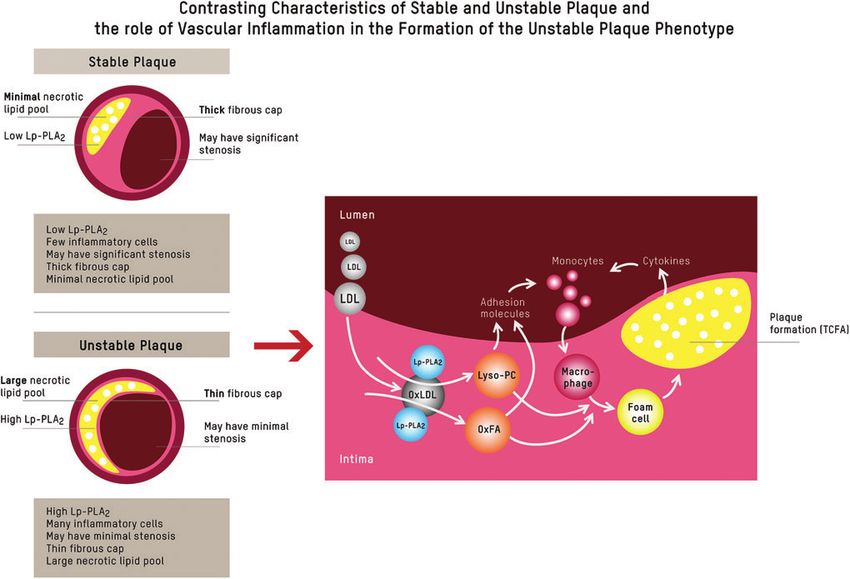
HIGH-RISK ATHEROSCLEROSIS AND METABOLIC PHENOTYPE 177
Introduction The role of body composition in ASCVD
Body composition, in particular accumulation of dys-
D espite ongoing advances in cardiovascular medicine,
acute complications of atherosclerosis remain the lead-
ing causes of death worldwide.1 Of concern, common ap-
functional adipose tissue (AT)9,10 and loss of skeletal
muscle,11,12 is at the core of a cluster of local and systemic
proaches relying on clustered risk factors and surrogate pathophysiological changes that have been linked to high-
biomarkers fail to identify a sizeable proportion of indi- risk atherosclerosis (Fig. 1A, B).
viduals at high risk for cardiovascular events.2 Furthermore, As depicted in Figure 1, excess hepatic fat production
despite management of low-density lipoprotein cholesterol (i.e., de novo lipogenesis) may be an early common path-
(LDL-C) and other conventional risk factors, significant way of non-alcoholic fatty liver disease (NAFLD), athero-
residual risk remains. genic dyslipidemia, pancreatic b cell dysfunction, insulin
One explanation for this observation is the high preva- resistance, and associated ASCVD risk in the high-risk
lence of individuals with metabolic disorders where plasma phenotype.10,14
LDL-C and atherogenic lipoprotein particle concentration, AT secretome. While subcutaneous AT is largely neutral,
as assessed by apoB or direct particle measurement, become or in the case of lower body AT even protective with respect
discordant, rendering LDL-C less predictive of atheroscle- to cardiovascular risk,15 expansion of visceral and/or ectopic
rotic cardiovascular disease (ASCVD) events.3 This results dysfunctional AT is closely linked to poor cardiometabolic
in missed opportunities for prevention and intensification of health and MetS9,16 (Fig. 1A). Factors that promote AT
lifestyle intervention and/or pharmacotherapy in the sub- dysfunction are chronic positive energy balance in conjunc-
group of the population with metabolic syndrome (MetS), tion with biochemical stressors, including physical inactiv-
prediabetes, and diabetes, which is at highest risk for vas- ity,9 poor diet quality,9 active/passive exposure to cigarette
cular complications. Thus, assessment of adjunctive bio- smoke,17 and sleep deprivation.18 Free fatty acid-induced
markers for the presence and severity of subclinical ASCVD cellular stress causes remodeling of AT and encompasses
is of utmost importance for taking appropriate measures to a set of changes, including AT inflammation and altered
prevent adverse clinical outcomes and reduce the associated secretome and modulation of the browning phenotype.
economic burden of this disease. Dysfunctional AT is characterized by an infiltration of
This review discusses the biochemical principles under- macrophages and lymphocytes, and an increased abundance
lying high-risk atherosclerosis within the conceptual frame- of senescent cells. These cells release fatty acids and
work of the phenotype characterized by excess ectopic fat. proinflammatory and chemotactic compounds, which is re-
It furthermore provides perspective on clinical biomarkers ferred to as a senescence-associated secretory phenotype. In
indicative of this high-risk metabolic condition, which may a vicious cycle, this promotes ectopic fat accumulation and
add incremental information to standard risk factor algo- contributes to chronic inflammation, metabolic disturbances,
rithms for detection of high-risk patients who are candi- sarcopenia, and accelerated cardiovascular aging.19 Epi-
dates for interventions aimed at preventing major adverse cardial AT (Fig. 1A1) is regarded as a paracrine transducer
cardiovascular events. of the adverse effects of systemic inflammation and meta-
bolic dysregulation on adjacent tissues, such as the under-
lying coronary arteries, and has accordingly been linked to
Pathophysiological Mechanisms Underlying arrhythmia/atrial fibrillation, accelerated coronary athero-
High-Risk Atherosclerosis sclerosis, and left ventricular diastolic dysfunction.20 Hepatic
An integrative view on atherosclerosis fat accumulation/NAFLD (Fig. 1A2) causally contributes to
atherogenic dyslipidemia [high plasma triglyceride and re-
Atherosclerosis is a lifelong process, and it progresses at duced high-density lipoprotein cholesterol (HDL-C)]. In-
various rates depending on genetic and nongenetic factors.4 creased hepatic de novo lipogenesis is furthermore associated
It is initiated by the penetration and retention of apoB- with higher hepatic palmitic acid (C16:0) flux and enrich-
containing lipoproteins within the subendothelial intima of ment of palmitic acid in very low-density lipoprotein parti-
arteries. This process is gradient driven, providing biological cles (VLDL-P).14 Palmitic acid contributes to vascular
plausibility for the concept that the number of apoB- inflammation through dimerization and activation of toll-like
containing lipoprotein particles, not their aggregate choles- receptor (TLR) 2/4 as explained further below.21 These
terol content (LDL-C), more closely tracks with risk.5–7 mechanisms provide some plausibility for the observation
It is, however, worth noting that numerous factors modify that NAFLD is closely linked to subclinical atherosclero-
the causality of lipoprotein-driven disease risk in ASCVD.8 sis.22 Pancreatic fat (Fig. 1A3) has been linked to b cell
High-risk atherosclerosis is closely associated with a clus- dysfunction23 and concomitant postprandial and fasting hy-
ter of metabolic and inflammatory features of a phenotype perglycemia. Chronically elevated serum glucose levels,
characterized by an increase of ectopic body fat.9,10 Figure 1 and postprandial glucose spikes in particular, result in sym-
depicts a conceptual framework of how the accumulation of pathetic hyperactivity and the formation of advanced gly-
dysfunctional, ectopic fat in the abdominal cavity (visceral cation end products (AGEs). AGE, through interaction with
fat) and in organs (pericardium, liver, pancreas, and skeletal receptor for AGEs, activate proinflammatory signaling
muscle)—in particular if accompanied by sarcopenia11,12— pathways, which promote oxidative stress, chronic vascular
contributes to a proatherogenic and procoagulatory state9,10,13 inflammation, endothelial dysfunction, and accelerated car-
and drives high-risk atherosclerosis through multiorgan/tissue diovascular aging in this phenotype.24
involvement. Figure 1 furthermore maps the links between Lifestyle link. AT phenotype can be modified upon lifestyle
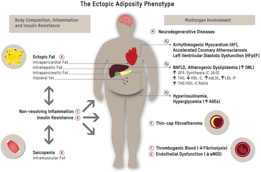
each of the elements within this framework. interventions. Physical exercise,9 intermittent fasting,25,26 diet178 LECHNER ET AL. FIG. 1. The ectopic adiposity phenotype. Ectopic fat accumulation in the abdominal cavity (visceral fat) and in organs like pericardium, liver, and pancreas (A), and muscle wasting/intramuscular fat accumulation (B) are bidirectionally linked to chronic inflammation (C) and insulin resistance (D), both of which have been linked to conventional and novel pathways of cardiometabolic risk.9 Ectopic fat is a major driver of atherosclerosis and its acute complications: epicardial fat (A1) has been linked to AF, accelerated coronary atherosclerosis, and left ventricular diastolic dysfunction20; hepatic fat (NAFLD) (A2) causally contributes to atherogenic dyslipidemia and is closely linked to subclinical atherosclerosis22; and pancreatic fat (A3) has been linked to beta-cell dysfunction23 and concomitant postprandial and fasting hyperglycemia. Chronically elevated serum glucose levels, and postprandial glucose spikes in particular, promote oxidative stress/chronic inflammation, endothelial dysfunction, and sympathetic hyperactivity, and result in the formation of AGEs. Glycation damage has been pathophysiologically linked to numerous chronic disease states such as cardiovascular aging.24 Down arrows indicate decreased levels, and up arrows indicate increased levels. AF, arrhythmia/atrial fibrillation; AGEs, advanced glycation end products; NAFLD, non-alcoholic fatty liver disease. Color images are available online. quality,9 and regular circadian rhythms/restorative sleep18 teract with remote tissues and organs (endocrine function). promote the preservation of a healthy AT phenotype and The muscle secretome consists of several hundred myokines have the potential to reverse AT dysfunction and related that communicate with other organs, such as AT, liver, pan- cardiometabolic risk. These effects occur largely indepen- creas, vasculature, immune system, bones, and brain12 dent of body mass index (BMI).9,16 The CENTRAL-MRI (Fig. 1B). Myokines such as irisin, fibroblast growth factor trial demonstrated that in a group of 278 sedentary adults 21, interleukin (IL)-6, and IL-15 can improve cardiometa- (age = 48 years, 89% men, BMI 30.8 kg/m2) with abdomi- bolic health through several mechanisms: (1) AT browning nal obesity (75%) or dyslipidemia, a Mediterranean low- and maintenance of a functional white AT phenotype, (2) carbohydrate dietary pattern was superior to a low-fat diet in preservation of muscle mass, (3) improved endothelial func- decreasing intrahepatic, intrapericardial, and pancreatic fat tion and myocardial contractility, (4) decreased inflamma- (P < 0.05 for all), and that exercise had an independent tion, and (5) improved metabolic status, including increased contribution to visceral AT loss.27 Further to that, mobili- insulin sensitivity and glucose homeostasis.11,12,16 This un- zation of ectopic fat depots was associated with improved derpins the importance of maintaining or increasing muscle cardiometabolic surrogate markers such as decreased ex- mass to attenuate cardiovascular aging. pression of atherogenic dyslipidemia.27,28 Skeletal muscle secretome. Upon contraction, skeletal mus- Nonresolving inflammation and plaque phenotype cle fibers express and release cytokines and other peptides, which encompass a group of hormone-like substances re- Inflammatory processes at the endothelial layer of the ferred to as myokines.12 Myokines exert their effects within arterial wall play a fundamental role in the initiation, pro- the muscle itself (autocrine and paracrine function) and in- gression, and in particular, the clinical complications of
HIGH-RISK ATHEROSCLEROSIS AND METABOLIC PHENOTYPE                                                                           179

high-risk atherosclerosis. The acute inflammatory response       TLR-mediated immune responses.21 EPA and DHA can
is divided into the two phases of initiation and resolution.29   be supplemented or obtained by the consumption of
Mounting evidence points to defects in inflammation reso-        oily fish.
lution as a key causal factor in atherosclerosis (Fig. 1C).         In an inflammatory resolving environment, vascular smooth
   During the initiation phase, apolipoprotein B-containing      muscle cells that migrate from the medial layer undergo a
lipoproteins, when retained and modified (oxidized) in the       phenotypic shift, forming a collagenous fibrous cap that over-
subendothelial vascular wall, serve as damage-associated         lies the lipid-rich plaque core. Plaque with net resolution is
molecular patterns (DAMPs). DAMPs activate cytokine syn-         smaller, has a thicker fibrous cap (with predominantly anti-
thesis through pattern recognition receptors, for example,       inflammatory M2-like macrophages, smooth muscle cells, and
TLRs.30 Activation of TLR2 and the NLRP3-inflammasome            intact collagen), and has a smaller necrotic lipid core.32
by apolipoprotein C3 (ApoC3) provides one link between              In the setting of continued inflammation, advanced lesions
atherogenic lipoprotein patterns as seen in MetS and im-         develop. They are larger, have a thinner fibrous cap (with
mune activation/inflammation. It is furthermore worth not-       predominantly proinflammatory M1-like macrophages, few
ing that certain saturated fatty acids (SFA) such as palmitic    smooth muscle cells, and cleaved collagen), and have a
acid (C16:0) promote TLR activation.21,31 In the ectopic         larger necrotic lipid core.32 Cells in advanced atherosclerotic
adiposity phenotype, excess dietary starch, sugar, and pro-      plaques express a senescence-associated secretory pheno-
tein are converted into fatty acids (FA)—in particular pal-      type, producing metalloproteinases that degrade the extra-
mitic acid (C16:0)—during a metabolic process referred to        cellular matrix, which promotes inflammation, while further
as hepatic de novo lipogenesis.14,31 This is another mecha-      weakening the fibrous cap, thus fostering a milieu conducive
nism by which proinflammatory signaling pathways are             for a vulnerable plaque phenotype.19,29 This is commonly
activated in the phenotype with ectopic adiposity and            referred to as thin-cap fibroatheroma (TCFA) (reviewed in
NAFLD in particular. Inflammasome activation within              Kasikara et al.29, and Bäck et al.32) (Fig. 1E). One indica-
macrophages leads to the release of proinflammatory cyto-        tor for the presence of TCFA, and concomitant vascular
kines, such as IL-1b, which are chemotactic for other in-        inflammation, is lipoprotein-associated phospholipase A2
flammatory cells; this includes T cells and B cells, which       (Lp-PLA2), which plays a key role in the degradation of
further sustain the chronic inflammatory response by ex-         proinflammatory oxidized phospholipids to generate lyso-
pression of proinflammatory cytokines and eicosanoids.29,30      phosphatidylcholine (Lyso-PC) and oxidized fatty acids,34 as
   During the resolution phase, in a process referred to as      depicted in Figure 2. It is, however, important to note that,
efferocytosis, biosynthesis of proinflammatory mediators         while Lp-PLA2 activity is a marker of risk, pharmacologi-
(e.g., leukotrienes) transitions to synthesis of specialized     cal lowering of Lp-PLA2 in patients with stable coronary
proresolving mediators (SPMs).29 Efferocytosis—the phago-        heart disease (CHD) has not been shown to reduce clinical
cytic removal of apoptotic cells—is a crucial step in the        cardiovascular endpoints.35
resolution of lesional inflammation in chronic nonresolving         Longitudinal observational studies have demonstrated
inflammatory diseases.29 A mismatch between SPMs (low)           that elevated blood levels of proinflammatory biomarkers,
and proinflammatory lipids such as leukotrienes (high)           including high-sensitivity C-reactive protein (hsCRP) and
results in efferocytosis failure, which is, in large part,       IL-6, predict the risk of ASCVD.36 Proof of principle that
mediated by a shift in macrophage phenotype from the anti-       lowering inflammation with pharmacotherapy reduces car-
inflammatory M2-like to proinflammatory M1-like macro-           diovascular events in the absence of lipid lowering came
phages.32 SPMs, which are predominantly produced by M2           from the Canakinumab Anti-inflammatory Thrombosis Out-
macrophages during efferocytosis, include lipoxins bio-          comes Study (CANTOS). In CANTOS, treatment with ca-
synthesized from arachidonic acid and E-series resolvins         nakinumab, a human monoclonal antibody directed against
from the eicosapentaenoic acid (EPA) and docosahexaenoic         the proinflammatory cytokine IL-1b, significantly reduced
acid (DHA)-derived D-series resolvins, protectins, and           IL-1b, IL-6, and hsCRP and demonstrated a cardiovascular
maresins.33 These anti-inflammatory mediators support ef-        (CV) benefit.37,38 Unlike CANTOS, the Cardiovascular In-
ferocytosis by (1) limiting further neutrophil granulocyte       flammation Reduction Trial (CIRT), which tested low-dose
recruitment to the site of injury and (2) enhancing macro-       methotrexate among patients with established CAD and di-
phage uptake of cellular debris and apoptotic neutrophil         abetes and/or MetS, did not reduce IL-1b, IL-6, or C-reactive
granulocytes.33 One subgroup of maresins is involved in          protein (CRP), and did not result in fewer cardiovascular
switching macrophage phenotype from the proinflammatory/         events compared with placebo.39
proatherogenic M1 to the anti-inflammatory/antiatherogenic          Collectively, the evidence from CANTOS and CIRT
M2 phenotype.33 Failure to clear apoptotic cells from ath-       suggests that the prognostic value of anti-inflammatory
erosclerotic plaques results in secondary necrosis, which        agents might strongly depend on the inflammatory pathway
generates inflammation owing to the release of DAMPs             targeted. While inhibition of IL-1b and IL-6-signaling,
from necrotic cells.29                                           which are initiated at the level of the NLRP3 inflammasome,40
   Lifestyle link. One group of SPMs that supports resolu-       effectively reduced cardiovascular events in CANTOS—
tion of inflammation are derivatives of the long-chain ma-       with human genetic data implicating these pathways as
rine n-3 fatty acids EPA and DHA.32 It is furthermore worth      causal in atherothrombosis40—a treatment not targeting the
noting that DHA inhibits TLR2/4 dimerization and activa-         IL-1b and IL-6-signaling pathway (i.e., CIRT) failed to
tion. TLRs—as discussed above—are pattern recognition            show prognostic benefit.
receptors that can be activated by both pathogen-associated         Lifestyle link. As further development and evaluation of
molecular patterns and nonmicrobial endogenous molecules.        pharmacological agents targeting IL-6, IL-1b, and the
The inhibition of TLR2/4 dimerization and activation by          NLRP3 inflammasome are under way, possible alternative
DHA suggest a role for dietary components to modulate            strategies to support resolution of inflammation to lower risk
180                                                                                                        LECHNER ET AL.

FIG. 2. Inflammation and plaque phenotype. When LDLs penetrate the intima of the vessel wall from the lumen, the Lp-
PLA2 residing there uses oxLDL as a substrate, hydrolyzing it to Lyso-PC and OxFA.34 Lyso-PC and OxFA act as
secondary messengers that stimulate the upregulation of adhesion molecules on the lumen surface, act as chemoattractants
for circulating inflammatory cells, and play a role in the activation and transformation of local macrophages within the
plaque lesion. As activated local macrophages take up oxidized (phospho)lipids, they transform to foam cells and subse-
quently express more Lp-PLA2, creating a vicious proinflammatory cycle. The expression of other cytokines like MCP-1
and adhesion molecules creates a feedback loop by attracting more monocytes to the plaque. This feedback loop generates a
vicious cycle of attracting more inflammatory cells to the plaque lesion, resulting in an infiltration with an abundance
of inflammatory cells, a thinning of the fibrous cap, and a growing necrotic lipid core. Although this process often results
in limited luminal narrowing, it leads to the main clinical complications of ASCVD.29 ASCVD, atherosclerotic cardio-
vascular disease; LDL, low-density lipoprotein; Lp-PLA2, lipoprotein-associated phospholipase A2; Lyso-PC, lyso-
phosphatidylcholine; MCP-1, monocyte chemoattractant protein-1; OxFA, oxidized fatty acids; oxLDL, oxidized LDL.
Color images are available online.

of cardiovascular disease (CVD) are (1) to increase the          plasma insulin level, an effect involving suppression of
precursors of SPMs through dietary supplementation of            endogenous glucose production, suppression of lipolysis,
the marine n-3 fatty acids EPA and/or DHA19,29 and/or (2)        cellular uptake of available plasma glucose, and net glyco-
to address chronic inflammation by lifestyle intervention.41     gen synthesis.44 Compensatory hyperinsulinemia is the con-
Vigorous physical activity, intermittent caloric restriction,    sequence of insulin resistance. It means that when insulin
and carbohydrate restriction can be anti-inflammatory due to     resistance is present, fasting and postprandial plasma insulin
elevations of the ketone b-hydroxybutyrate, an endogenous        levels stay chronically elevated and increase further fol-
inhibitor of the NLRP3 inflammasome.42 Furthermore,              lowing a glycemic load.44 This chronic state of exaggerated
contracting muscle secretes hormone-like substances termed       postprandial dysmetabolism, including elevated plasma in-
myokines, which have the potential to attenuate immuno-          sulin – glucose and free fatty acids, creates a milieu con-
senescence through altered tissue crosstalk.43 These effects     ducive to the development of high-risk atherosclerosis and
occur largely independent of body weight.                        type 2 diabetes mellitus (T2DM), one of the major risk
                                                                 factors for ASCVD.45
Insulin resistance/compensatory                                     As depicted in Figure 1, impaired insulin signaling affects
hyperinsulinemia/prediabetes and diabetes                        processes in various tissues and organs relevant to ASCVD,
                                                                 including dysregulation of glucose and lipoprotein metab-
  Insulin resistance is the inability of target tissues to co-   olism and ectopic fat accumulation44 (Fig. 1A1–A3). Ad-
ordinate a normal glucose-lowering response at a normal          ditional features that have been associated with insulin
HIGH-RISK ATHEROSCLEROSIS AND METABOLIC PHENOTYPE 181
resistance include inflammation/oxidative stress and in- syndrome. This discordance can result in significant un-
flammasome activation as discussed above46 (Fig. 1C), derestimation of ASCVD risk by reliance on LDL-C, and
procoagulation/impaired fibrinolysis (Fig. 1F), and endo- failure to adequately manage this risk in individuals with
thelial dysfunction (Fig. 1G).47 The procoagulatory state atherogenic dyslipidemia, and, more broadly, those with
associated with diabetes has been linked to higher concen- visceral adiposity and other features of MetS [5–7].
trations of plasminogen activator inhibitor-1, which reflects Pathophysiologically, the sequence of lipoprotein
a state of fibrinolytic dysfunction in this phenotype. This changes in atherogenic dyslipidemia is induced primarily by
provides biological plausibility for the increased predis- abnormalities of hepatic fat metabolism. Increased rates of
position of individuals with MetS to develop athero- de novo lipogenesis lead to an increased hepatic triglyceride
thrombosis.48,49 Endothelial dysfunction, a key antecedent pool and overproduction of large, triglyceride-enriched
of age-related CVD risk, is attributed to selective vascular VLDL-P. The accumulation of these particles in plasma
insulin resistance, increased superoxide-related oxidative results in the formation of increased levels of lipolytic
stress, and inflammation mediated by, for example, remnants and ultimately smaller LDL-P, as well as de-
ApoC3.30 Endothelial dysfunction results in decreased bio- creased HDL-C due to increased HDL-P catabolism.59 The
availability of the vascular protective vasodilatory mole- consequences with respect to the initiation and progression
cule nitric oxide.47 Furthermore, hyperinsulinemia affects of high-risk atherosclerosis are twofold. First, the presence
kidney function in a way that promotes uric acid and sodium of small LDL-P in atherogenic dyslipidemia contributes to
retention and hypertension.50 a greater total number of circulating LDL-P, even when
T2DM constitutes the tip of the iceberg of this vicious LDL-C is normal or low. Second, there is clinically relevant
cycle of insulin resistance and compensatory hyperinsulin- evidence that small LDL-P have properties that may render
emia. It represents a model for accelerated cardiovascular them more pathologic per se.61–63 Mechanistically, this has
aging and leads to a substantial increase in risk for ASCVD, been linked to a longer residence time of small LDL-P in the
the leading cause of death in people with T2DM.45 In this circulation due to reduced receptor-mediated clearance,
regard, it should, however, be noted that insulin resistance which exposes the endothelial lining to proinflammatory
and compensatory hyperinsulinemia have been linked to the and proatherogenic particle components such as ApoC3.30,55
pathogenesis of CHD and cardiometabolic endpoints even in This concept is supported by the notion that a common
the absence of diabetes mellitus.51,52 underlying trait connecting lipoprotein metabolism to heart
Lifestyle link. Numerous lifestyle factors, dietary and disease risk is the extent to which the condition influences
nondietary, negatively affect insulin sensitivity.41 En- the duration of exposure to arterial tissue, due to either the
couragingly, insulin resistance and compensatory hyper- properties of the LDL-P (e.g., small LDL) or to reduced
insulinemia, even in overt T2DM, are reversible upon hepatic LDL receptor expression (e.g., in familial hypercho-
reduction of intrahepatic, intrapancreatic, and intramuscular lesterolemia). Further traits that have been associated with
fat storage pools as a result of dietary modification and in- potentially higher atherogenicity of small LDL-P include
creased physical activity.23,27,53,54 compositional and conformational changes that render them
more prone to retention,64 aggregation,65,66 and oxidation64 in
Lipoprotein metabolism the arterial wall. Overall, there is not incontrovertible evi-
dence supporting the concept that particle for particle, a small
LDL-C, the concentration in plasma of cholesterol in LDL-P (182 LECHNER ET AL.
triglycerides, and HDL-C.68 Collectively, these data support surements of plasma apoB5–7 and/or LDL-P subclasses55
the concept of greater atherogenic risk associated with provide more specific measures of atherogenic lipoprotein
smaller versus larger LDL-P. burden that can also serve as guides for therapeutic man-
In patients living with MetS, sdLDL particles are strong agement of ASCVD risk. In this regard, LDL-P subclass
predictors of cardiovascular and cerebrovascular events analysis has the potential to detect atherogenic lipoprotein
beyond traditional cardiovascular risk factors.69 In this sub- patterns, which are below the discrimination level of stan-
group, improving the quality of lipoproteins may represent dard lipid testing in MetS.60
an independent target to reduce cardiovascular risk beyond Another metabolic index that can be derived from standard
lowering the quantity of lipoproteins.70 laboratory assays is triglyceride to HDL-C (TG/HDL-C) ra-
Lifestyle link. Notably, levels of small LDL-P are pri- tio, which is associated with both insulin resistance and
marily responsive to dietary carbohydrate intake (increase measures of atherogenic dyslipidemia, including smaller
with higher carbohydrate consumption), while large LDL-P LDL-P diameter (also designated LDL subclass phenotype
are more responsive to dietary saturated fat (increase with B)81 and higher remnant lipoprotein particle cholesterol.61
higher saturated fat consumption). Both weight loss and Furthermore, the TG/HDL-C ratio has been linked to plaque
carbohydrate restriction decrease the expression of the phenotype and clinical stability in coronary artery disease:
small LDL-P pathway.71 These considerations provide some the ratio was significantly higher in patients with TCFAs
biological plausibility for the observation that in large than in those without, and it was nonsignificantly higher in
populations, higher dietary saturated fat consumption is patients with multiple recurrent acute coronary syndromes
associated with higher LDL-C, but not with higher all-cause than in those with long-standing stable angina.82–84 Of note,
or CVD mortality.72 LDL-C might thus provide misleading triglycerides and the TG/HDL-C ratio do not reliably pre-
information as to the effect of diet on ASCVD risk and dict insulin resistance in African Americans. This has been
may therefore be an inappropriate marker for informing linked to the observation that insulin resistance does not
dietary advice.73,74 A further level of complexity regarding impair lipoprotein lipase in this subgroup and thus does not
recommendations for dietary fat intake is the fact that die- induce hypertriglyceridemia.85
tary fats comprise heterogeneous molecules with diverse
structures even within conventional fat classes (i.e., satu- Hypertriglyceridemic waist
rated, monounsaturated, and polyunsaturated fatty acids).74
For example, odd-chain SFAs (C15:0 and C17:0) are rela- At any given BMI, an elevated waist circumference is
tively unique to dairy fat, are not synthesized by humans, predictive of visceral adiposity.13 Using waist circumfer-
and are therefore regarded as reasonable biomarkers of ence as a proxy, increased ectopic and hepatic fat22 in
dairy fat consumption. Cohort studies measuring these particular, is an independent risk factor for high-risk ath-
biomarkers of dairy fat intake show associations with pro- erosclerosis9,10 and coronary artery disease and death in
tection against diabetes, and meta-analytic evidence from prospective studies.45 Mendelian randomization analyses
prospective studies suggest that C17:0, but not C15:0, intake indicate that triglyceride-related risk is causal in ASCVD
is inversely associated with the risk of CVD.74 Collectively, and considering triglyceride levels along with waist cir-
this does not speak to restricting all food sources of dietary cumference improves risk prediction.8,78 Hypertriglyceri-
saturated fat for cardiometabolic health and supports food- demic waist, a visceral adiposity marker combining elevated
based, instead of nutrient-based dietary recommendations.73 waist circumference (‡90 cm) and elevated fasting plasma
HDL-C and HDL subclasses. While clinical and epide- triglycerides (‡2 mmol/L), thus has good diagnostic accu-
miological evidence have consistently shown an inverse racy for the identification of individuals at high risk of
association between HDL-C levels and ASCVD,75,76 the ASCVD.86
failure of clinical trials targeting HDL-C to show prognostic Lifestyle link. Adequate intake of EPA+DHA and/or EPA
benefit,77 in conjunction with genetic evidence,78 supports only at doses of >3 and 4 g/day, respectively, is an effective
the notion that HDL-C is not causal in ASCVD. It is, and safe option for lowering triglycerides87 and hepatic fat
however, worth mentioning that features of HDL other content in NAFLD.41,88
than its cholesterol content are of pathophysiological im-
portance, most notably, the capacity of its major apoprotein Inflammatory markers
component, apoAI, to promote cellular cholesterol efflux.79 While CRP is not thought to have a causal role in ASCVD,
While there is limited evidence that clinical measurement its measurement may augment overall ASCVD risk assess-
of HDL size subclasses provides information bearing on ment, as it is a downstream marker of the IL-1b–IL-6
specific functional properties of HDL, it is of interest that pathway, for which there is evidence of causality.40 Thus,
small HDL-P have recently been reported to be associated measurement of high-sensitivity (hs) CRP may have a role
with increased coronary plaque stability.80 in assessing ASCVD risk in patients with features of MetS,
in whom other measurements are inconclusive.
Clinical assessment of ASCVD risk—beyond LDL-C
Conclusions
Lipid and lipoprotein measurements. As noted above, a
measurement derived from a standard lipid panel that is While atherosclerosis is in large measure a lipoprotein-
more strongly related to ASCVD risk than LDL-C, partic- driven disease, there are numerous factors that modify that
ularly in patients with features of the ectopic fat phenotype, causality. In particular, in the setting of excess ectopic fat
is non-HDL-C (total cholesterol minus HDL-C), which accumulation, metabolic stress fosters a milieu conducive
comprises the cholesterol content of VLDL and atherogenic for high-risk atherosclerosis and its clinical complications:
remnant lipoproteins, in addition to LDL.72 However, mea- dysfunctional AT phenotype and secretome, chronic low-HIGH-RISK ATHEROSCLEROSIS AND METABOLIC PHENOTYPE 183
grade inflammation, atherogenic dyslipidemia, impaired fi- 7. Sniderman AD, Thanassoulis G, Glavinovic T, et al.
brinolysis, and endothelial dysfunction.89 This combination Apolipoprotein B particles and cardiovascular disease: A
of high-risk metabolic and inflammatory traits can be in- narrative review. JAMA Cardiol 2019 [Epub ahead of
expensively detected by combining anthropometric mea- print]; DOI: 10.1001/jamacardio.2019.3780.
sures and clinically available biomarker panels. 8. Ganda OP, Bhatt DL, Mason RP, et al. Unmet need for
adjunctive dyslipidemia therapy in hypertriglyceridemia
Authors’ Contributions management. J Am Coll Cardiol 2018;72:330–343.
9. Oikonomou EK, Antoniades C. The role of adipose tissue
All authors listed have contributed sufficiently to the article in cardiovascular health and disease. Nat Rev Cardiol 2019;
to be included as authors, and all those who are qualified to be 16:83–99.
authors are listed in the author byline. K.L. did the literature 10. Shulman GI. Ectopic fat in insulin resistance, dyslipidemia,
search, and drafted the article. N.K., C.v.S., N.W., U.N., B.L., and cardiometabolic disease. N Engl J Med 2014;371:
J.S., and O.W. reviewed and edited the article. A.L.M. and 2237–2238.
R.M.K contributed significantly to the writing process. 11. Fiuza-Luces C, Santos-Lozano A, Joyner M, et al. Exercise
R.M.K. supervised the writing process and did major revi- benefits in cardiovascular disease: Beyond attenuation of
sions. All authors approved the final version of the article. traditional risk factors. Nat Rev Cardiol 2018;15:731–743.
12. Pedersen BK, Febbraio MA. Muscles, exercise and obesity:
Skeletal muscle as a secretory organ. Nat Rev Endocrinol
Acknowledgment
2012;8:457–465.
The authors are indebted to Nicola Bernhart for graphical 13. Despres JP. Body fat distribution and risk of cardiovascular
design. disease: An update. Circulation 2012;126:1301–1313.
14. Al-Mrabeh A, Zhyzhneuskaya SV, Peters C, et al. Hepatic
Author Disclosure Statement lipoprotein export and remission of human type 2 diabetes
after weight loss. Cell Metab 2019 [Epub ahead of print];
K.L., N.K., N.W., U.N., B.L., J.S., and O.W. declare DOI: 10.1016/j.cmet.2019.11.018.
that no competing financial interests exist with respect to 15. Chen GC, Arthur R, Iyengar NM, et al. Association be-
this article. A.L.M. is employed by Virta Health and has tween regional body fat and cardiovascular disease risk
been offered stock options. C.v.S. operates Omegametrix, a among postmenopausal women with normal body mass
laboratory for fatty acid analyses. He consults for BASF/ index. Eur Heart J 2019;40:2849–2855.
Pronova, and Huntsworth Medical, and received speaker’s 16. Priest C, Tontonoz P. Inter-organ cross-talk in metabolic
honoraria from Abbott, DSM, and Norsan. R.M.K. is on the syndrome. Nat Metab 2019;1:1177–1188.
Scientific Advisory Board of Virta Health and Day Two, has 17. Wang Z, Wang D, Wang Y. Cigarette smoking and adipose
grant support from Quest Diagnostics and Dairy Manage- tissue: The emerging role in progression of atherosclerosis.
ment, Inc., and has a licensed patent for lipoprotein particle Mediators Inflamm 2017;2017:11.
analysis by ion mobility. 18. Pagano ES, Spinedi E, Gagliardino JJ. White adipose tissue
and circadian rhythm dysfunctions in obesity: Pathogenesis
and available therapies. Neuroendocrinology 2017;104:
Funding Information
347–363.
No funding was received for this article. 19. Ferrucci L, Fabbri E. Inflammageing: Chronic inflamma-
tion in ageing, cardiovascular disease, and frailty. Nat Rev
References Cardiol 2018;15:505–522.
20. Packer M. Epicardial adipose tissue may mediate deleteri-
1. GBD 2015 Mortality and Causes of Death Collaborators. ous effects of obesity and inflammation on the myocar-
Global, regional, and national life expectancy, all-cause dium. J Am Coll Cardiol 2018;71:2360–2372.
mortality, and cause-specific mortality for 249 causes of 21. Hwang DH, Kim JA, Lee JY. Mechanisms for the activa-
death, 1980–2015: A systematic analysis for the Global tion of Toll-like receptor 2/4 by saturated fatty acids and
Burden of Disease Study 2015. Lancet 2016;388:1459– inhibition by docosahexaenoic acid. Eur J Pharmacol
1544. 2016;785:24–35.
2. Davidson MH, Ballantyne CM, Jacobson TA, et al. Clinical 22. Stahl EP, Dhindsa DS, Lee SK, et al. Nonalcoholic fatty
utility of inflammatory markers and advanced lipoprotein liver disease and the heart. J Am Coll Cardiol 2019;73:948.
testing: Advice from an expert panel of lipid specialists. 23. White MG, Shaw JA, Taylor R. Type 2 diabetes: The
J Clin Lipidol 2011;5:338–367. pathologic basis of reversible beta-cell dysfunction. Dia-
3. Mora S, Martin Seth S, Virani Salim S. Cholesterol insights betes Care 2016;39:2080–2088.
and controversies from the UK Biobank Study. Circulation 24. Fournet M, Bonté F, Desmoulière A. Glycation damage: A
2019;140:553–555. possible hub for major pathophysiological disorders and
4. Tabas I, Williams KJ, Boren J. Subendothelial lipoprotein aging. Aging Dis 2018;9:880–900.
retention as the initiating process in atherosclerosis: Update 25. de Cabo R, Mattson MP. Effects of intermittent fasting on
and therapeutic implications. Circulation 2007;116:1832– health, aging, and disease. N Engl J Med 2019;381:2541–
1844. 2551.
5. Sniderman AD, Pencina M, Thanassoulis G. ApoB. Circ 26. Di Francesco A, Di Germanio C, Bernier M, et al. A time to
Res 2019;124:1425–1427. fast. Science 2018;362:770.
6. Sniderman AD, Toth PP, Thanassoulis G, et al. An 27. Gepner Y, Shelef I, Schwarzfuchs D, et al. Effect of dis-
evidence-based analysis of the National Lipid Association tinct lifestyle interventions on mobilization of fat storage
recommendations concerning non-HDL-C and apoB. J Clin pools: The CENTRAL MRI randomized controlled trial.
Lipidol 2016;10:1248–1258. Circulation 2018;137:1143–1157.184 LECHNER ET AL.
28. Tsaban G, Wolak A, Avni-Hassid H, et al. Dynamics of 47. Scholz GH, Hanefeld M. Metabolic vascular syndrome:
intrapericardial and extrapericardial fat tissues during long- New insights into a multidimensional network of risk fac-
term, dietary-induced, moderate weight loss. Am J Clin tors and diseases. Visc Med 2016;32:319–326.
Nutr 2017;106:984–995. 48. Anand SS, Yi Q, Gerstein H, et al. Relationship of meta-
29. Kasikara C, Doran AC, Cai B, et al. The role of non- bolic syndrome and fibrinolytic dysfunction to cardiovas-
resolving inflammation in atherosclerosis. J Clin Invest cular disease. Circulation 2003;108:420–425.
2018;128:2713–2723. 49. Meigs JB, Mittleman MA, Nathan DM, et al. Hyper-
30. Zewinger S, Reiser J, Jankowski V, et al. Apolipoprotein insulinemia, hyperglycemia, and impaired hemostasis The
C3 induces inflammation and organ damage by alternative Framingham Offspring Study. JAMA 2000;283:221–228.
inflammasome activation. Nat Immunol 2020;21:30–41. 50. Quinones-Galvan A, Ferrannini E. Renal effects of insulin
31. Lai HTM, de Oliveira Otto MC, Lee Y, et al. Serial plasma in man. J Nephrol 1997;10:88–191.
phospholipid fatty acids in the de novo lipogenesis pathway 51. Reaven G. Insulin resistance and coronary heart disease in
and total mortality, cause-specific mortality, and cardio- nondiabetic individuals. Arterioscler Thromb Vasc Biol
vascular diseases in the cardiovascular health study. J Am 2012;32:1754–1759.
Heart Assoc 2019;8:e012881. 52. Caporaso NE, Jones RR, Stolzenberg-Solomon RZ, et al.
32. Bäck M, Yurdagul A, Tabas I, et al. Inflammation and its Insulin resistance in healthy U.S. adults: Findings from the
resolution in atherosclerosis: Mediators and therapeutic National Health and Nutrition Examination Survey
opportunities. Nat Rev Cardiol 2019;16:389–406. (NHANES). Cancer Epidemiol Biomarkers Prev 2020;29:
33. Dalli J, Serhan C. Macrophage proresolving mediators-the 157–168.
when and where. Microbiol Spectr 2016;4:10.1128/ 53. Taylor R, Al-Mrabeh A, Zhyzhneuskaya S, et al. Remission
microbiolspec.MCHD-0001-2014. of human type 2 diabetes requires decrease in liver and
34. Tselepis AD, John Chapman M. Inflammation, bioactive pancreas fat content but is dependent upon capacity for b
lipids and atherosclerosis: Potential roles of a lipoprotein- cell recovery. Cell Metab 2018;28:547–556.e3.
associated phospholipase A2, platelet activating factor- 54. Taylor R, Barnes AC. Translating aetiological insight into
acetylhydrolase. Atheroscler Suppl 2002;3:57–68. sustainable management of type 2 diabetes. Diabetologia
35. Wallentin L, Held C, Armstrong PW, et al. Lipoprotein- 2018;61:273–283.
associated phospholipase A2 activity is a marker of risk but 55. Krauss RM. All low-density lipoprotein particles are not created
not a useful target for treatment in patients with stable equal. Arterioscler Thromb Vasc Biol 2014;34:959–961.
coronary heart disease. J Am Heart Assoc 2016;5:e003407. 56. Krauss RM. Insulin resistance syndrome and dyslipidemia.
36. Ridker PM, Luscher TF. Anti-inflammatory therapies Endocrine Pract 2003;9 Suppl 2:67–72.
for cardiovascular disease. Eur Heart J 2014;35:1782– 57. Lemieux I, Pascot A, Couillard C, et al. Hypertriglyceri-
1791. demic waist: A marker of the atherogenic metabolic triad
37. Ridker PM, Everett BM, Thuren T, et al. Antiinflammatory (hyperinsulinemia; hyperapolipoprotein B; small, dense
therapy with canakinumab for atherosclerotic disease. LDL) in men? Circulation 2000;102:179–184.
N Engl J Med 2017;377:1119–1131. 58. Ronald M. Krauss M. Insulin resistance syndrome and dys-
38. Ridker PM, Libby P, MacFadyen JG, et al. Modulation of the lipidemia. Endocrine Pract 2003;9(Supplement 2):67–72.
interleukin-6 signalling pathway and incidence rates of ath- 59. Adiels M, Olofsson SO, Taskinen MR, et al. Over-
erosclerotic events and all-cause mortality: Analyses from production of very low-density lipoproteins is the hallmark
the Canakinumab Anti-Inflammatory Thrombosis Outcomes of the dyslipidemia in the metabolic syndrome. Arterioscler
Study (CANTOS). Eur Heart J 2018;39:3499–3507. Thromb Vasc Biol 2008;28:1225–1236.
39. Ridker PM, Everett BM, Pradhan A, et al. Low-dose 60. Rizzo M, Berneis K. Small, dense low-density-lipoproteins
methotrexate for the prevention of atherosclerotic events. and the metabolic syndrome. Diabetes Metab Res Rev
N Engl J Med 2019;380:752–762. 2007;23:14–20.
40. Libby P, Everett B. Novel antiatherosclerotic therapies. 61. Hoogeveen RC, Gaubatz JW, Sun W, et al. Small dense
Arterioscler Thromb Vasc Biol 2019;39:538–545. low-density lipoprotein-cholesterol concentrations predict
41. Lechner K, von Schacky C, McKenzie AL, et al. Lifestyle risk for coronary heart disease: The Atherosclerosis Risk In
factors and high-risk atherosclerosis: Pathways and me- Communities (ARIC) study. Arterioscler Thromb Vasc Biol
chanisms beyond traditional risk factors. Eur J Prev Car- 2014;34:1069–1077.
diol 2019 [Epub ahead of print]; DOI: 10.1177/20474873 62. St-Pierre AC, Cantin B, Dagenais GR, et al. Low-density
19869400. lipoprotein subfractions and the long-term risk of ischemic
42. Youm YH, Nguyen KY, Grant RW, et al. The ketone me- heart disease in men: 13-year follow-up data from the
tabolite beta-hydroxybutyrate blocks NLRP3 inflammasome- Quebec Cardiovascular Study. Arterioscler Thromb Vasc
mediated inflammatory disease. Nat Med 2015;21:263–269. Biol 2005;25:553–559.
43. Duggal NA, Niemiro G, Harridge SDR, et al. Can physical 63. Mora S, Caulfield MP, Wohlgemuth J, et al. Atherogenic
activity ameliorate immunosenescence and thereby reduce lipoprotein subfractions determined by ion mobility and first
age-related multi-morbidity? Nat Rev Immunol 2019;19:563– cardiovascular events after random allocation to high-
572. intensity statin or placebo: The justification for the use of
44. Petersen MC, Shulman GI. Mechanisms of insulin action statins in prevention: An intervention trial evaluating rosu-
and insulin resistance. Physiol Rev 2018;98:2133–2223. vastatin ( JUPITER) trial. Circulation 2015;132:2220–
45. Chia CW, Egan JM, Ferrucci L. Age-related changes in 2229.
glucose metabolism, hyperglycemia, and cardiovascular 64. Berneis KK, Krauss RM. Metabolic origins and clinical
risk. Circ Res 2018;123:886–904. significance of LDL heterogeneity. J Lipid Res 2002;43:
46. Shah MS, Brownlee M. Molecular and cellular mechanisms 1363–1379.
of cardiovascular disorders in diabetes. Circ Res 2016;118: 65. Laufs U, Weingartner O. Pathological phenotypes of LDL
1808–1829. particles. Eur Heart J 2018;39:2574–2576.HIGH-RISK ATHEROSCLEROSIS AND METABOLIC PHENOTYPE 185
66. Ruuth M, Nguyen SD, Vihervaara T, et al. Susceptibility of 79. Silbernagel G, Pagel P, Pfahlert V, et al. High-density li-
low-density lipoprotein particles to aggregate depends on poprotein subclasses, coronary artery disease, and cardio-
particle lipidome, is modifiable, and associates with future vascular mortality. Clin Chem 2017;63:1886–1896.
cardiovascular deaths. Eur Heart J 2018;39:2562–2573. 80. Wang X, Liu X, Xie Z, et al. Small HDL subclass is as-
67. Tehrani DM, Zhao Y, Blaha MJ, et al. Discordance of low- sociated with coronary plaque stability: An optical coher-
density lipoprotein and high-density lipoprotein cholesterol ence tomography study in patients with coronary artery
particle versus cholesterol concentration for the prediction disease. J Clin Lipidol 2019;13:326–334.e2.
of cardiovascular disease in patients with metabolic syn- 81. McLaughlin T, Reaven G, Abbasi F, et al. Is there a simple
drome and diabetes mellitus (from the Multi-Ethnic Study way to identify insulin-resistant individuals at increased risk
of Atherosclerosis [MESA]). Am J Cardiol 2016;117:1921– of cardiovascular disease? Am J Cardiol 2005;96:399–404.
1927. 82. Lechner K, Halle M. Are atherogenic lipoprotein pheno-
68. Tsai MY, Steffen BT, Guan W, et al. New automated assay type and inflammation indicative of plaque phenotype and
of small dense low-density lipoprotein cholesterol identifies clinical stability in coronary artery disease? JAMA Cardi-
risk of coronary heart disease: The Multi-ethnic Study of ology 2019 [Epub ahead of print]; DOI: 10.1001/jama
Atherosclerosis. Arterioscler Thromb Vasc Biol 2014;34: cardio.2019.2261.
196–201. 83. Vergallo R, Porto I, Crea F. Are atherogenic lipoprotein
69. Rizzo M, Pernice V, Frasheri A, et al. Small, dense low- phenotype and inflammation indicative of plaque pheno-
density lipoproteins (LDL) are predictors of cardio- and type and clinical stability in coronary artery disease?-reply.
cerebro-vascular events in subjects with the metabolic JAMA Cardiol 2019;4(9):951-952.
syndrome. Clin Endocrinol 2009;70:870–875. 84. Vergallo R, Porto I, D’Amario D, et al. Coronary athero-
70. Nikolic D, Katsiki N, Montalto G, et al. Lipoprotein sub- sclerotic phenotype and plaque healing in patients with re-
fractions in metabolic syndrome and obesity: Clinical sig- current acute coronary syndromes compared with patients
nificance and therapeutic approaches. Nutrients 2013;5: with long-term clinical stability: An in vivo optical coher-
928–948. ence tomography study. JAMA Cardiol 2019;4:321–329.
71. Krauss RM, Blanche PJ, Rawlings RS, et al. Separate ef- 85. Sumner AE, Finley KB, Genovese DJ, et al. Fasting tri-
fects of reduced carbohydrate intake and weight loss on glyceride and the triglyceride-HDL cholesterol ratio are not
atherogenic dyslipidemia. Am J Clin Nutr 2006;83:1025– markers of insulin resistance in African Americans. Arch
1031; quiz 205. Int Med 2005;165:1395–1400.
72. Mente A, Dehghan M, Rangarajan S, et al. Association of 86. LeBlanc S, Coulombe F, Bertrand OF, et al. Hyper-
dietary nutrients with blood lipids and blood pressure in 18 triglyceridemic waist: A simple marker of high-risk ath-
countries: A cross-sectional analysis from the PURE study. erosclerosis features associated with excess visceral
Lancet Diabetes Endocrinol 2017;5:774–787. adiposity/ectopic fat. J Am Heart Assoc 2018;7:e008139.
73. Astrup A, Bertram HC, Bonjour JP, et al. WHO draft 87. Skulas-Ray AC, Wilson PWF, Harris WS, et al. Omega-3
guidelines on dietary saturated and trans fatty acids: Time fatty acids for the management of hypertriglyceridemia: A
for a new approach? BMJ 2019;366:l4137. science advisory from the American Heart Association.
74. Wu JHY, Micha R, Mozaffarian D. Dietary fats and car- Circulation 2019;140:e673–e691.
diometabolic disease: Mechanisms and effects on risk 88. Yan JH, Guan BJ, Gao HY, et al. Omega-3 polyunsaturated
factors and outcomes. Nat Rev Cardiol 2019;16:581–601. fatty acid supplementation and non-alcoholic fatty liver
75. Di Angelantonio E, Sarwar N, Perry P, et al. Major lipids, disease: A meta-analysis of randomized controlled trials.
apolipoproteins, and risk of vascular disease. JAMA 2009; Medicine 2018;97:e12271.
302:1993–2000. 89. Ren J, Sowers JR, Zhang Y. Metabolic stress, autophagy,
76. Acharjee S, Boden WE, Hartigan PM, et al. Low levels of and cardiovascular aging: From pathophysiology to thera-
high-density lipoprotein cholesterol and increased risk of peutics. Trends Endocrinol Metab 2018;29:699–711.
cardiovascular events in stable ischemic heart disease pa-
tients: A post-hoc analysis from the COURAGE Trial
(Clinical Outcomes Utilizing Revascularization and Ag- Address correspondence to:
gressive Drug Evaluation). J Am Coll Cardiol 2013;62: Katharina Lechner, MD
1826–1833. Department of Cardiology
77. Keene D, Price C, Shun-Shin MJ, et al. Effect on cardio- German Heart Centre
vascular risk of high density lipoprotein targeted drug School of Medicine
treatments niacin, fibrates, and CETP inhibitors: Meta- Technical University Munich
analysis of randomised controlled trials including 117,411 Lazarettstraße 36
patients. BMJ 2014;349:g4379. Munich D-80636
78. Holmes MV, Asselbergs FW, Palmer TM, et al. Mendelian Germany
randomization of blood lipids for coronary heart disease.
Eur Heart J 2015;36:539–550. E-mail: contact@katharinalechner.netYou can also read