Saracatinib is an efficacious clinical candidate for fibrodysplasia ossificans progressiva - JCI Insight
←
→
Page content transcription
If your browser does not render page correctly, please read the page content below
Saracatinib is an efficacious clinical candidate for fibrodysplasia
ossificans progressiva
Eleanor Williams, … , Paul B. Yu, Alex N. Bullock
JCI Insight. 2021;6(8):e95042. https://doi.org/10.1172/jci.insight.95042.

 Research Article   Bone biology     Therapeutics

Currently, no effective therapies exist for fibrodysplasia ossificans progressiva (FOP), a rare congenital syndrome in which
heterotopic bone is formed in soft tissues owing to dysregulated activity of the bone morphogenetic protein (BMP)
receptor kinase ALK2 (also known as ACVR1). From a screen of known biologically active compounds, we identified
saracatinib as a potent ALK2 kinase inhibitor. In enzymatic and cell-based assays, saracatinib preferentially inhibited
ALK2, compared with other receptors of the BMP/TGF-β signaling pathway, and induced dorsalization in zebrafish
embryos consistent with BMP antagonism. We further tested the efficacy of saracatinib using an inducible ACVR1Q207D-
transgenic mouse line, which provides a model of heterotopic ossification (HO), as well as an inducible ACVR1R206H-
knockin mouse, which serves as a genetically and physiologically faithful FOP model. In both models, saracatinib was
well tolerated and potently inhibited the development of HO, even when administered transiently following soft tissue
injury. Together, these data suggest that saracatinib is an efficacious clinical candidate for repositioning in FOP treatment,
offering an accelerated path to clinical proof-of-efficacy studies and potentially significant benefits to individuals with this
devastating condition.

Find the latest version:
https://jci.me/95042/pdf
RESEARCH ARTICLE

                                         Saracatinib is an efficacious clinical
                                         candidate for fibrodysplasia ossificans
                                         progressiva
                                         Eleanor Williams,1 Jana Bagarova,2 Georgina Kerr,1 Dong-Dong Xia,2 Elsie S. Place,3 Devaveena Dey,2
                                         Yue Shen,2 Geoffrey A. Bocobo,2 Agustin H. Mohedas,2 Xiuli Huang,4 Philip E. Sanderson,4 Arthur Lee,4
                                         Wei Zheng,4 Aris N. Economides,5 James C. Smith,3 Paul B. Yu,2 and Alex N. Bullock1
                                         Centre for Medicines Discovery, University of Oxford, Oxford, United Kingdom. 2Department of Medicine, Cardiovascular
                                         1

                                         Division, Brigham and Women’s Hospital, Harvard Medical School, Boston, Massachusetts, USA. 3Developmental Biology
                                         Laboratory, Francis Crick Institute, London, United Kingdom. 4National Center for Advancing Translational Sciences, NIH,
                                         Bethesda, Maryland, USA. 5Regeneron Pharmaceuticals Inc., Tarrytown, New York, USA.

                                                 Currently, no effective therapies exist for fibrodysplasia ossificans progressiva (FOP), a rare
                                                 congenital syndrome in which heterotopic bone is formed in soft tissues owing to dysregulated
                                                 activity of the bone morphogenetic protein (BMP) receptor kinase ALK2 (also known as ACVR1).
                                                 From a screen of known biologically active compounds, we identified saracatinib as a potent ALK2
                                                 kinase inhibitor. In enzymatic and cell-based assays, saracatinib preferentially inhibited ALK2,
                                                 compared with other receptors of the BMP/TGF-β signaling pathway, and induced dorsalization in
                                                 zebrafish embryos consistent with BMP antagonism. We further tested the efficacy of saracatinib
                                                 using an inducible ACVR1Q207D-transgenic mouse line, which provides a model of heterotopic
                                                 ossification (HO), as well as an inducible ACVR1R206H-knockin mouse, which serves as a genetically
                                                 and physiologically faithful FOP model. In both models, saracatinib was well tolerated and potently
                                                 inhibited the development of HO, even when administered transiently following soft tissue injury.
                                                 Together, these data suggest that saracatinib is an efficacious clinical candidate for repositioning
Authorship note: EW, JB, and GK                  in FOP treatment, offering an accelerated path to clinical proof-of-efficacy studies and potentially
contributed equally to this work.                significant benefits to individuals with this devastating condition.
Conflict of interest: ANE is
an employee of Regeneron
Pharmaceuticals Inc. PBY is a
cofounder of and holds stock in
Keros Therapeutics, which develops       Introduction
therapies for hematologic and            Current estimates suggest that there are about 7000 inherited rare diseases (defined as affecting fewer than
musculoskeletal diseases targeting       1 in 2000 people in Europe or 1 in approximately 1600 in the United States) (1). Although their incidence
bone morphogenetic protein and           is low individually, rare diseases collectively affect more than 7% of the general population. Approxi-
TGF-β signaling pathways, including
                                         mately one-half of these conditions now have a recognized genetic cause, offering patients hope for new
fibrodysplasia ossificans progressiva.
                                         treatments (1, 2). However, the costs and timelines of drug development remain major obstacles for many
The interests of PBY are reviewed and
managed by Brigham and Women’s           smaller patient groups as well as for industry investment. Indeed, less than 5% of all rare diseases have an
Hospital in accordance with their        effective pharmacologic treatment, revealing a great unmet medical need (3). One attractive solution for
conflict-of-interest policies.           targeting protein kinases is drug repositioning, which enables well-characterized clinical compounds to be
                                         tested in novel indications, usually based on clear hypotheses and strong target validation (4–6).
Copyright: © 2021, Williams et
al. This is an open access article            Most inherited rare diseases are chronic monogenic conditions that present in early life and cause
published under the terms of the         severely debilitating symptoms as well as reduced life expectancy (7). Fibrodysplasia ossificans pro-
Creative Commons Attribution 4.0         gressiva (FOP) is a particularly devastating example with significant opportunity for drug repositioning
International License.                   (8). Individuals with FOP become severely disabled owing to episodes of heterotopic ossification (HO)
Submitted: May 9, 2017                   that progressively restrict skeletal mobility and reduce life span to a median of 56 years (9). Mus-
Accepted: March 10, 2021                 cle-resident interstitial Mx1+ cells mediate intramuscular HO that is dependent upon trauma, whereas
Published: April 22, 2021                tendon-derived Scx+ progenitor cells mediate spontaneous and progressive HO in tendons, ligaments,
                                         and fascia (10). All cases arise from an autosomal dominant germline mutation in the gene ACVR1
Reference information: JCI Insight.
2021;6(8):e95042.                        encoding the type I bone morphogenetic protein (BMP) receptor kinase ALK2 (11). Approximately
https://doi.org/10.1172/jci.             97% of patients with the classic FOP phenotype harbor the recurrent gain-of-function mutation R206H
insight.95042.                           (c.617G>A) (12). This substitution alters the structure of the receptor’s intracellular domain causing

                                                                                                                                                                    1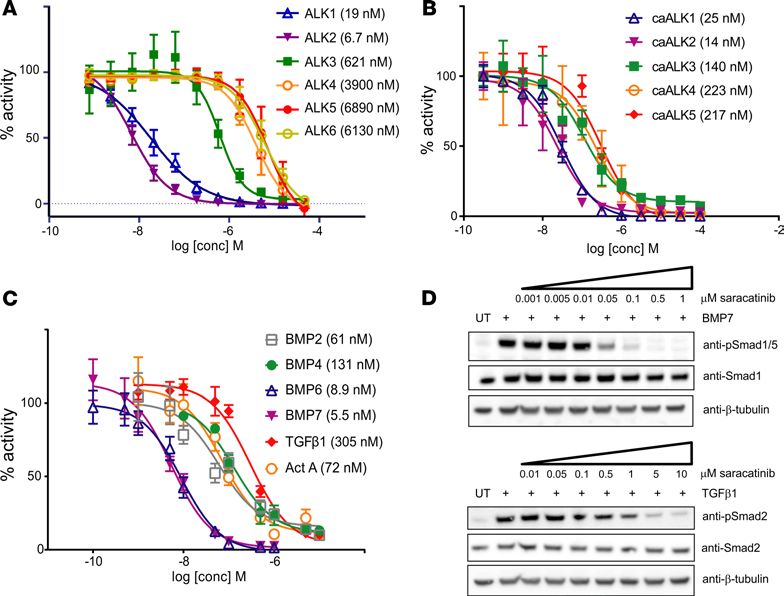
RESEARCH ARTICLE

                                 aberrant kinase activation and SMAD1/5/8 phosphorylation in response to activin A as well as hyper-
                                 sensitivity to BMP ligands (13–15).
                                      There are currently no effective treatments for FOP, and surgical resection of HO has only proved to
                                 exacerbate the condition (8). The discovery that all FOP cases are caused by mutations in ALK2 (also known
                                 as ACVR1) has ignited great interest in this pathway as a therapeutic target (11). Prophylactic treatments tar-
                                 geting the ALK2 pathway (13, 16, 17), or associated transcriptional effectors (18, 19), have demonstrated
                                 preclinical efficacy in FOP mouse models. The activated kinase domain of ALK2 was first targeted by the
                                 small molecules dorsomorphin and LDN-193189 (16, 20). Although these tool compounds are not suitable
                                 for clinical use, they have demonstrated the potential to inhibit BMP receptor–dependent phosphorylation
                                 of SMAD1/5/8 while largely sparing the phosphorylation of SMAD2/3 by the activin (ALK4, also known
                                 as ACVR1B, and ALK7, also known as ACVR1C) and TGF-β (ALK5, also known as TGFBR1) receptors
                                 (20). A number of derivatives have also shown selectivity for ALK2 over the other type I BMP receptors
                                 ALK1 (also known as ACVRL1), ALK3 (also known as BMPR1A), and ALK6 (also known as BMPR1B) (17,
                                 21, 22), but considerable time and resources would be needed to optimize these molecules for clinical use.
                                      The protein kinase family provides one of the most successful group of targets for drug discovery. To
                                 date, more than 70 small-molecule kinase inhibitors have been approved by the FDA, and more than 150
                                 are in clinical trials (23, 24). We reasoned that some of these molecules may harbor unreported activity
                                 against ALK2, and our unbiased screen proved to reveal a number of clinically tested compounds that
                                 bound to ALK2. By far, the most potent was the dual SRC/ABL inhibitor saracatinib (AZD0530), which
                                 demonstrated low nanomolar inhibitory activity against ALK2. In cells, this compound also showed
                                 30-fold selectivity for inhibition of BMP6 over TGF-β signaling pathways. Most important, however, sara-
                                 catinib blocked HO in 2 relevant mouse models. Thus, saracatinib is a promising investigational drug to test
                                 in clinical trials as a frontline therapy for FOP.

                                 Results
                                 Saracatinib is a potent inhibitor of the protein kinase ALK2. To identify potential drug candidates for application
                                 in FOP, we used the kinase domain of the BMP type I receptor ALK2 to screen a library of clinically tested
                                 small-molecule inhibitors via differential scanning fluorimetry (DSF). Ligands in this assay increase the
                                 melting temperature (Tm shift) of a protein by an amount proportional to their binding affinity (25). The
                                 most potent hit was the dual SRC/ABL inhibitor saracatinib (AZD0530, Figure 1A), which induced a large
                                 Tm shift of 13.9°C, comparable to that of the leading tool compound LDN-193189 (26, 27) (Figure 1B and
                                 Supplemental Table 1; supplemental material available online with this article; https://doi.org/10.1172/
                                 jci.insight.95042DS1). By contrast, the next hit, ASP3026, produced a Tm shift of just 10.7°C, equivalent
                                 to the weaker tool compound dorsomorphin (20, 26). As a control for inhibitor selectivity, a counter screen
                                 was performed using the kinase domain of TGF-β type I receptor ALK5. Importantly, saracatinib induced
                                 a significantly lower Tm shift with ALK5 of 10.2°C, suggesting that this inhibitor was both potent and
                                 selective for BMP versus TGF-β signaling (Figure 1B and Supplemental Table 1). We also tested saracati-
                                 nib against ALK2 mutants implicated in FOP disease, finding that all mutants displayed Tm shifts above
                                 14.5°C, confirming their strong interaction (Table 1). Interestingly, the mutant proteins were all destabilized
                                 in their apo states in solution, consistent with structural models showing that FOP-causing mutations break
                                 interactions that normally stabilize the inactive conformation of receptors (14).
                                      Saracatinib binds inactive ALK2 in an αC-in conformation. Although still in phase III trials, saracatinib
                                 is based on the quinazoline scaffold, which has yielded numerous approved drugs, including gefitinib
                                 and vandetanib (28). Interestingly, none of the other quinazoline-based kinase inhibitors tested showed
                                 any notable Tm shift against ALK2 or ALK5 (Tm shift of 4.5°C or less), indicating that this was a specific
                                 property of saracatinib (Supplemental Figure 1, A–C). To explore the molecular basis of this interaction,
                                 we solved the crystal structure of this inhibitor in complex with the ALK2 kinase domain and refined the
                                 structure at 2.66 Å resolution (protein data bank [PDB] accession 6ZGC, Figure 1C, see Supplemental
                                 Table 2 for diffraction data collection and refinement statistics). Within the ATP-binding pocket, the bound
                                 ligand was well resolved by the electron density map (Supplemental Figure 2). Although the quinazoline
                                 of saracatinib formed a hydrogen bond to the hinge residue His286, specificity was likely derived from the
                                 large chlorobenzodioxole moiety, which nicely complemented the size, shape, and hydrophobicity of the
                                 back pocket (Figure 1D). The tetrahydropyran ring was located in the ribose pocket below the glycine-rich
                                 loop (β1-β2), whereas the methylpiperazine moiety was extended largely into solvent (Figure 1D).

JCI Insight 2021;6(8):e95042 https://doi.org/10.1172/jci.insight.95042                                                                            2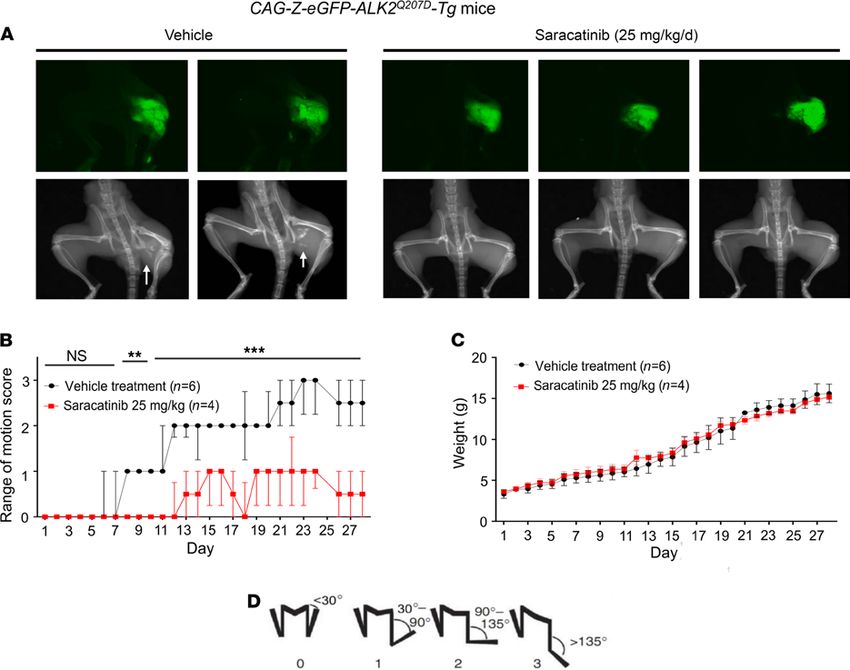
RESEARCH ARTICLE

                                 Figure 1. DSF screening identifies saracatinib as a potent inhibitor of ALK2. (A) Chemical structure of saracatinib.
                                 (B) Plot comparing the thermal shift (ΔTm) of ALK2 and ALK5 in response to different clinical compounds. Selected
                                 compounds of interest are labeled, including saracatinib. A red dashed line is drawn as an approximate guide to mark
                                 equipotency for ALK2 and ALK5. (C) Overview of the ALK2 co-crystal structure with saracatinib. (D) Interactions of
                                 saracatinib with the ATP-binding pocket of ALK2.

                                      Structural differences between the ALK2 and SRC complexes with saracatinib were evident from the
                                 different conformations of their catalytic domains. The previously determined SRC complex structure dis-
                                 played an inactive αC-out conformation that widened the ATP pocket and rotated the αC residue Glu310
                                 away to solvent, as well as the glycine-rich loop residue Phe278 (Supplemental Figure 3) (29). By contrast,
                                 the ALK2 complex exhibited an inactive conformation in which the αC helix was swung inwards to
                                 compress the ATP pocket. As a result, the equivalent αC (Glu248) and glycine-rich loop (Tyr219) residues
                                 were packed inside the ATP pocket where they mediated additional interaction with the chlorobenzodiox-
                                 ole and tetrahydropyran groups, respectively (Supplemental Figure 3). Thus, further optimization of this
                                 chemotype could yield molecules with increased selectivity for ALK2 over SRC family kinases.
                                      Saracatinib was developed to be relatively selective for SRC/ABL kinases compared with some other
                                 multitargeted kinase inhibitors used in oncology today (29). We extended the kinase inhibition profiling
                                 for this compound to a panel of 252 human kinases using the Caliper technology (Nanosyn Inc.). Using
                                 100 nM saracatinib, LCK and ALK2 ranked first and second as the most strongly inhibited kinases, fol-
                                 lowed by SRC, RIPK2, and ABL (Supplemental Table 3). Importantly, ALK2, RIPK2, and TNIK were
                                 the only serine/threonine kinases showing a greater than 50% inhibition at this concentration, whereas
                                 the others were all tyrosine kinases, including paralogs such as ARG (ABL2) and the SRC-family kinases
                                 HCK, FYN, and LYN. These observations are consistent with determined IC50 values for saracatinib
                                 against purified activin receptor-like kinases (IC50: ALK2, 6.7 nM < ALK1, 19 nM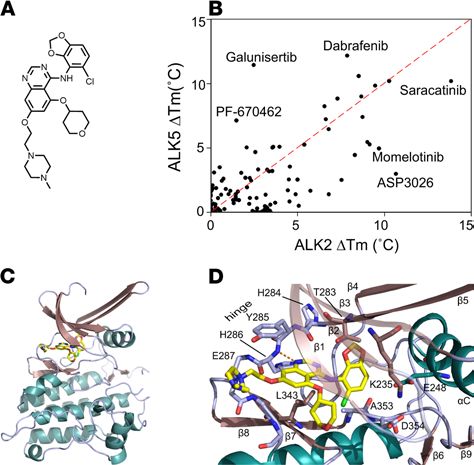
RESEARCH ARTICLE

                                          Table 1. DSF results for ALK2 variants

                                                                                       Tm (°C)
                                                ALK2 protein                                                              ΔTm (°C)
                                                                            Apo                  + saracatinib
                                                    WT                      42.4                     56.4                   13.9
                                                   L196P                    40.0                     57.6                   17.6
                                                   R206H                    40.9                     55.4                   14.5
                                                   Q207E                    42.2                     57.6                   15.4
                                                   R258S                    38.1                     56.4                   18.3
                                                   G328E                    37.0                     56.0                   19.0
                                          ΔTm, thermal shift value.

                                 140 nM), whereas inhibition of the activin/TGF-β receptors caALK4 and caALK5 was weaker (IC50
                                 approximately 220 nM) (Figure 2B).
                                      For subsequent investigations of ligand-dependent receptor signaling we used MDA-MB-231 cells sta-
                                 bly expressing either the BRE-Luc or CAGA-Luc construct. Again, the inhibition profile of saracatinib
                                 showed marked selectivity toward ALK2 and its preferred ligands BMP6 and BMP7. Saracatinib potently
                                 inhibited signaling downstream of BMP6 and BMP7 with IC50 values of 8.9 and 5.5 nM, respectively,
                                 whereas it was less potent against signaling downstream of BMP2 (IC50 = 61 nM) and BMP4 (IC50 = 131
                                 nM), as shown in Figure 2C. Canonical activin A and TGF-β signaling were inhibited to a lesser extent
                                 (IC50 = 72 and 305 nM, respectively, Figure 2C). Thus, there was a 30-fold increase in the concentration of
                                 saracatinib required to inhibit TGF-β signaling compared with that of preferred ALK2 ligands. A similar
                                 pattern was observed in C2C12 cells using specific antibodies to detect receptor-mediated phosphorylation
                                 of the substrate SMAD molecules. Western blot analyses revealed that BMP7-induced phosphorylation of
                                 SMAD1/5 was completely inhibited by 100 nM saracatinib, whereas the TGF-β–dependent phosphoryla-
                                 tion of SMAD2 was only blocked at an inhibitor concentration of 5 μM (Figure 2D). SMAD phosphory-
                                 lation induced by other BMP and activin ligands was inhibited at intermediate saracatinib concentrations
                                 consistent with the LUC reporter assays (Supplemental Figure 5).
                                      FOP-causing mutations in ACVR1 induce neofunction in transducing activin A via BMP receptor–
                                 associated SMAD1/5/8 (13, 15), and this gain of function appears to be the major pathogenetic mech-
                                 anism for the formation of heterotopic bone. To test whether saracatinib could also inhibit activin A–
                                 induced activation of SMAD1/5, we used primary dermal fibroblast cells derived from FOP patients with
                                 the classic ACVR1R206H mutation or WT control cells. Western blot analysis confirmed that phosphorylation
                                 of SMAD1/5 in response to activin A was observed only in the FOP patient-derived cells and not in WT
                                 (Figure 3). In the presence of 100 nM saracatinib, this phosphorylation was inhibited with similar efficacy
                                 to that shown using the control ligand BMP6 (Figure 3A). An IC50 of 15 nM was determined using an
                                 in-cell immunofluorescent assay (Supplemental Figure 6), further confirming the ability of this molecule to
                                 block the neofunction of ALK2 implicated in the development of FOP.
                                      Saracatinib induces dorsalization of zebrafish embryos. Dorsoventral axis specification during embryogenesis
                                 is regulated by the activity of BMP signaling agonists and antagonists (30). In fact, dorsalization of zebrafish
                                 embryos is a developmental phenotype that is highly specific for BMP inhibition and was the basis for the phe-
                                 notypic screens that resulted in the identification of dorsomorphin (20). Incubating embryonic zebrafish with
                                 saracatinib starting at 0.5 hours after fertilization resulted in a dorsalized phenotype that increased in severity
                                 in a dose-dependent manner (Supplemental Figure 7). We observed no evidence of cyclopia or phenotypes
                                 suggesting inhibition of mesoderm specification, consistent with the interpretation that saracatinib selectively
                                 inhibits BMP signaling in vivo without significantly impacting TGF-β or activin type I receptor activity.
                                      Saracatinib prevents heterotopic bone formation in the ACVR1Q207D-Tg mouse model of HO. To evaluate the ther-
                                 apeutic potential of saracatinib for FOP treatment, we first employed one of the most studied models of HO,
                                 a Cre-inducible transgenic mouse that conditionally expresses the constitutively active ACVR1Q207D mutation
                                 (CAG-Z-eGFP-caALK2-Tg) (16, 31). In these animals, activation of the ACVR1Q207D transgene and associat-
                                 ed GFP reporter is mediated by a single intramuscular injection of Adenovirus expressing Cre recombinase
                                 (Ad.Cre) in the left hindlimb on P7. This induces the expression of the human ACVR1Q207D transgene, as well
                                 as muscle necrosis and inflammation, which together result in the formation of heterotopic bone lesions

JCI Insight 2021;6(8):e95042 https://doi.org/10.1172/jci.insight.95042                                                                            4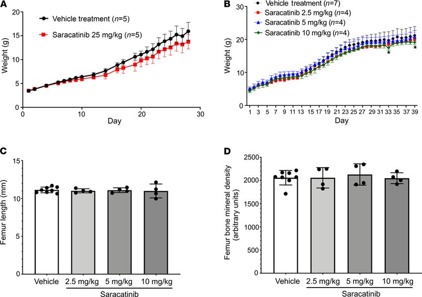
RESEARCH ARTICLE

                                 Figure 2. Saracatinib is a selective inhibitor of BMP versus TGF-β type I receptor activity in cells. (A) IC50 inhibition curves
                                 for saracatinib against purified recombinant ALK1-6 kinase domains were determined using a LANCE Ultra TR-FRET kinase
                                 assay (PerkinElmer). Reactions contained 10 nM kinase, 50 μM peptide substrate, and 10 μM ATP. Data shown are plotted as
                                 mean ± SD. (n = 3 independent replicates). (B) Representative inhibition curves for saracatinib against constitutively active
                                 BMP (caALK1, 2 and 3) and activin/TGF-β (caALK4 and 5) type I receptors, based on the activity of BMP responsive promoter
                                 element LUC (BRE-Luc) and TGF-β responsive LUC (CAGA-Luc) reporters in C2C12 and 293T cells, respectively. Data shown are
                                 representative of more than 3 independent experiments, with data plotted as mean ±SEM. (n = 3 replicates). (C) IC50 inhibi-
                                 tion curves for saracatinib against the signaling induced by indicated ligands based on the activity of BRE-Luc (BMP ligands)
                                 and CAGA-Luc (activin/TGF-β ligands) reporters stably expressed in MDA-MB-231 cells. Data shown are plotted as mean ± SD.
                                 (n ≥ 3 independent replicates). (D) Western blot analyses showing the inhibitory activity of saracatinib against BMP7-induced
                                 phosphorylation of SMAD1/5, as well as TGF-β–dependent phosphorylation of SMAD2 in C2C12 cells.

                                 within 7–10 days following Ad.Cre injection (16). This model is believed to recapitulate aspects of clinical
                                 HO and FOP, in which muscle injury and inflammation potentiate the formation of heterotopic bone in
                                 soft tissues. Untreated, this process normally leads to progressive loss of passive and active range-of-motion
                                 of the hip, knee, and ankle joints over 3–4 weeks, which is accompanied by the formation of intramuscular
                                 heterotopic bone lesions visible by x-ray (Figure 4).
                                      Protocols for saracatinib use in mice are well established from oncology studies and typically use a
                                 clinically relevant dose of 25 mg/kg/d (29, 32–35). CAG-Z-eGFP-caALK2-Tg mice treated similarly with
                                 saracatinib by oral gavage (25 mg/kg once daily for 28 days) demonstrated significantly improved range
                                 of motion, and markedly reduced HO at the site of Ad.Cre injection by x-ray compared with mice treated
                                 with vehicle control (Figure 4). GFP expression in the left hind limb confirmed efficient recombination of
                                 the ACVR1Q207D transgene at the injection site in all drug- and vehicle-treated animals (Figure 4A).
                                      Notably, saracatinib did not impact the normal growth of the pups based on weight gain curves, sug-
                                 gesting that saracatinib can be well tolerated at doses effective for inhibiting HO (Figure 4C). By contrast,
                                 ALK2 inhibitor tool compounds LDN-193189 and LDN-212854 (6 mg/kg i.p. twice daily) (17), caused
                                 a 10%–25% weight loss relative to vehicle-treated animals under these regimens (Supplemental Figure 8).
                                      Other SRC inhibitors are ineffective in blocking HO. Saracatinib has known activity against osteoclasts
                                 and bone turnover through SRC that could potentially contribute to the phenotypic response (35, 36).
                                 To investigate the potential for an SRC-mediated effect, we tested vandetanib (AZD6474) as a control
                                 compound in the same ACVR1Q207D-Tg mouse model. Vandetanib is an analogous quinazoline-based Src
                                 kinase inhibitor that was approved for clinical use in advanced medullary thyroid cancer (28). Although

JCI Insight 2021;6(8):e95042 https://doi.org/10.1172/jci.insight.95042                                                                                         5
RESEARCH ARTICLE
Figure 3. Saracatinib inhibits the neofunction of ALK2R206H. Western blot analysis of phospho-SMAD1/5 levels follow-
ing treatment with saracatinib and either BMP6 or activin A in (A) FOP patient-derived fibroblasts cells (GM00513) or
(B) WT fibroblasts cells (ND34770). Cell lines were validated by DNA sequencing (top panels). Data are representative
of multiple experiments using fibroblasts from 2 independent FOP patients (GM00513 female 16 years of age and
GM00783 male age unknown, Coriell Institute).
it has proven potency against SRC and other tyrosine kinases, including VEGFR2, EGFR, and RET (37),
vandetanib does not show any notable binding to ALK2 (Supplemental Figure 1C). When administered at
25 mg/kg i.p. twice daily for 28 days, vandetanib did not block HO formation in ACVR1Q207D-Tg mice fol-
lowing injection with Ad.Cre (Supplemental Figure 9), despite an exposure higher than previously shown
to demonstrate in vivo efficacy against other target kinases in rodent models (38). Thus, the beneficial
effects of saracatinib in the FOP mouse model were not from inhibition of SRC.
Saracatinib prevents heterotopic bone formation in the Acvr1[R206H]FlEx/+ mouse model of FOP. To test the effect
of saracatinib in a more faithful model of FOP disease, we employed a more recently described conditional
Acvr1R206H-knockin mouse, in which the Acvr1R206H mutant allele (Acvr1[R206H]FlEx/+]) is conditionally expressed
within the mouse Acvr1 locus following Cre-loxP–mediated recombination (13). Acvr1[R206H]FlEx/+]-knockin
mice were challenged with Ad.Cre (1 × 10⁸ PFU intramuscularly) on P7 to induce Cre-mediated recombi-
nation as well as muscle injury. For initial testing, mice were then treated daily with 25 mg/kg saracatinib or
vehicle by oral gavage for 28 days and monitored up to 90 days without further treatment. The rate of HO
induction in this FOP mouse model based on x-ray or range-of-motion loss was notably reduced, consistent
with the milder gain of function of Acvr1R206H compared with ACVR1Q207D (Figure 5, A and B). Vehicle-treated
animals showed the first evidence of decreased range of motion at day 13, which subsequently deteriorated
up to 90 days, when abundant HO was detected (Figure 5, A and B). In contrast, animals receiving an initial
28-day course of saracatinib were protected for the duration of the study and only developed mild loss of
JCI Insight 2021;6(8):e95042 https://doi.org/10.1172/jci.insight.95042 6RESEARCH ARTICLE
Figure 4. In vivo efficacy of saracatinib in the ACVR1Q207D-transgenic mouse model of FOP. Neonatal CAG-Z-eGFP-caALK2-transgenic mice were injected
with Ad.Cre (1 × 10⁸ PFU i.m. P7) and treated with 25 mg/kg/d saracatinib or vehicle orally for 28 days. (A) All mice expressed the eGFP reporter in the
injected left hindlimb, and 100% of vehicle-treated mice (6 of 6) developed radiographic HO and severe loss of passive range of motion (original magnifica-
tion, ×0.8). (B) Treatment with saracatinib essentially abrogated radiographic HO in 4/4 mice, preserving range of motion (2-way ANOVA with Sidak’s test
for multiple comparisons; P = NS, days 1–7; **P < 0.01, days 8–10; ***P < 0.001, days 11–28; all vs. vehicle treatment), data depicted as median ± IQR, n as
indicated. (C) Treatment with saracatinib had no significant impact on normal growth based on weight gain as compared with controls (2-way ANOVA, P =
NS), data depicted as mean ± SEM, n as indicated. (D) Representative hindlimb scoring system. All range of motion scoring was performed by 2 separate
operators who were blinded to the treatment condition of the animal.
range of motion between 60 and 90 days after completing treatment (Figure 5, A and B). These initial results
confirmed that saracatinib attenuates HO mediated by dysregulated Acvr1R206H signaling in vivo and may be
an effective transient therapy following acute trauma, as supported by the prolonged protection observed
after treatment withdrawal and the lack of rebound ossification.
Chronic use of saracatinib as a SRC family inhibitor was recently explored in 2 phase II clinical studies
in Alzheimer’s disease and lymphangioleiomyomatosis (ClinicalTrials.gov identifiers NCT02167256 and
NCT02737202, respectively). Both studies used lower dosing regimens of 100–125 mg saracatinib daily
for 9–12 months (4, 39, 40), corresponding to exposures of approximately 8 mg/kg/d orally in mice based
on comparable levels of drug achieved in plasma and brain (41). Therefore, further experiments in the
ACVR1[R206H]FlEx/+]-knockin mice were performed using saracatinib in the range 2.5–10 mg/kg/d. A higher
dose of Ad.Cre injection (5 × 10⁹ PFU) was administered to all animals to promote more rapid HO induc-
tion. Importantly, we observed that these reduced doses of saracatinib were also effective in preventing
range-of-motion loss over a treatment course of 40 days (Figure 5, C–E).
The efficacy observed at moderate doses of AZD0530 did not appear to impact neonatal growth
assessed by weight gain, femur length, and bone mineral density (Figure 6). Thus, saracatinib may be suf-
ficiently potent and well tolerated for long-term administration in FOP, and in this manner could suppress
acute flares as well as disease that progresses without known flares or antecedent injury.
JCI Insight 2021;6(8):e95042 https://doi.org/10.1172/jci.insight.95042 7RESEARCH ARTICLE
Figure 5. Dose-dependent in vivo efficacy of saracatinib in the FOP-knockin mouse model. Neonatal Acvr1[R206H]FlEx/+] mice were injected with a low dose of
Ad.Cre (1 × 10⁸ PFU i.m. P7) and treated with 25 mg/kg/d saracatinib or vehicle orally for 28 days (grey shaded region) and observed for a total of 90 days. Rep-
resentative mice are shown with x-ray radiography, Micro-CT, and Alizarin red hindlimb prep. (A) Sixty percent of vehicle-treated mice (3/5) developed HO and
severe loss of passive range of motion at 90 days (original magnification, ×0.7). Treatment with saracatinib prevented radiographic HO in 5/5 treated mice, and
(B) preserved range of motion in 4/5 mice at 90 days (2-way ANOVA with Sidak’s test for multiple comparisons, **P < 0.01, day 61; ****P < 0.0001, day 90 vs.
control), data depicted as median ± IQR, n as indicated. ACVR1[R206H]FlEx/+-knockin mice were injected with a high dose of Ad.Cre (5 × 10⁹ PFU i.m. P7) and treated
with varying doses of saracatinib or vehicle orally for 40 days. Radiographic HO and associated impaired range of motion were observed to progress over 40
days of treatment. Treatment with saracatinib at 2.5, 5, and 10 mg/kg/d protected mice from radiographic HO, and preserved passive range of motion (C–E).
Two-way with Dunnett’s test for multiple comparisons. (C) *P < 0.05, days 14–15, 24–25, and 27–28; ***P < 0.001, days 19–23; ****P < 0.0001, days 29–39 vs.
vehicle treatment. (D) ***P < 0.001, days 19–24; **P < 0.01, days 29–34; ****P < 0.0001, days 35–39 vs. vehicle treatment. (E) *P < 0.05, days 19–24; **P <
0.01, days 29–33; ****P < 0.0001, days 34–39 vs. vehicle treatment. Data depicted as median ± IQR, n as indicated.
Discussion
Saracatinib (AZD0530) is a potent, orally bioavailable SRC/ABL inhibitor originally developed by Astra-
Zeneca for treatment of ovarian adenocarcinoma (29). Saracatinib exhibits excellent pharmacokinetic
parameters (t1/2 approximately 40 hours) and is safe in healthy volunteers up to a daily dose of 250 mg (36).
Despite extensive testing in the clinic, with more than 30 registered trials in more than 600 patients, saraca-
tinib has shown only limited efficacy in cancer therapy. However, saracatinib has proven to be of significant
interest for drug repositioning studies and now forms part of an open-innovation portfolio of industry
assets made available to NIH and the Medical Research Council in the United Kingdom (4).
Based on a screen of known biologically active compounds that are approved or in clinical testing, we
identified saracatinib as a multikinase inhibitor that potently inhibits ALK2 in addition to the SRC family
JCI Insight 2021;6(8):e95042 https://doi.org/10.1172/jci.insight.95042 8RESEARCH ARTICLE
Figure 6. Saracatinib treatment preserves neonatal and juvenile orthotopic skeletal growth. (A) Treatment of neonatal mice with saracatinib at 25 mg/kg/d
exerted a mild impact on weight gain during the first 28 days of treatment (P < 0.0001 compared with vehicle treatment, 2-way ANOVA), data depicted as mean ±
SEM, n as indicated. (B) Treatment of neonatal mice with saracatinib at 2.5—10 mg/kg/d generally did not impact weight gain in comparison to vehicle, except for
the 10 mg/kg/d dose at days 33 and 39 (P < 0.05, 2-way ANOVA with Dunnett’s test for multiple comparisons), nor did it impact neonatal and juvenile orthotopic
skeletal growth, based on preserved femur length (C), and preserved femur bone mineral density (D) at 2.5, 5, and 10 mg/kg/d oral doses of saracatinib versus
vehicle treatment (data shown as mean ± SD, n = 8, 4, and 4 femurs analyzed, respectively; P = NS, 1-way ANOVA with Dunnett’s test for multiple comparisons).
of human kinases. Within the BMP/TGF type I receptor family, saracatinib exhibits useful selectivity; it
discriminates between the signaling of ALK2 and its cognate ligands in WT cells (BMP6 and BMP7) and
mutant cells (activin A) versus other receptors and ligands of this family. In fact, saracatinib and the pre-
viously characterized pyrazolo-pyrimidine compound LDN-193189 display comparable cellular potency
against caALK2 (IC50 values of 14 and 11 nM, respectively), as well as selectivity against caALK5 (IC50
values of 217 and 213 nM, respectively) (17). The ability of saracatinib to inhibit ALK2 signaling in a
selective manner in vivo was confirmed by its ability to induce dorsalization in embryonic zebrafish, with-
out inducing other phenotypes owing to the suppression of TGF-β or activin signaling.
Historically, much drug discovery effort has focused on tyrosine kinases of relevance to cancer (28). The
BMP/TGF-β receptors form the only family of serine/threonine transmembrane receptor kinases but map
favorably for drug repositioning to the tyrosine kinase-like branch of the human kinome (42). Therefore,
ALK2 shares sequence and structural similarity with the tyrosine kinases, including a conserved gatekeeper
threonine residue in the hinge region of the ATP-binding pocket (14). In contrast, our screens identify little
saracatinib activity against most other serine/threonine kinases. This specificity appears to derive from the
large chlorobenzodioxole moiety of saracatinib, which complements the size and shape of the back pocket
in ALK2. Notably, these regions differ in the costructures of ALK2 and SRC owing to the different positions
of the αC helix in their inactive states. Based on these differences, it is possible that further optimization of
this chemotype could yield molecules that bind with increased selectivity for ALK2 over SRC family kinases.
Importantly, our findings demonstrate that when FOP is modeled in an authentic and physiologic man-
ner by expressing the classic FOP-causing ACVR1R206H allele under endogenous promoter control, short-term
dosing of saracatinib is sufficient to suppress the development of HO for an extended period following injury.
JCI Insight 2021;6(8):e95042 https://doi.org/10.1172/jci.insight.95042 9RESEARCH ARTICLE
One might extrapolate from these findings that acute prophylactic pharmacologic inhibition of ALK2 activity
could be sufficient to prevent HO in individuals with FOP, although the specific window of time during which
transient therapy is effective would need to be determined in humans. BMP receptor signaling has also been
implicated in the formation of HO in the general population without ACVR1 mutation, including burn- and
trauma-associated HO, or HO associated with severe neurologic injuries (43, 44). Thus, saracatinib is an
interesting clinical candidate to test as an acute therapy in these disease models as well as in FOP patients.
Although several classes of ALK2 kinase inhibitors have been previously reported (20, 26), saracatinib
has potential advantages for clinical translation attributed to the detailed characterization of its tolerability and
side-effect profile in humans. In the case of FOP, the high risk and large investment associated with developing
a novel compound, coupled with the extremely small numbers of patients eligible for trials and treatment,
could further hinder drug development efforts. In addition to being used in numerous oncology clinical trials,
saracatinib has been explored as a FYN kinase inhibitor in a phase II study of patients with mild-to-mod-
erate Alzheimer’s disease at doses up to 125 mg daily for 12 months (4, 39, 40) (NCT02167256). Another
phase II study is investigating this molecule as a SRC inhibitor in lymphangioleiomyomatosis, with a regimen
of 125 mg daily for 9 months (4) (NCT02737202). The primary side effect of saracatinib at these doses has
been gastrointestinal discomfort (40), whereas previous trials using higher doses in oncology were associated
with neutropenia and thrombocytopenia (45–48). The finding that saracatinib is tolerable when administered
chronically at moderate doses suggests that its application to FOP could be extended beyond the prophylactic
treatment of acute episodes of HO. It has been generally supposed that much of FOP disease progression
occurs episodically, in acute flares characterized by soft tissue swelling, pain, and warmth (49). Indeed, phase
II clinical trials for FOP using the RARγ agonist palovarotene were designed to treat acute flare episodes in an
abortive manner (NCT02190747). However, recent natural history studies have suggested that nearly one-half
of individuals with FOP report the insidious progression of disease without known flares or antecedent injury
(50). Thus, more chronic suppression of the underlying disease process outside of acute flares may be needed.
We observed that reduced doses of saracatinib (2.5–10 mg/kg/d) that were similar to or lower than exposures
in phase II studies in Alzheimer’s disease and lymphangioleiomyomatosis were effective in preventing HO in
the ACVR1R206H FOP mouse model. Thus, if saracatinib is sufficiently tolerable, its chronic use for the long-
term suppression of ACVR1R206H activity might be a promising option for maintaining optimal control of FOP
disease, suppressing its progression even in the absence of flares or acute episodes, and potentially preventing
flares altogether. Based on the evidence of target engagement and efficacy in preclinical models at doses equiv-
alent to or lower than those previously tolerated in humans, a phase II clinical trial using 100 mg/d has been
launched to explore the use of saracatinib as prophylaxis for FOP progression (NCT04307953).
Reassuringly, the efficacy of saracatinib has been confirmed by independent study. Hino and colleagues
have demonstrated that saracatinib can efficiently inhibit chondrogenic differentiation and provided addi-
tional efficacy data in FOP mouse models (51). Moreover, Hildebrandt et al. have reported further mecha-
nistic insight showing by RNA-Seq that saracatinib can block the transcriptional program driven by mutant
ALK2 to restore WT expression patterns (52).
The discovery that all cases of FOP are caused by gain-of-function mutations in the intracellular
domain of ALK2 has ignited great interest in ALK2 as a therapeutic target. Repositioning saracatinib for
FOP offers an accelerated path to clinical proof-of-efficacy studies and potentially significant benefits to
individuals with this devastating condition.
Methods
Protein expression and purification. All constructs for protein expression were cloned into the pFastBac-
derived vector pFB-LIC-Bse, which provides an N-terminal hexahistidine tag to enable purification. For
DSF, constructs included ALK2 GS and kinase domain residues 172–499 (WT or R206H) or 172–509
(L196P, Q207E, R258S, or G328E), whereas the ALK5 construct included residues 162–503. For crystal-
lography, the ALK2 construct comprised residues 201–499 with the Q207D mutation. Baculoviral expres-
sion was performed in Sf9 insect cells at 27°C, shaking at 110 rpm. Cells were harvested at 72 hours after
infection and resuspended in 50 mM HEPES, pH 7.5, 500 mM NaCl, 5 mM imidazole, 5% glycerol, and
0.1 mM Tris(2-carboxyethyl)phosphine (TCEP), supplemented with protease inhibitor set V (Calbiochem).
Cells were lysed using sonication (Vibra-cell). Subsequently, 0.15% polyethylenimine was added to precipi-
tate DNA before the insoluble material was excluded by centrifugation at 53,200g. Initial purifications were
performed by Ni-affinity chromatography. Eluted proteins were cleaved with protease and further purified by
JCI Insight 2021;6(8):e95042 https://doi.org/10.1172/jci.insight.95042 10RESEARCH ARTICLE
size-exclusion chromatography using a S200 HiLoad 16/60 Superdex column buffered in 50 mM HEPES,
pH 7.5, 300 mM NaCl, and 1 mM TCEP. Excess protein was flash frozen and stored at −80°C.
Structure determination. Frozen ALK2 protein was thawed, purified from aggregates on a S200 HiLoad
16/60 Superdex column, and concentrated to 10 mg/mL buffered in 5 mM HEPES, pH 7.5, and 100 mM
NaCl. Crystallization was performed using the sitting drop vapor diffusion method at 4°C. Viable crystals
of ALK2 in complex with saracatinib grew in a 150 nL drop mixing 10 mg/mL protein, preincubated with
1 mM compound, with a reservoir solution containing a 1:1 volume ratio mix of 1.26 M sodium phosphate
monobasic and 0.14 M potassium phosphate dibasic. Crystals were transferred into a cryoprotective solution
prepared from the mother liquor supplemented with 25% ethylene glycol. Diffraction data were collected
at Diamond Light Source, beamline I04-1 at a temperature of 100 K using a wavelength of 0.9174 Å. Data
were processed and scaled with MOSFLM and SCALA from CCP4 suite (53, 54). The structure was solved
by molecular replacement using PHASER (55) and the coordinates of ALK2 from the ALK2-FKBP12-
dorsomorphin complex (14) as a search model. Subsequent manual model building was performed using
COOT (56) alternated with refinement in REFMAC (57) and Phenix (58). TLS-restrained refinement was
applied in the latter cycles using the input thermal motion parameters determined by the TLSMD server
(59). The final model was verified for geometry correctness with MOLPROBITY (60) and deposited in the
PDB (accession 6ZGC). The Ramachandran statistics after refinement were 99.8% preferred and 98.0%
allowed. Data collection and refinement statistics are summarized in Supplemental Table 2.
DSF screening. DSF was performed as described by Fedorov et al. (10). Purified proteins containing the
GS and kinase domains of ALK2 or ALK5 were screened at 2 μM concentration against a library of approx-
imately 150 clinically tested kinase inhibitors (Selleckchem) in a 96-well plate format. Compounds were
added to a final concentration of 12.5 μM in an assay buffer containing 10 mM HEPES, pH 7.4, 150 mM
NaCl, and SYPRO Orange (1:1000 dilution, MilliporeSigma). Samples were heated from 25°C to 96°C in a
Mx3005P real-time PCR instrument (Stratagene). Fluorescence was monitored with excitation and emission
filters set to 465 and 590 nm, respectively. Data were analyzed with MxPro software. Thermal shift values
induced by inhibitor binding were calculated relative to control wells containing protein and 2.5% DMSO.
Recombinant ALK1-6 enzyme assays. Recombinant ALK1 was obtained from Life Technology (catalog
PV4883). Recombinant ALK2 to ALK6 were purchased from Carna Bioscience (catalog 09-134). A LANCE
Ultra TR-FRET kinase assay was used for measurements of enzyme activities that included a ULight-
labeled (Thr-1342) peptide substrate (catalog TRF0130-M, PerkinElmer) and a Europium-labeled anti-phos-
pho peptide antibody (catalog TRF0218-M). The kinase reaction was carried out in a buffer containing 10
mM MgCl2, 3 mM MnCl2, 0.005% Tween-20, 2 mM DTT, and 50 mM HEPES, pH 7.0. The details of the
enzyme assays will be reported in a separate paper (our unpublished observations). Briefly, 2.5 μL/well
kinase protein (10 nM final) was dispensed to a white solid 1536-well plate followed by addition of 23 nL/
well compound diluted in DMSO using a Pin-tool station (Wako-Kalypsis). After a 10-minute incubation
at room temperature, 2.5 μL/well substrate solution (50 μM peptide and 10 μM ATP, final) was added, and
the assay plate was incubated for 60 minutes at room temperature. The enzyme reaction was stopped by
addition of 5 μL/well detection solution containing the Europium-labeled anti-phospho peptide antibody.
The signals of time-resolved fluorescence resonance energy transfer (TR-FRET) assay were measured in an
Envision plate reader (PerkinElmer) with a TR-FRET mode (Ex = 320, Em1 = 665, and Em2 = 615 nm).
Cell viability. Cell viability was assayed with an MTT (3-[4,5-dimethylthiazol-2-yl]-2,5-diphenyl tetra-
zolium bromide) colorimetric assay (Promega) per the manufacturer’s instructions.
Western blotting. Bovine aortic endothelial cells were purchased from Lonza and maintained in high-
glucose DMEM supplemented with 10% FBS. C2C12 myofibroblast cells from the European Collection
of Cell Cultures were grown in DMEM/F12 with 10% FCS. Primary human fibroblasts GM00513 (FOP
R206H), GM00783 (FOP R206H), and ND34770 (WT) were obtained from Coriell (https://catalog.cori-
ell.org/1/NINDS) and grown in MEM with 2 mM L-glutamine and 10% FCS. Western blotting was per-
formed essentially as described in Sanvitale et al. (26). Briefly, 1 × 10⁵ cells were seeded into 6-well plates.
The following day, cells were starved in medium containing 1% FCS for 5 hours before treatment with
indicated concentrations of saracatinib and growth factor for 1 hour. Cells were lysed, proteins separated
by SDS-PAGE, and analyzed by Western blotting using relevant antibodies (Cell Signaling: anti-SMAD1
9743), anti-P-SMAD1/5 9516, anti-SMAD2 5339, and anti-P-SMAD2 3101).
In-cell immunofluorescent assay of phospho-SMAD1/5 levels. Human FOP fibroblasts (Coriell Institute)
were grown to confluence in DMEM supplemented with 10% FBS (MilliporeSigma) and then seeded into
JCI Insight 2021;6(8):e95042 https://doi.org/10.1172/jci.insight.95042 11RESEARCH ARTICLE
96-well plates (Costar 3610; Corning). The cells were incubated at 37°C and 10% CO2, allowed to attach,
then starved overnight in 1% FBS-DMEM. Saracatinib (Biotang Inc.) was dissolved in DMSO (Fisher Sci-
entific), whereas LDN-193189 (Yu Lab) was diluted in 0.1% PBS-BSA. The cells were then preincubated
with increasing molar concentrations of saracatinib and LDN-193189 for 15 minutes. Activin A (R&D
Systems) was diluted in 0.1% PBS-BSA (MilliporeSigma) to a working concentration of 250 ng/mL. After
addition of activin A, the cells were allowed to incubate for 45 minutes. Fixation of the cells was performed
with ice-cold methanol and 0.5% glutaraldehyde (Fisher Scientific) before the addition of primary antibody
(Phospho-Smad1/5 (Ser463/465) rabbit mAb 9516; Cell Signaling Technologies) and secondary antibody
(Anti-rabbit IgG, HRP-linked antibody; Cell Signaling Technologies). Finally, the cells were developed
with BioFX Chemiluminescent Ultra Sensitive HRP Microwell Substrate solution (Surmodics) and read on
a Spectra Max L plate reader (Molecular Devices). Softmax Pro software was used, on the Ready to Glow
LUC Assay setting, with an integration time at 0.25 seconds and a wavelength of 470 nm.
Dual LUC assays of constitutively active receptors. C2C12 cells stably expressing firefly LUC under the
control of BMP-responsive promoter element (BRE-Luc) were generously provided by Peter ten Dijke
(Leiden University Medical Center, Leiden, Netherlands). Human embryonic kidney (HEK) 293T cells
stably transfected with the TGF-β–responsive element fused to LUC gene (CAGA-Luc) were a gift of How-
ard Weiner (Brigham and Women’s Hospital). C2C12 BRE-Luc and 293T CAGA-Luc cells were seeded at
20,000 cells in DMEM supplemented with 2% FBS per well in tissue culture treated 96-well plates (Costar
3610; Corning). The cells were incubated for 1 hour (37°C and 10% CO2) and allowed to settle and attach.
Saracatinib was diluted in DMSO, and diluted drug or DMSO vehicle only added to cells at final compound
concentrations of 1 nM to 10 μM and a final concentration of DMSO of 2%. Cells were then incubated for
30 minutes. Adenovirus expressing constitutively active BMP and TGF-β type I receptors (Ad.caALK1-5),
generously provided by Akiko Hata (University of California at San Francisco), were added to achieve
a MOI of 100. Plates were incubated overnight at 37°C. Media was discarded and firefly LUC activity
was measured (Promega) according to the manufacturer’s protocol. Light output was measured using a
Spectramax L luminometer (Molecular Devices) with an integration time of 1 second per well. Data were
normalized to 100% of incremental BRE-Luc activity owing to adenoviruses specifying caALK1, 2, or 3,
or the incremental CAGA-Luc activity owing to adenoviruses specifying caALK4 or 5, and also normal-
ized to total cell counts based on MTT cell viability assay. Graphing and regression analysis by sigmoidal
dose-response with variable Hill coefficient were performed using GraphPad Prism software.
Dual LUC assays of ligand-induced signaling. MDA-MB-231 cells stably expressing BRE-Luc or CAGA-
Luc were generously provided by Caroline Hill (Francis Crick Institute). Some 4000 cells were seeded into
each well of a 96-well plate in DMEM/F12 with 10% FCS. The following day the medium was replaced
with DMEM/F12 and 1% FCS, and then saracatinib added at the indicated concentrations to 6 wells,
followed by overnight stimulation with growth factor. LUC activity was measured using the Promega Dual
LUC Reporter Assay system and percentage activity determined compared with cells treated with growth
factor and no inhibitor. Growth factors (Peprotech) were used at the following concentrations: 500 ng/mL
BMP2, 10 ng/mL BMP4, 500 ng/mL BMP6, 500 ng/mL BMP7, 4 ng/mL TGF-β, and 200 ng/mL activin
A. Experiments were repeated at least 3 times and plotted as mean ± SD.
Zebrafish dorsalization. Zebrafish embryos were treated with varying inhibitor concentrations from less
than or equal to 0.5 hours after fertilization. Inhibitor stocks (20 mM) were diluted in fish water from
DMSO. Embryos were scored according to Mullins et al. (61).
ACVR1Q207D-Tg and Acvr1[R206H]FlEx/+ mouse HO models. Mice were maintained in accordance with IACUC
guidelines under approved experimental protocols. Cre-inducible ACVR1Q207D (CAG-Z-eGFP-caACVR1-
Tg) transgenic mice were a gift from Yuji Michina (University of Michigan, Ann Arbor, Michigan, USA)
as previously described (31). Acvr1[R206H]FlEx/+ mice were generated as previously described (13). Heterotopic
bone formation in these mice was introduced via single retro-popliteal injection of adenoviral Cre-recom-
binase at 1 × 10⁸ PFU at P7. For initial experiments mice (n = 3–4 per group) were treated once a day for 4
weeks with saracatinib at 25 mg/kg by oral gavage dissolved in a vehicle consisting of 5% DMSO and 95%
peanut oil, or vehicle alone. Bone formation as a function of a loss of passive range of motion, via dorsi-
flexion of the left ankle joint was assessed daily. Scores were assessed by 2 independent observers blinded
to genotype and treatment. The observers scored the minimum angle formed by the ankle and the tibia
with passive dorsiflexion under light manual pressure as follows: 0, normal flexion with a minimal angle
ofRESEARCH ARTICLE
with a minimal angle of ≥90° butRESEARCH ARTICLE
Brigham and Women’s Hospital, Harvard Medical School, 75 Francis Street, Boston, Massachusetts 02115,
USA. Phone 857.307.0395; Email: pbyu@partners.org.
1. Boycott KM, et al. Rare-disease genetics in the era of next-generation sequencing: discovery to translation. Nat Rev Genet.
2013;14(10):681–691.
2. Melnikova I. Rare diseases and orphan drugs. Nat Rev Drug Discov. 2012;11(4):267–268.
3. Braun MM, et al. Emergence of orphan drugs in the United States: a quantitative assessment of the first 25 years. Nat Rev Drug
Discov. 2010;9(7):519–522.
4. Frail DE, et al. Pioneering government-sponsored drug repositioning collaborations: progress and learning. Nat Rev Drug Discov.
2015;14(12):833–841.
5. Ekins S, et al. In silico repositioning of approved drugs for rare and neglected diseases. Drug Discov Today. 2011;16(7–8):298–310.
6. Nosengo N. Can you teach old drugs new tricks? Nature. 2016;534(7607):314–316.
7. Boycott KM, et al. Identification of genes for childhood heritable diseases. Annu Rev Med. 2014;65:19–31.
8. Kaplan FS, et al. A new era for fibrodysplasia ossificans progressiva: a druggable target for the second skeleton. Expert Opin Biol Ther.
2007;7(5):705–712.
9. Kaplan FS, et al. Early mortality and cardiorespiratory failure in patients with fibrodysplasia ossificans progressiva. J Bone Joint
Surg Am. 2010;92(3):686–691.
10. Dey D, et al. Two tissue-resident progenitor lineages drive distinct phenotypes of heterotopic ossification. Sci Transl Med.
2016;8(366):366ra163.
11. Shore EM, et al. A recurrent mutation in the BMP type I receptor ACVR1 causes inherited and sporadic fibrodysplasia ossificans
progressiva. Nat Genet. 2006;38(5):525–527.
12. Kaplan FS, et al. Classic and atypical fibrodysplasia ossificans progressiva (FOP) phenotypes are caused by mutations in the
bone morphogenetic protein (BMP) type I receptor ACVR1. Human Mutation. 2009;30(3):379–390.
13. Hatsell SJ, et al. ACVR1R206H receptor mutation causes fibrodysplasia ossificans progressiva by imparting responsiveness to
activin A. Sci Transl Med. 2015;7(303):303ra137.
14. Chaikuad A, et al. Structure of the bone morphogenetic protein receptor ALK2 and implications for fibrodysplasia ossificans
progressiva. J Biol Chem. 2012;287(44):36990–36998.
15. Hino K, et al. Neofunction of ACVR1 in fibrodysplasia ossificans progressiva. Proc Natl Acad Sci U S A. 2015;112(50):15438–15443.
16. Yu PB, et al. BMP type I receptor inhibition reduces heterotopic [corrected] ossification. Nat Med. 2008;14(12):1363–1369.
17. Mohedas AH, et al. Development of an ALK2-biased BMP type I receptor kinase inhibitor. ACS Chem Biol. 2013;8(6):1291–1302.
18. Agarwal S, et al. Inhibition of Hif1α prevents both trauma-induced and genetic heterotopic ossification. Proc Natl Acad Sci U S A.
2016;113(3):E338–E347.
19. Shimono K, et al. Potent inhibition of heterotopic ossification by nuclear retinoic acid receptor-γ agonists. Nat Med. 2011;17(4):454–460.
20. Yu PB, et al. Dorsomorphin inhibits BMP signals required for embryogenesis and iron metabolism. Nat Chem Biol. 2008;4(1):33–41.
21. Mohedas AH, et al. Structure-activity relationship of 3,5-diaryl-2-aminopyridine ALK2 inhibitors reveals unaltered binding
affinity for fibrodysplasia ossificans progressiva causing mutants. J Med Chem. 2014;57(19):7900–7915.
22. Engers DW, et al. Synthesis and structure-activity relationships of a novel and selective bone morphogenetic protein receptor
(BMP) inhibitor derived from the pyrazolo[1.5-a]pyrimidine scaffold of dorsomorphin: the discovery of ML347 as an ALK2
versus ALK3 selective MLPCN probe. Bioorg Med Chem Lett. 2013;23(11):3248–3252.
23. Fabbro D, et al. Targeting cancer with small-molecular-weight kinase inhibitors. Methods Mol Biol. 2012;795:1–34.
24. Wu P, et al. Small-molecule kinase inhibitors: an analysis of FDA-approved drugs. Drug Discov Today. 2016;21(1):5–10.
25. Fedorov O, et al. Kinase inhibitor selectivity profiling using differential scanning fluorimetry. Methods Mol Biol. 2012;795:109–118.
26. Sanvitale CE, et al. A new class of small molecule inhibitor of BMP signaling. PLoS One. 2013;8(4):e62721.
27. Cuny GD, et al. Structure-activity relationship study of bone morphogenetic protein (BMP) signaling inhibitors. Bioorg Med
Chem Lett. 2008;18(15):4388–4392.
28. Wu P, et al. FDA-approved small-molecule kinase inhibitors. Trends Pharmacol Sci. 2015;36(7):422–439.
29. Hennequin LF, et al. N-(5-chloro-1,3-benzodioxol-4-yl)-7-[2-(4-methylpiperazin-1-yl)ethoxy]-5- (tetrahydro-2H-pyran-4-
yloxy)quinazolin-4-amine, a novel, highly selective, orally available, dual-specific c-Src/Abl kinase inhibitor. J Med Chem.
2006;49(22):6465–6488.
30. Wu MY, Hill CS. Tgf-beta superfamily signaling in embryonic development and homeostasis. Dev Cell. 2009;16(3):329–343.
31. Fukuda T, et al. Generation of a mouse with conditionally activated signaling through the BMP receptor, ALK2. Genesis.
2006;44(4):159–167.
32. Chang YM, et al. Src family kinase oncogenic potential and pathways in prostate cancer as revealed by AZD0530. Oncogene.
2008;27(49):6365–6375.
33. Green TP, et al. Preclinical anticancer activity of the potent, oral Src inhibitor AZD0530. Mol Oncol. 2009;3(3):248–261.
34. Cavalloni G, et al. Antitumor activity of Src inhibitor saracatinib (AZD-0530) in preclinical models of biliary tract carcinomas.
Mol Cancer Ther. 2012;11(7):1528–1538.
35. Heusschen R, et al. SRC kinase inhibition with saracatinib limits the development of osteolytic bone disease in multiple myeloma.
Oncotarget. 2016;7(21):30712–30729.
36. Hannon RA, et al. Effects of the Src kinase inhibitor saracatinib (AZD0530) on bone turnover in healthy men: a randomized,
double-blind, placebo-controlled, multiple-ascending-dose phase I trial. J Bone Miner Res. 2010;25(3):463–471.
37. Jia HY, et al. ZD6474 inhibits Src kinase leading to apoptosis of imatinib-resistant K562 cells. Leuk Res. 2009;33(11):1512–1519.
38. Gule MK, et al. Targeted therapy of VEGFR2 and EGFR significantly inhibits growth of anaplastic thyroid cancer in an orthotopic
murine model. Clin Cancer Res. 2011;17(8):2281–2291.
39. Nygaard HB, et al. A phase Ib multiple ascending dose study of the safety, tolerability, and central nervous system availability
of AZD0530 (saracatinib) in Alzheimer’s disease. Alzheimers Res Ther. 2015;7(1):35.
JCI Insight 2021;6(8):e95042 https://doi.org/10.1172/jci.insight.95042 14You can also read