Cancer as a disease of epithelial-mesenchymal interactions and extracellular matrix regulation
←
→
Page content transcription
If your browser does not render page correctly, please read the page content below

Differentiation (2002) 70:547–560                                                                                 C Blackwell Verlag 2002

  REVIEW

Donald E. Ingber

Cancer as a disease of epithelial – mesenchymal interactions and
extracellular matrix regulation

Accepted in revised form: 26 August 2002

Abstract Carcinogenesis – the process of cancer forma-                       normal growth patterns can be observed in benign tu-
tion – is commonly discussed in terms of genetic alter-                      mors, and certain normal tissues, such as bone marrow
ations that lead to deregulation of cell growth. Recently,                   and intestine, exhibit higher cell turnover rates than seen
there has been a resurgence of interest in epigenetic fac-                   in most cancers. The reality is that cancer is not just a
tors and, in particular, the role of the stromal micro-                      disease of the cell. In addition to increased growth, clas-
environment and angiogenesis in tumor formation. In                          sic hallmarks of malignancy include loss of normal
this article, cancer is presented as a disease of the devel-                 tissue architecture, breakdown of tissue boundaries,
opmental processes that govern how cells organize into                       stromal changes, angiogenesis, and compromise of dis-
tissues and tissues into organs. This histogenetic per-                      tant organs through metastatic spread. Cancer therefore
spective raises the possibility that epithelial – mesenchy-                  may be viewed to result from deregulation of the finely
mal interactions and the extracellular matrix (basement                      coordinated processes that normally govern how indi-
membrane) that is deposited through these interactions                       vidual cells are integrated into tissues, tissues into or-
may actively contribute to the carcinogenic process. Ex-                     gans, and organs into a functional living organism
perimental work is reviewed that confirms that extracel-                     (Ingber and Jamieson, 1982). For this reason, we must
lular matrix plays a key role in normal histodifferenti-                     go beyond current reductionist approaches that focus on
ation during both epitheliogenesis and angiogenesis, and                     analysis of the abnormal properties of individual tumor
that epigenetic deregulation of cell – matrix interactions                   cells. Instead, we need to examine the process of carcino-
may actively promote tumor initiation and progression.                       genesis in context of normal tissue formation and devel-
The contributions of integrins, cytoskeleton, tensegrity                     opmental control. Only in this way will we gain full in-
and local variations in extracellular matrix mechanics to                    sight into how tissues undergo malignant transform-
these processes are discussed, as are the implications of                    ation and hence, how this process may be controlled or
this work for future studies on cancer formation.                            even reversed.
                                                                                In this article, I explore cancer as a disease of tissue
Key words carcinogenesis ¡ tumor formation ¡                                 development. First, older (perhaps even ‘‘lost’’) literature
basement membrane ¡ integrin ¡ cytoskeleton ¡                                will be reviewed that suggest cancer may result from de-
mechanics                                                                    regulation of the normal process of histodifferentiation
                                                                             by which multiple cells collectively generate functional
                                                                             tissue architecture. As the large majority of cancers are
Introduction                                                                 epithelial in origin, this perspective leads to a view of
                                                                             cancer as a breakdown of epithelial – mesenchymal inter-
Cancer is commonly characterized as a disease that re-                       actions. In the embryo, active interactions between these
sults from unrestricted cell proliferation. However, ab-                     neighboring tissues drive epitheliogenesis and determine
                                                                             the tissue’s characteristic three-dimensional (3-D) form.
Donald E. Ingber ( )    ✉                                                    Epithelial – stromal interactions also play a key role in
Departments of Pathology and Surgery, Enders 1007, Harvard                   control of angiogenesis in the surrounding stroma and
Medical School and Children’s Hospital, 300 Longwood Ave.,
Boston, MA 02115, USA                                                        thereby guide the formation of a functional vasculature
Tel: π1 617 355 8031, Fax: π1 617 232 7914                                   that is required to feed the growing organ. As will be
e-mail: donald.ingber/tch.harvard.edu                                        described, the extracellular matrix (ECM) that accumu-

U. S. Copyright Clearance Center Code Statement:   0301–4681/2002/709–547 $ 15.00/0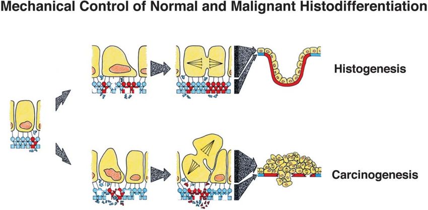
548

lates along the epithelial – mesenchymal interface as a re-    rived matrix metalloproteinases (Simian et al., 2001).
sult of these interactions is a critical control element in    Thus, the stability of epithelial tissue form depends on
these developmental processes. More recent experimen-          the presence of an intact BM, whereas changes in tissue
tal results will be summarized that provide insight into       pattern are driven by spatial differentials in BM turn-
how ECM acts locally to regulate individual cell re-           over. Most importantly, both activities are governed
sponses to soluble growth factors and thereby control          through active interactions between closely apposed epi-
tissue patterning. A model of cancer formation will then       thelial and mesenchymal tissues.
be presented along with supporting experimental data              Similar developmental controls are apparently sus-
that suggest deregulation of tissue growth and form dur-       tained throughout adult life. Mature epithelial tissues re-
ing cancer formation may result from progressive break-        tain the ability to undergo normal morphogenesis when
down of ECM-dependent developmental constraints.               mixed with embryonic mesenchyme (Sakakura et al.,
                                                               1979; Cunha et al., 1983; Chung et al., 1984) and to
                                                               switch histologic form when combined with different
Cancer as a disease of epithelial – mesenchymal                types of stromal tissue (e.g. epidermal grafts in different
interactions                                                   dermal sites; Tarin, 1972a). Studies of teratocarcinoma
                                                               cells that exhibit multiple differentiated lineages suggest
Genesis of characteristic epithelial tissue form (e.g. aci-    that tumors may mimic their tissue of origin in both
nar, tubular, branched, planar) is determined through          appearance and mode of development (Pierce et al.,
complex interactions between the epithelium and its            1978). Given that over 90 % of tumors are carcinomas
underlying mesenchyme in the embryo (Dodson and                (epithelial in origin), then formation of most cancers
Hay, 1971; Banerjee et al., 1977). Studies in which em-        may involve deregulated interactions between epithelial
bryonic epithelia and mesenchyme were isolated inde-           cells and underlying mesenchymally derived stromal
pendently of different tissues and then recombined het-        tissue. In fact, examination of the microscopic anatomy
erotypically revealed that the precise 3-D form that the       of various cancers supports this hypothesis (Leighton,
tissue will express (‘‘histodifferentiation’’) is determined   1969). Examples include the finding that tumor architec-
by the source of the mesenchyme (Sakakura et al., 1976).       ture varies depending on the source of connective tissue
In contrast, the epithelium governs what specialized           (Foley et al., 1968), whereas production of stromal colla-
products the cells will produce (‘‘cytodifferentiation’’).     gen by host fibroblasts depends on the epithelial tumor
   One of the key products of epithelial – mesenchymal         cell type (Gullino, 1966). The finding that an epithelial
interactions is the accumulation of a specialized ECM          neoplasm must gain the ability to induce angiogenesis
scaffold, called the ‘‘basement membrane’’ (BM) along          within its surrounding stroma to switch from hyper-
the epithelial – mesenchymal interface (Grobstein, 1967).      plasia to cancer (Folkman et al., 1989) and to progres-
The BM functions as a extracellular complex of in-             sively expand in size (Ingber et al., 1990; Folkman, 1996)
formative molecules that guides the differentiation, po-       is another clear example of how epithelial – stromal in-
larization, and growth of adjacent adherent cells, in ad-      teractions are critical to cancer progression.
dition to stabilizing the tissue’s characteristic 3-D form        Epithelial – mesenchymal interactions also may con-
(Grobstein, 1967; Dodson and Hay, 1971; Bernfield et           tribute to tumor initiation. For instance, chemical car-
al., 1972; Sakakura et al., 1976; Banerjee et al., 1977;       cinogenesis of epidermis requires the presence of closely
Bernfield and Banerjee, 1978). Localized differentials in      apposed carcinogen-treated dermis (Orr and Spencer,
BM turnover also play a central role in tissue patterning.     1972). Malignant transformation of embryonic mouse
The epithelium physically stabilizes tissue morphology         submandibular gland by polyoma virus also only occurs
by producing BM; the mesenchyme actively induces               in intact or reconstituted glands; transformation cannot
histogenic changes in form by degrading BM at selective        take place in isolated submandibular epithelium or mes-
sites (Bernfield et al., 1972; Bernfield and Banerjee,         enchyme, even though the resultant tumor is epithelial
1978). The highest cell growth rates are observed in re-       in origin (Dawe et al., 1966, 1971). Moreover, once
gions that exhibit the most rapid BM turnover, such as         transformed, these epithelial tumor cells can then substi-
the tips of growing epithelial lobules during salivary         tute for mesenchyme in either normal epithelial mor-
morphogenesis (Bernfield and Banerjee, 1978) and in re-        phogenesis or viral transformation of isolated embry-
gions of capillary sprout formation during angiogenesis        onic epithelia (Dawe et al., 1968). Grafted human epi-
(Ausprunk and Folkman, 1977). At the same time, the            thelial tumors also recruit normal murine stromal cells
mesenchyme slows matrix turnover and induces BM ac-            to become tumorigenic in nude mice (Goldenberg and
cumulation by depositing fibrillar collagen in slower          Pavia, 1981). But most impressive is the finding that
growing regions of the same tissue (e.g. in clefts between     combination of various disorganized epithelial cancers
growing lobules) (David and Bernfield, 1979). Branching        with normal embryonic mesenchyme results in reversal
morphogenesis in mammary gland also can be either              of the malignant phenotype, as evidenced by restoration
stimulated or inhibited by increasing or decreasing ECM        of normal epithelial organization and histodifferenti-
turnover, respectively, using modulators of stromal-de-        ation (Ellison et al., 1969; Lakshmi and Sherbet, 1974;
549

DeCosse et al., 1973; Fujii et al., 1982; Cunha et al.,       taneously metastatic (Pitelka et al., 1980), but the small-
1991; Wong et al., 1992; Chung et al., 1990). Thus, con-      est metastatic tumor in this study displayed multiple BM
tinued epithelial – mesenchymal interactions appear to        breaks. This observation emphasizes the dynamic nature
play an important role in the maintenance of normal           of BM metabolism and epithelial architecture as well as
tissue architecture in adults and deregulation of these       the heterogeneity of tumor cell populations (Fidler,
interactions may contribute to both early and late stages     1978; Pierce et al., 1978) within different microenviron-
of cancer formation.                                          ments. In fact, it is likely that local environmental cues
                                                              may cause intermittent changes in BM dissolution and
                                                              resynthesis. For instance, while BM was absent from one
ECM as a mediator of epithelial – mesenchymal                 primary squamous cell carcinoma, it was present sur-
interactions during tumor formation                           rounding metastatic tumor cells that grew at a distant
                                                              site (Tarin, 1972b). The finding that the ability of a var-
The BM serves as an in vivo adhesive scaffold that en-        iety of tumors to enzymatically degrade BM collagen
sures orderly tissue renewal and maintenance of normal        correlates closely with their metastatic potential (Liotta
tissue form in the adult (Vracko, 1974) as it does in the     et al., 1980) and that tumor angiogenesis also involves
embryo (Grobstein, 1967; Dodson and Hay, 1971;                local microenvironmental control of BM structure (Aus-
Bernfield et al., 1972; Sakakura et al., 1976; Bernfield      prunk and Folkman, 1977; Ingber, 1992) further empha-
and Banerjee, 1978). Thus, changes in ECM metabolism          sizes that dynamic changes in BM may play important
could mediate the effects of deregulated epithelial – mes-    roles at many points during the carcinogenic process.
enchymal interactions during neoplastic transformation
in mature tissues. In fact, local breakdown of the BM
boundary is used as a histologic hallmark of malignant        Experimental confirmation of the active role that ECM
conversion. However, the more fundamental question is:        plays in tumor formation
do subtle changes in ECM structure that precede its
complete breakdown actively contribute to cancer initia-      Many years ago we demonstrated the importance of BM
tion and progression?                                         for neoplastic disorganization of tissue architecture in
   In support of this possibility, ultrastructural changes    studies using a rat pancreatic acinar cell carcinoma
in the BM are commonly observed during early phases           (Ingber et al., 1981, 1985, 1986b). The experiments re-
of cancer formation, prior to development of a palpable       vealed that these cancer cells failed to produce a com-
tumor (Tarin, 1972b; Li et al., 2001; Lu et al., 2000).       plete BM within the tumor parenchyma and they grew in
Early stages of skin carcinogenesis are characterized by      a disorganized array. Yet, the same cells spontaneously
BM gaps, thickening and reduplication as well as              accumulated intact BM and reformed into a polarized
loosening of basal cells from one another and from            epithelium wherever they contacted stromal tissue of the
neighboring connective tissue (Tarin, 1972b), probably        tumor vasculature and surrounding connective tissue
as a result of repeated stages of BM breakdown and            capsule (Fig. 1). The cells were then mechanically iso-
attempted repair. Long-term treatment of prostate with        lated from the parenchyma of the pancreatic tumor and
steroids that leads to neoplastic transformation results      cultured in vitro on intact BM or collagenous stroma
in altered expression of ECM proteins and matrix              from human amnion. The exogenous acellular BM re-
metalloproteinases in the stroma of dysplastic lesions as     versed the neoplastic disorganization process and re-
well as newly formed tumors (Li et al., 2001). The epi-       stored normal epithelial organization in vitro, whereas
thelial BM of carcinogen-treated thyroid gland becomes        tumor cells grew in a disorganized form on the amniotic
discontinuous and is eventually completely lost during        stroma (Ingber et al., 1986b). Induction of pancreatic
progression of lesions from prenodular to nodular forms       epithelial tumor cell reorganization by BM is also ac-
and finally overt carcinomas (Lu et al., 2000). Studies of    companied by restoration of normal cell proliferation
the invasive growth of normal epithelium (Vasilie, 1958)      rates within the adherent tumor cells (Watanabe et al.,
and endothelium (Ausprunk and Folkman, 1977) also             1984). These findings led us to propose that BM nor-
show that local dissolution of BM and growth of under-        mally functions as a spatial organizer of polarized epi-
lying connective tissue occur before the onset of cell pro-   thelia and that progressive loss of BM may actively con-
liferation.                                                   tribute to the neoplastic disorganization of epithelial
   In later stages of cancer formation, a direct corre-       cell – cell relations that is the hallmark of tumor forma-
lation exists between carcinoma cell invasion and local       tion (Ingber et al., 1981, 1985, 1986b).
BM disruption, whereas cells in tumors that retain an            These early experimental studies were provocative in
intact BM do not penetrate into surrounding tissue (Oz-       that they suggested that changes in ECM may play an
zello, 1959; Luibel et al., 1960; Rubio and Biberfeld,        active role during early stages of tumor formation prior
1979). This relationship may be confused at times, how-       to the onset of malignant invasion. However, this epige-
ever. For example, BM discontinuities were rare in a          netic hypothesis was recently confirmed in a series of
virally induced murine mammary tumor that was spon-           elegant studies from the laboratories of Bissell and Werb
550 Fig. 1 Schematic summary of the epithelial organization process along its interface with underlying mesenchyme or stroma. See text within embryonic tissue and in an adult, pancreatic acinar cell tu- for details. mor. The solid blue line represents BM beneath the epithelium that showed that deregulation of ECM metabolism can actively promote cancer development. Specifically, they Architectural control of tissue development and demonstrated that targeting of autoactivating or tetracy- tumor formation by ECM cline-inducible forms of the matrix metalloproteinase stromelysin-1 to the mammary epithelium leads to for- The literature described above indicates that BM pro- mation of a reactive stroma (Thomasset et al., 1998) and vides critical cues for growth and differentiation during promotes both tumor initiation and malignant trans- embryogenesis, and its continued maintenance is formation in transgenic mice (Lochter et al., 1997; mandatory for the normal functioning and growth regu- Sternlicht et al., 1999, 2000). Interestingly, stromelysin-1 lation of adult tissues. But how could a macromolecular is a natural product of the stromal cells and enhanced complex like the BM influence normal tissue morpho- ECM degradation is again observed in early phases of genesis or cancer formation? This is a question that has this process prior to full expression of the invasive plagued the field of epithelial – stromal interactions for phenotype and BM disruption (Thomasset et al., 1998). many years. Moreover, once transformed, mammary epithelial cells BM may influence cell behavior in many ways. For lose the ability to downregulate their production of stro- example, BM serves as the filtration barrier in the kid- melysin-1 in response to cell adhesion to BM (Lochter ney glomerulus (Farquhar, 1978) and thus it may pro- et al., 1999). vide a similar ability to selectively control epithelial ac- Analysis of transgenic mouse models of cancer also cess to soluble molecules in other organs. Physical has revealed that early stages in cancer formation are breakdown of this differentially permeable barrier, as is accompanied by changes in the expression of cell surface observed in regions of most rapid growth in embryonic ECM receptors, known as ‘‘integrins’’ (Tennenbaum et tissues or during early tumor formation, could result in al., 1992). Moreover, cancer formation can be reversed increased flow of chemical factors between the stromal in vitro and in vivo by changing integrin expression levels and epithelial microcompartments. Certain cytokines, (Ruoslahti, 1996), modulating integrin binding (Weaver such as basic fibroblast growth factor, are stored within et al., 1997) or altering integrin signaling activities the basement membrane (Folkman et al., 1988) and thus (McLean et al., 2001). Integrin antagonists also inhibit increased BM turnover may also release growth factors angiogenesis (Brooks et al., 1994) and tumor metastasis locally. However, normal cells will not grow in response (Ruoslahti, 1996). Thus, ECM and its integrin receptors to growth factor stimulation when they are free of ad- clearly play an active role in both carcinogenesis and hesion to ECM (Guadagno and Assoian, 1991) or when tumor progression. they are physically crowded (compressed) within a tight
551

Fig. 2 Schematic diagram of a mechanical model of normal and          tion, and associated growth. However, because net BM accumu-
malignant tissue differentiation based on BM remodeling and as-       lation does not occur, piling up of cells and tissue disorganization
sociated tension-driven restructuring of tissue form. Circles, tri-   result. If the growth stimulus (e.g. altered BM turnover) were to
angles, and squares indicate different BM components; broken fig-     cease at this point, cells that lack contact with the BM would un-
ures, molecules removed by degradation; closed red figures, newly     dergo apoptosis or stop growing and thus this hyperplastic process
added molecules. Top In normal histogenesis, increased BM turn-       would be reversible. Continued stimulation of cell proliferation
over results in thinning of the BM and an associated increase in      may eventually lead to selection of a subpopulation of cells that
mechanical compliance (flexibility), which promotes cell distortion   gain the ability to survive and grow free of adhesion to BM; at this
and growth locally. Because increased cell division is accompanied    point the process of tumor formation would become irreversible.
by net BM expansion, the tissue branches outward and initiates        If progressive deregulation of BM metabolism leads to physical
pattern formation. Bottom During carcinogenesis, an increase in       compromise of the BM barrier, tumor cell invasion may occur and
BM turnover may lead to similar thinning of the BM, cell distor-      hence the tumor would become malignant.

epithelial monolayer (Stoker and Rubin, 1967; Folkman                 structure and mechanics (Fig. 2, top). In this model, the
and Moscona, 1978). Thus, release of growth factors                   local regions of BM at tips of growing epithelial glands
alone is not sufficient to explain local cell growth induc-           and new capillary sprouts that become thinner due to
tion or why proliferating cells are observed in suprabasal            high ECM turnover (Bernfield et al., 1972; Ausprunk
regions of preneoplastic epithelium. Instead, these ob-               and Folkman, 1977) would be expected to become more
servations suggest that ECM-dependent growth regula-                  compliant. All soft tissues experience isometric tension
tion and normal cell ‘‘crowd controls’’ also must be de-              or a tensile ‘‘pre-stress’’ based on the generation of trac-
regulated in order for tumor formation to proceed                     tional forces by their constituent cells. Because it is
(Schwartz and Ingber, 1994). To explain how this pro-                 under tension, a weak spot in the BM would stretch out
cess may be involved in cancer formation, we must first               more than the neighboring tissue, just like a ‘‘run’’ in
understand how ECM acts to regulate cell growth and                   a woman’s stocking. This alteration in ECM mechanics
function during normal morphogenesis.                                 would change the balance of forces that are transferred
                                                                      across cell surface integrin receptors that physically con-
                                                                      nect the ECM to the internal cytoskeleton (CSK) within
Normal histogenesis                                                   adjacent adherent cells. Increased tension on these ad-
                                                                      hesion receptors would, in turn, promote cell and CSK
Twenty years ago, we proposed that ECM may contrib-                   distortion in these distended regions and thereby alter
ute to control of both morphogenesis and tumor forma-                 cell sensitivity to soluble cytokines, resulting in the local-
tion based on its mechanical properties; that is, its ability         ized growth that drives tissue patterning (Ingber and Ja-
to physically resist cell tractional forces (Ingber et al.,           mieson, 1982, 1985). If this increase in cell division were
1981; Ingber and Jamieson, 1982, 1985). The concept                   paralleled by a commensurate local increase in BM
was that the local variations in ECM remodeling that                  expansion (due to net BM accumulation), then coordi-
are observed during morphogenesis would change ECM                    nated budding and branching would result (Fig. 2, top).
552 Fig. 3 Microscopic images of capillary endothelial cells (A, C, E) switching between growth, differentiation, and apoptosis that has whose shape, size and position were controlled using the corre- been demonstrated using micropatterned substrates of different sponding micropatterned adhesive substrate designs shown in (B), size, as described in the text. H A fluorescent micrograph of a (D), and (F), respectively. Note that when cells are cultured on fibroblast cultured on a square, fibronectin-coated adhesive island different-sized islands in the same dish that are coated with a con- (40 ¿ 40 mm) and stimulated with the motility factor, PDGF, after stant fibronectin density and in the same growth factor-containing staining for F-actin with rhodamine-phalloidin. Note that the cell medium, cell distortion can be varied independently, as shown in preferentially extends actin-containing lamellipodia and filopodia (E, F). G A schematic summary of cell distortion-dependent from its corners. Experimental results from many laboratories have chemically defined medium containing a saturating confirmed that ECM exerts this form of mechanical amount of growth factor, cell growth increased in paral- control over epithelial and endothelial cell behavior both lel as island size was raised and cell spreading was pro- in vitro and in vivo (Folkman and Moscona, 1978; Ben moted (Fig. 3G). We also confirmed that both small and Ze’ev et al., 1980, 1988; Ingber et al., 1986a, 1987; Li et large ECM islands induce similar levels of integrin sig- al., 1987; Ingber and Folkman, 1988, 1989; Opas, 1989; naling (Yan et al., 2000), and that when cells spread over Ingber, 1990; Mochitate et al., 1991; Mooney et al., multiple focal adhesion-sized islands (3 to 5 mm in diam- 1992; Singhvi et al., 1994; Chen et al., 1997; Huang et eter), growth scaled with cell spreading and not the total al., 1998; Dike et al., 1999; Flusberg et al., 2001; Huang area of ECM contact formation (Chen et al., 1997). and Ingber, 2002; Parker et al., 2002). For example, in When the island area was decreased further and cell our studies, we used a microfabrication method to vary growth was shut off, apoptosis – the cellular ‘‘suicide’’ cell distortion independently of growth factor stimula- program – was switched on in capillary cells (Fig. 3G), tion and ECM binding. This was accomplished by even though the cells still remained adherent to the adapting a micropatterning technology that was de- ECM substrate (Chen et al., 1997; Flusberg et al., 2001). veloped by George Whitesides’ Laboratory (Dept. of Moreover, treatment of growing embryonic tissues with Chemistry and Chemical Biology, Harvard U.) as a drugs that produce BM breakdown and associated cell method for fabricating microchips for the computer in- retraction (rounding) resulted in inhibition of endo- dustry (Singhvi et al., 1994; Chen et al., 1997, 2000). thelial cell growth and induction of capillary regression This technique was then used to create adhesive islands (i.e. angiogenesis inhibition) in vivo (Ingber et al., 1986a; of predefined size, shape and position on the micrometer Ingber and Folkman, 1988). Similar treatments that re- scale coated with a saturating density of immobilized sult in complete dissolution of BM also induce re- ECM molecules; the surrounding barrier regions were gression of growing mammary epithelium (Wicha et al., made nonadhesive by exposing polyethlylene glycol moi- 1980) and müllerian duct (Trelstad et al., 1982). eties on their surface that prevented protein adsorption Interestingly, when cells were cultured on intermedi- (Fig. 3). ate-sized ECM islands that supported a moderate degree When primary liver epithelial cells were cultured on of spreading and promoted neither growth nor rectangular islands coated with laminin that ranged in apoptosis, a differentiation program was induced (Fig. diameter from 10 to 50 mm, they spread to the limits of 3G). Moderate-sized hepatocytes secrete higher levels of the island and thus took on rectangular shapes (Sinhvi liver-specific blood proteins, including albumin and fi- et al., 1994). Similar shape control was observed when brinogen (Mooney et al., 1992; Singhvi et al., 1994), and capillary endothelial cells were plated on square or circu- endothelial cells form hollow capillary tubes (Ingber and lar islands of similar size coated with fibronectin or Folkman, 1989; Dike et al., 1999). More recently, we also other ECM ligands (Chen et al., 1997) (Fig. 3A – F). found that if a motility factor, such as PDGF or FGF, When both types of cells were cultured on different-sized is added to a cell adherent to a square ECM island, the adhesive islands coated with a high ECM density in cell will extend out lamellipodia, filopodia, and
553

microspikes that drive cell migration. However, forma-         monly remains physically intact during early stages of
tion of these new cell processes is physically constrained     tumor formation (i.e. prior to malignant transformation
in that they preferentially extend from the corners of the     and invasion), but a reduction in BM thickness or subtle
square cells, where cells form focal adhesions and exert       decreases in the levels of certain ECM constituents often
their tractional forces on ECM (Fig. 3H) (Parker et al.,       can be detected. In contrast to changes observed during
2002; Wang et al., 2002a). In other words, the geometry        normal tissue development, these neoplastic changes are
of the substrate and orientation in which the cell             not restricted in space or time and hence tissue disorgan-
stretches dictates the direction of cell movement.             ization results (Fig. 2, bottom). In fact, it is this loss of
   These results, taken together with findings from other      tissue pattern that usually catches the eye of the pathol-
experiments (Folkman and Moscona, 1978; Ben Ze’ev et           ogist who recognizes it as abnormal (Clark, 1995). As in
al., 1980, 1988; Li et al., 1987; Opas, 1989; Mochitate et     the embryo, continued changes in BM structure that
al., 1991; Dembo and Wang, 1999; Wang et al., 2002a),          lead to increased mechanical compliance (e.g. thinning)
demonstrate that cell distortion per se governs whether        may promote cell distortion or increase CSK tension
individual cells will grow, differentiate or die (Fig. 3G)     locally and thereby increase the sensitivity of adjacent
when stimulated by soluble mitogens as well as the direc-      cells to mitogenic stimuli. But in this case, a piling up of
tion in which they move (Fig. 3H). These data support          cells would result because the BM does not expand in
our model in which mechanical changes in ECM compli-           parallel to match increases in cell number (Fig. 2, bot-
ance that affect cell shape and structure play a central       tom), as occurs in normal development (Fig. 2, top).
role during normal histogenesis (Fig. 2, top). This ten-          If these changes are maintained over many years and
sion-driven remodeling hypothesis for morphogenetic            the growth stimulus is sustained, cells that grow free of
regulation (Ingber et al., 1981; Ingber and Jamieson,          anchorage in vivo may spontaneously emerge just as con-
1982, 1985; Huang and Ingber, 1999) is also consistent         tinued culturing of normal cells may lead to spon-
with the finding that variations in the pattern-directing      taneous transformation in vitro. This transformation
behavior of different mesenchyme correlate with their          process would require that the cells gain the ability to
ability to generate mechanical tension (Nogawa and             grow independent of both ECM adhesion and cell dis-
Nakanishi, 1987). This is important because, as de-            tortion to fully overcome normal crowd controls. Natu-
scribed above, the mesenchyme determines tissue-speci-         ral selection and expansion of this autonomous cell
fic pattern formation in epithelium (Sakakura et al.,          would result in the ‘‘clonal’’ origin of proliferating tu-
1976). Moreover, pharmacologic inhibition of tension           mor cells, yet the evolutionary process that led to cre-
generation inhibits morphogenesis in developing salivary       ation of this cancer cell would have taken place at the
gland (Ash et al., 1973) and lung (Moore et al., 2002),        tissue level. Cell growth and survival free of contact with
while increasing cytoskeletal tension through activation       the BM is then sufficient to explain the disorganization
of the rho/ROCK pathway actually accelerates                   of normal cell – cell relations that is observed during
branching morphogenesis (Moore et al., 2002).                  subsequent stages of neoplastic transformation (Ingber
   Reliance on local distortion for spatial control of cell    et al., 1981, 1985).
growth may explain why expressing high stromal levels             In support of this concept, the ability of cells to form
of growth factors in transgenic mice increases branching       3-D organotypic structures and to maintain normal cell
of mammary epithelium, rather than producing prolifer-         shapes suppresses expression of the malignant pheno-
ation in all cells and amorphous tissue growth (Joseph         type, including production of stromelysin-1 (a promoter
et al., 1999). While soluble growth factors act to regulate    of tumor progression), as shown in studies involving cul-
overall tissue and organ size, their effect may be spatially   ture of normal mammary epithelial cells and their trans-
restricted in a tissue-specific manner through this ten-       formed counterparts on BM gels (Peterson et al., 1992;
sion-molding mechanism to generate distinct histologic         Wang et al., 1998; Sternlicht et al., 1999, 2000). Some
patterns. The key point here is that this spatial restric-     studies attribute the ability of BM gels (e.g. Matrigel) to
tion may involve localized changes in ECM mechanics            modulate epithelial cell behavior to be due to the pres-
and cell structure as well as localized production of sol-     ence of specific ECM molecules (e.g. laminin) within
uble growth modulators (Metzger and Krasnow, 1999).            these gels (Grant et al., 1989; Streuli et al., 1995). How-
                                                               ever, similar differentiation can be induced by culturing
                                                               the cells on rigid 2-D substrates coated with various
Carcinogenesis                                                 ECM molecules (e.g. laminin, fibronectin, types I and IV
                                                               collagens) that restrain cell spreading to an intermediate
If cancer results from a breakdown of the rules that           degree similar to that observed on the flexible ECM gels
guide normal histogenesis, then loss of this form of ten-      (Ingber and Folkman, 1989; Mooney et al., 1992). In
sion-driven structural remodeling could contribute to          this latter case, it is the ability of the ECM to resist cell
neoplastic disorganization of tissue architecture (Ingber      tension and promote cell distortion, and not the precise
et al., 1981; Ingber and Jamieson, 1982, 1985; Huang           3-D arrangement of the ECM ligand, that controls cell
and Ingber, 1999). As described above, the BM com-             behavior. Moreover, this control can be exerted in the
554
complete absence of cell – cell contact formation Ingber, 1993a; Chen and Ingber, 1999). Living cells or-
(Mooney et al., 1992; Singhvi et al., 1994; Chen et al., ganized within this type of tensegrity array would be
1997). expected to respond to physical alterations in their en-
In fact, BM gels inhibit stromelysin-1 production and vironment as an integrated unit due to distribution of
promote normal differentiation in normal mammary forces across interconnected load-bearing elements and
epithelium based on their mechanics: similar inhibition resulting stress-dependent restructuring of the lattice.
can be obtained by preventing cell spreading through Changes in the CSK may then produce changes in cellu-
other means or culturing cells in suspension (round) lar biochemistry through modulation of any one of the
(Roskelley et al., 1994; Lochter et al., 1999). Mammary many signaling molecules, enzymes, and biochemical in-
epithelial cells also progressively lose sensitivity to this termediates that physically associate with these insoluble
shape-dependent control of metalloproteinase produc- load-bearing scaffolds (Ingber and Jamieson, 1982;
tion as they become more highly transformed and malig- 1985; Bissell et al., 1982; Ingber, 1993b, 1997).
nant (Roskelley et al., 1994), just as many cell types pro- Recent experimental and computational modeling
gressively become more resistant to shape-dependent studies confirm that individual cells use tensegrity to sta-
growth control (Wittelsberger et al., 1981; Tucker et al., bilize their shape (Wang et al., 1993, 2001, 2002b; Sta-
1981; Schwartz et al., 1990). Thus, the local structural menovic et al., 1996, 2002; Maniotis et al., 1997; Pourati
and mechanical context of the cell may represent a criti- et al., 1998; Coughlin and Stamenovic, 1998; Stamenovic
cal epigenetic safeguard against neoplasia in vivo, in ad- and Coughlin, 1999, 2000; Wang and Stamenovic, 2000).
dition to guiding normal developmental patterning. The possibility that living tissues also use tensegrity re-
Loss of this mechanical form of growth control at the mains to be demonstrated directly; however, whole
level of tissue architecture may therefore represent a key tissues exhibit mechanical behaviors that are character-
step in the multistep process of cancer formation. istic of both individual living cells and tensegrity struc-
tures (Wang et al., 1993). The important point here is
that the tensegrity model explains why the importance
Mechanochemical transduction of normal tissue architecture for cell regulation may not
be based on the presentation of adhesive ligands in a
This path of investigation leads us to yet another ques- precise 3-D orientation. Rather, tensegrity predicts that
tion: how can changes in ECM mechanics and cell dis- ECM gels may promote different cell behaviors based
tortion alter cell behavior? In essence, our hypothesis is on their mechanics (gels are softer and more compliant
that the architectural form of a tissue may itself regulate than rigid dishes). As discussed above, many studies
the shape, orientation, and growth of its cells through confirm this point: flexible ECM gels promote cell differ-
transmission of the physical forces of tension and com- entiation and suppress growth, whereas the same gels
pression characteristic for a given 3-D configuration stimulate cell proliferation if they are chemically fixed
(Ingber et al., 1981; Ingber and Jamieson, 1982, 1985; with cross-linking reagents or immobilized on a plastic
Huang and Ingber, 1999). This system may function in substrate and hence mechanically stiffened (Li et al.,
a manner analogous to the way in which other tissues, 1987; Opas, 1989; Mochitate et al., 1991).
such as bone, cartilage, blood vessels, and skin, remodel The importance of the tensegrity model is that it sug-
themselves in response to physical stress (Ingber, 1997). gested that cell surface receptors that physically link
To link mechanics and biochemistry, this model also ECM to the CSK may function as mechanoreceptors
assumed that the epithelial tissue is organized and mech- and mediate mechanochemical transduction (Ingber and
anically stabilized through use of ‘‘tensegrity’’ (tensional Jamieson, 1985; Ingber, 1991, 1997). For example, the
integrity) architecture (Ingber et al., 1981; Ingber and model predicted that integrins would provide preferred
Jamieson, 1985; Ingber, 1993a, 1997, 1998). A tensegrity paths for mechanical signal transfer across the cell sur-
structure is comprised of a series of compression-resis- face, whereas other transmembrane receptors that do
tant members that resist the inward pull exerted by a not link to the internal CSK should dissipate stress loc-
surrounding network of tensile elements and thereby ally and thus fail to transmit the same signals. In fact,
create an internal pre-stress (isometric tension) that there is now a large body of literature that clearly shows
mechanically stabilizes the entire system. The human that alterations in the level of mechanical stress trans-
body with its compression-resistant bones resisting the mitted across cell surface integrin receptors can directly
pull of a continuous series of tensile muscle, tendons and alter signal transduction inside the cell in a specific man-
ligaments is a simple example. In tissues, CSK microtu- ner (Ingber, 1991, 1997; Wang et al., 1993; Davies, 1995;
bules, cross-linked bundles of microfilaments, and extra- Shyy and Chien, 1997; Chicurel et al., 1998a, 1998b;
cellular cross-linked collagen bundles are assumed to Meyer et al., 2000; Alenghat and Ingber, 2002). This ap-
function as compression struts; the tension elements are pears to be mediated through restructuring of molecular
represented by contractile microfilaments, intermediate linkages between integrins and the CSK in the focal ad-
filaments, individual epithelial and mesenchymal cells, hesion (Wang et al., 1993; Chicurel et al., 1998b; Meyer
elastin fibrils and the BM (Ingber and Jamieson, 1985; et al., 2000; Alenghat and Ingber, 2002). In this manner,555
local changes in ECM compliance may directly alter cel- deregulated epithelium also may alter stromal cell be-
lular biochemistry and thereby modulate cell responses havior and further compromise ECM regulation.
to soluble cytokines and hormones during morphogen- In this manner, a positive feedback system may de-
esis. Deregulation of ECM mechanics, integrin linkages velop that would move the tissue along a spectrum of
to the CSK, integrin signaling, or any other element of progressive deregulation and eventually result in in-
this mechanochemical regulation scheme also could lead vasion of epithelial cells through the BM (i.e. malig-
to abnormal tissue growth and remodeling, and hence nant conversion) (Fig. 2, bottom). This may be mani-
contribute directly to tumor formation. fested either through increased BM degradation rela-
tive to synthesis, or through acquisition of some new
transformed cell product that in some way further
compromises ECM-dependent developmental control.
Mechanical control of normal and malignant While progressive BM dissolution and altered CSK
tissue differentiation: an overview structure may be directly involved in early carcino-
genesis in certain tumors, other cancers may enter this
The most important point of this discussion is that can- positive feedback loop at a later stage after gaining
cer represents more than uncontrolled cell growth; it is the ability to proliferate independently of anchorage
a disease of tissue structure that results from a break- by chemical, genetic or viral means (Wirth et al., 1992;
down of normal epithelial – mesenchymal interactions. Boyd et al., 1995; Wang et al., 1996; Jung et al., 2000).
The structural coordination, homogeneity of cell form, In this manner, gene mutations for growth signaling,
and intercellular communication required for successful adhesive (integrin, cadherin) signaling, and mechanical
tissue function are maintained by normally constant (cell shape) signaling may all need to occur for full
architectural relationships. In the tension-driven re- malignant conversion of a benign neoplasm (Schwartz
modeling hypothesis for developmental control pre- and Ingber, 1994).
sented here, local thinning of the BM scaffold (which It is important to note that the changes in the epi-
resists cell tractional forces and stabilizes tissue form) thelial BM that are observed during neoplastic trans-
locally increases CSK tension within the adjacent epi- formation of the epithelium are quite similar to those
thelial cells. Due to the use of tensegrity for control of induced within the BM of nearby capillaries when they
shape stability, this local change in the mechanical forces are induced to grow by epithelial tumor-derived angio-
balanced across integrins would produce cell and CSK genic factors (Ausprunk and Folkman, 1977). Tumor
distortion that, in turn, would alter cellular biochemis- angiogenesis is required for progressive growth and ex-
try and increase cell growth. As long as accelerated cell pansion of the tumor mass (Folkman et al., 1989; Folk-
division was matched by a commensurate increase in man, 1976; Ingber et al., 1990). Thus, altered epithelial –
BM expansion (net BM accumulation), then orderly stromal interactions that compromise ECM regulation
tissue expansion and morphogenetic remodeling would in the local tissue microenvironment may actively con-
proceed (Fig. 2, top). tribute to all stages of cancer development, including
In certain situations in which epithelial – mesenchymal early tumor initiation, the onset of malignant invasion,
interactions become deregulated, accelerated BM re- and the final switch to the angiogenic phenotype that
modeling may lead to a continued release of mechanical represents the end of tumor dormancy.
constraints and an associated increase in cell growth
without commensurate BM extension (i.e. no net BM
accumulation) (Fig. 2, bottom). Cancer formation Implications for the future
would be prevented as along as cell viability and pro-
liferative capacity remained dependent upon continued Where do we go from here? If structure is as important
anchorage to ECM. Thus, this hyperplastic state would for cancer formation as individual oncogenes, sup-
be reversible; if the stimulus ceased, cells no longer in pressor genes and signaling molecules, as suggested by
contact with the BM would undergo apoptosis and nor- our group and others (Ingber et al., 1981; Ingber and
mal tissue form would return. On the other hand, if the Jamieson, 1982, 1985; Pienta et al., 1989; Huang and
conditions that led to release of tensile constraints Ingber, 1999; Bissell et al., 1999), then how can we use
within the tissue were sustained over an extended period this information to advance cancer therapy? First, by
of time (years), then this continued stimulus for cell divi- advancing our understanding of the role of tissue struc-
sion may lead to selection of an anchorage-independent ture in the carcinogenic process, we may be able to better
population that, by definition, could proliferate autono- develop a scientific basis for approaches that are cur-
mously. As the tumor grew in size, autonomous epi- rently used in pathology laboratories to diagnose and
thelial cells would become separated from the stroma by stage cancer. These approaches commonly rely on histo-
large distances and so would become less susceptible to logic alterations in BM, cell shape, nuclear size, and
the normal regulatory influences of the mesenchymally chromatin organization. Yet, almost all accepted expla-
derived connective tissue. Loss of normal cues from the nations of cancer formation focus exclusively on556
changes in the expression of individual genes. Given that stand the behavior of the biological network as a whole,
work on ECM-dependent control of cell shape and func- rather than focusing on the properties of the individual
tion provide a link between changes in ECM and result- elements.
ant alterations in cell, CSK, nuclear, and chromatin Our work on tensegrity represents a first step in this
structure, this seems like an exciting path for future in- direction. We can now predict mechanical behaviors of
vestigation (Ingber et al., 1986a, 1987; Pienta et al., living mammalian cells by viewing the CSK as a pre-
1989; Pienta and Coffey, 1992; Sims et al., 1992; Mani- stressed tensegrity network comprised of compressed
otis et al., 1997; LeLievre et al., 1998; Hagios et al., microtubules and ECM adhesions interconnected by
1998; Chicurel et al., 1998a, 1998b; Bissell et al., 1999; tensed microfilaments and intermediate filaments (Wang
Wang and Stamenovic, 2000; Wang et al., 2001, 2002b). et al., 1993, 2001, 2002b; Maniotis et al., 1997). The
Second, the microengineering methods we developed properties of the whole cell cannot be predicted by
to analyze the structural basis of cell growth control by analysis of any individual element; mechanical behaviors
ECM (Chen et al., 2000) may prove valuable in future emerge from a combination of network architecture and
efforts focused on development of in vitro models of pre-stress as well as the material properties of the indi-
cancer development. We have used these methods to vidual components. Similarly, recent insights from Com-
create ‘‘minimal’’ systems that retain all the information plex Systems Science suggest that cell phenotypes repre-
necessary to exert various forms of physiologic control sent mathematical ‘‘attractors’’ (Huang and Ingber,
in vitro. However, we also have demonstrated that these 2000). This observation may provide a handle with
systems may be modified and combined with microflu- which to approach the question of how these structural
idic systems and other microengineered substrates (Chiu networks impact on information processing networks in
et al., 2000; Takayama et al., 2001; Ostuni et al., 2001; normal cells and how cell behavior and mechanochemis-
Wang et al., 2002a) to create cellular microchips that try become deregulated through progressive loss of
permit analysis of cell – cell interactions, cell tractional structure during cancer formation.
forces, directional migration, and histodifferentiation.
Thus, this microengineering approach may prove to be Acknowledgements The model for normal and malignant tissue dif-
useful for analysis of the cellular and molecular basis of ferentiation presented in this article could not have been developed
without the earlier seminal contributions to this field by the late
epithelial – stromal interactions in tumor development.
Mert Bernfield. I also would like to recognize the assistance of
Third, the finding that changes in ECM remodeling, multiple talented students, fellows, technicians and collaborators
integrin signaling, and CSK structure appear to be criti- who have contributed enormously to this work, which was sup-
cal for tumor formation and progression suggests that ported by NIH grants CA-55833, CA-45548, and HL67669.
the molecules that contribute to these processes may rep-
resent potential targets for anticancer therapy. Tissue in-
hibitors of matrix metalloproteinases and integrin an- References
tagonists that interfere with cancer metastasis as well as
angiogenesis have already entered human clinical trials. Alenghat, F.J. and Ingber, D.E. (2002) Mechanotransduction: all
It is possible that more focused and effective therapies signals point to cytoskeleton, matrix, and integrins. Science
STKE Feb. 12(119):PE6.
may be created by developing inhibitors of integrin-sig- Ash, J.F., Spooner, B.S. and Wessells, N.K. (1973) Effects of pa-
naling molecules or cell type-specific CSK proteins that paverine and calcium-free medium on salivary gland morpho-
mediate ECM-dependent and mechanical stress-depend- genesis. Dev Biol 33:463–469.
ent developmental controls. Ausprunk, D.H. and Folkman, J. (1977) Migration and prolifer-
ation of endothelial cells in preformed and newly formed blood
Finally, our work on mechanochemical control of cell
vessels during tumor angiogenesis. Microvasc Res 14:53–65.
behavior and tissue development by ECM raises an even Banerjee, S.D., Cohn, R.H. and Bernfield, M.R. (1977) Basal lam-
more fundamental question. How can a stimulus as non- ina of embryonic salivary epithelia. Production by the epithelium
specific as cell distortion produce identical cell fates and role in maintaining lobular morphology. J Cell Biol 73:445–
(growth, differentiation, apoptosis) as specific growth 463.
Ben-Ze’ev, A., Farmer, S.R. and Penman, S. (1980) Protein syn-
factors and hormones that bind to distinct high affinity thesis requires cell-surface contact while nuclear events respond
receptors? A related question is how can the same ‘‘criti- to cell shape in anchorage-dependent fibroblasts. Cell 21(2):365–
cal’’ growth factor (e.g. FGF, PDGF, EGF), signaling 372.
molecule (e.g. Ras, MAPK, rho, etc.), or environmental Ben-Ze’ev, A., Robinson, G.S., Bucher, N.L. and Farmer, S.R.
(1988) Cell–cell and cell–matrix interactions differentially regu-
stress (e.g. UV light, heat, carcinogens) produce entirely
late the expression of hepatic and cytoskeletal genes in primary
different functional outputs (growth or differentiation or cultures of rat hepatocytes. Proc Natl Acad Sci USA 85(7):2161–
death) in different microenvironments. The answer to 2165.
these questions will require us to confront the ultimate Bernfield, M.R. and Banerjee, S.D. (1978) The basal lamina in epi-
challenge in cell biology: how complex behaviors emerge thelial–mesenchymal interactions. In: Kefalides, N. (ed.) Biology
and Chemistry of Basement Membranes. Academic Press, New
through collective interactions among thousands of dif- York, pp 137–148.
ferent molecular elements. This is the beginning of Bernfield, M.R., Banerjee, S.D. and Cohn, R.H. (1972) Depend-
‘‘Complex Systems Biology’’ in which we strive to under- ence of salivary epithelial morphology and branching morpho-557 genesis upon acid mucopolysaccharide-protein proteoglycan at R.E. (eds) Epithelial–mesenchymal interactions. Williams & Wil- the epithelial surface. J Cell Biol 52:674–689. kin, Baltimore, MD, pp 293–312. Bissell, M.J., Hall, H.G. and Parry, G. (1982) How does the extra- Dawe, C.J., Whang-Peng, J., Morgan, W.D., Hearon, E.C. and cellular matrix direct gene expression? J Theor Biol 99(1):31–68. Knutsen, T. (1971) Epithelial origin of polyoma salivary tumors Bissell, M.J., Weaver, V.M., Lelievre, S.A., Wang, F., Petersen, O.W. in mice: evidence based on chromosome-marked cells. Science and Schmeichel, K.L. (1999) Tissue structure, nuclear organiza- 171:394–397. tion, and gene expression in normal and malignant breast. Can- DeCosse, J.J., Gossens, C.L. and Kuzma, J.F. (1973) Breast cancer: cer Res 59:1757–1763. induction of differentiation by embryonic tissue. Science Boyd, J., Risinger, J.I., Wiseman, R.W., Merrick, B.A., Selkirk, 181:1057–1058. J.K. and Barrett, J.C. (1995) Regulation of microfilament organ- Dembo, M. and Wang, Y.L. (1999) Stresses at the cell-to-substrate ization and anchorage-independent growth by tropomyosin 1. interface during locomotion of fibroblasts. Biophys J 76:2307– Proc Natl Acad Sci USA 92:11534–11538. 2316. Brooks, P.C., Clark, R.A. and Cheresh, D.A. (1994) Requirement Dike, L., Chen, C.S., Mrkisch, M., Tien, J., Whitesides, G.M. and of vascular integrin alpha v beta 3 for angiogenesis. Science Ingber, D.E. (1999) Geometric control of switching between 264:569–571. growth, apoptosis, and differentiation during angiogenesis using Chen, C.S. and Ingber, D.E. (1999) Tensegrity and mechanoregul- micropatterned substrates. In Vitro Cell Dev Biol Anim 35:441– ation: from skeleton to cytoskeleton. Osteoarthritis Cartilage 448. 7:81–94. Dodson, J.W. and Hay, E.D. (1971) Secretion of collagenous Chen, C.S., Mrksich, M., Huang, S., Whitesides, G. and Ingber, stroma by isolated epithelium grown in vitro. Exp Cell Res D.E. (1997) Geometric control of cell life and death. Science 65:215–220. 1276:1425–1428. Ellison, M.L., Ambrose, E.J. and Easty, G.C. (1969) Differen- Chen, C.S., Ostuni, E., Whitesides, G.M. and Ingber, D.E. (2000) tiation in a transplantable rat tumour maintained in organ cul- Using self-assembled monolayers to pattern ECM proteins and ture. Exp Cell Res 55:198–204. cells on substrates. Methods Mol Biol 139:209–219. Farquhar, M.G. (1978) Structure and function in glomeruler capil- Chicurel, M., Chen, C.S. and Ingber, D.E. (1998a) Cellular control laries: role of the basement membrane in glomerular filtration. lies in the balance of forces. Curr Opin Cell Biol 10:232–239. In: Kefalides, N.A. (ed.) Biology and chemistry of basement Chicurel, M.E., Singer, R.H., Meyer, C. and Ingber, D.E. (1998b) membranes. Academic Press, New York, pp 43–80. Integrin binding and mechanical tension induce movement of Fidler, I.J. (1978) Tumor heterogeneity and the biology of cancer mRNA and ribosomes to focal adhesions. Nature 392:730–733. invasion and metastasis. Cancer Res 38:2651–2660. Chiu, D.T., Jeon, N.L., Huang, S., Kane, R., Wargo, C., J., Choi, Foley, J.F., Aftonomos, B.T. and Heidrick, M.L. (1968) Influence I., S., Ingber, D.E. and Whitesides, G.M. (2000) Patterned depo- of fibroblast collagen and mucopolysaccharides on HeLa cell sition of cells and proteins onto surfaces using three-dimensional colonial morphology. Life Sci 7:1003–1008. microfluidic systems. Proc Nat Acad Sci USA 97:2408–2413. Folkman, J. (1996) Fighting cancer by attacking its blood supply. Chung, L.W., Matsuura, J. and Runner, M.N. (1984) Tissue inter- Sci Am 275:150–154. actions and prostatic growth. I. Induction of adult mouse pros- Folkman, J., Klagsbrun, M.K., Sasse, J., Wadzinski, M., Ingber, tatic hyperplasia by fetal urogenital sinus implants. Biol Reprod D.E. and Vlodavsky, I. (1988) A heparin-binding angiogenic pro- 31:155–163. tein – basic fibroblast growth factor – is stored within basement Chung, L.W., Zhau, H.E. and Ro, J.Y. (1990) Morphologic and membrane. Am J Pathol 130:393–400. biochemical alterations in rat prostatic tumors induced by fetal Folkman, J. and Moscona, A. (1978) Role of cell shape in growth urogenital sinus mesenchyme. Prostate 17(2):165–174. control. Nature 273:345–349. Clark, W.H. Jr (1995) The nature of cancer: morphogenesis and Folkman, J., Watson, K., Ingber, D.E. and Hanahan, D. (1989) progressive (self)-disorganization in neoplastic development and Induction of angiogenesis during the transition from hyperplasia progression. Acta Oncol 34:3–21. to neoplasia. Nature 339:58–61. Coughlin, M.F. and Stamenovic, D. (1998) A tensegrity model of Flusberg, D.A., Numaguchi, Y. and Ingber, D.E. (2001) Cooper- the cytoskeleton in spread and round cells. J Biomech Eng ative control of Akt phosphorylation and apoptosis by cyto- 120(6):770–777. skeletal microfilaments and microtubules. Mol Biol Cell Cunha, G.R., Fujii, H., Neubauer, B.L., Shannon, J.M., Sawyer, 12:3087–3094. L. and Reese, B.A. (1983) Epithelial–mesenchymal interactions Fujii, H., Cunha, G.R. and Norman, J.T. (1982) The induction of in prostatic development. I. morphological observations of pros- adenocarcinomatous differentiation in neoplastic bladder epi- tatic induction by urogenital sinus mesenchyme in epithelium of thelium by an embryonic prostatic inductor. J Urol 128:858–861. the adult rodent urinary bladder. J Cell Biol 96:1662–1670. Goldenberg, D.M. and Pavia, R.A. (1981) Malignant potential of Cunha, G.R., Hayashi, N. and Wong, Y.C. (1991) Regulation of murine stroma cells after transplantation of human tumors in differentiation and growth of normal adult and neoplastic epi- nude mice. Science 212:65–67. thelial by inductive mesenchyme. In: Isaacs, J.T. (ed.) Prostate Grant, D.S., Tashiro, K., Segui-Real, B., Yamada, Y., Martin, G.R. Cancer: Cell and Molecular Mechanisms in Diagnosis and Treat- and Kleinman, H.K. (1989) Two different laminin domains me- ment. Cold Spring Harbor Laboratory Press, Cold Spring Har- diate the differentiation of human endothelial cells into capil- bor, NY, pp 73–90. lary-like structures in vitro. Cell 58:933–943. David, G. and Bernfield, M.R. (1979) Collagen reduces glycosami- Grobstein, C. (1967) Mechanisms of organogenetic tissue interac- noglycan degradation by cultured mammary epithelial cells: tion. Natl Cancer Inst Monogr 26:279–299. possible mechanism for basal lamina formation. Proc Natl Acad Guadagno, T.M. and Assoian, R.K. (1991) G1/S control of anchor- Sci USA 76:786–790. age-independent growth in the fibroblast cell cycle. J Cell Biol Davies, P.F. (1995) Flow-mediated endothelial mechanotransduc- 115:1419–1425. tion. Physiol Rev 75:519–560. Gullino, P.M. (1966) The internal milieu of tumors. Prog Exp Tu- Dawe, C.J., Morgan, W.D. and Slatnick, M.S. (1966) Influence of mor Res 8:1–25. epithelio-mesenchymal interactions on tumor induction by poly- Hagios, C., Lochter, A. and Bissell, M.J. (1998) Tissue architecture: oma virus. Int J Cancer 1:419–450. the ultimate regulator of epithelial function? Philos Trans R Soc Dawe, C.J., Morgan, W.D. and Slatnick, M.S. (1968) Salivary Lond B Biol Sci 353:857–870. grand neoplasms in the role of normal mesenchyme during sali- Huang, S., Chen, C.S. and Ingber, D.E. (1998) Control of cyclin vary gland morphogenesis. In: Fleischmayer, R. and Billingham, D1, p27Kip1 and cell cycle progression in human capillary endo-
You can also read