STRUCTURED REPORTING PROTOCOL - Neoplasia of the Testis - Orchidectomy (2nd Edition 2018) - The Royal College of Pathologists of Australasia
←
→
Page content transcription
If your browser does not render page correctly, please read the page content below
Neoplasia of the Testis -
            Orchidectomy
       STRUCTURED REPORTING
             PROTOCOL
              (2nd Edition 2018)

Incorporating the:
International Collaboration on Cancer Reporting (ICCR)
Neoplasia of the Testis - Orchidectomy Dataset
www.ICCR-Cancer.org

                              1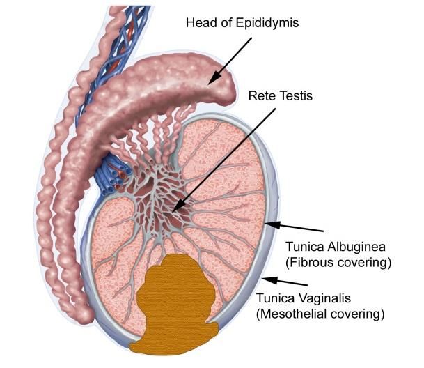
Core Document versions:

  •   ICCR dataset: Neoplasia of the Testis - Orchidectomy specimen 1st edition
  •   AJCC Cancer Staging Manual 8th edition
  •   World Health Organization (WHO). Classification of tumours. Pathology and
      genetics of the urinary system and male genital organs. 4 th edition.

                                          2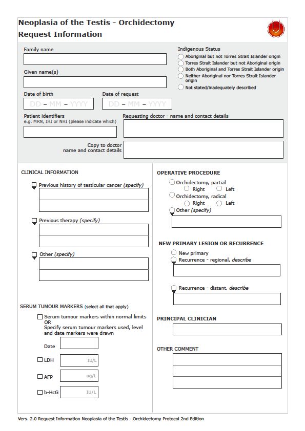
ISBN: 978‐1‐76000‐925‐0
Publications number (SHPN): (CI) 180562.

Online copyright

© RCPA 2018

This work (Protocol) is copyright. You may download, display, print and reproduce the
Protocol for your personal, non-commercial use or use within your organisation subject
to the following terms and conditions:

 1.       The Protocol may not be copied, reproduced, communicated or displayed, in whole
          or in part, for profit or commercial gain.

 2.       Any copy, reproduction or communication must include this RCPA copyright notice
          in full.

 3.       With the exception of Chapter 6 - the checklist, no changes may be made to the
          wording of the Protocol including any Standards, Guidelines, commentary, tables
          or diagrams. Excerpts from the Protocol may be used in support of the checklist.
          References and acknowledgments must be maintained in any reproduction or copy
          in full or part of the Protocol.

 4.       In regard to Chapter 6 of the Protocol - the checklist:

      o    The wording of the Standards may not be altered in any way and must be
           included as part of the checklist.

      o    Guidelines are optional and those which are deemed not applicable may be
           removed.

      o    Numbering of Standards and Guidelines must be retained in the checklist, but can
           be reduced in size, moved to the end of the checklist item or greyed out or other
           means to minimise the visual impact.

      o    Additional items for local use may be added but must not be numbered as a
           Standard or Guideline, in order to avoid confusion with the RCPA checklist items.

      o    Formatting changes in regard to font, spacing, tabulation and sequencing may be
           made.

      o    Commentary from the Protocol may be added or hyperlinked to the relevant
           checklist item.

Apart from any use as permitted under the Copyright Act 1968 or as set out above, all
other rights are reserved. Requests and inquiries concerning reproduction and rights
should be addressed to RCPA, 207 Albion St, Surry Hills, NSW 2010, Australia.

First published: July 2018, 1st Edition (Version 1.0)

                                                 3
Disclaimer
The Royal College of Pathologists of Australasia ("College") has developed these
protocols as an educational tool to assist pathologists in reporting of relevant information
for specific cancers. Each protocol includes “standards” and “guidelines” which are
indicators of ‘minimum requirements’ and ‘recommendations’, which reflect the opinion
of the relevant expert authoring groups. The use of these standards and guidelines is
subject to the clinician’s judgement in each individual case.
The College makes all reasonable efforts to ensure the quality and accuracy of the
protocols and to update the protocols regularly. However subject to any warranties,
terms or conditions which may be implied by law and which cannot be excluded, the
protocols are provided on an "as is" basis. The College does not warrant or represent
that the protocols are complete, accurate, error-free, or up to date. The protocols do
not constitute medical or professional advice. Users should obtain appropriate medical
or professional advice, or where appropriately qualified, exercise their own professional
judgement relevant to their own particular circumstances. Users are responsible for
evaluating the suitability, accuracy, currency, completeness and fitness for purpose of
the protocols.
Except as set out in this paragraph, the College excludes: (i) all warranties, terms and
conditions relating in any way to; and (ii) all liability (including for negligence) in respect
of any loss or damage (including direct, special, indirect or consequential loss or
damage, loss of revenue, loss of expectation, unavailability of systems, loss of data,
personal injury or property damage) arising in any way from or in connection with; the
protocols or any use thereof. Where any statute implies any term, condition or warranty
in connection with the provision or use of the protocols, and that statute prohibits the
exclusion of that term, condition or warranty, then such term, condition or warranty is
not excluded. To the extent permitted by law, the College's liability under or for breach
of any such term, condition or warranty is limited to the resupply or replacement of
services or goods.
4Contents
Scope .................................................................................................................6
Abbreviations .....................................................................................................7
Definitions..........................................................................................................8
Introduction .....................................................................................................11
Authority and development ..............................................................................16
1 Pre-analytical .........................................................................................20
2 Specimen handling and macroscopic findings ........................................22
3 Microscopic findings ...............................................................................27
4 Ancillary studies findings .......................................................................36
5 Synthesis and overview .........................................................................37
6 Structured checklist ...............................................................................39
7 Formatting of pathology reports ............................................................54
Appendix 1 Pathology request information and surgical handling
procedures ........................................................................55
Appendix 2 Guidelines for formatting of a pathology report ................60
Appendix 3 Example of a pathology report for testicular cancer ..........61
Appendix 4 WHO Classification of Tumours of the testis and
paratesticular tissue .........................................................64
Appendix 5 Serum tumour markers ......................................................66
References .......................................................................................................67
5Scope
This protocol contains standards and guidelines for the preparation of structured
reports for testicular tumours in adults and children. The guidelines can be used
in the reporting of both partial and radical orchidectomy specimens, and applies
to all germ cell and sex cord-stromal tumours of the testis. Paratesticular
malignancies, haematological malignancies and metastases to the testes are
excluded. This dataset does not include information on the excision of residual
masses after chemotherapy. A separate protocol is available for the reporting of
retroperitoneal lymphadenectomy specimens which deals with resections at this
site and other metastatic sites, particularly in the post-treatment setting. For
bilateral tumours, complete a separate checklist for each.
Structured reporting aims to improve the completeness and usability of pathology
reports for clinicians, and improve decision support for cancer treatment. The
protocol provides the framework for the reporting of any testicular tumour,
whether as a minimum data set or fully comprehensive report.
6Abbreviations
AFP alpha feto-protein
AJCC American Joint Committee on Cancer
b-hCG Beta subunit of human chorionic gonadotropin
CG Commentary for a guideline
CS Commentary for a standard
FISH Fluorescent in-situ hybridization
GCNIS germ cell neoplasia in situ
ICCR International Collaboration on Cancer Reporting
ISUP International Society of Urological Pathology
LDH lactate dehydrogenase
LIS Laboratory Information System
LVI lymphovascular invasion
PBS Pharmaceutical Benefits Scheme
RCPA Royal College of Pathologists of Australasia
TNM tumour-node-metastasis
UICC International Union Against Cancer
WHO World Health Organization
7Definitions
The table below provides definitions for general or technical terms used in this
protocol. Readers should take particular note of the definitions for ‘standard’,
‘guideline’ and ‘commentary’, because these form the basis of the protocol.
Ancillary An ancillary study is any pathology investigation that may form part
study of a cancer pathology report but is not part of routine histological
assessment.
Clinical Patient information required to inform pathological assessment,
information usually provided with the specimen request form, also referred to as
“pre-test information”.
Commentary Commentary is text, diagrams or photographs that clarify the
standards (see below) and guidelines (see below), provide examples
and help with interpretation, where necessary (not every standard or
guideline has commentary).
Commentary is used to:
• define the way an item should be reported, to foster
reproducibility
• explain why an item is included (e.g. how does the item assist
with clinical management or prognosis of the specific cancer).
• cite published evidence in support of the standard or guideline
• state any exceptions to a standard or guideline.
In this document, commentary is prefixed with ‘CS’ (for commentary
on a standard) or ‘CG’ (for commentary on a guideline), numbered to
be consistent with the relevant standard or guideline, and with
sequential alphabetic lettering within each set of commentaries (e.g.
CS1.01a, CG2.05b).
General General commentary is text that is not associated with a specific
commentary standard or guideline. It is used:
• to provide a brief introduction to a chapter, if necessary
• for items that are not standards or guidelines but are included in
the protocol as items of potential importance, for which there is
currently insufficient evidence to recommend their inclusion.
(Note: in future reviews of protocols, such items may be
reclassified as either standards or guidelines, in line with
diagnostic and prognostic advances, following evidentiary review).
8Guideline Guidelines are recommendations; they are not mandatory, as
indicated by the use of the word ‘should’. Guidelines cover items that
are unanimously agreed should be included in the dataset but are not
supported by NHMRC level III-2 evidence.1 These elements may be
clinically important and recommended as good practice but are not
yet validated or regularly used in patient management.
Guidelines include key information other than that which is essential
for clinical management, staging or prognosis of the cancer such as
macroscopic observations and interpretation, which are fundamental
to the histological diagnosis and conclusion e.g. macroscopic tumour
details, block identification key, may be included as either required or
recommended elements by consensus of the expert committee. Such
findings are essential from a clinical governance perspective, because
they provide a clear, evidentiary decision-making trail.
Guidelines are not used for research items.
In this document, guidelines are prefixed with ‘G’ and numbered
consecutively within each chapter (e.g. G1.10).
Macroscopic Measurements, or assessment of a biopsy specimen, made by the
findings unaided eye.
Microscopic In this document, the term ‘microscopic findings’ refers to histo-
findings morphological assessment.
Predictive A predictive factor is a measurement that is associated with response
factor or lack of response to a particular therapy.
Prognostic A prognostic factor is a measurement that is associated with clinical
factor outcome in the absence of therapy or with the application of a
standard therapy. It can be thought of as a measure of the natural
history of the disease.
Standard Standards are mandatory, as indicated by the use of the term ‘must’.
Standards are essential for the clinical management, staging or
prognosis of the cancer. These elements will either have evidentiary
support at Level III-2 or above (based on prognostic factors in the
NHMRC levels of evidence1 (document). In rare circumstances,
where level III-2 evidence is not available an element may be made a
Standard where there is unanimous agreement in the expert
committee. An appropriate staging system e.g. Pathological TNM
staging would normally be included as a required element. These
elements must be recorded and at the discretion of the pathologist
included in the pathology report according to the needs of the
recipient of the report.
The summation of all standards represents the minimum dataset for
the cancer.
In this document, standards are prefixed with ‘S’ and numbered
consecutively within each chapter (e.g. S1.02).
Structured A report format which utilises standard headings, definitions and
report nomenclature with required information.
9Synoptic A structured report in condensed form (as a synopsis or precis).
report
Synthesis Synthesis is the process in which two or more pre-existing elements
are combined, resulting in the formation of something new.
The Oxford dictionary defines synthesis as “the combination of
components or elements to form a connected whole”.
In the context of structured pathology reporting, synthesis represents
the integration and interpretation of information from two or more
modalities to derive new information.
10Introduction
Testicular tumours are uncommon, with 721 new cases diagnosed in Australia in
2013, giving an estimated incidence of 6.3 cases per 100,000 men. 2 Testicular
tumours tend to occur in younger men, with 50% of cases in men less than 35
years of age, and the peak incidence in the 20 to 40 year age range. Only 25% of
cases occur in men over 40 years. There has been a gradual increase in the
incidence of testicular tumours in Australia at 2% a year throughout the last 100
years, but the cause of this increase is unknown. Despite the increasing
incidence, the mortality has been declining, with a 5-year survival rate of 98%.
Germ cell tumours in their myriad types and combinations make up 95% of
testicular tumours. Germ cell tumours are usually malignant, but the vast
majority can be cured with current therapies. Therefore, accurate diagnosis is
essential to ensure the selection of the most appropriate therapy. Diagnosis and
staging in this group of tumours depends not only on histopathological
assessment, but also imaging and the use of serum markers, specifically alpha
fetoprotein (AFP), the beta subunit of human chorionic gonadotropin (b-hCG) and
lactate dehydrogenase (LDH). The serum markers are included in determination
of “S” stage.
While the sex cord-stromal cell tumours derived from the supporting and
interstitial cells make up only 5% of tumours, they may be more difficult to
diagnose. This group of tumours have normal serum markers, but may be
associated with a variety of clinical syndromes. Knowledge of these associations
is important, as diagnosis of particular testicular tumours may lead to the
diagnosis of previously unrecognised syndromes, such as the occurrence of Large
cell calcifying Sertoli cell tumour in Carney’s syndrome.3 Although less common,
other tumour types involving the testis, such as lymphoma and soft tissue
tumours, must also be considered in the differential diagnosis of a testicular
tumour.
Paediatric testicular tumours should be considered separately from germ cell
tumours occurring after adolescence. This group of tumours consists principally
of yolk sac tumours and teratomas, and the histogenesis appears to be different.4
In this age group, the tumours are usually diploid with no karyotypic
abnormalities, and there is usually no associated germ cell neoplasia in situ
(GCNIS).
Importance of histopathological reporting
Information in the pathology report of the macroscopic and microscopic findings
in orchidectomy specimens is of both clinical and prognostic utility. This
information is interpreted with the results of imaging and serum markers to guide
clinical management of patients, particularly in relation to the role of adjuvant
therapy and surveillance.
While the report must contain all information necessary for tumour staging, the
treating clinician will often look for additional information in the report, such as
tumour size or hilar invasion to further refine the patient’s likely prognosis and
optimal treatment.
11Benefits of structured reporting
The pathology report lays the foundation for a patient’s cancer journey and
conveys information which:
• Provides the definitive diagnosis
• Includes critical information for Tumour-Node-Metastasis (TNM) staging
• Evaluates the adequacy of the surgical excision
• Provides morphological and biological prognostic markers which determine
personalised cancer therapy
However, the rapid growth in ancillary testing such as immunohistochemistry,
flow cytometry, cytogenetics, and molecular studies, have made the task of
keeping abreast of advances on specific cancer investigations extremely difficult
for pathologists. The use of structured reporting checklists by pathologists
ensures that all key elements are included in the report specifically those which
have clinical management, staging or prognostic implications. Consequently
minimum or comprehensive datasets for the reporting of cancer have been
developed5,6 around the world. Both the United Kingdom,7 and United States8
have produced standardised cancer reporting protocols or “datasets” for national
use for many years.
The use of cancer reporting checklists improves completeness and quality of
cancer reporting and thereby ensures an improved outcome for cancer patients.
This has long term cost implications for public health by ensuring the most
effective and timely treatment based on accurate and complete information.
The use of a structured reporting format also facilitates easy extraction of the
necessary information by secondary users of the information i.e. cancer
registries.
International Collaboration on Cancer Reporting
The International Collaboration on Cancer Reporting (ICCR), founded in 2011 by
the Australasian (RCPA), US (CAP) and UK (RCPath) Colleges of Pathology and
the Canadian Association of Pathology (CAP-ACP) in association with the Canadian
Partnership Against Cancer (CPAC), was established to explore the possibilities of
a collaborative approach to the development of common, internationally
standardised and evidence-based cancer reporting protocols for surgical
pathology specimens.
The ICCR, recognising that standardised cancer datasets have been shown to
provide significant benefits for patients and efficiencies for organisations through
the ease and completeness of data capture9-12 undertook to use the best
international approaches and the knowledge and experience of expert
pathologists, and produce cancer datasets which would ensure that cancer reports
across the world will be of the same high quality – ensuring completeness,
consistency, clarity, conciseness and above all, clinical utility.
Representatives from the four countries participating in the initial collaboration
undertook a pilot project in 2011 to develop four cancer datasets - Lung,
Melanoma, Prostate (Radical Prostatectomy), and Endometrium. Following on
from the success of this pilot project, the ICCR was joined by the European
Society of Pathology (ESP) in 2013 and in 2014 incorporated a not-for-profit
12organisation focussed on the development of internationally agreed evidence-
based datasets developed by world leading experts. The ICCR Datasets are made
freely available from its website www.ICCR-Cancer.org
Design of this protocol
This structured reporting protocol has been developed using the ICCR dataset on
neoplasia of the testis - orchidectomy specimen as the foundation.
This protocol includes all of the ICCR cancer dataset elements as well as
additional information, elements and commentary as agreed by the RCPA expert
committee. It provides a comprehensive framework for the assessment and
documentation of pathological features of testicular cancer in orchidectomy
specimens.
ICCR dataset elements for testicular cancer in orchidectomy specimens are
included verbatim. ICCR required elements are mandatory and therefore
represented as standards in this document. ICCR Recommended elements, that
is, those which are not mandatory but are recommended, may be included as
guidelines or upgraded to a standard based on the consensus opinion of the local
expert committee.
The ICCR elements are identified in each chapter with the ICCR logo placed
before the Standard or Guideline number or bullet and the ICCR element
description and commentary is boarded by a grey box as shown below:
G3.02 The intraglandular extent should be recorded as a percentage.
Additional commentary by the RCPA expert committee may be added to an ICCR
element but is not included in the grey bordered area nor indicated with an ICCR
logo e.g.
G2.03 If present, the laterality of the lymph nodes submitted may be
recorded as left, right or bilateral.
CS2.03a If present, record site and number. All lymph node
tissue should be submitted for histological
examination.
Further information on the ICCR is available at www.iccr-cancer.org
Checklist
Consistency and speed of reporting is improved by the use of discrete data
elements recorded from the checklist. Items suited to tick boxes are distinguished
from more complex elements requiring free text or narrative. A structured or
discrete approach to responses is favoured, however the pathologist is
13encouraged to include free text or narrative where necessary to document any
other relevant issues, to give reasons for coming to a particular opinion and to
explain any points of uncertainty.
Report format
The structure provided by the following chapters, headings and subheadings
describes the elements of information and their groupings, but does not
necessarily represent the format of either a pathology report (Chapter 7) or
checklist (Chapter 6). These, and the structured pathology request form
(Appendix 1) are templates that represent information from this protocol,
organised and formatted differently to suit different purposes.
14Key documentation
• Guidelines for Authors of Structured Cancer Pathology Reporting Protocols,
Royal College of Pathologists of Australasia, 200913
• World Health Organization (WHO). Classification of tumours. Pathology and
genetics of the urinary system and male genital organs. Humphrey PA, Moch
H, Reuter VE, Ulbright TM editors. 4th edition. Lyon, France: IARC
Press;2016.14
• AJCC Cancer Staging Manual, 8th edition, American Joint Committee on
Cancer, 201615
Updates since last edition
Inclusion of all ICCR agreed REQUIRED and RECOMMENDED elements.
15Authority and development
This section provides information about the process undertaken to develop this
protocol.
This 2nd edition of the protocol is an amalgam of two separate processes:
1. This protocol is based on the ICCR dataset – Neoplasia of the Testis -
Orchidectomy specimen 1st edition. All ICCR elements from this dataset,
both required (mandatory) and recommended (optional), are included in
this protocol, verbatim. (It should be noted that RCPA feedback from all
Anatomical Pathology fellows and specifically the local expert committee
was sought during the development process of the ICCR dataset.) Details
of the ICCR development process and the international expert authoring
committee responsible for the ICCR dataset are available on the ICCR
website: iccr-cancer.org.
2. Additional elements, values and commentary have been included as
deemed necessary by the local expert committee. In addition, the
standard inclusions of RCPA protocols e.g. example reports, request
information etc., have also been added.
Authorship – 2nd edition
Dr David Clouston (Chair and Lead author), Pathologist
Dr Jessica Ng, Paediatric Pathologist
Professor Brett Delahunt, Pathologist
Professor Ian Davis, Medical Oncologist
Adjunct Professor Warick Delprado, Pathologist
Dr Andrew See, Radiation Oncologist
Clinical Professor James Kench, Pathologist
Associate Professor Nathan Lawrentschuk, Urologist
Professor Hemamali Samaratunga, Pathologist
Authorship – 1st edition (2011)
Dr David Clouston (Lead author), Pathologist
Dr Adrian Charles, Paediatric Pathologist
Professor Brett Delahunt, Pathologist
Associate Professor Ian Davis, Medical Oncologist
Professor Warick Delprado, Pathologist
Dr Thomas Eade, Radiation Oncologist
Professor James Kench, Pathologist
Associate Professor Nathan Lawrentschuk, Urologist
Associate Professor Hemamali Samaratunga, Pathologist
16Editorial manager
Meagan Judge, Royal College of Pathologists of Australasia
Acknowledgements
The testicular tumour expert committee wish to thank all the pathologists and
clinicians who contributed to the discussion around this document.
17Stakeholders
ACT Health
ACT Cancer Registry
Australian Cancer Network
Australian Commission on Safety and Quality in Health Care
Australian Digital Health Agency
Australian Institute of Health and Welfare
Cancer Australia
Cancer Council ACT
Cancer Council Queensland
Cancer Council Victoria
Cancer Council Western Australia
Cancer Institute NSW
Cancer Services Advisory Committee (CanSAC)
Cancer Voices NSW
Clinical Oncology Society of Australia (COSA)
Department of Health, Australia
Department of Health, New Zealand
Faculty of Radiation Oncology Genito-Urinary Group (FROGG)
Health Informatics Society of Australia (HISA)
Independent Review Group of Pathologists
Medical Software Industry Association (MSIA)
National Pathology Accreditation Advisory Council (NPAAC)
New Zealand Cancer Registry
Northern Territory Cancer Registry
Pathology Australia
Public Pathology Australia
Queensland Cooperative Oncology Group (QCOG)
RCPA Anatomical Pathology Advisory Committee (APAC)
Representatives from laboratories specialising in anatomical pathology across
Australia
Royal Australasian College of Physicians (RACP)
South Australia Cancer Registry
Standards Australia
Tasmanian Cancer Registry
The Australian and New Zealand Urogenital and Prostate Cancer Trials Group
(ANZUP)
The Medical Oncology Group of Australia
The Prostate Cancer Foundation of Australia (PCFA)
18The Prostate Cancer Foundation of New Zealand (PCFNZ)
The Royal Australasian College of Surgeons (RACS)
The Royal Australian and New Zealand College of Radiologists (RANZCR)
The Royal Australian College of General Practitioners (RACGP)
The Royal College of Pathologists of Australasia (RCPA)
The Urological Society Of Australia And New Zealand (USANZ)
Western Australia Clinical Oncology Group (WACOG)
Development process
This protocol has been developed following the ten-step process set out in
Guidelines for Authors of Structured Cancer Pathology Reporting Protocols.13
Where no reference is provided, the authority is the consensus of the local expert
group for local inclusions and the ICCR Dataset Authoring Committee for ICCR
components denoted with the ICCR logo.
191 Pre-analytical
This chapter relates to information that should be recorded on receipt of the
specimen in the laboratory.
The pathologist is reliant on the quality of information received from the clinicians
or requestor. Some of this information may be received in generic pathology
request forms, however, the additional information required by the pathologist
specifically for the reporting of testicular tumours is outlined in Appendix 1.
Appendix 1 also includes a standardised request information sheet that may be
useful in obtaining all relevant information from the requestor.
Surgical handling procedures affect the quality of the specimen and
recommendations for appropriate surgical handling are included in Appendix 1.
S1.01 All demographic information provided on the request form and
with the specimen must be recorded.
CS1.01a The Royal College of Pathologists of Australasia (RCPA) The
Pathology Request-Test-Report Cycle — Guidelines for
Requesters and Pathology Providers must be adhered to.16
This document specifies the minimum information to be
provided by the requesting clinician for any pathology test.
CS1.01b Whether or not the patient identifies as Aboriginal and/ or
Torres Strait Islander. This is in support of a government
initiative to monitor the health of indigenous Australians
particularly in relation to cancer.
CS1.01c The patient’s health identifiers may include the patient’s
Medical Record Number as well as a national health number
such as a patient’s Individual Healthcare Identifier (IHI)
(Australia) or the National Healthcare Identifier (New
Zealand).
S1.02 All clinical information as documented on the request form must
be recorded verbatim.
CS1.02a The request information may be recorded as a single text
(narrative) field or it may be recorded in a structured format.
CS1.02b The copy doctors requested on the request form must be
recorded.
S1.03 The pathology accession number of the specimen must be
recorded.
S1.04 The principal clinician involved in the patient’s care and
responsible for investigating the patient must be recorded.
CS1.04a Knowledge of the clinical presentation is an essential part
of the WHO classification yet it may not be available for a
number of reasons:
• The clinical assessment and staging may be incomplete
20at the time of biopsy.
• The pathology request is often authored by the clinician
performing the surgical excision/biopsy rather than the
clinician who is investigating and managing the patient.
• The identity of this clinician is often not indicated on the
pathology request form
In practice therefore, it is important in such cases that the
reporting pathologist should be able to communicate with
the managing clinician for clarification.
CS1.04b The Australian Healthcare identifiers i.e. Healthcare
Provider Identifier - Individual (HPI-I) and Healthcare
Provider Identifier - Organisation (HPI-O) should be
included, where possible, to identify the principal clinician
involved in the patient's care.
G1.01 Any clinical information received in other communications from the
requestor or other clinician should be recorded together with the source
of that information.
212 Specimen handling and macroscopic
findings
This section relates to the procedures required after the information has been
handed over from the requesting clinician, and the specimen has been received in
the laboratory.
Tissue Banking
➢ Pathologists may be asked to provide tissue samples from fresh specimens
for tissue banking or research purposes. The decision to provide tissue should
only be made if the pathologist is sure that the diagnostic process will not be
compromised. As a safeguard, research use of the specimen may be put on
hold until the diagnostic process is complete.
Specimen handling
➢ Detailed fixation and specimen handling instructions are available from the
RCPA online Cut-up Manual:
www.rcpa.edu.au/Library/Practising-Pathology/Macroscopic-Cut-Up
➢ The specimen must be handled in a systematic and thorough fashion to
ensure completeness and accuracy of pathological data.
➢ If Lymph nodes are submitted please follow the Retroperitoneal lymph node
dissection (RPLND) protocol.
Macroscopic findings
S2.01 The labelling of the specimen(s) must be clearly recorded.
S2.02 The operative procedure must be recorded.
CS2.02a Whether the surgical procedure is a radical or partial
orchidectomy must be stated, as this will influence the
assessment of surgical margins. For bilateral tumours,
complete a separate dataset for each testis.
G2.01 The specimen should be measured in three dimensions.
G2.02 The testis should be measured in 3 dimensions without the tunical sac
(in mm).
G2.03 The length of the spermatic cord should be given in millimetres.
G2.04 Any abnormalities of the spermatic cord should be recorded.
22G2.05 Any abnormalities of the surface of the tunica vaginalis or tunica
albuginea should be recorded.
G2.06 The quantity and nature of any intratunical fluid should be recorded.
S2.03 Tumour focality must be recorded.
CS2.03a There is no specific paper dealing with mutifocality in germ
cell tumours, however many show multifocal tumours
which may coalesce together to form a complex multifocal
nodule. The noting of mutifocality is important, as the
separate nodules may contain different tumour elements
which may affect prognosis.17 Secondly, the determination
of maximum tumour diameter depends on whether the
tumours are multifocal or unifocal. Rare testicular tumours
may be associated with mutifocality and suggest a variety
of syndromes.18
S2.04 The maximum tumour dimension of the largest tumour must be
recorded (in mm).
CS2.04a It has been shown in a number of studies that the
maximum tumour dimension has prognostic significance,
especially in seminomas.19 In a pooled analysis of data
from four large cohort studies (638 patients) of patients
with stage I seminoma, size (tumour size >4 cm) was
independently predictive of recurrence at five years on
multivariate analysis. If the tumour was >4 cm, there was
a two-fold increased risk of recurrence. In another study
on multivariable analysis, tumour size above median (cut-
point of 3 cm) was a predictor for relapse, HR 1.87 (95%
Confidence Interval (CI) 1.15– 3.06)).20 The 3-year relapse
risk based on the primary tumour size alone increased
from 9% for a 1 cm primary tumour to 26% for an 8 cm
tumour.20 This is supported by other studies, especially for
seminoma.21
The evidence for the importance of size in non-
seminomatous germ cell tumours is less well established,
as other factors (vascular invasion) are more important.
However, as it is often not apparent whether the tumour is
a seminoma or non-seminoma on macroscopy, size
measurement is required. We suggest that when there is
multifocality, the longest diameter of the largest focus be
recorded and that the maximum diameter of the additional
nodules also be recorded. Where the nodules coalesce, this
may be difficult to calculate. Evidence for the relevance of
this is disputed but we suggest that tumours should be
counted as separate if there is intervening parenchyma.
S2.05 Record the additional two dimensions of the largest tumour (in
mm).
G2.07 The tumour dimensions of additional tumour nodules may be recorded
23(in mm).
G2.08 The colour, consistency, and heterogeneity of the tumour should be
recorded.
G2.09 The presence of cysts and bone should be recorded, as well as any
other areas which look different macroscopically.
G2.10 The presence or absence of necrosis or haemorrhage should be
recorded.
S2.06 The macroscopic extent of invasion must be recorded.
CS2.06a The relationship of the tumour to the tunica vaginalis,
tunica albuginea and the testicular hilum is important.
CS2.06b The macroscopic extent of the disease may be difficult to
discern even on close inspection of the testis and hilar
structures. The vast majority of radical orchidectomies will
not include the scrotum unless the surgeon finds evidence
of invasion at surgery. The testis parenchyma is bound by
the tunica albuginea except in the region where the rete
testis connects with the epididymis and vas deferens.
Adjacent to the hilum in this area is a small amount of hilar
fat. The tunica vaginalis is bound by a double layer of
mesothelium, with the visceral (inner) layer forming the
tunica albuginea (See Figure 1 below). Involvement of the
hilar soft tissue, epididymis or tunica vaginalis may be
challenging to detect. Also diffusely infiltrative tumours
such as intertubular seminoma which infiltrate between the
tubules may not be easy to detect, meaning that the size
of the tumour may in fact be larger than that suspected
macroscopically. Therefore all suspected areas of invasion
seen macroscopically should be confirmed microscopically
by appropriate sampling for confirmation (see below).
G2.11 Features of the uninvolved testicular parenchyma and epididymis
should be described, such as fibrosis, other nodularity, and any other
features.
S2.07 A block identification key listing the nature and origin of all
tissue blocks must be recorded.
CS2.07a The origin/designation of all tissue blocks should be
recorded and it is preferable to document this information
in the final pathology report. This is particularly important
should the need for internal or external review arise. The
reviewer needs to be clear about the origin of each block in
order to provide an informed specialist opinion. If this
information is not included in the final pathology report, it
should be available on the laboratory computer system and
relayed to the reviewing pathologist. Recording the
origin/designation of tissue blocks also facilitates retrieval
24of blocks, for example for further immunohistochemical or
molecular analysis, research studies or clinical trials.
CS2.07b Tumour sampling should be generous to ensure
documentation of all tumour types present. Germ cell
tumours should, as a minimum be sampled at 1 block per
cm of tumour. However while this may be adequate for a
non seminomatous germ cell tumour, to represent different
elements, it has been recommended that seminomas are
more generously sampled than this, as small foci of non
seminoma will change patient management; if the tumour
is small (less than 2 cm) it can be completely sampled.22
Pure seminomas should be sampled especially thoroughly
to exclude small areas on non-seminomatous germ cell
tumour. It is important that blocks include the adjacent
testicular parenchyma to allow for the assessment of
lymphovascular invasion (LVI) and germ cell neoplasia in
situ (GCNIS).
Different areas of the tumour must be sampled,
particularly including haemorrhagic and necrotic areas and
solid/fleshy areas. All of the haemorrhagic tumour must be
blocked, as choriocarcinoma is often haemorrhagic with
little residual viable tumour.
Sections of tumour should include at least one section
showing the relation of the tumour to the testicular hilum.
If the tumour is well away from the hilum, there should be
a separate section of the hilum clearly showing this region
is free of tumour.
Sections of tumour should include the adjacent tunica
albuginea and vaginalis and adjacent testicular
parenchyma. Sections of uninvolved testicular parenchyma
should be included. A block from the cord resection margin
should be taken. This block should be taken prior to
incision of the tumour to avoid contamination.23
Orchidectomy specimens for clinically localised
disease
Blocks are selected to represent:
• the cord resection margin and base of cord (further
cord blocks depending on macroscopy)
• the relationship of the tumour(s) to the rete testis,
epididymis and cord
• the minimum distance of the tumour to the nearest
inked resection margin for partial orchidectomies
• all areas of the tumour(s) with different
macroscopic appearances (solid, cystic, pale or
haemorrhagic)
• adjacent testis including the tunica albuginea (and
vaginalis), a common site for vascular invasion
• uninvolved testis.
25It is recommended that a record is kept of a good
representative paraffin block of tumour and whether frozen
tissue has been stored.
G2.12 A descriptive or narrative field should be provided to record any
macroscopic information that is not recorded in the above standards
and guidelines, and that would normally form part of the macroscopic
description.
CG2.12a The traditional macroscopic narrative recorded at the time
of specimen dissection is often reported separately from
the cancer dataset. Although this remains an option, it is
recommended that macroscopic information be recorded
within the overall structure of this protocol.
CG2.12b Much of the information recorded in a traditional
macroscopic narrative is covered in the standards and
guidelines above and in many cases, no further description
is required.
Figure 1. Diagrammatic representation of a tumour (Tumour A) invading the
tunica vaginalis, perforating through the mesothelium, and another
tumour (Tumour B) partly involving the rete testis and invading the
hilar soft tissue.
Figure courtesy of Satish K. Tickoo. MD. Source: College of American Pathologists
(CAP) Protocol for the examination of specimens from patients with malignant
germ cell and sex cord-stromal tumors of the testis (October 2013).
263 Microscopic findings
This section relates to purely histological (morphological) assessment.
Information derived from multiple investigational modalities, or from two or more
chapters, is described in Chapter 5.
S3.01 The histological tumour type must be recorded.
CS3.01a State whether the tumour is a Germ Cell tumour or
other tumour type which must be specified.
CS3.01b Germ cell tumours often contain several tumour
types, and for mixed germ cell tumours, each of the
different tumour types must be listed.
CS3.01c The classification of testicular tumours is taken from
the World Health Organisation (WHO) 201614
classification. (Refer to Appendix 4).
CS3.01d Percentage of different tumour components in
mixed germ cell tumours must be specified.
The percentage of the different tumour elements has
been shown to be predictive of the relapse risk in
non-seminomatous germ cell tumours (NSGCT),
especially the percentage of embryonal carcinoma. As
well as the percentage of embryonal carcinoma as a
core data item, the approximate percentages of other
tumour elements should also be given. The presence
of LVI, embryonal carcinoma and yolk sac tumour
were risk factors for relapse in a study of 132
patients.24 A second study showed that 25/85 men
who had predominantly embryonal carcinoma
histology relapsed.25 Of 93 men with stage I NSGCTs
who were placed in a surveillance study following
orchidectomy, 81 men had predominantly embryonal
carcinoma component in their primary tumour and a
third of these developed metastases, whereas none
of the men lacking an embryonal carcinoma
component developed metastases (p=0.05).26 An
older surveillance study in 373 men with stage I
NSGCT suggested that ‘undifferentiated cells’ and the
absence of yolk sac elements in the primary tumour
were able to identify men with a high risk of
relapse.27
Giving ‘exact’ percentages in a mixed non-
seminomatous germ cell tumour may be challenging,
as some elements may be extremely small, and it
may occasionally be difficult to distinguish closely
intermingled elements (such as yolk sac tumour and
embryonal carcinoma). We suggest that only basic
‘eyeball’ style quantitation is required. For example,
27the difference between 10% embryonal carcinoma
and 90% embryonal carcinoma may be important in
determining the need to adjuvant therapy. However a
difference of 5 or 10% is likely insignificant. For
NSGCTs which are of pure type, then the percentage
of the pure type should be listed as 100%.
Mention of areas of scarring is helpful, particularly in
pure seminoma or teratoma cases as they may
indicate areas of regression, which might have
represented other tumour types. These findings can
explain the occasional discordance between the
orchidectomy tumour type and that seen in
metastatic deposits.
G3.01 The presence or absence of scarring should be documented.
CG3.01a Testicular scars may represent regressed germ cell
tumour. The scarring may be partial or complete,
and may be all that remains in patients presenting
with metastatic disease and clinically inapparent
testicular primaries. Other features associated with
tumour regression are the presence of haemosiderin
laden macrophages, lymphoplasmacytic infiltrate and
the presence of GCNIS or intratubular calcifications.
CG3.01b Rarely the tumour may be completely necrotic. In
this instance, necrotic tumour can be recognised by
the loss of normal architecture and the ghost outlines
of large tumour cells. It is important to report the
tumour as a germ cell tumour which cannot be
further typed due to complete necrosis. The
differential diagnosis is testicular infarction, which
can be recognised from the preservation of normal
architecture.
G3.02 The presence or absence of syncytiotrophoblastic giant cells
should be documented.
CG3.02a Syncytiotrophoblast may occur in germ cell tumours,
with the syncytiotrophoblast present as single cells or
very small groups, with no associated cytotrophoblast
to indicate associated choriocarcinoma.
S3.02 Maximum tumour size must be specified (in mm).
CS3.02a The tumour size microscopically should be correlated
with the macroscopic dimension. In some cases, the
microscopic size may be larger than the macroscopic
size, in particular with seminomas which may have a
more extensive interstitial growth pattern. Therefore
it is important to assess and record the microscopic
size separately from the macroscopic size.
28S3.03 The microscopic extent of tumour must be reported.
CS3.03a Extent must document the presence or absence of
tumour in each of the following structures:
• Rete testis of stromal/interstitial type
• Epididymis
• Hilar fat
• Tunica vaginalis (both mesothelial layers
covering the tunica albuginea)
• Spermatic cord
• Scrotal wall
CS3.03b Rete testis
Rete testis invasion is the direct invasion of tumour
into the stroma of the rete testis and does not include
pagetoid spread of GCNIS into the tubules of the
rete.19 In the pooled cohort surveillance study of pure
seminomas, rete testis invasion was independently
predictive of recurrence at five years on multivariate
analysis, conferring an increased risk of recurrence
by a factor of 1.7 (95% CI 1.1–2.6).19 Other studies
of pure seminoma show differing results. Two cohort
analyses of 425 and 744 patients respectively
confirmed this.21,28 However, two other studies of 685
patients20 and 1954 patients29 showed that rete testis
invasion was not a significant predictor for relapse
when compared with tumour size.
For NSGCTs, there is less evidence that rete testis
invasion is an important prognostic factor,30 probably
because other factors such as the percentage of
embryonal carcinoma and vascular invasion are more
important.
Rete testis invasion and tumour size are also
interdependent. It should be noted that most of the
studies listed above did not include formal
prospective pathological review and were a
retrospective assessment of pathological reports by
clinicians. Data on rete testis involvement was
missing in many cases, and there is doubt in some
studies whether pagetoid invasion of the rete was
assessed. A survey of recent practice in Europe
showed many pathologists did not distinguish
between pagetoid and interstitial invasion of the
rete.31 Rete testis and tumour size were not part of
the TNM 7th edition32,33 however tumour size using a
cut off of 3 cm has now been incorporated into the
American Joint Committee on Cancer (AJCC) 8th
29edition15 for pure seminomas only, separating the
pT1 stage into pT1a and pT1b. Both rete testis
invasion and size are used by many clinicians to
determine adjuvant chemotherapy and are part of
existing European clinical guidelines.34,35
CS3.03c Hilar soft tissue invasion
Invasion of the hilar soft tissues is a common mode
of extratesticular spread.36 One study has shown that
it predicts high stage at presentation,30 but there has
been previously no consensus on the correct way to
stage hilar soft tissue invasion31 Following a
consultation conference by the International Society
of Urological Pathologists (ISUP)37 and adoption by
the AJCC 8th edition15 it has been decided to stage
soft tissue invasion as pT2. Soft tissue invasion has
been defined as ‘invasion of the adipose tissue and
soft fibrous connective tissue present…beyond the
boundaries of the rete testis.15
CS3.03d Epididymal invasion
There is no evidence on the prognostic significance of
epididymal invasion. Although in previous editions of
AJCC32 and Union for International Cancer Control
(UICC)33 manuals (7th editions) it has been
designated as pT1, the evidence and consensus for
pT2 staging of soft tissue has necessitated a
redesignation of epididymal invasion as pT2 in the
AJCC 8th edition15 as it is normally secondary to this.
CS3.03e Direct invasion of the cord
This is regarded as a core data item as it is required
for TNM staging but evidence on its prognostic
significance in seminoma is lacking. In a large cohort
study of stage I seminoma, spermatic cord invasion
was not found to be independently prognostic for
recurrence.28 In contrast, it was identified as an
adverse prognostic factor in another study.38 In a
review of 326 testicular germ cell tumours, of which
79 had tumour in the spermatic cord, most cases
(72%) were thought to be due to contamination
compared to 19% cases of true involvement and with
8.9% showing both contamination and true
involvement.23 Spermatic cord contamination was
most frequently seen with seminomas. To
differentiate cord invasion from hilar soft tissue
invasion, it has been defined as ‘tumour extending
beyond the angle between the epididymis and
spermatic cord proper or tumour surrounding the vas
deferens’.15
30CS3.03f For partial orchidectomy specimens, the relationship
of the tumour to structures which are not included in
the resection can be recorded as “Not submitted”.
G3.03 The microscopic extent of invasion into the tunica albuginea
(white fibrous capsule around testicular parenchyma) may be
reported.
CG3.03a The tunica albuginea is the white fibrous outer coat of
the testis and is covered by a mesothelial layer which
forms the tunica vaginalis. Refer to Figure 2 and 3.
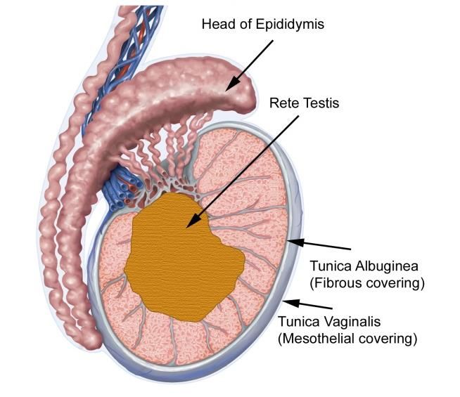
Assessment of testicular tumour extent.
Figure 2:
Figure 3:
Figures 2 and 3 show the anatomical landmarks used to assess the extent of a
testicular tumour. In figure 2, the tumour is involving the rete testis (stage pT1).
In figure 3, the tumour has invaded through the tunica albuginea and breached
the tunica vaginalis (stage pT2).
31S3.04 The presence or absence of lymphovascular invasion
(LVI) must be specified.
CS3.04a In several studies, the presence of vascular space
has been correlated with a significantly elevated risk
for distant metastasis, particularly in NSGCTs. Some
clinicians manage the patients with clinical stage I
disease that lack evidence of lymphatic or vascular
invasion in their orchidectomy specimens by
surveillance.
Most of the previous studies on LVI appear not to use
immunochemistry routinely in its diagnosis. Although
one recent paper suggests that the routine use of
immunochemistry to identify LVI may be helpful,
further studies are needed and at present we
recommend that diagnosis should be made on H&E
backed up by immunochemistry for lymphovascular
vessels in challenging cases.39
We recommend that vascular invasion be called
either present or ‘not identified’ as equivocation in
the report is unhelpful to the clinician. We advise
restricting the definition of vascular invasion so that
those cases which are equivocal are assigned as ‘not
identified’. Vascular invasion is much more likely to
be seen at the periphery of the tumour than within
the centre of solid tumour masses. It is often seen in
fibrous bands surrounding or intersecting the main
tumour mass, as well as in the region of rete testis.
LVI may be seen in the tunica albuginea, spermatic
cord vessels or the parenchyma of the testis. All
warrant a stage of pT2.
In seminoma, although vascular invasion is a
statistically significant factor for predicting for relapse
in occasional small historical cohort studies,40 it has
not proved independently statistically significant in
stage I seminoma in large cohort pooled studies;19,28
however, it was found significant in a recent cohort of
1954 patients.29 This may be secondary to the
frequent presence of tumour smearing artefact in
seminoma, making identification of genuine LVI
challenging.
For NSGCTs, LVI has been shown in multiple studies
to be a powerful predictor of metastatic disease and
recurrence.24,26,41-46
If LVI is present in a mixed or combined germ cell
tumour, it is good practice to state which subtype of
tumour is showing the LVI as this may alter clinical
management if it was an embryonal carcinoma
component showing LVI versus classical seminoma.
Indicating that a case is ‘uncertain’ for vascular
invasion is unhelpful for the treatment of patients
32with germ cell tumours.
G3.04 If present, the type of lymphovascular invasion should be
recorded.
S3.05 The presence or absence of germ cell neoplasia in situ
(GCNIS) must be specified.
CS3.05a The term germ cell neoplasia in situ (GCNIS) has
replaced the previous terms, carcinoma in situ (CIS),
intratubular germ cell neoplasia, unclassified (IGCNU)
and testicular intraepithelial neoplasia (TIN). None of
the previous terms was entirely correct and led to
much confusion in the literature. GCNIS is not a
‘carcinoma’ nor is it ‘intra-epithelial’, and the term
IGCNU, was confusing due to the use of the term
‘unclassified’ which many replaced by
‘undifferentiated’.
In fact, the true in situ area for the development of
germ cell tumours is in a specific intratubular
location, the ‘spermatogonial niche’ between the
basement membrane and the tight junctions between
adjacent Sertoli cells.
GCNIS is the precursor lesion for the most common
variants of invasive germ cell tumours. Although not
a prognostic factor, it should be a core item, as its
absence may raise the suspicion of a non- GCNIS
associated tumour, which has differing prognosis and
treatments, as well as the possibility that the tumour
is a non-germ cell tumour mimic of a germ cell
tumour (notably some Sertoli cell tumours).
‘Pagetoid’ invasion of the rete testis occurs when
GCNIS-like cells infiltrate the epithelial cells of the
rete but do not invade the rete stroma. The
significance of pagetoid type rete invasion is
unknown but is generally accepted that these
represent infiltration of GCNIS rather than invasive
seminoma.
G3.05 Extent of GCNIS should be specified, and can be listed as
widespread or focal.
G3.06 The presence or absence of other intratubular lesions should be
specified.
S3.06 The surgical margin status must be reported.
CS3.06a Whether the surgical procedure is a radical or partial
orchidectomy must be stated, as this will influence
the assessment of surgical margins. Specifically, in
the case of partial orchidectomy specimens, it is
33important that the intratesticular surgical margin is
carefully evaluated to ensure that no residual tumour
is present in the remaining testis.
For radical orchidectomies there is little evidence that
surgical margin status has been studied as an
independent prognostic factor separately from stage
and other known indicators. The only true surgical
margin is the spermatic cord margin in a usual radical
orchidectomy and involvement with stromal invasion
is rare. Very rarely in a widely invasive tumour,
scrotal skin may be included. This should be easily
apparent in such cases, and it would be appropriate
to state whether the scrotal skin margin was invaded
by tumour.
Occasionally the spermatic cord margin may include
vessels showing vascular invasion by tumour. This is
vascular invasion, and does not represent a positive
margin.
G3.07 For negative margins in a partial orchidectomy specimen, the
distance between tumour and the closest surgical margin should
be given in mm.
G3.08 The presence or absence of GCNIS at a surgical margin should
be documented.
S3.07 The nature of any relevant coexisting pathology must be
described.
CS3.07a ‘Burnt out’ germ cell tumours may present as
scarring, with the presence of hemosiderin-laden
macrophages, and intratubular calcification, with
surrounding GCNIS and must be carefully evaluated.
Signs of testicular dysgenesis, androgen insensitivity,
Klinefelter’s syndrome or other intersex conditions
may be identified or suggested by close examination
of the testicular parenchyma. These might include
residual gonadoblastoma or ovarian type tissue for
intersex conditions. Leydig cell hyperplasia which
may be correlated with b-hCG elevation and
testicular atrophy may also be seen in dysgenetic
gonads (e.g. dysgenesis or androgen-insensitivity
syndrome).47,48
It may be helpful to give the status of the
surrounding parenchyma to the tumour: especially
the amount of spermatogenesis present and degree
of atrophy. The status of the parenchyma is of great
importance in some types of testicular neoplasm
(prepubertal type teratoma in particular) and also
may indicate the functioning status of the
contralateral testis.47,48
34You can also read