Tauopathy and Movement Disorders-Unveiling the Chameleons and Mimics - Frontiers
←
→
Page content transcription
If your browser does not render page correctly, please read the page content below
REVIEW
                                                                                                                                      published: 05 November 2020
                                                                                                                                    doi: 10.3389/fneur.2020.599384

                                               Tauopathy and Movement
                                               Disorders—Unveiling the
                                               Chameleons and Mimics
                                               Jacky Ganguly and Mandar Jog*

                                               Movement Disorder Centre, London Health Sciences Centre, University of Western Ontario, London, ON, Canada

                                               The spectrum of tauopathy encompasses heterogenous group of neurodegenerative
                                               disorders characterized by neural or glial deposition of pathological protein tau. Clinically
                                               they can present as cognitive syndromes, movement disorders, motor neuron disease,
                                               or mixed. The heterogeneity in clinical presentation, genetic background, and underlying
                                               pathology make it difficult to classify and clinically approach tauopathy. In the literature,
                                               tauopathies are thus mostly highlighted from pathological perspective. From clinical
                                               standpoint, cognitive syndromes are often been focussed while reviewing tauopathies.
                                               However, the spectrum of tauopathy has also evolved significantly in the domain of
                                               movement disorders and has transgressed beyond the domain of primary tauopathies.
                                               Secondary tauopathies from neuroinflammation or autoimmune insults and some other
                                               “novel” tauopathies are increasingly being reported in the current literature, while some
                                               of them are geographically isolated. Because of the overlapping clinical phenotypes,
                             Edited by:        it often becomes difficult for the clinician to diagnose them clinically and have to wait
                        Sanjay Pandey,
               University of Delhi, India
                                               for the pathological confirmation by autopsy. However, each of these tauopathies has
                       Reviewed by:
                                               some clinical and radiological signatures those can help in clinical diagnosis and targeted
          Prachaya Srivanitchapoom,            genetic testing. In this review, we have exposed the heterogeneity of tauopathy from a
          Mahidol University, Thailand
                                               movement disorder perspective and have provided a clinical approach to diagnose them
           Rukmini Mridula Kandadai,
          Nizam’s Institute of Medical         ante mortem before confirmatory autopsy. Additionally, phenotypic variability of these
                      Sciences, India          disorders (chameleons) and the look-alikes (mimics) have been discussed with potential
                 *Correspondence:              clinical pointers for each of them. The review provides a framework within which new and
                        Mandar Jog
              Mandar.Jog@lhsc.on.ca
                                               as yet undiscovered entities can be classified in the future.
                                               Keywords: tauopathy, movement disorders, chameleons, mimics, MAPT
                    Specialty section:
          This article was submitted to
                  Movement Disorders,
                a section of the journal
                                               INTRODUCTION
                 Frontiers in Neurology
                                               Tauopathies are a heterogeneous group of neurodegenerative disorders, pathologically
         Received: 27 August 2020              characterized by neuronal and/or glial inclusions of the microtubule-binding protein, tau.
      Accepted: 30 September 2020
                                               Heterogeneity spans many domains from the clinical presentation, anatomical localization, genetic
      Published: 05 November 2020
                                               variations, and radiological and pathological signs. Neuroanatomical vulnerability may be a key
                             Citation:         to the heterogeneity (the concept of “molecular nexopathies”) (1). Many factors can be implicated
          Ganguly J and Jog M (2020)
                                               including “strain” specificity of the tau protein, biochemical property of the abnormal protein
            Tauopathy and Movement
Disorders—Unveiling the Chameleons
                                               according to its post-translational modification, “prion-like” propagation capacity, interaction
                          and Mimics.          with other co-existent proteins like alpha-synuclein or TDP43, seeding or “permissive templating”
            Front. Neurol. 11:599384.          property, intrinsic vulnerability of the affected structure, genetic, and epigenetic factors and
     doi: 10.3389/fneur.2020.599384            environmental influences (1, 2).

Frontiers in Neurology | www.frontiersin.org                                       1                                     November 2020 | Volume 11 | Article 599384
Ganguly and Jog Tauopathy and Movement Disorders
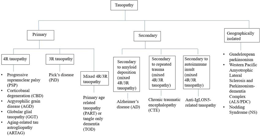
The spectrum of tauopathy is still unfolding and transcending Secondary tauopathies where additional etiologies (e.g.,
beyond the domain of primary tauopathies. While secondary amyloid, trauma, and autoimmune) are involved for tau
tauopathy from autoimmune insult like in anti-IgLON5 deposition (Figure 1) (8). However, some tauopathies are
disease brings up the topic of complex interaction between geographically isolated like Guadeloupean parkinsonism (9),
autoimmunity and neurodegeneration (3), geographically Western pacific amyotrophic lateral sclerosis and parkinsonism-
isolated tauopathies highlight the environmental impact. Apart dementia complex (ALS/PDC) (10), and Nodding syndrome
from this, some novel tauopathies are also increasingly being of northern Uganda (11). While exact etiopathogenesis of
described in the literature (4). these geographically isolated tauopathies are still unknown,
Clinically, tauopathies present as movement disorders, environmental impact (discussed later) has been highlighted in
dementia, and motor neuron disease, either in isolation many studies. Thus, whether to include them in the group of
or in varied combinations (5), based on the vulnerable secondary tauopathies or not, is still a matter of debate.
anatomical structures being affected by the pathological protein Recently, Hõglinger et al. (12) have highlighted syndromic
accumulation. In terms of genetics, MAPT gene containing N classification of tauopathy based on the predominant domain
terminal domain (N1, N2) and microtubule binding domain (R1, affected (cognitive or motor):
R2, R3, R4), on chromosome 17q21 encodes the protein tau. Due
1. Cognitive syndromes: Behavioral variant of frontotemporal
to alternative splicing of the MAPT gene, three repeat (2N3R,
dementia (bvFTD), non-fluent agrammatic variant of primary
1N3R, 0N3R) or four repeat (2N4R, 1N4R, 0N4R) tau isoforms
progressive aphasia (nfavPPA), semantic variant of primary-
are formed (6). On the other hand, depending upon numerous
progressive aphasia (svPPA), and amnestic syndrome of
single nucleotide polymorphisms (SNPs) and a 900kb inversion,
hippocampal type (AS).
H2 and H1 haplotypes of MAPT gene are formed and have
2. Motor syndromes: Richardson syndrome (RS), Parkinson
impact on the phenotypic presentation (7).
syndrome (P), corticobasal syndrome (CBS), primary gait
In the literature, tauopathy has been discussed mostly as
freezing (PGF), cerebellar syndrome (C), and primary lateral
a pathological entity with its detailed pathological intricacies.
sclerosis (PLS).
Pathological confirmation of the diagnosis of tauopathy mostly
depends on autopsy findings. However, pathological diagnosis is However, a primary tauopathy like progressive supranuclear
often confounded by the presence of multiple other proteins and palsy (PSP) or corticobasal degeneration (CBD) can present
thus it becomes difficult to determine whether the accumulated with different cognitive and motor syndromes (chameleons) and
tau is pathological or an innocent bystander. In vivo biomarkers many a times there is phenotypic overlap of cognitive and motor
like CSF tau and tau-PET imaging are still research-based tools. syndrome like in familial FTD with parkinsonism linked to
Additionally, each of these tauopathies has some clinical and MAPT (FTDP-17).
radiological signature that can predict the underlying genetics Apart from these, tauopathies can be classified based on
and pathology. the etiology like genetic (e.g., MAPT related), autoimmune
In order to clarify this complexity of heterogeneity of (e.g., anti IgLON5 related), traumatic (e.g., chronic traumatic
tauopathies, in this review, we have approached tauopathy encephalopathy), etc. It can also be classified based on the
from a clinical standpoint highlighting mainly the movement area of brain predominantly involved like frontal cortex (e.g.,
disorder perspective, focused on the clinical presentations behavioral variant frontotemporal dementia/bvFTD, progressive
(chameleons) and their phenotypic look-alikes (mimics). A supranuclear palsy-frontal variant/PSP-F), parietal cortex (e.g.,
critical review of the current status of the classification of corticobasal syndrome/CBS), peri-sylvian (e.g., progressive
tauopathies will be followed by the clinical spectrum of primary, nonfluent aphasia/PNFA), limbic (e.g., argyrophilic grain
secondary, and geographically isolated tauopathies to understand disease/AGD), brainstem (e.g., progressive supranuclear palsy-
the heterogeneity. Potential clinical and radiological clues will Richardson’s type/PSP-RS, anti IgLON5 related), or cerebellum
be discussed for each of them. Finally, a practical approach (e.g., PSP-C).
is presented to guide the clinician in day to day practice.
Specifically, the phenotype of familial frontotemporal dementia Primary 4R- and 3R-Tauopathies
with parkinsonism has been clinically dissected further at the In this large group of primary tauopathies, CBD, GGT, AGD, and
end, because it is one of the commonest overlapping phenotypes PiD are primarily pathological diagnosis where as corticobasal
of tauopathies. syndrome (CBS) is a clinical term. PSP can be described as both, a
pathological or a clinical entity. In this review, PSP and CBS have
been discussed with their phenotypic presentations (chameleons)
CLASSIFICATION OF and look-alikes (mimics). PNFA/PPA-G has also been discussed
TAUOPATHIES—CURRENT STATUS AND because it is primarily a clinical diagnosis and its pathology is
PITFALLS mostly FTLD-tau. In the literature, GGT, AGD, and PiD have
been traditionally discussed from a pathological standpoint. We
Tauopathies have been conventionally classified from a have highlighted the clinical and radiological clues for suspecting
pathological perspective into two groups—(A) Primary GGT, AGD, and PiD clinically before confirmatory autopsy.
tauopathies where tau is the predominant pathology including We have sub-classified primary tauopathies according to the
three repeat (3R-) and four repeat (4R-) tauopathies, (B) predominant clinical presentation like, movement disorder (PSP,
Frontiers in Neurology | www.frontiersin.org 2 November 2020 | Volume 11 | Article 599384Ganguly and Jog Tauopathy and Movement Disorders
FIGURE 1 | Classification of tauopathies.
CBS), language dysfunction (PNFA) and cognitive dysfunction and “Hummingbird,” “Penguin silhouette” signs on sagittal MR
or mixed (GGT, AGD, and PiD). However, movement disorders images (17). Midbrain/Pons (M/P) ratio < 0.52, midbrain AP
can be associated with the second and the third subtype in diameter measurement < 9.35 mm (18), MR Parkinsonism
varied proportions. Index (MRPI) > 13.55 (19), and MRPI 2.0 > 2.18 for PSP-
P, > 2.50 for PSP-RS (20) are other helpful radiological
Predominant Movement Disorder signs. MAPT H1 haplotype, specially H1c sub-haplotype and
recently described H1d, H1g, and H1o sub-haplotypes of
Presentation
MAPT are associated with increased risk of PSP. Classic PSP
Progressive Supranuclear Palsy (PSP)
pathology is characterized by “tufted astrocytes” and “globose”
Axial rigidity, facial dystonia, retrocollis, vertical supranuclear
neurofibrillary tangles. Predominantly PSP pathology is seen
gaze palsy (VSGP), early postural instability, and pseudobulbar
in the progressive gait freezing phenotype (PSP-PGF) and in
palsy are clinical pointers for classic PSP or Richardson’s
Richardson’s phenotype (PSP-RS), whereas in the other variants
phenotype (PSP-RS). Apart from this, PSP can have varied
of PSP, the pathology is often mixed or of non-PSP pathology
phenotypic presentations (chameleons) like parkinsonian type
(21). Alzheimer’s disease (AD) pathology and argyrophilic grains
(PSP-P), progressive gait freezing (PSP-PGF), etc. (Table 1) (13–
(AG) are commonly associated co-pathologies (22). Overall, the
15). According to the latest MDS criteria, four core clinical
phenotypic presentation of PSP depends on the brain area that
features should be assessed for varying levels of certainty for PSP
is more vulnerable to the pathological protein accumulation like
pathology: (i) oculomotor dysfunction (e.g., VSGP, slow vertical
frontal lobe in PSP-F, parietal lobe in PSP-CBS, temporal lobe in
saccades, square wave jerks, and eyelid apraxia), (ii) postural
PSP-SL, midbrain in PSP-RS, basal ganglia (post-synaptic striatal)
instability within 3 years (e.g., spontaneous loss of balance,
in PSP-P, pons in PSP-PGF and cerebellum in PSP-C (Table 1)
unprovoked falls, tendency to fall on pull-test), (iii) akinesia
(23, 24).
(e.g., progressive gait freezing, akinetic rigid, predominantly
axial parkinsonism), and (iv) cognitive dysfunction (e.g., non-
fluent aphasia, apraxia of speech, frontal cognitive/behavioral Corticobasal Syndrome (CBS)
presentation) (16). Levodopa resistance (Ganguly and Jog Tauopathy and Movement Disorders
TABLE 1 | Chameleons of PSP and CBS.
Disease entity Phenotypic presentations (Chameleons) Clinical clues
Progressive PSP-Richardson’s syndrome (PSP-RS) Vertical supranuclear gaze palsy (VSGP), slowing of vertical saccades,
supranuclear palsy early postural instability within 3 years, axial rigidity, retrocollis,
(PSP) (13, 16, 25) hyperactivity of frontalis and procerus muscle (“Reptilian stare,”
“Procerus sign”)
PSP-parkinsonism (PSP-P) Initially mimics Parkinson’s disease (PD), prominent axial symptoms,
attenuated response to levodopa, along with VSGP or slow vertical
saccade, hypokinesia without decrement, micrographia without
decrement in script size (26), freezing of swallowing (27) are helpful
clinical clues
PSP-Progressive gait freezing (PSP-PGF) Gait ignition failure, start hesitation, progressive freezing of gait (FOG)
within 3 years, along with VSGP or slow vertical saccade, stuttering or
stammering speech, axial rigidity without appendicular rigidity, fast
micrographia, rapid hypophonia, or tachyphemia
PSP-Corticobasal syndrome (PSP-CBS) VSGP/slow vertical saccade with features of CBS like delayed initiation
of horizontal saccade, limb apraxia, dystonia, myoclonus, cortical
sensory loss
PSP-speech/language disorder (PSP-SL) or PSP-PNFA VSGP or slow vertical saccade with features of PNFA like progressive
apraxia of speech (AOS), agrammatism, phonemic errors
PSP-frontal variant (PSP-F) VSGP/slow vertical saccade with frontal cognitive/behavioral
presentation like apathy, dysexecutive syndrome, reduced phonemic
verbal fluency, impulsivity, disinhibition, perseveration
PSP-postural instability (PSP-PI) Isolated postural instability within 3 years (repeated unprovoked falls or
fall during pull test)
PSP-ocular motor (PSP-OM) Isolated VSGP/slow vertical saccade/macro square wave jerk or eyelid
opening apraxia
PSP-primary lateral sclerosis (PSP-PLS) (28) PSP phenotype with marked upper motor neuron (UMN) signs (may be
a clinical clue for underlying GGT pathology)
PSP-cerebellar ataxia (PSP-C) (29) Progressive truncal and limb ataxia
Can mimic multisystem atrophy (MSA-C) or idiopathic late onset
cerebellar ataxia (ILOA); early falls, VSGP, no dysautonomia, cognitive
dysfunction are helpful clinical clues for PSP-C
Corticobasal syndrome Classic CBS phenotype (CBD-CBS) (30) Asymmetric parkinsonism, limb dystonia, myoclonus, saccadic apraxia,
(CBS) ideomotor apraxia, cortical sensory deficits, alien limb phenomena
Non fluent/agrammatic variant Primary progressive aphasia Apraxia of speech (AOS), agrammatism
phenotype (CBD-PNFA) (30)
Frontal behavioral-spatial syndrome (FBS/ CBD-bvFTD) (30) Executive dysfunction, disinhibited behavior, personality changes
Mimics bvFTD, but with additional visuospatial and visuoconstructive
deficits
PSP-RS-like phenotype (PSPS/CBD-RS/CBD-PSP) (30) VSGP or slowing of vertical saccade, axial rigidity, postural instability,
early falls
Amnestic phenotype (31) Mimics AD like dementia at onset, additional asymmetric
motor/sensory signs, hyperreflexia, gait impairment, parkinsonism,
dystonia are clinical clues
Posterior variants clinically presenting with Posterior cortical Symmetric bi-parietal syndromic presentation with asymmetric
atrophy (CBD-PCA), Gerstmann-variant or Balint syndrome progression, progressive visuospatial impairments, fluent aphasia,
(32) posterior alien hand, apraxia, agraphia, acalculia, optic ataxia,
oculomotor apraxia, simultagnosia with parkinsonism, myoclonus
Progressive dysarthria and orofacial apraxia variant (33) Presents with progressive loss of speech output, orofacial apraxia
(OFA) for lower facial and tongue movements, later development of
myoclonus, limb apraxia, akinetic-rigid parkinsonism
Prominent pseudobulbar effect and dysarthria, emotional Presents with spastic dysarthria, pathological laughter/crying, later
lability variant (34) development of asymmetric rigidity, dystonic posturing
Progressive conduction aphasia (35) Presents with progressive language problem with preserved fluency
and comprehension but with paraphasia and marked impairment in
repetitions of words or phrases
Frontal-type gait impairment (36) Presents with difficulty to initiate gait, imbalance during walking, marked
anxiety for falling, upper limb dyspraxia, paratonia, frontal release signs
Frontiers in Neurology | www.frontiersin.org 4 November 2020 | Volume 11 | Article 599384Ganguly and Jog Tauopathy and Movement Disorders
have been described, certain other presentations have been (109). Clinically, PPA-G must be differentiated from the semantic
reported in the literature (Table 1). Cognitive presentation of variant PPA-S (single word comprehension and object knowledge
CBD (CBD-Cog) mimicking bvFTD or AD is an increasingly is affected with intact repetition, commonly TDP43 pathology)
recognized phenotype with apathy, executive dysfunction, and logopenic variant PPA-L (word finding difficulty with “tip-
language, and visuospatial problems (37). Asymmetric cortical of-the-tongue” hesitation and impaired repetition, commonly
atrophy predominantly affecting peri-rolandic region, posterior AD pathology) (111). Orofacial apraxia is a common association
frontal, and parietal lobes (38) is the radiological hallmark of with PPA-G. Signs of PSP or CBS may arise as the disease
CBS, but the predominant area of atrophy varies with underlying evolves (109). Predominately left peri-sylvian atrophy involving
pathology. CBS-CBD and CBS-PSP pathology: focal atrophy left posterior fronto-insular region (inferior frontal gyrus and
involving premotor cortex, posterior superior frontal lobe and insula) is seen in MRI brain (111). Around 30% of the cases are
supplementary motor area (SMA), CBS-TDP-43, and CBS- genetic and association with MAPT, PGRN and C9orf72 have
AD pathology: more widespread gray matter loss, CBS-TDP43 been reported (112). PSP pathology is common in AOS variant
pathology: fronto-temporal involvement (particularly prefrontal with more dysarthric presentation and CBD pathology with more
cortex) and CBS-AD pathology: temporo-parietal involvement sentence comprehension deficit (113). Sometimes PiD pathology,
(particularly parietal cortex) (39). In 25–56% of cases, clinical TDP43-A pathology if there is associated ALS (nfvPPA-ALS) or
diagnosis of CBS correlates with classic CBD pathology (CBD– AD pathology is also seen (112–114).
CBS) (40). Astrocytic plaques, ballooned, or achromatic neurons
and argyrophilic threads are pathological hallmarks of classic Predominant Cognitive or Mixed
CBD pathology. However, CBS phenotype can be associated Presentation
with various other pathological entities apart from classic CBD Three other primary tauopathies namely globular glial tauopathy
pathology like, AD pathology, PSP pathology, and FTLD-TDP43 (GGT), argyrophilic grain disease (AGD) and Pick’s disease (PiD)
pathology (40). present as cognitive or mixed (cognitive and movement disorder
Because of the overlapping phenotypes, whether PSP and CBD overlap). Clinical, radiological, and pathological features of these
are two different disorders or are part of a spectrum, is a matter entities have been described in Table 3.
of debate (41, 42). Phenotypically, PSP and CBD pathology both
can present as speech/language (SL) dysfunction (agrammatic Secondary Tauopathies
non-fluent aphasia/speech apraxia), frontal cognitive/behavioral Anti IgLON5 Disease
presentation (F), Richardson’s syndrome, and corticobasal Anti IgLON5 mediated secondary tauopathy stands at a
syndrome (43). Interestingly, if a patient presents with VSGP critical juncture of autoimmunity and neurodegeneration, where
or slowing of vertical saccade, axial or symmetric limb rigidity deposition of hyperphosphorylated tau (both 3R and 4R) occurs
or akinesia, limb apraxia, and postural instability, the patient mainly in the hypothalamus, brainstem, and hippocampus.
can be classified as PSP-CBS or CBD-PSP. To get rid of this Initially, it was described as an antibody mediated sleep
conundrum, Movement Disorder Society (MDS) criteria (2017) disorder (132, 133). Subsequently, many other phenotypes
for PSP have introduced the novel diagnostic category “probable (chameleons) have emerged and most of the times they
4R-tauopathy” for joint clinical recognition of the patients with overlap (51, 134–137) (Table 4). MRI brain is mostly normal
PSP and CBD pathology and to facilitate the research on 4R-tau or may show cerebellar, brainstem atrophy (138). Recognition
targeted therapeutic strategies (16, 44). Probable 4R-tauopathy of these clinical phenotypes of anti IgLON5 are necessary
includes “possible PSP with SL” and “possible PSP with CBS” because of its treatability with immunomodulators can prevent
apart from all “probable PSP.” further neurodegeneration.
On top of this, vertical gaze palsy can be seen in a lot of other
disorders apart from PSP-RS and asymmetric dystonic stiff limb Chronic Traumatic Encephalopathy (CTE)
presentation can be seen in other disorders besides CBS. Thus, CTE is mainly a neurocognitive syndrome related to repeated
clinicians should always be aware of these look-alikes (“mimics”) traumatic brain injury (TBI) where both 3R- and 4R- tau
of PSP and CBS (45, 46) (Table 2). deposition is seen (like AD). TBI likely ignites a vicious
cycle of neuroinflammation and tau phosphorylation, deposition
(139). Susceptibility depends on multiple factors like carrying
Predominant Language Dysfunction ApoE4 allele, cognitive reserve, etc. It was initially described in
Presentation boxers and named as “punch-drunk syndrome” or “dementia
Progressive Nonfluent Aphasia (PNFA) or Agrammatic puglistica.” Subsequently, the disease got noticed among
Variant of Primary Progressive Aphasia (PPA-G) athletes like football players and war veterans. Gardner
PNFA/PPA-G can clinically present with apraxia of speech et al. (140) has classified the older ones as “classic CTE”
(AOS), agrammatism, or mixed. AOS manifests as slow, labored, (parkinsonism followed by cognitive symptoms) and the
effortful, hesitant speech with inconsistent speech sound error recent ones as “modern CTE” (behavioral symptoms affecting
and aprosody. “Groping after the target sound” is characteristic. mood/affect followed by cognitive symptoms). From clinical
Patients have difficulty to utter polysyllabic words and sequences perspective, Jordan et al. (141) have divided CTE into
of syllables (e.g., “puh-tuh-kuh”) (109). Phonemic speech sound three phenotypes: (1) Behavioral and psychiatric (aggression,
errors are more common (110). Errors in grammar mainly impulsivity, delusions, depression, suicidality) that can mimic
affects syntax, function words, use of conjunction and verb bvFTD; (2) Cognitive (affecting attention, executive, memory,
Frontiers in Neurology | www.frontiersin.org 5 November 2020 | Volume 11 | Article 599384Ganguly and Jog Tauopathy and Movement Disorders
TABLE 2 | Mimics of PSP and CBS.
Clinical entity Look-alikes (Mimics) Clinical clues Radiological clues
Progressive Niemann-Pick type C (NPC) (47, 48) Splenomegaly, ataxia, dystonia, chorea, cognitive, and Frontal and cerebellar atrophy, white
supranuclear palsy psychiatric symptoms, downgaze palsy, epilepsy, history matter T2 hyperintensities in
(PSP) (for classic of gelastic cataplexy, usual age of onset earlier than PSP parieto-occipital periventricular regions
PSP-RS with VSGP) (though can be late onset)
Anti Ma2 related paraneoplastic Hypothalamic- pituitary endocrine dysfunction, weight T2 FLAIR hyperintensities in mesial
syndrome (49, 50) gain, sleep disorders (e.g., hypersomnia, narcolepsy, temporal, dorsal midbrain, medial
REM sleep behavioral disorders), rapid progression, thalamus and hypothalamus
history of testicular cancer
Anti IgLON5 related autoimmune NREM and REM parasomnia, gait instability, cognitive Mostly normal, may show brainstem,
disease (51, 52) impairment with or without chorea, autonomic cerebellar and hippocampal atrophy, T2
dysfunction, bulbar dysfunction, sleep apnoea, and FLAIR hyperintensities in hypothalamus
stridor and brainstem
Anti LGI1 related autoimmune Rapidly progressive dementia, facio-brachial dystonic T2 FLAIR hyperintensities in bilateral
disease (53, 54) seizure, hyponatremia, episodic bradycardia, humming hippocampus and medial temporal lobes
Whipple’s disease (55, 56) Oculomasticatory myorhythmia, dementia, myoclonus, T2 FLAIR hyperintensities and mildly
ataxia, history of frequent diarrhea, weight loss, arthralgia contrast enhancing lesions in midbrain,
mesial temporal lobe, hypothalamus and
corticospinal tracts
Frontotemporal lobar degeneration Family history of FTD-parkinsonism Symmetric fronto-temporal atrophy
with MAPT gene mutation
(FTLD-MAPT) (57, 58)
Kufor-Rakeb disease (mutations in Juvenile onset, spasticity, facial-faucial-finger Diffuse cerebral and cerebellar atrophy,
ATP13A2) (59, 60) mini-myoclonus, upgaze palsy, oculogyric crisis, increased iron accumulation can be seen
dementia, psychiatric features, levodopa responsive in caudate and putamen in T2*/SWI MRI
parkinsonism
Mitochondrial disorders (Polymerase Deafness, ataxia, epilepsy, migraine, neuropathy, positive Cerebellar atrophy, T2 hyperintensities in
gamma/POLG1 gene mutations) (61) family history cerebellar white matter, dorsal thalamus
and inferior olivary nucleus
Perry syndrome (mutations in DCTN1, Unexpected weight loss, respiratory problem Mostly normal, frontotemporal and
TDP-43 proteinopathy) (62–64) (hypoventilation), central sleep apnoea, midbrain atrophy can be seen
apathy/depression, family history of parkinsonism or
respiratory problems
Gaucher disease (Type 3) (mutations Hepatosplenomegaly, horizontal > vertical gaze palsy Normal or mild diffuse cortical and
in GBA) (65) and slow saccade, head thrusts, epilepsy, cognitive midbrain atrophy
decline, usual age of onset earlier than PSP, ataxia,
spasticity
Prion diseases like familial Rapid progression, cognitive decline, myoclonus, ataxia T2 FLAIR hyperintensity and DWI
Creutzfeldt-Jakob disease (66, 67) or restriction in caudate, putamen and
Gerstmann-Straussler-Scheinker thalamus, cortical ribboning in DWI MRI,
disease (GSS) (mutations in PRNP) cerebellar atrophy
(68, 69)
Cerebral autosomal dominant History of migraine, transient ischemic attacks/stroke, Periventricular white matter T2
arteriopathy with subcortical infarcts positive family history, cognitive decline (executive hyperintensities and characteristic
and leukoencephalopathy (CADASIL) dysfunction), apathy or depression, subcortical white hyperintensities of anterior temporal lobe
(70, 71) matter hyperintensities (mainly anterior temporal lobe, and external capsule
external capsule) in MRI brain
Spastic paraplegia type 7 (SPG7) Spastic ataxia, optic neuropathy, bladder dysfunction Cerebellar atrophy
(72–74) (multisystem atrophy-cerebellar type /MSA-C mimicker)
Spinocerebellar ataxia type 2, type 3, Ataxia with parkinsonism, slow horizontal saccade (in Cerebellar atrophy
type 17 (SCA2, SCA3, SCA17) SCA2), bulging eyes with upgaze palsy (in SCA3),
(75–78) autonomic dysfunction, cognitive decline and chorea (in
SCA17), positive family history
Autosomal recessive parkinsonism Early onset parkinsonism, dystonia, with vertical Diffuse cortical atrophy, thinning of
due to Synaptojanin 1 (SYNJ1) gene supranuclear gaze palsy, history of seizure, cognitive quadrigeminal plate, hippocampal
mutation (45, 79) decline sclerosis
Corticobasal syndrome Frontotemporal lobar degeneration Frontotemporal dementia associated with amyotrophic Asymmetric fronto-temporal atrophy with
(CBS) with Progranulin gene mutation lateral sclerosis (FTD-ALS phenotype), family history of temporo-parietal, parieto-occipital
(FTLD-PGRN) (57, 58) early onset dementia/ALS, language dysfunction, involvement
hallucination, prominent parietal signs like apraxia,
dyscalculia, visuospatial impairment
(Continued)
Frontiers in Neurology | www.frontiersin.org 6 November 2020 | Volume 11 | Article 599384Ganguly and Jog Tauopathy and Movement Disorders
TABLE 2 | Continued
Clinical entity Look-alikes (Mimics) Clinical clues Radiological clues
Frontotemporal lobar degeneration Frontotemporal dementia associated with amyotrophic Frontotemporal atrophy, additional
with FUS, C9orf72 and TANK-binding lateral sclerosis (FTD-ALS phenotype), family history of caudate atrophy in FUS and cerebellar,
kinase 1 (TBK1) gene mutation dementia or ALS, history of hallucination, psychosis in thalamic atrophy in C9orf72
(80–84) C9orf72
Primary progressive aphasia (CBS-PNFA) in TBK1
mutation,
Familial and sporadic Rapid progression, cognitive decline, myoclonus, ataxia T2 FLAIR hyperintensity and DWI
Creutzfeldt-Jakob disease (CJD) restriction in caudate, putamen and
(85–88) thalamus, cortical ribboning in DWI MRI
Vascular insults like multi infarct state History of transient ischemic attacks or stroke, dementia, MR evidence of multiple brain infarcts of
(vascular CBS) (89–91) history of dyslipidemia, ischemic heart disease, atrial different stages, stenosis of internal carotid
fibrillation, peripheral vascular disease arterial system in MR Angiography
Antiphospholipid antibody syndrome History of repeated pregnancy loss, deep vein Multiple T2 hyperintensities in subcortical
(APLA) with or without cerebral thrombosis, chorea white matter
infarction (92–94)
Presenilin 1 (PSEN1) mutation (95) Family history of dementia, earlier age of onset than Diffuse cortical atrophy including
(gene responsible for early onset classic CBS, seizure, cognitive impairment temporo-parietal lobe, subcortical and
Alzheimer’s disease/EOAD) periventricular white matter T2
hyperintensities
Amyloid precursor protein (APP) gene Family history of dementia and/or parkinsonism, earlier Medial temporal/Hippocampal atrophy
mutation (96, 97) (gene responsible age of onset than classic CBS, prominent cognitive
for early onset Alzheimer’s impairment, with or without seizure
disease/EOAD)
Cerebrotendinous xanthomatosis Usual age of onset early than CBS, ataxia, tendon Dentate and peri-dentate cerebellar white
(CTX) (98, 99) (mutation in CYP27A1) xanthoma, early cataract, cognitive decline, spasticity matter T2 hyperintensities
Fahr’s disease (Primary familial brain Usual age of onset early than CBS, history of seizure, Evidence of bilateral brain calcification
calcification/PFBC) (100) neuropsychiatric features including dementia, executive (basal ganglia, dentate, centrum
dysfunction and psychosis, positive family history semiovale) in CT or MRI
Stiff limb syndrome (focal variant of Fluctuating stiffness (more with activity), tonic spasms Mostly normal, T2 FLAIR hyperintensities
stiff person syndrome) (101, 102) provoked by tactile stimuli, anti GAD antibody positivity, in medial temporal lobes can be seen
ataxia, history of autoimmune diseases like type 1
diabetes, thyroiditis
Anti glycine receptor (anti-GlyR) Rapid progression, hyperekplexia (excessive startle), Mostly normal, subcortical and
antibody mediated (103, 104) progressive encephalomyelitis with rigidity and periventricular white matter T2
myoclonus (PERM), trigeminal/facial disturbance, ataxia hyperintensities can be seen
Diffuse Lewy body disease (DLB) Fluctuating cognition, visual hallucination, delusion, Diffuse cortical atrophy (with relatively
(105, 106) neuroleptic sensitivity, autonomic dysfunction preserved medial temporal) in MRI and
occipital hypoperfusion with “cingulate
island sign” (preserved metabolism of the
posterior cingulate) on SPECT/PET
Adult-onset leukoencephalopathy Relative earlier onset than CBS, psychiatric symptoms Dilation of the lateral ventricles, bilateral
with axonal spheroids and pigmented with personality change, progressive cognitive decline, white matter T2 FLAIR hyperintensities
glia (ALSP) due to CSF1R gene frontal executive dysfunction, pyramidal signs, history of with diffusion restriction, thinning of corpus
mutation (107, 108) seizure, rapid disease course callosum, abnormal signal intensities in
corpus callosum and pyramidal
tract, calcifications in the white matter
visuospatial domains) that can mimic FTD or AD; (3) Motor deposition is more in AD (142). Additionally, tau filaments
(parkinsonism, ataxia, dysarthria, spasticity). Chronic post- in CTE have a unique ß-helix region with a hydrophobic
concussive syndrome (CPCS) comes as differential but its cavity, containing cofactors necessary for tau aggregation and
temporal relation with the acute concussive event and the propagation (143).
presence of headache are the helpful differentiating features
(139). Pathologically CTE differs from AD, though both are
secondary tauopathy with mixed 3R- and 4R-tau deposition. Alzheimer’s Disease (AD)
Perivascular deposition of tau positive NFTs along the depth AD is the most common cause of dementia worldwide.
of cortical sulci is the pathological hallmark of CTE. TDP- Pathologically, extracellular Aß amyloid plaques and intracellular
43 inclusions are more common in CTE, while Aß amyloid tau (mixed 3R and 4R) positive neurofibrillary tangles are
Frontiers in Neurology | www.frontiersin.org 7 November 2020 | Volume 11 | Article 599384Ganguly and Jog Tauopathy and Movement Disorders
TABLE 3 | Clinical, radiological, and pathological clues for GGT, AGD, and PiD.
Disease entity Clinical features Radiology Pathology
GGT (28, 115, 116) • Can clinically present with bvFTD • Frontotemporal atrophy with T2 FLAIR hyperintensities Tau-immunoreactive globular inclusions
(4R-tauopathy) (Type 1), PSP/CBS with MND/PLS in white matter involving cortical-white matter in astrocytes (GAI) and oligodendrocytes
spectrum (Type 2) and mixed (Type junctions, subcortical and periventricular areas, (GOI) (117, 122)
3), based on topographic location anterior commissure, posterior horn of lateral • Type 1: frontotemporal involvement
of white matter deposits of tau ventricles, cerebral peduncle, basis pontis (regions • Type 2: motor cortex and/or corticospinal
immunoreactive globular glial corresponding to traversing corticospinal fibers) tract involvement
inclusions (117) (28, 120, 121) • Type 3: frontotemporal, motor cortex
• Atypical PSP with marked upper and/or corticospinal tract involvement
motor neuron (UMN) signs
(PSP-PLS phenotype) can be a
clinical clue
• Other phenotypes: PPA-G with
chorea (118), PPA-S (119), Mill’s
hemiplegic variant of MND (120)
AGD (123, 124) • Limbic predominant 4R-tauopathy • Medial temporal lobe atrophy • Argyrophilic grains spindle- or comma-
(4R-tauopathy) that commonly presents with very • Midbrain atrophy if associated PSP pathology shaped Gallyas positive, 4R tau in
late onset (>75 year) slowly neuronal dendrites and axons
progressive mild cognitive • CBD, PSP, and AD pathology are
impairment with prominent commonly associated
psychiatric symptoms (limbic
involvement), disinhibited
behaviors, change of appetite and
eating disorders (hypothalamic
involvement), late-onset
schizophrenia and delusional
disorders, late-onset
bipolar disorder
• AGD can be seen in very late onset
CBD, PSP
• CBD-Cog patients have found to
have more AGD pathology than
CBD-CBS (37)
• Other phenotypes: Late onset
Parkinson’s disease with dementia,
hallucination, delusion, mimicking
Lewy body dementia (DLB)
(125, 126), bvFTD presentation with
diffuse cortical involvement (127)
• Can rarely present with rapid
cognitive decline, seizure,
psychotic episodes, urinary
incontinence in younger population
(temporal atrophy (131)
• PPA-S: mainly left anterior temporal lobe, with
involvement of inferior temporal gyrus, fusiform gyrus,
anterior hippocampal region (113, 131)
seen (144). Apart from classic amnestic presentation, non- Framework criteria (2018), AD has been re-defined based on the
amnestic phenotypes of AD are increasingly being recognized underlying pathology (amyloid pathology/A, tau pathology/T
like, language variant (e.g., logopenic aphasia with word and neurodegeneration/N) that can be documented in vivo
finding difficulty), visuospatial variant (posterior cortical by biomarkers or by post-mortem examination (147). From a
atrophy/PCA with impaired spatial cognition), behavioral movement disorder perspective, many studies have reported
variant (executive dysfunction with impaired reasoning, problem extrapyramidal signs including parkinsonism in AD with
solving), and mixed cognitive-motor presentation (atypical widely varied prevalence (20–100%) (148). Parkinsonism in
parkinsonism) (145, 146). In the latest National Institute AD is mostly unresponsive to levodopa and the patients with
on Aging and Alzheimer’s Association (NIA-AA) Research AD-parkinsonism phenotype usually show relatively rapid
Frontiers in Neurology | www.frontiersin.org 8 November 2020 | Volume 11 | Article 599384Ganguly and Jog Tauopathy and Movement Disorders
TABLE 4 | Phenotypic presentations (chameleons) of IgLON5 disease. the disease course in early onset familial AD and in AD with
faster progression (156). Small amplitude postural jerky tremor
Sleep disorders NREM and REM parasomnias (commonly
vocalization, simple or finalistic limb
(minipolymyoclonus) has also been reported in AD (157).
movements, RBD), sleep apnea and
stridor, excessive daytime somnolence
Geographically Isolated
Bulbar dysfunction Dysphagia, dysarthria, laryngeal stridor,
recurrent acute respiratory failure
Tauopathies—Sociocultural and
(mimicking ALS or myasthenia) Environmental Impact
PSP phenotype VSGP and gait instability (restriction in Guadeloupean Parkinsonism
upgaze is more than downgaze in contrast High frequency of atypical parkinsonism with PSP like
to PSP)
presentation is seen in French-Caribbean islands of Guadeloupe
MSA phenotype Parasomnia, dysautonomia (urinary and Martinique (158). Two phenotypes have been described: (1)
dysfunction, episodic profuse sweating),
stridor, parkinsonism, ataxia
Guadeloupean PSP-like syndrome (Gd-PSP) with levodopa-
resistant parkinsonism, early postural instability, and
Acute or subacute
encephalopathy
supranuclear gaze palsy (differs from classic PSP phenotype
Huntington’s disease (HD) Cognitive impairment with chorea
because of the high frequency of tremor, dysautonomia, and
phenotype hallucination); (2) Guadeloupean Parkinsonism-dementia
Orofacial dyskinesia Facial myokymia and orolingual complex (Gd-PDC) with levodopa-resistant parkinsonism,
myorhythmia (mimicking Whipple’s subcortical dementia, and hallucination (9). Eating the fruits
disease) and infusions of the leaves of Annona muricata (soursop),
Motor neuron disease Distal muscle atrophy, fasciculation containing Annonacin (toxic inhibitors of the mitochondrial
(MND) phenotype respiratory chain complex I) has been proposed as a risk factor.
Stiff-person syndrome Peripheral nerve hyperexcitability with Apart from supratentorial atrophy, 3rd ventricular dilatation
spectrum (SPS) phenotype cramps, hyperekplexia, stiffness,
(in both subgroups) and midbrain atrophy in Gd-PSP (like
myokymia, neuromyotonia
classic PSP), hypointense signals noted in T2 FLAIR, T2∗
Cerebellar ataxia phenotype Postural and intention tremor, titubation,
gait, and limb ataxia
sequences over substantia nigra, red nucleus, globus pallidus,
and putamen in both subgroups (an important radiological
Cervical and truncal
dystonia
clue) (9). Pathologically, they can mimic PSP or may have some
atypical features like absence of tufted astrocytes and more tau
positive neurons than true NFT (159).
progression, severe deficits on neuropsychological testing and Western Pacific Amyotrophic Lateral Sclerosis and
high frequency of major depression and dysthymia (149). Parkinsonism-Dementia Complex (ALS/PDC)
However, in these scenarios of cognitive-motor overlap, the Historically, epidemiologist Kurland and neurologist Mulder
clinicians must differentiate cortical “pseudo-parkinsonian” described the high incidence of atypical parkinsonism and
features like ideomotor apraxia, paratonic rigidity, and familial ALS in Guam (southernmost of the Mariana islands)
frontal/higher level gait disorders from true parkinsonian in native Chamorro tribe. Subsequently, Hirano et al. termed it
(nigrostriatal) features like bradykinesia, lead-pipe rigidity, as “Parkinsonism-dementia complex of Guam (PDC)” because
and parkinsonian/middle-level gait disorders (148). Dementia of the common association of dementia (160). Three high-
with Lewy bodies/DLB (occurrence of dementia prior to incidence foci have been described so far in the literature: (1)
or within a year of onset of motor symptoms, cognitive Guam, USA (“Lytico-bodig” disease in Chamorro tribe), (2)
fluctuation, well-formed visual hallucination, neuroleptic PapuaNew Guinea, Indonesia (Auyu and Jakai tribe) (161),
sensitivity, and autonomic dysfunction), Parkinson’s disease and (3) Hohara and Kozagawa regions of Kii Peninsula,
dementia/PDD (onset of dementia after 1 year of parkinsonian Honshu Island, Japan (“Muro” disease) (10). Though the clinical
motor symptoms) and Creutzfeldt–Jakob disease/CJD (rapid description varied in literature, most common presentation
progression, cerebellar ataxia, seizure, and chorea) can mimic described was rapidly progressive, familial, symmetric akinetic-
AD-parkinsonism phenotype with dementia, rigidity, and rigid parkinsonism (PSP or “Bodig” phenotype) along with
myoclonus (149). Lastly, genes responsible for early onset AD distal muscle atrophy (ALS or “Lytico” phenotype), hyperreflexia,
(EOAD) like presenilin 1 (PSEN1) and amyloid precursor protein vertical gaze palsy, and dementia (160). While overall the
(APP) can give rise to atypical parkinsonism like corticobasal incidence of ALS/PDC has decreased, in Kii peninsula it is still
syndrome (CBS) (95, 96). Besides the CBS phenotype, dystonia being reported because of the use of traditional medicines (10).
in AD can also be drug-induced (e.g., rivastigmine, mirtazapine, Ophthalmomyiasis-like pigmentary retinopathy (“criss-crossed
neuroleptics) (150–152). Choline esterase inhibitors (ChEIs) can tracks of depigmentation” of the retinal pigment epithelium)
induce truncal dystonia in the form of Pisa syndrome (tonic has been reported in these patients of Kii peninsula (162). MRI
lateral flexion of the trunk) in patients of AD (153–155). Apart brain shows rapidly progressive frontotemporal atrophy mainly
from parkinsonism, cortical reflex myoclonus is common in in PDC subtype (163). Typical “Hummingbird sign” (164) has
advanced AD (in around 50%). Myoclonus can appear early in been reported too. Pathologically, ALS/PDC can be called a
Frontiers in Neurology | www.frontiersin.org 9 November 2020 | Volume 11 | Article 599384Ganguly and Jog Tauopathy and Movement Disorders
“multiple proteinopathy” because apart from widespread 3R- and (BPAN), Benign hereditary chorea (BHC) Type 2, Huntington’s
4R-tau positive neuronal and glial inclusions, NFTs throughout disease (HD), Progressive ataxia, and palatal tremor (PAPT)
the gray and white matter, including in the lower motor neurons and Spinocerebellar ataxia (SCA 11, 31). These are mostly
in the spinal cord, TDP-43 deposits and accumulation of alpha isolated case reports and whether there is any pathological
synuclein deposition as Lewy bodies and Lewy neurites were significance of tau in these disorders or tau is just an
also noted in the amygdala, substantia nigra and locus coeruleus innocent bystander, is largely unknown. Presence of tau
(165). Several etiological hypotheses exist in the literature. may be because of co-existing other known tauopathy,
Ingestion of toxic chemicals in the flour from seed of cycad plants as a part of “mixed proteinopathy” or be just because
containing toxic β-methylamino-l-alanine (l-BMAA) and cycasin of old age. Similarly, tau pathology is also seen in post
has been proposed in “cycad hypothesis.” Japanese folk medicine encephalitic parkinsonism (172), Niemann-Pick type C disease
(Kampo) also contains Sotetsu seed (cycad) (10). Cyanobacteria (173) subacute sclerosing panencephalitis (SSPE) (174) and
(Blue-green algae) containing toxic BMAA reaches the cycad in prion disease like Gerstmann-Straussler-Scheinker disease
seeds via the roots and these native people cook bats who eat these (GSS) (175) although the clinico-pathological significance is
cycad seeds (160). Interestingly, C9orf mutation has recently still unknown.
been reported in some of these people of Kii peninsula (166), but
not in Guam (167).
A PRACTICAL CLINICAL APPROACH TO
Nodding Syndrome (NS) TAUOPATHIES AND MOVEMENT
Children in the East Africa, mostly the Acholi tribe in DISORDERS
northern Uganda (“lucluc”), Wapogoro tribe of Tanzania
(“kifafa cha kusinzia”) and South Sudan suffer from a Considering tauopathies from a movement disorder perspective,
deleterious syndrome initially presenting as stereotypical head interlacing clinical phenotypes and pathological, genetic
dropping movements (triggered by food, cold weather) that heterogeneity often make it difficult to diagnose them clinically.
gradually leads to cognitive impairment, malnutrition, impaired Some clinical pointers like VSGP, frontal disinhibited behavior,
growth, seizures, epileptic encephalopathy, and parkinsonism amyotrophy, prominent language involvement, chorea, and
in late stage (168, 169). Often the affected children die by cerebellar ataxia can be helpful clues for the clinician on this
accidental drowning and burns. Clinically, NS overlaps with sub- regard (Figure 2).
Saharan Nakalanga syndrome (NLS) with pituitary dwarfism
(169). Several etiological hypotheses exist in the literature,
autoimmune reaction toleiomodin-1 epitope of Onchocerca FAMILIAL FTD WITH PARKINSONISM—A
volvulus (nematode causing river blindness) has been mostly PHENOTYPIC OVERLAP
mentioned. Recently, widespread tau-immunoreactive NFTs
and pre-tangles have been noted, mostly in the gyral crests The overlap of familial frontotemporal dementia and
of the frontal and temporal cortex, brainstem, substantia parkinsonism needs special attention because it is one of
nigra, and locus coeruleus (11). MRI shows varying degree the commonest presenting phenotypes baffling the movement
of cortical atrophy mainly involving fronto-temporal regions disorder specialists. Frontotemporal dementia and parkinsonism
(170). Presence of tau pathology in NS is a pathological factor linked to chromosome 17 (FTDP-17) has recently been
for the disease or just an effect of repeated seizure, is still to described as “familial FTLD-tau” because of the similarity of
be determined. neuropathological features and disease progression between
patients of familial FTLD-tau with MAPT mutations and
Cluster of PSP in Northern France sporadic FTLD-tau subtypes (PiD, PSP, CBD, and GGT)
Caparros-Lefebvre et al. (171) have reported a cluster of older (176). Parkinsonism associated with familial FTD and MAPT
onset (mean age 74 years) PSP cases (53% PSP-P, 33% PSP-RS) mutation varies from mild to the aggressive form in severity
from suburban towns centered on Wattrelos and Leers, northern and can occur early or late in this spectrum (57). Chromosome
France. Etiopathogenesis has been linked to the environmental 17 carries another gene named progranulin (PGRN), that is
toxic exposure from the industrial dumping of phosphate and also linked with the spectrum of frontotemporal dementia-
chromate ores in the territory. parkinsonism, but with TAR DNA binding protein 43 (TDP43)
Being familiar with these disease phenotypes are needed inclusions instead of tau. Apart from these two common
because many a times patients migrate and a so-called genetic associations (MAPT and PGRN), parkinsonism in
“geographically isolated” tauopathy can present to a clinician familial FTD can also be liked with chromosome 9 open reading
practicing far away. frame 72 (C9orf72) gene where overlap with motor neuron
disease (FTD-MND) is commonly seen (177, 178) (Table 6).
Novel Tauopathies—Are They? Four other less common genetic links reported in familial
Mulroy et al. (4) in their recent paper on “novel tauopathies,” have FTD with parkinsonism cases are—chromatin modifying
highlighted how pathological tau deposition is interestingly being protein 2B (CHMP2B), transactive response DNA-binding
noted in some other movement disorders like ADCY5 related protein (TARDBP), valosin-containing protein (VCP), and
dyskinesia, Beta-propeller protein associated neurodegeneration fused-in-sarcoma (FUS) genes (179–181).
Frontiers in Neurology | www.frontiersin.org 10 November 2020 | Volume 11 | Article 599384Ganguly and Jog Tauopathy and Movement Disorders FIGURE 2 | Clinical pointers for diagnosing tauopathies. *Novel tauopathies, +DDPAC, disinhibition–dementia–parkinsonism–amyotrophy complex related to MAPT mutation (intron 10 + 14). Phenotypically, MAPT and PGRN both can present with involvement while PGRN presents with corticobasal syndrome akinetic-rigid variant of parkinsonism. But MAPT commonly (CBS) like phenotype with asymmetric involvement and presents with PSP like phenotype with symmetric motor parietal lobe signs like apraxia, dyscalculia, visuoperceptual, Frontiers in Neurology | www.frontiersin.org 11 November 2020 | Volume 11 | Article 599384
Ganguly and Jog                                                                                                                 Tauopathy and Movement Disorders

TABLE 5 | Clinical and radiological clues for familial FTD with Parkinsonism.

Clinical clues                                                  Radiological clues                                Targeted gene                 Suspected
                                                                                                                                                pathology

Early onset (3rd or 4th decade), PSP                            Symmetric fronto-temporal atrophy                 MAPT                          Tau
phenotype, vertical supranuclear gaze palsy
Late onset (5th or 6th decade), CBS                             Asymmetric fronto-temporal atrophy,               PGRN                          TDP43
phenotype, FTD-MND overlap, language                            more posterior involvement
involvement, apraxia, dyscalculia, visuospatial                 (temporo-parietal, parieto-occipital),
impairment, episodic memory involvement,                        significant white matter
hallucination                                                   hyperintensities
FTD-MND overlap, early cognitive, and/or                        Symmetric fronto-temporal and                     C9orf72                       TDP43, Ubiquitin
behavioral symptoms, psychosis, hallucination,                  cerebellar atrophy
chorea (Huntington’s disease phenocopy),
positive family history of MND or FTD

TABLE 6 | Clinical phenotypes associated with MAPT gene mutations.                       CONCLUSION
Clinical Phenotype                                    MAPT gene mutation                 Tauopathy is a complex clinico-pathological hub encompassing
                                                                                         multiple facets of movement disorders, dementia, and motor
Early prominent personality                           intron 10 + 14 (190, 191)
change with                                                                              neuron disease. Topographic localization of tau accumulation
disinhibition–dementia–                                                                  shapes the clinical phenotype with varied combination of
parkinsonism–amyotrophy                                                                  these domains. They can present with diverse overlapping
complex (DDPAC)                                                                          phenotypes and their presentation can be mimicked by a
Early onset aggressive                                N279K, P301S, intron 10 +          lot of other diseases. Recognizing these “chameleons” and
parkinsonism                                          16,G389R, intron 10 + 13
                                                      (57)
                                                                                         “mimics” are necessary from clinical and therapeutic standpoints.
                                                                                         Emerging secondary tauopathies and geographically isolated
CBS phenotype                                         N410H, P301S (192, 193)
                                                      G389R, C291R (96)                  tauopathies, many a time having relation with secondary
Rest tremor (uncommon in                              K317M,G389R, Q336H                 environmental factors, are continuously invoking more and
FTD-parkinsonism)                                     (194, 195)                         more research on the pathogenesis of tauopathy. Obviously,
                                                                                         we can’t stamp a disorder as “tauopathy” by mere presence
                                                                                         of tau pathology in the brain. But is there a reliable clinical
                                                                                         criterion for tagging a disorder as “tauopathy” ? MDS-PSP
and visuospatial dysfunction (57, 58, 182). Penetrance is                                criteria has introduced the diagnostic category of “probable 4R-
100% in MAPT, while it is age dependent in PGRN and                                      tauopathy” for ante mortem diagnosis of patients with PSP
reaches about 90% at the age of 70 (58). So, if there                                    or CBD pathology (16). However, the spectrum of tauopathy
is no family history, MAPT is unlikely but PGRN can                                      is expanding far beyond these two pathological subtypes and
still be a possibility. Progression of disease is relatively                             tau deposition is being seen in different disease entities.
faster in PGRN (131) and hallucinations (183) are more                                   Confirming the role of tau as a pathogenic factor for these
common. Radiologically, symmetric fronto-temporal atrophy                                disorders is the unmet need of the hour. The crosstalk between
is seen in MAPT involving anteromedial temporal lobe and                                 autoimmunity and neurodegeneration or neuroinflammation
orbitofrontal region (184) while caudate atrophy (185) (also                             and tau aggregation also demands further research on this
common in FUS) (186) can also be seen. However, PGRN                                     regard (196).
commonly presents with asymmetric fronto-temporal atrophy                                    Traditionally, tauopathy has been depicted in the literature
and more prominent posterior atrophy involving temporo-                                  either as a pathological construct or from a cognitive perspective,
parietal, parieto-occipital regions (58, 182). White matter hyper-                       while the evolving movement disorder domain of tauopathy is
intensities are more frequent in PGRN (187). Additional                                  often neglected. Intertwining of the clinical, radiological, genetic,
cerebellar and thalamic atrophy can be seen in C9orf72                                   and pathological domains of movement disorder makes the
along with symmetric frontotemporal atrophy (188, 189)                                   spectrum intriguing but creates diagnostic confusion. Subtle
(Table 5).                                                                               clinical and radiological clues are the keys here to navigate
    In addition to this, specific mutations in the MAPT gene can                         through this conundrum. They are not only helpful for
present with subtle phenotypic differences (57) (Table 6). On the                        targeted genetic testing and predicting the pathology before
other hand, Forrest et al. have noted pathological variability with                      autopsy, but also can open the door for utilizing newer
specific mutations in MAPT like PSP pathology in S305S, CBD                              biomarkers like ligand gated imaging or CSF biochemistry more
pathology in S305S, IVS10+16 and R406W, PiD pathology in                                 efficiently and encourage further research on protein based
K257T and GGT pathology in P301L, IVS10+16 mutation (176).                               therapeutic strategies.

Frontiers in Neurology | www.frontiersin.org                                        12                                  November 2020 | Volume 11 | Article 599384
Ganguly and Jog Tauopathy and Movement Disorders
AUTHOR CONTRIBUTIONS FUNDING
JG: organization and execution of the research project and No authors have received any funding from any institution,
writing of the first draft of the manuscript. MJ: conception of including personal relationships, interests, grants, employment,
the research project and review and critique of the manuscript. affiliations, patents, inventions, honoraria, consultancies,
Both authors contributed to the article and approved the royalties, stock options/ownership, or expert testimony for the
submitted version. last 12 months.
REFERENCES 17. Jalal MJ, Menon M. “Humming bird sign”, “Mickey Mouse sign”, and
“morning glory sign” in progressive supranuclear palsy. Menoufia Med J.
1. Warren JD, Rohrer JD, Schott JM, Fox NC, Hardy J, Rossor MN. Molecular (2017) 30:325. doi: 10.4103/mmj.mmj_204_16
nexopathies: a new paradigm of neurodegenerative disease. Trends Neurosci. 18. Massey LA, Jager HR, Paviour DC, O’Sullivan SS, Ling H, Williams
(2013) 36:561–9. doi: 10.1016/j.tins.2013.06.007 DR, et al. The midbrain to pons ratio: a simple and specific MRI
2. Sanders DW, Kaufman SK, DeVos SL, Sharma AM, Mirbaha H, Li A, et al. sign of progressive supranuclear palsy. Neurology. (2013) 80:1856–61.
Distinct tau prion strains propagate in cells and mice and define different doi: 10.1212/WNL.0b013e318292a2d2
tauopathies. Neuron. (2014) 82:1271–88. doi: 10.1016/j.neuron.2014.04.047 19. Morelli M, Arabia G, Novellino F, Salsone M, Giofre L, Condino F, et al. MRI
3. Landa J, Gaig C, Planagumà J, Saiz A, Antonell A, Sanchez-Valle measurements predict PSP in unclassifiable parkinsonisms: a cohort study.
R, et al. Effects of IgLON5 antibodies on neuronal cytoskeleton: a Neurology. (2011) 77:1042–7. doi: 10.1212/WNL.0b013e31822e55d0
link between autoimmunity and neurodegeneration. Ann Neurol. (2020). 20. Quattrone A, Morelli M, Nigro S, Quattrone A, Vescio B, Arabia G, et al. A
doi: 10.1002/ana.25857. [Epub ahead of print]. new MR imaging index for differentiation of progressive supranuclear palsy-
4. Mulroy E, Jaunmuktane Z, Balint B, Erro R, Latorre A, Bhatia KP. Some new parkinsonism from Parkinson’s disease. Park Relat Disord. (2018) 54:3–8.
and unexpected tauopathies in movement disorders. Mov Disord Clin Pract. doi: 10.1016/j.parkreldis.2018.07.016
(2020) 7:616–26. doi: 10.1002/mdc3.12995 21. Boxer AL, Yu J-T, Golbe LI, Litvan I, Lang AE, Höglinger GU.
5. Murley AG, Coyle-Gilchrist I, Rouse MA, Jones PS, Li W, Wiggins Advances in progressive supranuclear palsy: new diagnostic criteria,
J, et al. Redefining the multidimensional clinical phenotypes of biomarkers, and therapeutic approaches. Lancet Neurol. (2017) 16:552–63.
frontotemporal lobar degeneration syndromes. Brain. (2020) 143:1555–71. doi: 10.1016/S1474-4422(17)30157-6
doi: 10.1093/brain/awaa097 22. Jecmenica Lukic M, Kurz C, Respondek G, Grau-Rivera O, Compta Y, Gelpi
6. Ling H. Untangling the tauopathies: Current concepts of tau E, et al. Copathology in progressive supranuclear palsy: does it matter? Mov
pathology and neurodegeneration. Park Relat Disord. (2018) 46:S34–S8. Disord. (2020) 35:984–93. doi: 10.1002/mds.28011
doi: 10.1016/j.parkreldis.2017.07.031 23. Coughlin DG, Litvan I. Progressive supranuclear palsy: advances in
7. Caffrey TM, Wade-Martins R. Functional MAPT haplotypes: bridging the diagnosis and management. Parkinsonism Relat Disord. (2020) 73:105–16.
gap between genotype and neuropathology. Neurobiol Dis. (2007) 27:1–0. doi: 10.1016/j.parkreldis.2020.04.014
doi: 10.1016/j.nbd.2007.04.006 24. Kovacs GG, Lukic MJ, Irwin DJ, Arzberger T, Respondek G, Lee EB, et al.
8. Rösler TW, Tayaranian Marvian A, Brendel M, Nykänen NP, Höllerhage Distribution patterns of tau pathology in progressive supranuclear palsy.
M, Schwarz SC, et al. Four-repeat tauopathies. Prog Neurobiol. (2019) Acta Neuropathol. (2020) 140:99–119. doi: 10.1007/s00401-020-02158-2
180:101644. doi: 10.1016/j.pneurobio.2019.101644 25. Ling H. Clinical Approach to Progressive Supranuclear Palsy. J Mov Disord.
9. Lannuzel A, Höglinger GU, Verhaeghe S, Gire L, Belson S, Escobar- (2016) 9:3–13. doi: 10.14802/jmd.15060
Khondiker M, et al. Atypical parkinsonism in Guadeloupe: a common 26. Ling H, Massey LA, Lees AJ, Brown P, Day BL. Hypokinesia without
risk factor for two closely related phenotypes? Brain. (2007) 130:816–27. decrement distinguishes progressive supranuclear palsy from Parkinson’s
doi: 10.1093/brain/awl347 disease. Brain. (2012) 135:1141–53. doi: 10.1093/brain/aws038
10. Spencer PS, Palmer VS, Kihira T, Yoshida S, Reis J, Yabushita M, 27. Maetzler W, Rattay TW, Hobert MA, Synofzik M, Bader A, Berg D,
et al. Kampo medicine and Muro disease (Amyotrophic Lateral Sclerosis et al. Freezing of Swallowing. Mov Disord Clin Pract. (2016) 3:490–3.
and Parkinsonism-Dementia Complex). eNeurologicalSci. (2020) 18:100230. doi: 10.1002/mdc3.12314
doi: 10.1016/j.ensci.2020.100230 28. Liu AJ, Chang JE, Naasan G, Boxer AL, Miller BL, Spina S. Progressive
11. Pollanen MS, Onzivua S, Robertson J, McKeever PM, Olawa F, Kitara DL, supranuclear palsy and primary lateral sclerosis secondary to globular glial
et al. Nodding syndrome in Uganda is a tauopathy. Acta Neuropathol. (2018) tauopathy: a case report and a practical theoretical framework for the
136:691–7. doi: 10.1007/s00401-018-1909-9 clinical prediction of this rare pathological entity. Neurocase. (2020) 26:91–7.
12. Höglinger GU, Respondek G, Kovacs GG. New classification of tauopathies. doi: 10.1080/13554794.2020.1732427
Rev Neurol (Paris). (2018) 174:664–8. doi: 10.1016/j.neurol.2018.07.001 29. Ando S, Kanazawa M, Onodera O. Progressive supranuclear palsy
13. Armstrong MJ. Progressive supranuclear palsy: an update. Curr Neurol with predominant cerebellar ataxia. J Mov Disord. (2020) 13:20–6.
Neurosci Rep. (2018) 18:1–9. doi: 10.1007/s11910-018-0819-5 doi: 10.14802/jmd.19061
14. Picillo M, Erro R, Cuoco S, Tepedino MF, Manara R, Pellecchia 30. Armstrong MJ, Litvan I, Lang AE, Bak TH, Bhatia KP, Borroni B, et al.
MT, et al. MDS PSP criteria in real-life clinical setting: motor and Criteria for the diagnosis of corticobasal degeneration. Neurology. (2013)
cognitive characterization of subtypes. Mov Disord. (2018) 33:1361–5. 80:496–503. doi: 10.1212/WNL.0b013e31827f0fd1
doi: 10.1002/mds.27408 31. Day GS, Sung Lim T, Hassenstab J, Goate AM, Grant EA, Roe
15. Iankova V, Respondek G, Saranza G, Painous C, Cámara A, Compta CM, et al. Differentiating cognitive impairment due to corticobasal
Y, et al. Video-tutorial for the Movement Disorder Society criteria degeneration and Alzheimer disease. Neurology. (2017) 88:1273–81.
for progressive supranuclear palsy. Parkinsonism Relat Disord. (2020). doi: 10.1212/WNL.0000000000003770
doi: 10.1016/j.parkreldis.2020.06.030. [Epub ahead of print]. 32. Abbate C, Trimarchi PD, Manzoni L, Quarenghi AM, Salvi G Pietro, Inglese
16. Höglinger GU, Respondek G, Stamelou M, Kurz C, Josephs KA, Lang AE, S, et al. A posterior variant of corticobasal syndrome: evidence from a
et al. Clinical diagnosis of progressive supranuclear palsy: the movement longitudinal study of cognitive and functional status in a single case. Cogent
disorder society criteria. Mov Disord. (2017) 32:853–64. Psychol. (2018) 5. doi: 10.1080/23311908.2018.1452868
Frontiers in Neurology | www.frontiersin.org 13 November 2020 | Volume 11 | Article 599384You can also read