A missense in HSF2BP causing primary ovarian insufficiency affects meiotic recombination by its novel interactor - C19ORF57/BRME1 - eLife
←
→
Page content transcription
If your browser does not render page correctly, please read the page content below
RESEARCH ARTICLE

                                     A missense in HSF2BP causing primary
                                     ovarian insufficiency affects meiotic
                                     recombination by its novel interactor
                                     C19ORF57/BRME1
                                     Natalia Felipe-Medina1†, Sandrine Caburet2,3†, Fernando Sánchez-Sáez1,
                                     Yazmine B Condezo1, Dirk G de Rooij4, Laura Gómez-H1,
                                     Rodrigo Garcia-Valiente1, Anne Laure Todeschini2,3, Paloma Duque1,
                                     Manuel Adolfo Sánchez-Martin5,6, Stavit A Shalev7,8, Elena Llano1,9,
                                     Reiner A Veitia2,3,10*, Alberto M Pendás1*
                                     1
                                       Molecular Mechanisms Program, Centro de Investigación del Cáncer and Instituto
                                     de Biologı́a Molecular y Celular del Cáncer (CSIC-Universidad de Salamanca),
                                     Salamanca, Spain; 2Universitede Paris, Paris Cedex, France; 3Institut Jacques
                                     Monod, Universitede Paris, Paris, France; 4Reproductive Biology Group, Division of
                                     Developmental Biology, Department of Biology, Faculty of Science, Utrecht
                                     University, Utrecht, Netherlands; 5Transgenic Facility, Nucleus platform, Universidad
                                     de Salamanca, Salamanca, Spain; 6Departamento de Medicina, Universidad de
                                     Salamanca, Salamanca, Spain; 7The Genetic Institute, "Emek" Medical Center, Afula,
                                     Israel; 8Bruce and Ruth Rappaport Faculty of Medicine, Technion, Haifa, Israel;
                                     9
                                       Departamento de Fisiologı́a y Farmacologı́a, Universidad de Salamanca, Salamanca,
                                     Spain; 10Université Paris-Saclay, Institut de Biologie F. Jacob, Commissariat à
                                     l’Energie Atomique, Fontenay aux Roses, France

*For correspondence:
reiner.veitia@ijm.fr (RAV);
amp@usal.es (AMP)                    Abstract Primary Ovarian Insufficiency (POI) is a major cause of infertility, but its etiology
†                                    remains poorly understood. Using whole-exome sequencing in a family with three cases of POI, we
 These authors contributed
                                     identified the candidate missense variant S167L in HSF2BP, an essential meiotic gene. Functional
equally to this work
                                     analysis of the HSF2BP-S167L variant in mouse showed that it behaves as a hypomorphic allele
Competing interests: The             compared to a new loss-of-function (knock-out) mouse model. Hsf2bpS167L/S167L females show
authors declare that no              reduced fertility with smaller litter sizes. To obtain mechanistic insights, we identified C19ORF57/
competing interests exist.
                                     BRME1 as a strong interactor and stabilizer of HSF2BP and showed that the BRME1/HSF2BP
Funding: See page 26                 protein complex co-immunoprecipitates with BRCA2, RAD51, RPA and PALB2. Meiocytes bearing
Received: 17 March 2020              the HSF2BP-S167L variant showed a strongly decreased staining of both HSF2BP and BRME1 at
Accepted: 26 August 2020             the recombination nodules and a reduced number of the foci formed by the recombinases RAD51/
Published: 26 August 2020            DMC1, thus leading to a lower frequency of crossovers. Our results provide insights into the
                                     molecular mechanism of HSF2BP-S167L in human ovarian insufficiency and sub(in)fertility.
Reviewing editor: Bernard de
Massy, CNRS UM, France
   Copyright Felipe-Medina et al.
This article is distributed under
the terms of the Creative
                                     Introduction
Commons Attribution License,         The process of gametogenesis is one of the most complex and highly regulated differentiation pro-
which permits unrestricted use       grams. It involves a unique reductional cell division, known as meiosis, to generate highly specialized
and redistribution provided that     cells: the gametes. Indeed, the outcome of meiosis is the production of oocytes and spermatozoa,
the original author and source are   which are the most distinctive cells of an adult organism and are essential for the faithful transmis-
credited.                            sion of the genome across generations.

Felipe-Medina et al. eLife 2020;9:e56996. DOI: https://doi.org/10.7554/eLife.56996                                                    1 of 39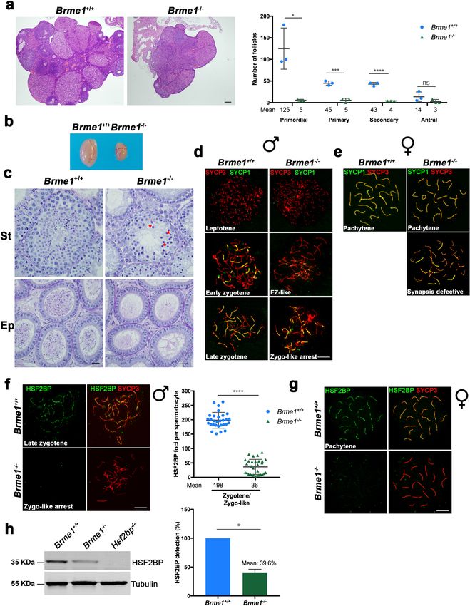
Research article Cell Biology
The meiotic division is an orderly process that results in the pairing and synapsis of homologous
chromosomes and crossover (CO) formation, which ultimately enable homologous chromosomes
segregation (Hunter, 2015; Loidl, 2016; Zickler and Kleckner, 2015). In mammals, pairing of
homologs is dependent on the repair of self-induced double-strand breaks (DSBs) during prophase I
by homologous recombination (Handel and Schimenti, 2010) and it leads to the intimate alignment
of homologous chromosomes (synapsis) through the zipper-like synaptonemal complex (SC)
(Cahoon and Hawley, 2016). The SC is a proteinaceous tripartite structure that provides the struc-
tural framework for DSBs repair (Baudat et al., 2013), as epitomized by the tight association of the
recombination nodules (RNs, multicomponent recombinogenic factories) and the axial elements of
the SC (Zickler and Kleckner, 2015).
Meiotic DSBs repair is an evolutionarily conserved pathway that is highly regulated to promote
the formation of at least one CO per bivalent. This chromosome connection between bivalents
through chiasmata is required for a correct reductional division. As other DNA repair processes,
proper meiotic recombination is essential for genome stability and alterations can result in infertility,
miscarriage and birth defects (Geisinger and Benavente, 2017; Handel and Schimenti, 2010;
Webster and Schuh, 2017).
Infertility refers to failure of a couple to reproduce and affects 10–15% of couples (Isaksson and
Tiitinen, 2004). Infertility can be due to female factors, male factors, a combination of both or to
unknown causes, each category representing approximately 25% of cases (Isaksson and Tiitinen,
2004; Matzuk and Lamb, 2008). There are several underlying causes and physiological, genetic and
even environmental and social factors can play a role. Forward and reverse genetic analyses in model
organisms have identified multiple molecular pathways that regulate fertility and have allowed to
infer reasonable estimates of the number of protein-coding genes essential for fertility (de Rooij and
de Boer, 2003; Schimenti and Handel, 2018).
Primary ovarian insufficiency (POI) is a major cause of female infertility and affects about 1–3% of
women under 40 years of age. It is characterized by cessation of ovarian function before the age of
40 years. POI results from a depletion of the ovarian follicle pool and can be isolated or syndromic.
Genetic causes of POI account for approximately 20% of cases (Rossetti et al., 2017). Although
infertility-causing pathogenic variants are inherently unlikely to spread in a population, they can be
observed within families, especially when there is consanguinity. Such cases provide crucial insights
into the function of the genes and molecular mechanisms that they disrupt. Over the last decade,
causative variants in several genes have been found using whole exome sequencing in ‘POI pedi-
grees’. In particular, pathogenic variants in genes involved in DNA replication, recombination or
repair, such as STAG3, SYCE1, HFM1, MSH5 and MEIOB have been formally implicated in this condi-
tion by ourselves and others (Caburet et al., 2014; Caburet et al., 2019a; de Vries et al., 2014;
Guo et al., 2017; Primary Ovarian Insufficiency Collaboration et al., 2014).
In this study, we have identified in a consanguineous family with POI the candidate S167L mis-
sense variant in HSF2BP, an essential yet poorly studied meiotic gene. HSF2BP encodes an interactor
of the heat-shock response transcription factor HSF2 (Yoshima et al., 1998). During the course of
this work and, in agreement with our results, two independent groups showed that HSF2BP is essen-
tial for meiotic recombination through its ability to interact with BRCA2 (Brandsma et al., 2019;
Zhang et al., 2019). Here, we report that the introduction of the missense variant HSF2BP-S167L in
mouse leads to subfertility and DNA repair defects during prophase I. In addition, we identified a
protein complex composed of BRCA2, HSF2BP, and the as yet unexplored C19ORF57/BRME1 (mei-
otic double-stranded break BRCA2/HSF2BP complex associated protein) as a key component of the
meiotic recombination machinery. Our studies show that a single substitution (S167L) in HSF2BP
leads to a reduced loading of both BRME1 and HSF2BP at the RNs. Furthermore, our results suggest
that meiotic progression requires a critical threshold level of HSF2BP/BRME1 for the ulterior loading
of the recombinases to the RNs.
Results
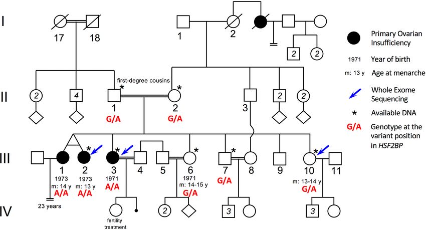
Clinical cases
The parents are first-degree cousins of Israeli Arab origin. Of the five daughters, three are affected
with POI and presented with early secondary amenorrhea. They had menarche at normal age (at 13–
Felipe-Medina et al. eLife 2020;9:e56996. DOI: https://doi.org/10.7554/eLife.56996 2 of 39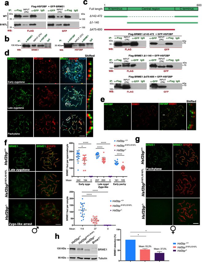
Research article                                                                                                                    Cell Biology

                                      14) but with irregular menses that stopped around 25. Only one of the patients affected by POI
                                      could have a child with the help of a fertility treatment (see pedigree in Figure 1). In order to identify
                                      the genetic basis of this familial POI case, we performed whole exome sequencing on genomic DNA
                                      from two POI patients, III-2 and III-3, and their fertile sister III-10 (Supplementary file 1a). Variants
                                      were filtered on the basis of (i) their homozygosity in the patients, (ii) their heterozygosity or absence
                                      in the fertile sister, (iii) their absence in unrelated fertile in-house controls and (iv) a minor allele fre-
                                      quency (MAF) below 0.01 in all available databases (Supplementary file 1b). This filtering process
                                      led to the identification of a missense substitution located in the HSF2BP gene: rs200655253
                                      (21:43630396 G > A, GRCh38). The variant lies within the sixth exon of the reference transcript
                                      ENST00000291560.7 (NM_007031.2:c.500C > T) and changes a TCG codon into a TTG
                                      (NP_008962.1:p.Ser167Leu). It is very rare (Variant Allele Frequency/VAF 0.0001845 in the GnomAD
                                      database and 0.0005 in the GME Variome dedicated to Middle-East populations) and absent in a

Figure 1. Pedigree of the consanguineous family with the variant HSF2BP-S167L. III-1 and III-2 are monozygotic twins, who appear phenotypically
dizygotic. Clinical investigation confirmed POI, with normal 46, XX karyotype (500 bands and SKY spectral karyotyping). Year of birth and age of
menarche are indicated when known. III-1 became amenorrheic at age 24 and III-2 at age 25, both after irregular menstruations since menarche. III-1
presents with a short stature (152 cm, within the 3–5 percentile), a normal neck, cubitus valgus and metacarpal shortening of 4–5. Ultrasound
investigation showed normal uterus and ovaries. Her g-banding karyotyping was normal 46, XX (500 bands) and variants in FMR1 gene were ruled out.
III-2 displays a normal secondary sexual development with no dysmorphic sign. Clinical investigation confirmed POI, with normal 46, XX karyotype (500
bands and SKY spectral karyotyping). The elder sister III-3 was also diagnosed with POI, with no further clinical information. She is 160.5 cm. She had
one normal pregnancy with the help of ‘fertility treatment’, and a second unsuccessful attempt. The two fertile sisters, III-6 and III-10 had their menarche
at 14–15 and 13–14 respectively, with regular menstruations ever since. They are respectively 150 cm and 151 cm, with no clinical sign, and each one
had several children without difficulties. The fertile brother III-7 is 171 cm and shows no health or fertility problem. He developed frontal baldness since
the age of 30. The genotype of each individual at the variant genomic position in HSF2BP is shown in red, as determined by Sanger sequencing for
available DNAs (See Figure 1—figure supplement 1).
The online version of this article includes the following figure supplement(s) for figure 1:
Figure supplement 1. Segregation of the S167L Variant in HSF2BP in the consanguineous family shows the chromatograms obtained by Sanger
sequencing of the HSF2BP-S167L variant in the family.
Figure supplement 2. Strong conservation of the Ser167 residue in HSF2BP protein in 99 mammals.
Figure supplement 3. Strong conservation of the Ser167 residue in HSF2BP in 48 birds and reptiles and 64 fish species.

Felipe-Medina et al. eLife 2020;9:e56996. DOI: https://doi.org/10.7554/eLife.56996                                                                  3 of 39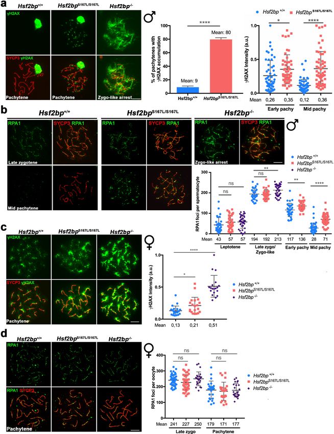
Research article                                                                                                     Cell Biology

                                   homozygous state from all available databases. The variant was verified by Sanger sequencing and
                                   was found to segregate in a Mendelian fashion within the family: the affected twin III-1 was homozy-
                                   gous for the variant and both parents and fertile siblings were heterozygous carriers (Figure 1—fig-
                                   ure supplement 1). Therefore, there was no homozygous males identified in this family, preventing
                                   the analysis of the impact of this variant on male fertility. Serine 167 is a highly conserved position
                                   and the S167L variant is predicted to be pathogenic or deleterious by 11 out of the 18 pathogenicity
                                   predictors available in dbNSFP 3.5. (Supplementary file 1c, Figure 1—figure supplement 2 and
                                   Figure 1—figure supplement 3).

                                   Mice with the HSF2BP S167L variant show a partial reduction of
                                   fertility
                                   During the course of this work, two independent groups showed that HSF2BP is essential for meiotic
                                   recombination through its ability to interact with the armadillo repeats of BRCA2 (Zhang et al.,
                                   2019). Both groups showed that genetic disruption of Hsf2bp in mouse leads to the accumulation in
                                   the chromosomes axes of DNA repair proteins such as gH2AX (ATR-dependent phosphorylation of
                                   H2AX marks DSBs) and the single stranded-DNA binding protein RPA, a strong reduction of the
                                   recombinases DMC1 and RAD51 at the RNs and a lack of COs as labelled by MLH1 (Baker et al.,
                                   1996). The end result is male sterility (Brandsma, 2006; Brandsma et al., 2019; Zhang et al.,
                                   2019). However, loss of HSF2BP in female mice showed a milder meiotic phenotype (Zhang et al.,
                                   2019 and our own data, see below) and a weak albeit non-statistical significant reduction of fertility
                                   (Brandsma et al., 2019) despite all of the mutants are nulls though in different genetic
                                   backgrounds.
                                       In order to confirm the causality of the S167L variant in this POI family, we generated a knock-in
                                   mouse Hsf2bpS167L/S167L by genome editing (Figure 2—figure supplement 1a). We also generated
                                   a loss-of-function model (Hsf2bp-/-) for direct comparison (Figure 2—figure supplement 1b–d).
                                   Hsf2bpS167L/S167L male and female mice were able to reproduce but females showed a significant
                                   reduction in the number of litters (Figure 2a), whilst males only showed a slight non-significant
                                   reduction in fertility (Figure 2a), suggesting that the S167L variant impacts murine fertility.
                                       Histological analysis of Hsf2bpS167L/S167L ovaries revealed no apparent differences in the number
                                   of follicles in comparison to wild-type (WT) animals (Figure 2b–c and Figure 2—figure supplement
                                   2a), in contrast with the drastic reduction of the follicle pool in Hsf2bp-/- ovaries (Figure 2b–c). Tes-
                                   tes from Hsf2bpS167L/S167L mice displayed a reduced size (21% reduction compared to WT mice; tes-
                                   tis/body weight ratio: S167L 0.26% ± 0.07 (n = 12) vs 0.33% ± 0.05 for WT controls (n = 14),
                                   **p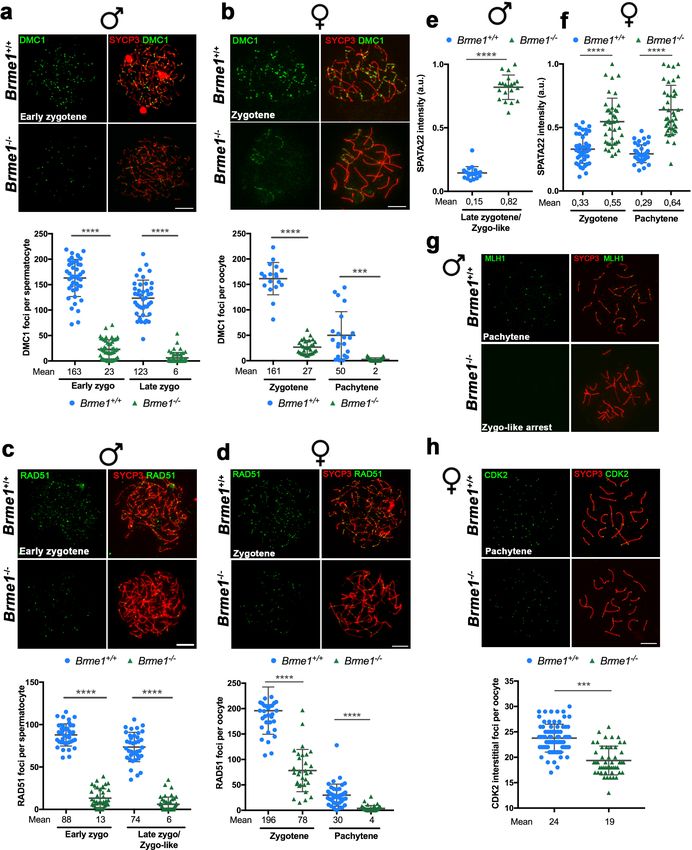
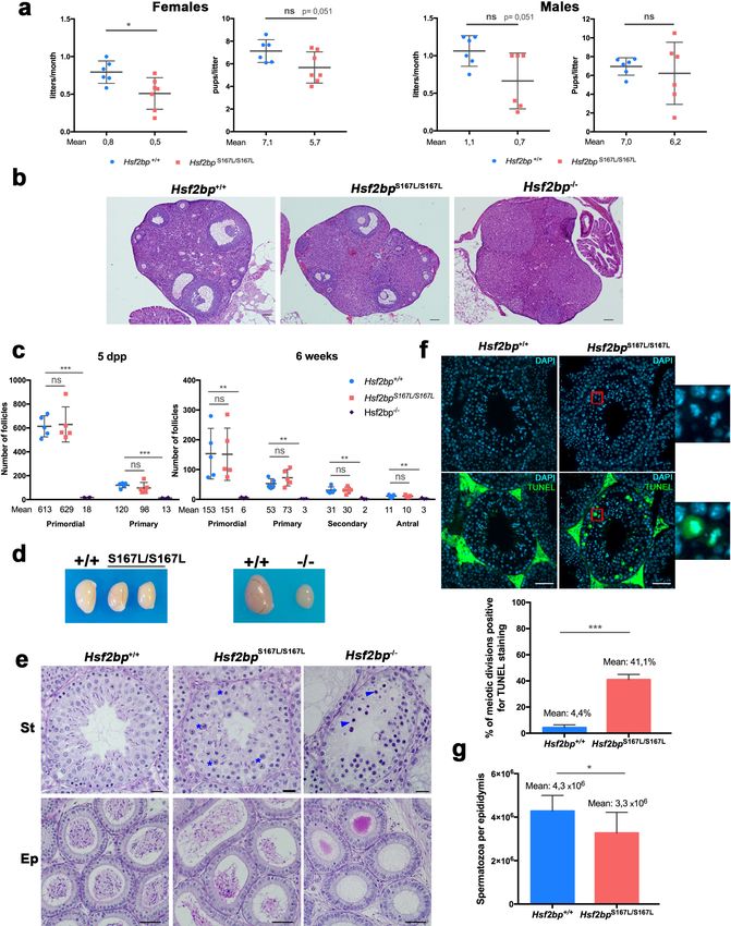
Research article                                                                                                                  Cell Biology

Figure 2. Mice carrying the HSF2BP S167L variant show a partial reduction of fertility. (a) Fertility assessment of males and female Hsf2bpS167L/S167L and
WT mice showing the number of litters per month and the number of pups per litter (see Materials and methods). Mice: Hsf2bp+/+n = 6 females/6
males, Hsf2bpS167L/S167Ln = 7 females/6 males. Two-tailed Welch’s t-test analysis: *p
Research article                                                                                                                       Cell Biology

Figure 2 continued
of follicles. Bar in panels 100 mm. (c) Quantification of the number of follicles (primordial, primary, secondary and antral follicles) per ovary in Hsf2bp+/+,
Hsf2bpS167L/S167L and Hsf2bp-/- females at 5 dpp and 6 weeks of age showing no differences between Hsf2bp+/+ and Hsf2bpS167L/S167L but a strong
reduction in the oocyte pool in Hsf2bp-/- females. Ovaries: five dpp/6 weeks = 5/5 ovaries from Hsf2bp+/+ and Hsf2bpS167L/S167L and 4/3 from Hsf2bp-/-.
Two-tailed Welch’s t-test analysis: ns, no significant differences, **p
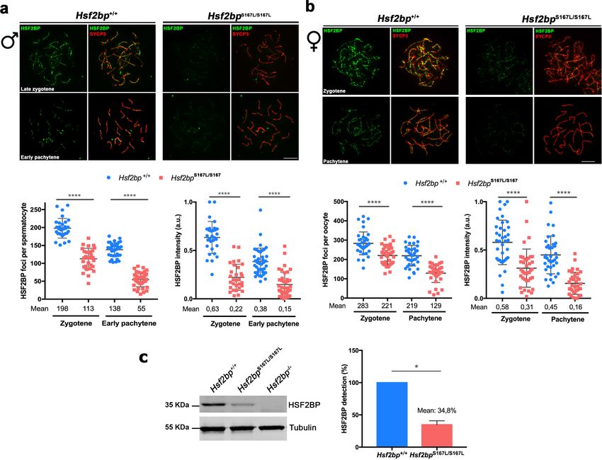
Research article                                                                                                                Cell Biology

Figure 3. Meiocytes from Hsf2bpS167L/S167L mice show a decrease in the expression of HSF2BP. (a–b) Double immunofluorescence of HSF2BP (green)
and SYCP3 (red) in Hsf2bp+/+and Hsf2bpS167L/S167L (a) spermatocyte and (b) oocyte spreads showing a strong reduction in the labeling of HSF2BP at the
chromosome axis. Plots under the panels show the quantification. Nuclei analyzed: 30 zygonemas and 40 pachynemas from two adult male mice of
each genotype. In females 38/39 zygonemas and 37/35 pachynemas from two 17.5 dpc embryos of Hsf2bp+/+ and Hsf2bpS167L/S167L, respectively. Two-
tailed Welch’s t-test analysis: ****p
Research article                                                                                                              Cell Biology

Figure 4. DNA repair in Hsf2bpS167L/S167L mice. (a, c) Double labeling of gH2AX (green) and SYCP3 (red) in (a) spermatocyte and (c) oocyte spreads
from WT, Hsf2bpS167L/S167L and Hsf2bp-/- mice. (a) Males display an accumulation of gH2AX patches in Hsf2bpS167L/S167L pachynemas and a strong
accumulation in the whole nucleus in Hsf2bp-/- zygotene-like arrested cells. Plots on the right of the panel represent the percentage of pachynemas
with gH2AX labeling (Nuclei: 364 Hsf2bp+/+ and 376 Hsf2bpS167L/S167L from three adult mice) and the quantification of gH2AX intensity on autosomes at
Figure 4 continued on next page

Felipe-Medina et al. eLife 2020;9:e56996. DOI: https://doi.org/10.7554/eLife.56996                                                            8 of 39
Research article                                                                                                                  Cell Biology

Figure 4 continued
early and mid-pachytene stages (Nuclei: 53 early and 60 mid pachynemas from three adult mice of each genotype). Two-tailed Welch’s t-test analysis:
*p
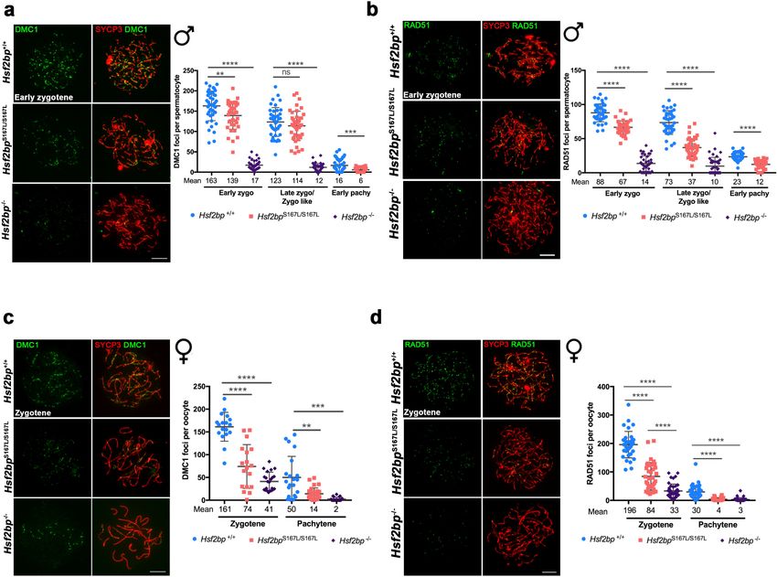
Research article                                                                                                             Cell Biology

Figure 5. The loading of recombinases is compromised in Hsf2bpS167L/S167L mice. Double immunolabeling of (a, c) DMC1 or (b, d) RAD51 (green) and
SYCP3 (red) in Hsf2bp+/+, Hsf2bp S167L/S167L and Hsf2bp-/- (a, b) spermatocytes and (c, d) oocytes showing a strong reduction (Hsf2bp-/-) and mild
reduction (Hsf2bp S167L/S167L) in the number of foci in comparison with their WT counterparts. Plots on the right of the panels represent the
quantification of foci on each genotype and stage. Male nuclei DMC1: Hsf2bp+/+/Hsf2bp S167L/S167L/Hsf2bp-/-, respectively, n = 43/38/37 early
zygonemas, 41/43/37 late zygonemas/zygonemas-like and 44/37 early pachynemas from two adult mice of each genotype. Male nuclei RAD51:
Hsf2bp+/+/Hsf2bp S167L/S167L/Hsf2bp-/- respectively n = 39 early zygonemas from all genotypes, 37/40/43 late zygonemas/zygonema-like and 37/39 early
pachynemas from two adult mice of each genotype. Oocyte nuclei DMC1: Hsf2bp+/+/Hsf2bp S167L/S167L/Hsf2bp-/- n = 18/18/22 zygonemas from two
embryos and n = 21/30/23 pachynemas from two embryos (17.5 dpc). Oocyte nuclei RAD51: Hsf2bp+/+/Hsf2bp S167L/S167L/Hsf2bp-/- n = 35/35/42
zygonemas and n = 42/42/40 pachynemas from two embryos (17.5 dpc). Two-tailed Welch’s t-test analysis: ns, no significant differences, **p
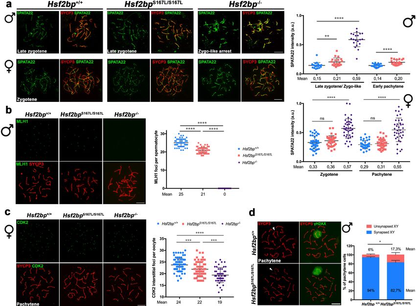
Research article                                                                                                                  Cell Biology

Figure 6. Recombination proficiency is decreased in Hsf2bpS167L/S167L mice. (a) Double labeling of SPATA22 (green) and SYCP3 (red) in spermatocyte
(upper panel) and oocyte (lower panel) spreads from WT, Hsf2bp-/- and Hsf2bpS167L/S167L mice. SPATA22 is accumulated in knock-out spermatocytes
and oocytes and shows a milder accumulation in the Hsf2bpS167L/S167L spermatocytes. Hsf2bpS167L/S167L oocytes show a slight but not significant
accumulation. Plots on the right of the panel represents the quantification of SPATA22 labeling. Males nuclei: n = 20 cells for each stage from two adult
mice of each genotype. Females nuclei: Hsf2bp+/+/Hsf2bp S167L/S167L/Hsf2bp-/- n = 41/40/40 zygonemas from two embryos and n = 40/39/38
pachynemas from two embryos (17.5 dpc). Two-tailed Welch’s t-test analysis: ns, no significant differences, **p
Research article                                                                                                               Cell Biology

Figure 7. BRME1, a novel HSF2BP interactor that colocalizes to the recombination nodules. (a) HEK293T cells were transfected with Flag-HSF2BP (WT,
upper panel; S167L, lower panel) and its novel interactor GFP-BRME1. Protein complexes were immunoprecipitated (IP: green text) with either anti-Flag
or anti-EGFP or IgGs (negative control) and analysed by immunoblotting with the indicated antibody (WB: red text). Both HSF2BP variants (WT and
S167L) co-immunoprecipitated similarly with BRME1. (b) IP of testis extracts with antibodies against BRME1, HSF2BP and IgGs as a negative control (IP:
Figure 7 continued on next page

Felipe-Medina et al. eLife 2020;9:e56996. DOI: https://doi.org/10.7554/eLife.56996                                                            12 of 39
Research article                                                                                                                  Cell Biology

Figure 7 continued
green text) and western blot with the indicated antibodies (WB: red text) (c) Schematic representation of full-length BRME1 protein and the
corresponding deletion (D) constructs (filled boxes) generated to decipher the essential BRME1 region for interacting with HSF2BP (green positive
interaction and red no interaction). Western blots under the scheme show the Co-IP experiments. HEK293T cells were transfected with GFP-HSF2BP
and the different delta constructs of Flag-BRME1. The D475–600 abolishes the interaction, indicating that the C terminus of BRME1 is the essential
region of interaction with HSF2BP. (d) Triple immunofluorescence of BRME1 (green), HSF2BP (red) and SYCP3 (blue) in WT spermatocyte spreads
showing high colocalization between BRME1 and HSF2BP at late zygotene and pachytene (See Supplementary file 1d for quantification). Bar in panel,
10 mm. (e) Double immunolabeling of spermatocyte spread preparations with HSF2BP (green) and BRME1 (red) analyzed by Stimulated emission
depletion (STED) microscopy. Bar in panel, 5 mm. (f–g) Double immunofluorescence of BRME1 (green) and SYCP3 (red) in (f) spermatocytes and (g)
oocyte spreads from Hsf2bp+/+, Hsf2bpS167L/S167L and Hsf2bp-/- showing a strong reduction of BRME1 staining in the S167L mutant and absence in the
Hsf2bp knock-out. Plots next to the panel represent the quantification. See also extended Figure 7—figure supplement 4a. Male nuclei: Hsf2bp+/+/
Hsf2bpS167L/S167L/Hsf2bp-/-: n = 16/16/20 early and 15/15/16 late zygonemas, 16/16 /- pachynemas from two adult mice. Female nuclei: n = 18
pachynemas from two embryos (17.5 dpc) of each genotype. Two-tailed Welch’s t-test analysis: *p
Research article                                                                                                                 Cell Biology

Figure 8. Brme1-/-mice show severe fertility defects. (a) Hematoxylin+eosin stained sections of ovaries from adult Brme1-/- females showing a strong
depletion of follicles. Plot on the right represents the quantification in 3 months-old females. Ovaries: n = 3 ovaries for each genotype. Two-tailed
Welch’s t-test analysis: *p
Research article                                                                                                                  Cell Biology

Figure 8 continued
Brme1-/- as shown in PAS+hematoxylin stained testis sections. Massive apoptosis of spermatocytes is indicated (red asterisks). The spermatogenic arrest
leads to empty epididymides and non-obstructive azoospermia. (St) Seminiferous tubules. (Ep) Epididymides. Bar in panels, 10 mm. (d–e) Double
labeling of (d) spermatocyte and (e) oocyte spreads from WT and Brme1-/- mice with SYCP3 (red) and SYCP1 (green). Brme1-/- spermatocytes arrest in a
zygotene-like stage and show synapsis between non-homologous chromosomes. (e) Brme1-/- females showed a subset of fully-synapsed pachynemas
(18.5 dpc) but increased numbers of synapsis-defective cells. See extended panel for females at Figure 8—figure supplement 1e. Bar in panels, 10
mm. (f–g) Double labeling with HSF2BP (green) and SYCP3 (red) of (f) spermatocyte and (g) oocyte spreads from Brme1-/- mice showing faint HSF2BP
labeling in spermatocytes and total absence of labeling in oocytes. Plot on the right of (f) panel represents de quantification of HSF2BP foci in Brme1-/-
spermatocytes. Nuclei: n = 30 zygonemas/zygonemas-like from two adult mice of each genotype (Brme1+/+ values from Figure 3a) Two-tailed Welch’s
t-test analysis: ****p
Research article                                                                                                          Cell Biology

Figure 9. BRME1 is essential for meiotic recombination. (a–b) Double immunofluorescence of DMC1(green) and SYCP3 (red) in Brme1+/+ and Brme1-/-
(a) spermatocytes and (b) oocytes showing a reduction in the number of DMC1 foci. Plots under each panel represent the quantification. See also
extended panels on Figure 9—figure supplement 2a–b. Male nuclei for DMC1: Brme1+/+/Brme1-/- n = 43/50 early and 41/52 late zygonemas/
zygonemas like from two adult mice of each genotype (Brme1+/+ values from Figure 5a). Female nuclei for DMC1: Brme1+/+/Brme1-/- n = 18/30
Figure 9 continued on next page

Felipe-Medina et al. eLife 2020;9:e56996. DOI: https://doi.org/10.7554/eLife.56996                                                      16 of 39
Research article                                                                                                              Cell Biology

Figure 9 continued
zygonemas and 21/31 pachynemas from two embryos (17.5 dpc) of each genotype (Brme1+/+ values from Figure 5c). Two-tailed Welch’s t-test analysis:
***p
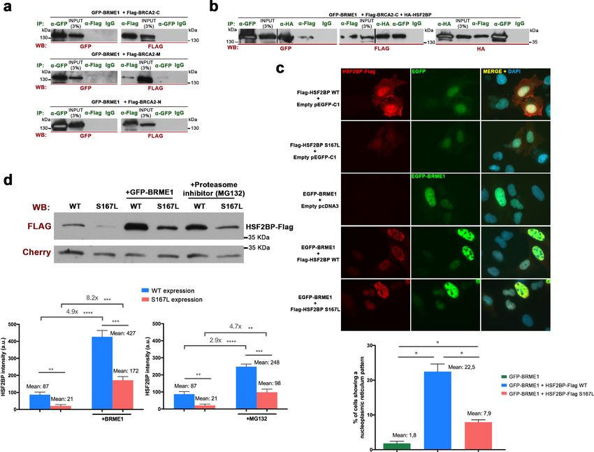
Research article                                                                                                              Cell Biology

Figure 10. BRME1 forms a complex with BRCA2 and HSF2BP and stabilizes HSF2BP. (a–b) HEK293T cells were co-transfected with GFP-BRME1, Flag-
BRCA2-C and HA-HSF2BP. Protein complexes were immunoprecipitated (IP: green text) with either an anti-Flag, anti-EGFP, anti-HA or IgGs, and
analyzed by western blot with the indicated antibody (WB: red text). (a) BRME1 does not co-immunoprecipitate with BRCA2-N, BRCA2-M or BRCA2-C.
(b) In the presence of HA-HSF2BP (triple co-transfection) BRCA2-C and BRME1 coimmunoprecipitate (co-IPs between HSF2BP and BRCA2-C are shown
in Figure 6—figure supplement 2a). (c) Transfected U2OS cells with plasmids encoding Flag-HSF2BP (WT or S167L) and EGFP-BRME1 alone or
together were immuno-detected with antibodies against Flag (red) and EGFP (green). Transfected HSF2BP (WT and S167L) labels the whole cell (S167L
less intense) whereas BRME1 shows nuclear localization. When co-expressed, BRME1 and HSF2BP change their patterns and form nuclear invaginations
that resemble nucleoplasmic reticulum. This phenotype is milder in the presence of HSF2BP-S167L than with the WT (graph under the panel:
quantification of the number of cells showing a nucleoplasmic reticulum pattern). n > 400 cells from two independent transfections of each condition.
Two-tailed Welch’s t-test analysis: *p
Research article                                                                                                     Cell Biology

                                   by the essential role that the meiotic cohesin subunit RAD21L plays in males but not in females
                                   (Herrán et al., 2011). In general, meiotic recombination mutants appear to proceed further in
                                   female than in male, because in males the asynapsis of the sex bivalent leads to a loss of silencing of
                                   the Y chromosome, and perhaps also because of the presence of less stringent checkpoints in
                                   oogenesis (Hunt and Hassold, 2002). A sexually dimorphic phenotype is also observed here in our
                                   HSF2BP-S167L mutant mice. Both sexes show a significant decrease in the number of COs and con-
                                   sequently an increase in the frequency of meiocytes showing bivalents without CO. This leads to a
                                   significant reduction of spermatozoa in the epididymis, while the number of oocytes and their distri-
                                   bution in the follicular pool are not affected in females. Our failure to detect any significant impact
                                   of the variant on male fertility could be due to the high variability in litter frequency and size in our
                                   study. However, it is known that a strong reduction of the spermatozoa count (up to 60%) does not
                                   affect male mouse fertility (Schürmann et al., 2002), which would explain the normal fertility of male
                                   mice bearing the HSF2BP-S167L variant despite the presence of overt meiotic alterations. By con-
                                   trast, female mice with the HSF2BP-S167L variant show a mild sub-fertility phenotype with a reduc-
                                   tion of litter frequency. This can be due to the very much fewer gametes available for fertilization in
                                   females in comparison to males but also to molecular differences in the meiotic recombination pro-
                                   cess in both sexes (Cahoon and Libuda, 2019), as displayed by the absence of RPA accumulation or
                                   the more pronounced decrease in DMC1 foci observed in HSF2BP-S167L oocytes.
                                       Although we cannot exclude that a POI-like phenotype would appear over time in HSF2BP-S167L
                                   female mice, the sub-fertility observed in HSF2BP-S167L females appears to be milder in comparison
                                   to the phenotype of the human patients. This could be explained by a lower sensitivity of mice to
                                   hypomorphic alleles compared to humans, in a similar manner to the known lower gene dosage sen-
                                   sitivity of the former in the context of genes that are haploinsufficient in human (Veitia, 2003). In
                                   addition, the initial events of human female meiosis appear to be more error-prone than in mice, or
                                   even than human males, as evidenced by the increased incidence of synaptic defects in the human
                                   oocytes or the fact that MLH1 foci appear much earlier in prophase I (Hassold et al., 2007). Further-
                                   more, it has recently been shown that human oocytes exhibit a specific CO maturation inefficiency
                                   (Wang et al., 2017). Indeed, despite a higher total number of COs in women, the frequency of biva-
                                   lents without CO is paradoxically higher in women than in men. Altogether, these observations could
                                   explain the stronger phenotype of the human POI patients compared to the Hsf2bpS167L/S167L female
                                   mice.
                                       The absence of homozygous male carriers in the consanguineous family studied here prevents
                                   the direct comparison of the impact of the HSF2BP-S167L variant on fertility phenotypes between
                                   human and mouse males. Although future studies might identify infertile men homozygous for the
                                   S167L variant, it is interesting to note that another variant of HSF2BP (G224*) was shown to affect
                                   recombination rate in males and that two siblings homozygous for this HSF2BP variant in the ana-
                                   lyzed Icelandic population were healthy but without descendants, suggesting they were infertile
                                   (Halldorsson et al., 2019). This reinforces a conserved function of HSF2BP in human male fertility.
                                       We have shown through a biochemical analysis that BRME1 immunoprecipitates mainly with its
                                   partner HSF2BP (constituting or belonging to a complex) but also with PALB2, RAD51, RPA and
                                   BRCA2 (in testis extracts). These interactions could possibly be mediated by the multidomain hub
                                   protein BRCA2 (Siaud et al., 2011) as HSF2BP interacts directly with BRCA2 (Brandsma et al.,
                                   2019) and with BRME1 (this work). In addition, BRCA2 also directly interacts with the DSBs recruiter
                                   PALB2, with the recombinases RAD51 and DMC1 (through different specific domains), and with
                                   DNA (Siaud et al., 2011). This BRCA2-containing complex participates in the orderly orchestration
                                   of events at DSBs such as the initial binding of RPA to the resected DNA, the exchange of RPA by
                                   RAD51/DMC1, and the loading of the MEIOB-SPATA22 complex to the RPA complexes
                                   (Martinez et al., 2016; Zhao et al., 2015). Interestingly, genes with recently identified variants in
                                   POI patients are implicated in the repair of induced DSBs at the early stages of meiosis and encode
                                   BRCA2-interacting factors, such as MEIOB, DMC1 or BRCA2 itself (Caburet et al., 2019a;
                                   Caburet et al., 2020; Caburet et al., 2019b; He et al., 2018). This highlights the crucial importance
                                   and the high sensitivity of this particular meiotic step, and the hub role of BRCA2 as a tightly regu-
                                   lated platform for correct meiotic recombination.
                                       We have also shown by several complementary approaches that the proteins HSF2BP/BRME1
                                   constitute in vivo a functional complex in which both subunits are essential for meiotic recombination
                                   and for their mutual protein expression and/or stability in vivo. Accordingly, the genetic depletion of

Felipe-Medina et al. eLife 2020;9:e56996. DOI: https://doi.org/10.7554/eLife.56996                                                  19 of 39
Research article Cell Biology
BRME1 or HSF2BP leads to similar if not identical phenotypes in which oogenesis is altered with
severe defects in chromosome synapsis that promotes premature loss of ovarian follicles and sper-
matogenesis is arrested at zygotene-like stage resulting in a lack of spermatozoa. These meiocytes
are not able to load the recombinases RAD51/DMC1, impairing the proper repair of DSBs leading
to the generation of no COs or very few in males and females, respectively. As a consequence, zygo-
nema-like spermatocytes accumulate the single strand binding proteins SPATA22 and RPA, whereas
oocytes accumulates only SPATA22. During the course of the reviewing of this work, three Brme1
knockouts have been described (Zhang et al., 2020; Shang et al., 2020; Takemoto et al., 2020).
All the described male mutants show strong fertility defects and similar molecular alterations
although with different severity. In females, the two works that address their analysis (Shang et al.
and Takemoto et al.), describe normal fertility which is in contrast with the strong reduction in the
follicle pool and meiotic defects observed in our Brme1-/- females. The higher severity of our male
and female mutants could be explained on the basis of the different genetic background of the mice
given that all of them are apparently similar.
The S167L human recessive POI variant behave as a hypomorphic allele in mice, which results in a
reduction of the protein expression/stability of itself and of its partner BRME1 in vivo and in trans-
fected cells. As a consequence, both male and female Hsf2bpS167L/S167 mice show a similar but
milder phenotype than that of the Hsf2bp-/-or Brme1-/-, consisting in a reduction in the spermatozoa
count while being fertile and a subtle reduction in female fertility. Molecularly, the reduction
observed in the meiocytes of the mutant Hsf2bpS167L/S167L mice of RAD51/DMC1, the reduction of
COs (both in males and females), the accumulation of RPA (only in males) and SPATA22 (in males
and females) are also weaker than in the null mutant. The observed accumulation of RPA in males is
likely to occur at the early stages of recombination because SPATA22 loading to the DSBs is also
increased in the mutants of HSF2BP and BRME1. RPA, as part of a trimeric replication protein com-
plex (RPA1-RPA2-RPA3), binds and stabilizes ssDNA intermediates that form during DNA repair. In
meiosis, RPA is also forming a complex with two other essential meiotic players MEIOB (homologue
of RPA) and SPATA22. However, the loading of this complex to DSBs is RPA-independent
(Shi et al., 2019). It has been postulated that RPA functions in meiosis at two different stages; (i)
during the early recombination stages when the DSBs ends are resected by the MRN complex and
(ii) during the strand invasion into the homologous duplex that is carried out by RAD51/DMC1 and
ssDNA is generated at the displacement loops (Shi et al., 2019). The observed lack of DNA-binding
ability of HSF2BP/BRME1 points towards a model in which the absence of the complex HSF2BP/
BRME1 through a direct interaction with BRCA2 impairs the replacement of RPA by RAD51/DMC1
in the foci that form on the DSBs of the spermatocytes. Similarly, the reduced expression at the pro-
tein level of HSF2BP/BRME1 as a consequence of the POI variant, which does not affect their hetero-
dimerization, would make them less proficient in replacing RPA in the spermatocytes by the
recombinases RAD51/DMC1 leading to a lower frequency of COs. Given the unknown function that
RPA plays in vivo during oogenesis (Shi et al., 2019), it is tempting to speculate that the role of RPA
in mediating the replacement of RAD51/DMC1 in female meiosis would be carried out by another
protein complex such as SPATA22/MEIOB in a HSF2BP/BRME1-dependent manner.
Very recently, a high-resolution genome-wide recombination map revealed novel loci involved in
the control of meiotic recombination and highlighted genes involved in the formation of the SC
(SYCE2, RAD21L, SYCP3, SIX6OS1) and the meiotic machinery itself as determinants of COs
(Halldorsson et al., 2019). Within the second category, variants of the SUMO ligase RNF212 and
the ubiquitin ligase HEI10 have been largely documented as genetic determinants of the recombina-
tion rate in humans and, importantly, so were variants of HSF2BP. Consequently, gene dosage of
RNF212 and HEI10 affects CO frequency through their activity in CO designation and maturation
(Lake and Hawley, 2013; Reynolds et al., 2013). We found that both BRME1 and HSF2BP localiza-
tion are unaffected in the loss-of-function mouse mutants of Rnf212 and Hei10 (Ccnb1ip1). This
observation together with the proper co-localization of BRME1/HSF2BP with RPA allows us to map
these proteins upstream in the recombination pathway.
It is worth noting that some of the genes affecting the recombination rate have also been
described as ‘fertility genes’, such as SYCP3, HFM1 and HSF2BP (Geisinger and Benavente, 2017;
Primary Ovarian Insufficiency Collaboration et al., 2014 and this work). Altogether, we propose
that different variants of the same meiotic gene (alleles responsible for mild or strong phenotypes)
can give rise to either an altered genome-wide recombination rate with no detrimental effect, or
Felipe-Medina et al. eLife 2020;9:e56996. DOI: https://doi.org/10.7554/eLife.56996 20 of 39Research article Cell Biology
cause infertility when the decreased recombination rate falls below the lower limit of one COs per
bivalent. In the present POI family, the S167L variant in HSF2BP seems to be below that limit. To our
knowledge, HSF2BP is one of the very few human genes with variants known to affect both the
genome-wide recombination rate in the human population and meiotic chromosome missegregation
(fertility) through a reduction of the recombination rate (Halldorsson et al., 2019). Along similar
lines, it is conceivable that variants with additive effects (Schimenti and Handel, 2018) can lead to a
genome-wide reduction of the recombination rate and thus to aneuploidy and infertility. Specifically,
variants in genes involved in meiotic recombination and SC constituents could be responsible for a
large fraction of genetic infertilities. These variants should be under purifying selection and would
be removed or substantially reduced from the population. However, this is not the case for genes
with sexual phenotypic dimorphism (Gershoni and Pietrokovski, 2014) as is apparent for a wide
number of meiotic genes (Cahoon and Libuda, 2019), including HSF2BP and BRME1, where individ-
uals of one of the sexes are fertile carriers.
In summary, we describe for the first time a human family where POI co-segregates with a genetic
variant in HSF2BP (S167L) in a Mendelian fashion. Humanized mice reveal that the HSF2BP variant is
a hypomorphic allele that promotes the lower protein expression and/or stability of the HSF2BP/
BRME1 complex and phenocopy in a milder manner the meiotic defects observed in mice lacking
either HSF2BP or its direct interactor BRME1.
Materials and methods
Whole exome sequencing
Written informed consent was received from participants prior to inclusion in the study and the insti-
tutions involved. Genomic DNA was extracted from blood samples by standards protocols.
For individuals III-3 and III-10, library preparation, exome capture, sequencing and initial data
processing were performed by Beckman Coulter Genomics (Danvers, USA). Exon capture was per-
formed using the hsV5UTR kit target enrichment kit. Libraries were sequenced on an Illumina HiSEQ
instrument as paired-end 100 bp reads. For individual III-2, library preparation, exome capture,
sequencing and data processing were performed by IntegraGen SA (Evry, France) according to their
in-house procedures. Target capture, enrichment and elution were performed according to manufac-
turer’s instructions and protocols (SureSelect Human All Exon Kits Version CRE, Agilent). The library
was sequenced on an Illumina HiSEQ 2500 as paired-end 75 bp reads. Image analysis and base call-
ing was performed using Illumina Real Time Analysis (RTA 1.18.64) with default parameters.
Bioinformatic analysis
For the three individuals, sequence reads were mapped onto the human genome build (hg38/
GRCh38) using the Burrows-Wheeler Aligner (BWA) tool. Duplicated reads were removed using sam-
bamba tools. Whole exome sequencing metrics are provided in Supplementary file 1a. Variant call-
ing, allowing the identification of SNV (Single Nucleotide Variations) and small insertions/deletions
(up to 20 bp) was performed via the Broad Institute GATK Haplotype Caller GVCF tool (3.7).
Ensembl VEP (Variant Effect Predictor, release 87) program was used for initial variant annotation.
This tool considers data available in dbSNP (dbSNP147), the 1000 Genomes Project
(1000G_phase3), the Exome Variant Server (ESP6500SI-V2-SSA137), the Exome Aggregation Con-
sortium (ExAC r3.0), and IntegraGen in-house databases. Additional annotation data was retrieved
using dbNSFP (version 3.5, https://sites.google.com/site/jpopgen/dbNSFP) and Varsome (https://
varsome.com/). Minor allele frequencies were manually verified on GnomAD (http://gnomad.broad-
institute.org), ISB Kaviar (http://db.systemsbiology.net/kaviar/), and Great Middle Eastern variant
database GME Variome (http://igm.ucsd.edu/gme/).
Variant filtering was performed on the following criteria:
. minimum depth at variant position of 10,
. correct segregation in the family, on the basis of homozygosity by descent: variants should be
homozygous in both affected sisters III-2 and III-3, and heterozygous or homozygous for Refer-
ence allele in the fertile sister III-10,
. absence in unrelated in-house fertile controls,
Felipe-Medina et al. eLife 2020;9:e56996. DOI: https://doi.org/10.7554/eLife.56996 21 of 39Research article Cell Biology
. Minor Allele Frequency (MAF) below 1% in global and in each population in the GnomAD
database,
. presence in the coding sequence (i.e not in UTRs, introns, intergenic,.)
. high predicted functional impact on the protein. Impact was evaluated based on the predictors
included in dbNSFP3.5 (Cahoon and Libuda, 2019; Carlosama et al., 2017; Cox and Mann,
2008) (considered as pathogenic when the majority of the predictors agreed).
The number of variants fulfilling those criteria is provided in Supplementary file 1b. Visual inspec-
tion of the variant was performed using the IGV viewer.
Sanger sequencing analysis
To confirm the presence and segregation of the variant, direct genomic Sanger DNA sequencing of
HSF2BP was performed in the patients, the parents and non-affected siblings using specific primers:
HSF2BP-ex6F: 5’-ctagaatcttctgtatcctgca-3’ and HSF2BP-ex6R2: 5’-ggtctggaagcaaacaggcaa-3’. The
resulting chromatograms are shown in Figure 1—figure supplement 1.
Predictions of pathogenicity and sequence conservation
The S167L variant was predicted to be pathogenic or deleterious and highly conserved by 11 out of
the 18 pathogenicity predictors available in dbNSFP 3.5 (Supplementary file 1c). Upon verification,
it appears that the conflicting interpretation of this variant might stem from the single occurrence of
a Leu at this position in zebrafish. As the change in zebrafish is the variant that we have in the human
family, we checked all the available sequences (Ensembl Release 99, January 2020, removing the
one-to-many relationships). Ser167 is very highly conserved in mammals, birds and reptiles and fish
and is present in 208 of 212 orthologous sequences (Figure 1—figure supplement 2 and Figure 1—
figure supplement 3).
Generation of CRISPR/Cas9-edited mice
For developing all the mutant mice models (Hsf2bp-/-, Hsf2bpS167L/S167L, Brme1D142-472/D D142-472,
Brme1-/-, Spo11-/-, Rnf212-/- and Hei10-/-) the different crRNAs were predicted at https://eu.idtdna.
com/site/order/ designtool/index/CRISPR_CUSTOM. The crRNAs, the tracrRNA and the ssODNs
were produced by chemical synthesis at IDT (crRNAs and ssODNs sequences are listed in
Supplementary files 1i-1j). For the Hsf2bpS167L we introduced a mutation in the mouse counterpart
residue (p.Ser171Leu) of the POI mutation found in the clinical case (p.Ser167Leu). However, for the
shake of simplicity, on this manuscript we refer to the mutant allele by the acronym of the human
mutation (S167L). The ssODN contains the mutation on the corresponding position of the mouse
sequence (c.512C > T, p.Ser171Leu, see character in red in Supplementary file 1j) and the PAM
mutations avoiding amino acid changes (see characters in bold in the Supplementary file 1j). For
the Spo11-/- mice generation, the ssODN contains the mutations in the active site
(TACTAC >TTCTTC p.YY137-138FF, see Supplementary file 1j) and the PAM mutations (bold char-
acters in Supplementary file 1j). In all cases the crRNA and tracrRNA were annealed to obtain the
mature sgRNA. A mixture containing the sgRNAs, recombinant Cas9 protein (IDT) and the ssODN
(30 ng/ml Cas9, 20 ng/ml of each annealed sgRNA and 10 ng/ml ssODN) were microinjected into B6/
CBA F2 zygotes (hybrids between strains C57BL/6J and CBA/J) (Singh et al., 2015) at the Trans-
genic Facility of the University of Salamanca. Edited founders were identified by PCR amplification
(Taq polymerase, NZYtech) with primers flanking the edited region (see Supplementary file 1k for
primer sequences). The PCRs products were direct sequenced or subcloned into pBlueScript (Strata-
gene) followed by Sanger sequencing, selecting the founders carrying the desired alelles. The
selected founders were crossed with wild-type mice to eliminate possible unwanted off-targets. Het-
erozygous mice were re-sequenced and crossed to give rise to edited homozygous. Genotyping was
performed by analysis of the PCR products produced from genomic DNA extracted from tail biop-
sies. The primers and the expected amplicon sizes are listed in the Supplementary file 1k. Mouse
mutants for Rec8, Six6os1 and Psma8 have been previously described (Bannister et al., 2004;
Gómez-H et al., 2019; Gómez-H et al., 2016).
Felipe-Medina et al. eLife 2020;9:e56996. DOI: https://doi.org/10.7554/eLife.56996 22 of 39Research article Cell Biology
Ethics statement
All the experiments were approved by the Ethics Committee for Animal Experimentation of the Uni-
versity of Salamanca (USAL) and the Ethics committee of the Spanish Research Council (CSIC) under
protocol #00–245. Accordingly, all the mouse protocols used in this work have been approved by
the Animal Experimentation committees mentioned above. Specifically, mice were always housed in
a temperature-controlled facility (specific pathogen free, spf) using individually ventilated cages,
standard diet and a 12 hr light/dark cycle, according to EU law (63/2010/UE) and the Spanish royal
law (53/2013) at the “Servicio de Experimentación Animal, SEA. In addition, animal suffering was
always minimized, and we made every effort to improve animal welfare during the life of the animals.
The mice analysed were between 2 and 4 months of age, except in those experiments where the
age is indicated.
Histology
For histological analysis, after the necropsy of the mice their testes or ovaries were removed and
fixed in Bouin´s fixative or formol 10%, respectively. They were processed into serial paraffin sections
and stained with haematoxylin-eosin (ovaries) or Periodic acid–Schiff (PAS) and hematoxylin (testes).
The samples were analysed using a microscope OLYMPUS BX51 and images were taken with a digi-
tal camera OLYMPUS DP70. For TUNEL assay, sections were deparaffinized and apoptotic cells
were detected with the In Situ Cell Death Detection Kit (Roche) and counterstained with DAPI.
Follicle counting
The inner third of each ovary was serially sliced into 5 mm thick sections and follicles were counted
every five sections and classified into four stages (primordial, primary, secondary and antral). Only
those follicles in which the nucleus of the oocyte was clearly visible were counted.
Epididymal sperm count
The epididymides were removed, minced and incubated in 1,5 ml of KSOM for 30 min at 37˚C to
release sperm into the medium. The suspension was incubated for 10 min at 60˚C and the total
sperm count was quantified by using a hemacytometer.
Fertility assessment
Hsf2bp+/+ and Hsf2bpS167L/S167L males and females (8 weeks old) were mated with WT females and
males, respectively, over the course of 4–12 months. six mice per genotype (seven mice for Hsf2bp
S167L/S167L
females) were crossed. The presence of copulatory plug was examined daily and the num-
ber of pups per litter was recorded.
Immunocytology and antibodies
Testes were detunicated and processed for spreading using a conventional ‘dry-down’ technique or
for squashing (Gómez-H et al., 2016). Oocytes from fetal ovaries (E16.5, E17.5 and E19.5 embryos)
were digested with collagenase, incubated in hypotonic buffer, disaggregated and fixed in parafor-
maldehyde. Rabbit polyclonal antibodies against HSF2BP and BRME1 were developed by Protein-
techTM against a fusion protein of poly-His with full length HSF2BP or BRME1 (pUC57 vector) of
mouse origin. Two antibodies (named R1 and R2) were generated against each protein (HSF2BP or
BRME1) by immunization of two different host rabbits. Rabbit polyclonal antibody against DMC1
was developed by ProteintechTM against a DMC1 peptide (EESGFQDDEESLFQDIDLLQKHGINMA-
DIKKLKSVGICTIKG). The primary antibodies used for immunofluorescence were rabbit aHSF2BP R2
(1:30, ProteintechTM), rabbit aBRME1 R2 (1:100, ProteintechTM), mouse aSYCP3 IgG sc-74569
(1:100, Santa Cruz), rabbit a-SYCP3 serum K921 (provided by Dr. José Luis Barbero, Centro de
Investigaciones Biológicas, Spain), rabbit aSYCP1 IgG ab15090 (1:200, Abcam), rabbit anti-gH2AX
(ser139) IgG #07–164 (1:500, Millipore), mouse aMLH1 51-1327GR (1:20, BD Biosciences), mouse
aCDK2 (1:20; Santa Cruz Sc-6248) rabbit aRAD51 PC130 (1:50, Calbiochem), rabbit aRPA1 serum
¨Molly¨ (1:30, provided by Dr. Edyta Marcon, Medical Research University of Toronto, Canada), rat
aRPA2 2208S (1:100, Cell Signaling), rabbit aDMC1 (1:500, ProteintechTM), rabbit aSPATA22
16989–1-AP (1:60, Proteintech), mouse aFlag IgG (1:100; F1804, Sigma-Aldrich).
Felipe-Medina et al. eLife 2020;9:e56996. DOI: https://doi.org/10.7554/eLife.56996 23 of 39Research article Cell Biology
Image acquisition and analysis
Slides were visualized at room temperature using a microscope (Axioplan 2; Carl Zeiss, Inc) with
63 objectives with an aperture of 1.4 (Carl Zeiss, Inc). Images were taken with a digital camera
(ORCA-ER; Hamamatsu) and processed with OPENLAB 4.0.3 and Photoshop (Adobe). The slides
from the different genotypes used for comparative analyses were all freshly prepared in parallel and
immunofluorescence were also carried out in parallel with the same freshly prepared cocktail of anti-
bodies. Slides were not frozen to avoid differences in the background and antigen reactivity. All the
images acquired were taken with constant exposure times for comparison. Quantification of foci and
fluorescence intensity were performed using Image J software. Only the axis-associated foci were
counted. For colocalization analysis, the same nucleus was quantified without rotation (experiment)
and after rotating 90 degrees one of the images. This condition allows to determine non-specific
colocalization (random). Background was subtracted for intensity quantification. Squashed prepara-
tions were visualized with a Delta vision microscopy station. Stimulated emission depletion (STED)
microscopy (SP8, Leica) was used to generate the super-resolution images. Secondary antibodies for
STED imaging were conjugated to Alexa 555 and 488 (Invitrogen) and the slides were mounted in
Prolong Antifade Gold without DAPI.
Generation of plasmids
Full-length cDNAs encoding HSF2BP, BRME1 (full length and delta constructs), RPA1, BRCA2 (N, M
and C constructs), PALB2, RAD51, and PSMA8 were RT-PCR amplified from murine testis RNA. The
cDNAs were cloned into the EcoRV pcDNA3-2XFlag, SmaI pcDNA3-2XHA or SmaI pEGFP-C1
expression vectors under the CMV promoter. In frame cloning was verified by Sanger sequencing.
Y2H assay and screening
Y2H assay was performed using the Matchmaker Gold Yeast Two-Hybrid System (Clontech) accord-
ing to the manufacturers’ instructions. Mouse Hsf2bp cDNA was subcloned into the vector pGBKT7
and was used as bait to screen a mouse testis Mate and Plate cDNA library (Clontech Laboratories
Inc). Positive clones were initially identified on double dropout SD (synthetic dropout)/–Leu /– Trp/X-
a-Gal/Aureobasidin A plates before further selection on higher stringency quadruple dropout SD /–
Ade /– His /– Leu /– Trp/X-a-Gal/Aureobasidin A plates. Pray plasmids were extracted from the can-
didate yeast clones and transformed into Escherichia coli. The plasmids from two independent bac-
teria colonies were independently grown, extracted and Sanger sequenced.
DNA pull-down assay
ssDNA/dsDNA pull down assays were performed using the protocol previously described by
Souquet et al., 2013. A HPLC-purified biotinylated oligonucleotide was used for the DNA pull
down assays: ss60-mer F: 50 -GAT CTG CACGACGCACACCGGACGTATCTGCTATCGCTCATG
TCAACCGCTCAAGCTGC/3’BiotinTEG/ (IDT) and ss60-mer R (No biotinylated): 50 - GCAGC
TTGAGCGGTTGACATGAGCGATAGCAGATACGTCCGGTGTGCGTCGTGCAGATC-3’. Double-
stranded DNA annealing was carried out in 50 mM NaCl, 25 mM Tris-HCl, pH 7.5 buffer with com-
plementary sequences at molecular equivalence by a denaturing step (5 min at 95˚C) and a slow
return to room temperature. DNA was immobilized onto Dynabeads M-280 Streptavidin (Dynal) fol-
lowing the manufacturer instructions (0.2 pmol per 1 mg of beads). Protein extracts were obtained
from in vitro coupled transcription/translation systems (TNT T7 Coupled Reticulocyte Lysate Sys-
tems, Promega) according to manufacturer’s protocol. 15 ml of Flag-tagged proteins from TNT
assays were pre-incubated on ice for 10 min in modified DBB (DBB: 50 mM Tris HCl, 100 mM NaCl,
10% (w/v) glycerol, Complete Protease inhibitor, 1 mM 2-mercaptoethanol pH 7,4 modified with 25
mM Tris-HCl, 1 mM EDTA plus 5 mg/ml BSA). After this preincubation 500 mg Dynabeads with
immobilized ss- or ds-DNA were added and incubated for 1 hr at 4˚C under agitation. Then the
beads were washed three times (5 min rotating at RT) in 700 ml of modified DBB without BSA, before
being washed once in 700 ml of rinsing buffer (modified DBB with 150 mM NaCl). Finally, DNA-bind-
ing proteins were eluted by resuspending the beads in 30 ml of Laemmli buffer boiling the samples
for 5 min. The samples were analyzed by western blot.
Felipe-Medina et al. eLife 2020;9:e56996. DOI: https://doi.org/10.7554/eLife.56996 24 of 39Research article Cell Biology
Cell lines and transfections
HEK293T and U2OS cell lines were obtained from the ATCC and transfected with Jetpei (PolyPlus)
according to the manufacturer protocol. Cell lines were tested for mycoplasma contamination using
the Mycoplasma PCR ELISA (Sigma).
Immunoprecipitation and western blotting
HEK293T cells were transiently transfected and whole cell extracts were prepared in a 50 mM Tris-
HCl pH 7,4, 150 mM NaCl, 1 mM EDTA, 1% Triton X-100 buffer supplemented with protease inhibi-
tors. Those extracts were cleared with protein G Sepharose beads (GE Healthcare) for 1 hr. The cor-
responding antibodies were incubated with the extracts for 2 hr and immunocomplexes were
isolated by adsorption to protein G-Sepharose beads o/n. After washing, the proteins were eluted
from the beads with 2xSDS gel-loading buffer 100 mM Tris-Hcl (pH 7), 4% SDS, 0.2% bromophenol
blue, 200 mM b-mercaptoethanol and 20% glycerol, and loaded onto reducing polyacrylamide SDS
gels. The proteins were detected by western blotting with the indicated antibodies. Immunoprecipi-
tations were performed using mouse aFlag IgG (5 mg; F1804, Sigma-Aldrich), mouse aGFP IgG (4
mg; CSB-MA000051M0m, Cusabio), ChromPure mouse IgG (5 mg/1 mg prot; 015-000-003). Primary
antibodies used for western blotting were rabbit aFlag IgG (1:2000; F7425 Sigma-Aldrich), goat
aGFP IgG (sc-5385, Santa Cruz) (1:3000), rabbit aMyc Tag IgG (1:3000; #06–549, Millipore), rabbit
aHSF2BP R2 (1:2000, ProteintechTM), rabbit aBRME1 R1 (1:3000, ProteintechTM), rat aRPA2
(1:1000, Cell Signaling (Cat 2208S)). Secondary horseradish peroxidase-conjugated a-mouse (715-
035-150, Jackson ImmunoResearch), a-rabbit (711-035-152, Jackson ImmunoResearch), a-goat (705-
035-147, Jackson ImmunoResearch) or a-rat (712-035-150, Jackson ImmunoResearch) antibodies
were used at 1:5000 dilution. Antibodies were detected by using Immobilon Western Chemilumines-
cent HRP Substrate from Millipore. Secondary DyLight conjugated a-mouse (DyLight 680, 35518
Thermo-Scientific) and a-rabbit (DyLight 800, 35571 Thermo-Scientific) were used at 1:10,000 dilu-
tion. Antiboides were detected using a LI-COR Oddysey fluorescent Imager.
Testis immunoprecipitation
Testis extracts were prepared in 50 mM Tris-HCl (pH8), 500 mM NaCl, 1 mM EDTA 1% Triton X100.
4 mg of protein were incubated with 10 mg of the specific antibody against the protein to be immu-
noprecipitated for 2 hr at 4˚C rotating. Then 50 ml of sepharose beads (GE Healthcare) were added
to the protein-Ab mixture and incubated overnight at 4˚C with rotation. After that, the protein-
bounded beads were washed four times with 500 ml of the extraction buffer by centrifugating 1 min
at 10,000 rpm and 4˚C. Finally, the co-immunoprecipitated proteins were eluted from the beads by
resuspending the beads in 50 ml Laemmli buffer and boiling for 5 min. The samples were analyzed
by western blot.
Testis immunoprecipitation coupled to mass spectrometry analysis
200 mg of antibodies R1 and R2 against BRME1 (two independent IPs) and IgG from rabbit (negative
control) were crosslinked to 100 ul of sepharose beads slurry (GE Healthcare). Testis extracts were
prepared in 50 mM Tris-HCl (pH8), 500 mM NaCl, 1 mM EDTA 1% Triton X100. 20 mg of protein
extracts were incubated o/n with the sepharose beads. Protein-bound beads were packed into col-
umns and washed in extracting buffer for three times. Proteins were eluted in 100 mM glycine pH3
and analysed by Lc-MS/MS shotgun in LTQ Velos Orbitrap at the Proteomics facility of Centro de
Investigación del Cáncer (CSIC/University of Salamanca).
Mass spectrometry data analysis
Raw data were analysed using MaxQuant v 1.6.2.6 (Cox and Mann, 2008) against SwissProt Mouse
database (UP000000589, Oct, 2019) and MaxQuant contaminants. All FDRs were of 1%. Variable
modifications taken into account were oxidation of M and acetylation of the N-term, while fixed
modifications included considered only carbamidomethylation of C. The maximum number of modi-
fications allowed per peptide was of 5. Proteins were quantified using iBAQ (Schwanhäusser et al.,
2011). Potential contaminants, reverse decoy sequences and proteins identified by site were
removed. Proteins with less than two unique peptides in the R1 and R2 groups were not considered
for ulterior analysis. Proteins with less than two unique peptides in the control group and more than
Felipe-Medina et al. eLife 2020;9:e56996. DOI: https://doi.org/10.7554/eLife.56996 25 of 39You can also read