Structural and functional analysis of the Francisella lysine decarboxylase as a key actor in oxidative stress resistance - Nature
←
→
Page content transcription
If your browser does not render page correctly, please read the page content below
www.nature.com/scientificreports
OPEN Structural and functional
analysis of the Francisella lysine
decarboxylase as a key actor
in oxidative stress resistance
Jan Felix1,7, Claire Siebert2,7, Julia Novion Ducassou3, Jérôme Nigou4, Pierre Simon Garcia5,6,
Angélique Fraudeau1, Karine Huard1, Caroline Mas1, Céline Brochier‑Armanet5,
Yohann Couté3, Irina Gutsche1* & Patricia Renesto2*
Francisella tularensis is one of the most virulent pathogenic bacteria causing the acute human
respiratory disease tularemia. While the mechanisms underlying F. tularensis pathogenesis are largely
unknown, previous studies have shown that a F. novicida transposon mutant with insertions in a gene
coding for a putative lysine decarboxylase was attenuated in mouse spleen, suggesting a possible
role of its protein product as a virulence factor. Therefore, we set out to structurally and functionally
characterize the F. novicida lysine decarboxylase, which we termed LdcF. Here, we investigate
the genetic environment of ldcF as well as its evolutionary relationships with other basic AAT-fold
amino acid decarboxylase superfamily members, known as key actors in bacterial adaptative stress
response and polyamine biosynthesis. We determine the crystal structure of LdcF and compare it
with the most thoroughly studied lysine decarboxylase, E. coli LdcI. We analyze the influence of ldcF
deletion on bacterial growth under different stress conditions in dedicated growth media, as well as
in infected macrophages, and demonstrate its involvement in oxidative stress resistance. Finally,
our mass spectrometry-based quantitative proteomic analysis enables identification of 80 proteins
with expression levels significantly affected by ldcF deletion, including several DNA repair proteins
potentially involved in the diminished capacity of the F. novicida mutant to deal with oxidative stress.
Taken together, we uncover an important role of LdcF in F. novicida survival in host cells through
participation in oxidative stress response, thereby singling out this previously uncharacterized protein
as a potential drug target.
The Gram-negative bacterium Francisella tularensis is the etiological agent of tularemia1. This zoonotic disease
can be contracted by humans through insect bites, contact with infected animal products, ingestion of polluted
food or water and inhalation of contaminated aerosols. Respiratory tularemia resulting from aerosol uptake
causes typical pneumonia symptoms with 30 to 60% fatality rate of untreated infections. Due to the ease of cul-
ture and the extremely high infectivity by airborne route, F. tularensis is considered as a dangerous bioweapon
classified as a category A bioterrorism agent by the Centers for Disease Control and Prevention (CDC)2. Indeed,
this bacterium, capable of surviving for weeks at low temperature in water, soil, grass or animal carcasses is one
of the most infectious pathogens known because inhalation of as few as a dozen of organisms can suffice to cause
illness and death. Yet, no licensed vaccine against tularemia is currently available, and the mechanisms underlying
pathogenesis of F. tularensis are still largely unknown. The virulent strains are classified as F. tularensis subsp.
tularensis and F. tularensis subsp. holarctica, whereas the closely related F. novicida is considered avirulent for
humans and is therefore a suitable working m odel3.
1
Institut de Biologie Structurale, Univ Grenoble Alpes, CNRS, CEA, IBS, 71 avenue des martyrs, 38044 Grenoble,
France. 2TIMC-IMAG UMR 5525 - CNRS, INP, Université Grenoble Alpes, Grenoble Cedex 9, France. 3Université
Grenoble Alpes, CEA, Inserm, IRIG, Grenoble, BGE, France. 4Institut de Pharmacologie et de Biologie Structurale,
Université de Toulouse, CNRS, Université Paul Sabatier, Toulouse, France. 5Univ Lyon, Université Lyon 1, CNRS,
UMR5558, Laboratoire de Biométrie et Biologie Evolutive, 43 bd du 11 novembre 1918, 69622 Villeurbanne,
France. 6Department of Microbiology, Stress Adaptation and Metabolism in Enterobacteria Unit, ERL CNRS 6002,
Institut Pasteur, 25‑28 Rue du Dr Roux, 75015 Paris, France. 7These authors contributed equally: Jan Felix and
Claire Siebert. *email: irina.gutsche@ibs.fr; patricia.renesto@univ‑grenoble‑alpes.fr
Scientific Reports | (2021) 11:972 | https://doi.org/10.1038/s41598-020-79611-5 1
Vol.:(0123456789)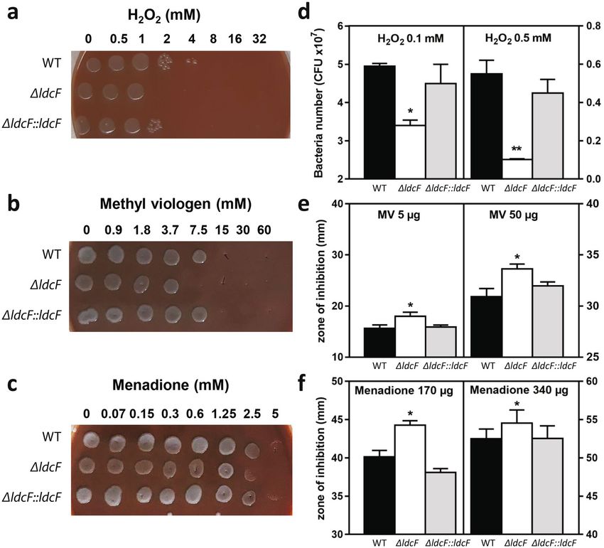
www.nature.com/scientificreports/
The pathogenicity of F. tularensis mainly relies on the Francisella Pathogenicity Island (FPI)4, a gene cluster
encoding 17 proteins comprising a type VI-like secretion system machinery (T6SS)5,6. The regulation of Franci-
sella virulence genes is complex and poorly understood but requires the expression of the macrophage growth
locus protein A (MglA)7,8. This major transcriptional regulator associates with the stringent starvation protein
A (SspA) to form a heterodimer able to interact with RNA polymerase and whose stability is tightly linked to
inorganic polyphosphate9. Another FPI transcriptional regulator essential for intracellular bacterial growth and
virulence is the Francisella effector of virulence regulation (FevR also known as PigR) which physically interacts
with the MglA/SspA complex10. MglA is also involved in the regulation of genes outside FPI and more specifically
in the Francisella oxidative stress response11.
Francisella is a facultative intracellular pathogen whose replication inside macrophages is mostly admitted to
be at the heart of the bacterial pathogenesis. However, this bacterium is also capable of invading many other cell
types such as dendritic cells and neutrophils1,12,13. Murine models of intranasal infection with different Francisella
species demonstrated that alveolar macrophages were predominantly infected at 4–24 h post-infection (hpi)
and that neutrophils serve as a replicative niche accounting for at least 50% of F. tularensis-infected cells from
day 3 post-infection. The intracellular life cycle exposes Francisella to oxidative stress upon bacterial uptake,
temporary residence in the phagosomes, and escape into the host cytoplasm for replication. Indeed, as a defense
mechanism for the clearance of phagocytosed microorganisms, both macrophages and neutrophils produce reac-
tive oxygen species (ROS), such as superoxide anions ( O2·−), hydrogen peroxide ( H2O2) and hydroxyl radicals
(OH·), which in turn trigger bacterial killing by causing damage to macromolecules including DNA, proteins
and membrane lipids14,15. Several factors including for example catalase (KatG), superoxide dismutase (SodB,
SodC) and peroxiredoxin (AhpC)16–20, have been identified as allowing Francisella to cope with such oxidative
stress and thus contributing to intracellular bacteria survival. Another factor that makes bacteria more resistant
to ROS killing is a lower iron c ontent21,22 .
A previous report showed that a F. novicida transposon mutant with insertions in cadA (FTT_0406), which
encodes a putative aspartate aminotransferase fold (AAT-fold) pyridoxal 5′-phosphate (PLP)-dependent lysine
decarboxylase (hereafter referred to as LdcF), was attenuated in mouse s pleen23. Its role as virulence factor was
further hypothesized from the comparative bioinformatic analysis of Francisella strains exhibiting different levels
of pathogenicity24. Considering the current knowledge on the other members of the superfamily of AAT-fold
PLP-dependent basic amino acid decarboxylases and their recognized involvement in bacterial physiology, stress
responses and virulence, we set out to investigate the structure and function of LdcF, using the F. novicida model
as a practicable surrogate of F. tularensis for experimental s tudies3.
Bacterial AAT-fold PLP-dependent basic amino acid decarboxylases are grouped into a superfamily termed
LAOdc because these enzymes decarboxylate lysine (LdcI, LdcC and LdcA), arginine (AdcI) and ornithine
(OdcI and OdcC) into corresponding polyamines (cadaverine, agmantine and putrescine) while consuming
protons and producing C O225–27. The main role of LdcI, AdcI and OdcI (with I standing for acid stress-inducible
LAOdcs) is to buffer the bacterial cytosol in acid stress response, while the primary function of LdcC, LdcA and
OdcC is polyamine biosynthesis. As polycations, polyamines bind negatively charged macromolecules such as
DNA, proteins and p hospholipids28,29, thereby contributing to a remarkable diversity of processes such as DNA
replication, gene expression, protein synthesis, stress and antibiotic resistance, siderophore synthesis, biofilm
formation and v irulence30. In this work, we investigate the genetic environment of Francisella ldcF and the evolu-
tionary relationships of LdcF with the other superfamily members in the light of a recent exhaustive phylogenetic
analysis of proteobacterial L AOdcs26. We determine its crystal structure and identify specific structural elements
which distinguish LdcF from E. coli LdcI, the most thoroughly-studied Ldc. We consider functional implications
of these structural differences, in particular in terms of nutrient stress response, and analyze the influence of
ldcF inactivation on bacterial growth under a variety of stress conditions. These experiments demonstrate the
involvement of LdcF in oxidative stress resistance in dedicated growth media, as well as in infected macrophages.
Finally, a comparative mass spectrometry (MS)-based quantitative analysis of the proteome of the wild-type F.
novicida versus the ΔldcF mutant provides elements for the explanation of LdcF involvement in defense against
oxidative stress, virulence and survival in macrophages. Taken together, this study provides a structural and
functional characterization of Francisella LdcF. It uncovers the important role of this previously uncharacterized
protein in survival in the host cells through participation in oxidative stress response, thereby identifying LdcF
as a potential drug target. Finally, the differential proteomic analysis opens up further avenues for mechanistic
investigations of the LdcF mode of action.
Results
Bioinformatic analysis of LdcF and the ldc genetic environment. A genomic survey of 4,467
prokaryote complete proteomes identified a single LAOdc sequence in Francisellaceae, which we termed LdcF.
LdcF sequences found in Francisellaceae strains display a high level of sequence identity (83%), and contain four
functional regions, corresponding to a wing domain (Pfam ID: PF03709), a PLP-binding domain and a AAT-like
domain (both corresponding to Pfam ID: PF01276), and a C-terminal domain (Pfam ID: PF03711) (Supple-
mentary Fig. S1). This corresponds to the canonical organization of the wing-containing LAOdc superfamily26.
Based on sequence comparison, LdcF proteins appear more similar to Escherichia coli LdcI (52.65% identity) and
LdcC (48.18%) than to Pseudomonas aeruginosa LdcA (36.44%), E. coli AdcI (33.03%), E. coli OdcI (30.34%) and
E. coli OdcC (27.28%). The phylogenetic analysis of 553 wing-containing LAOdcs present in 1,904 representa-
tive proteomes confirmed the specific relationship of Francisellaceae LdcF with the LdcI/C family (Fig. 1a and
Supplementary Fig. S2). More precisely, Francisellaceae LdcF grouped robustly with a sequence from Legionella
fallonii at the base of the clade corresponding to LdcI/C (ultrafast bootstrap = 100%, Fig. 1b). However, the long
Scientific Reports | (2021) 11:972 | https://doi.org/10.1038/s41598-020-79611-5 2
Vol:.(1234567890)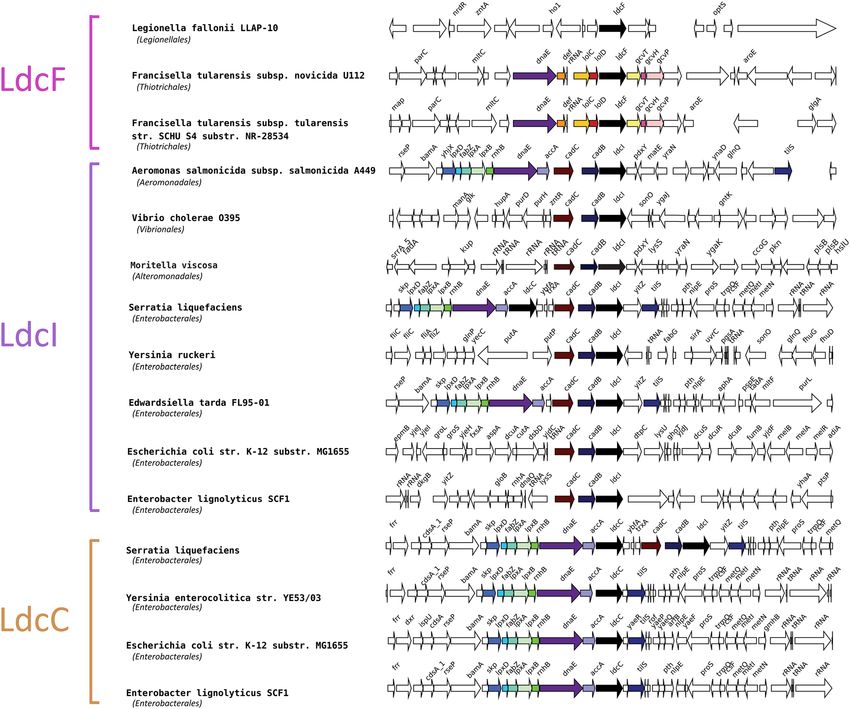
www.nature.com/scientificreports/
a OdcI
E. coli
b Tree scale: 0.1
LdcF
OdcI LdcF
F. tularensis
OdcC
OdcC
LdcI
E. coli
LdcC
E. coli LdcA
P. aeruginosa LdcI
E. coli
LdcC
LdcC
LdcI
E. coli
LdcA
LdcI
LdcF
LdcF
F. tularensis LdcC
Adc
subsp. novicida E. coli Adc E. coli
U112
Figure 1. Phylogenetic position of LdcF sequences within the wing-domain containing LAOdc family. (a) Tree
showing the relationships of 553 WING-containing AAT-fold decarboxylase sequences. The tree is a cladogram,
meaning that the length of the branches has no evolutionary significance. The cladogram is rooted according to
Carriel et al.26. The colour of leaves corresponds to the LdcI, LdcC, AdcI, OdcC, OdcI, and LdcA s ubfamilies26.
The group corresponding to Francisellaceae sequences (referred as to LdcF) is indicated in pink. F. tularensis, E.
coli, and P. aeruginosa sequences are indicated by grey arrows. Grey circles at branches correspond to ultrafast
bootstrap values > 95%. The taxonomy of species (Class) is represented by a coloured strip. (b) Phylogram
corresponding to the LdcI, LdcC, and LdcF subtree (122 sequences). The scale bar corresponds to the average
number of substitutions per site. The length of branches is proportional to genetic divergence.
stem of the LdcI/C cluster reflects the large evolutionary distance between LdcF and LdcI/C sequences and thus
their relative high divergence (Fig. 1b).
Therefore, LdcF sequences could represent a new Ldc family. In line with this hypothesis, the genomic context
of ldcF is very different of those of ldcI and ldcC (Fig. 2). In particular, in many genomes, ldcI and ldcC are present
in vicinity of lpxD, fabZ, lpxA, lpxB, rnhB, dnaE and accA genes involved in lipid synthesis and DNA replication.
Furthermore, most ldcI are clustered with cadB and cadC, encoding the lysine-cadaverine antiporter and the
transcriptional regulator of the cadBA operon respectively. In contrast, Francisellaceae ldcF are surrounded by
lolC and lolD on the one hand, and gcvT, gcvH, and gcvP on the other hand, involved in lipid transport and gly-
cine cleavage system, respectively. Altogether, these data underlie differences between LdcF and other LAOdcs.
Accordingly, LdcF may constitute a new family of LAOdcs phylogenetically related to LdcI/C but presenting a
different genomic context.
Structural characterization of Francisella novicida LdcF. Having shown that the LdcF family is dis-
tinct from LdcI/C and LdcA, and considering that the structures of E. coli LdcI, E. coli LdcC and P. aeruginosa
LdcA solved by either X-ray crystallography or cryo-EM are available, we decided to gain structural insights
into F. novicida LdcF and to compare its structure with those of the related families. LdcF was purified to homo-
geneity, and its lysine decarboxylase activity was assessed at pH 6.5 and 37 °C using a 2,4,6-trinitrobenzensul-
fonic acid colorimetric a ssay31(see “Methods”). The initial activity rate in nanomoles cadaverine produced per
minute and per microgram of enzyme was measured to be ~ 5 nmoles cadaverine min−1 µg−1 LdcF (Supple-
mentary Fig. S3). The observation that this activity rate is 30 times smaller than that of E. coli LdcI at the same
conditions31,32 may indicate that, similarly to LdcI and related enzymes33,34, optimal LdcF activity is pH, salt,
and temperature-dependent. We were able to determine the structure of F. novicida LdcF from X-ray diffrac-
tion data collected to a resolution of 3.4 Å (Supplementary Table S1 and Supplementary Fig. S4). The structure
was solved by molecular replacement (MR), using the crystal structure of the decameric E. coli LdcI (PDB ID:
3N75)32 as a starting model (see “Methods”). All Ldcs are pentamers of dimers arranged around a central pore,
thereby forming a D5-symmetric decamer32,35,36. The LdcF crystal structure contains one LdcF pentamer in the
Scientific Reports | (2021) 11:972 | https://doi.org/10.1038/s41598-020-79611-5 3
Vol.:(0123456789)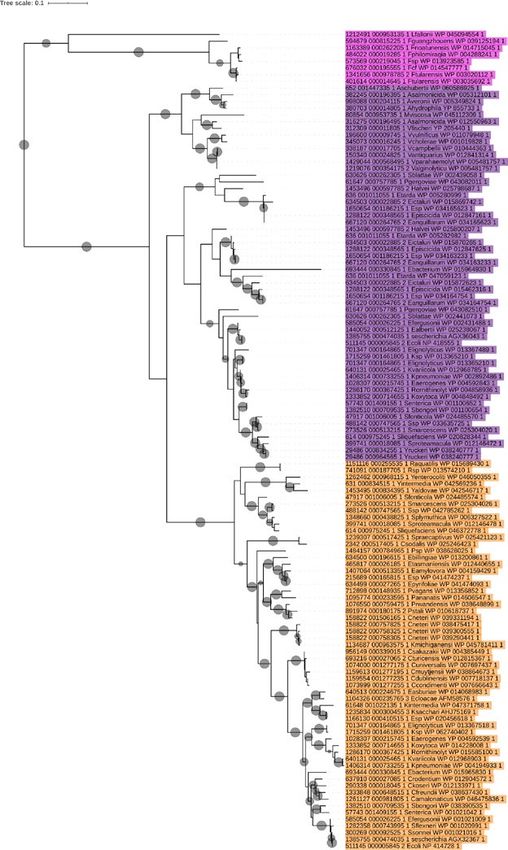
www.nature.com/scientificreports/
Figure 2. Genomic context of LdcI, LdcC, and LdcF coding genes (black arrows) in a subsample of
representative species. Other conserved neighbour genes are highlighted with colour. The taxonomy of species
(Class) is indicated in brackets.
crystallographic asymmetric unit, while an LdcF decamer is generated by a two-fold crystallographic symmetry
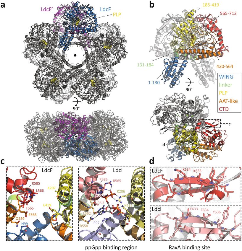
axis perpendicular to the pentamer pore (Fig. 3a).
In agreement with the relationship between LdcF and LdcI families disclosed by the phylogenetic analysis and
the relatively high level of sequence conservation (see above and Fig. 4), structural alignment between F. novicida
LdcF and E. coli LdcI dimers extracted from their respective decameric crystal structures demonstrates a high
overall similarity, with a root-mean-square-deviation (RMSD) of 1.043 Å over 1,223 aligned atoms. Like LdcI
and other LAOdcs, the LdcF monomer is organized in three different structural domains (Fig. 3b): A N-terminal
wing domain involved in stabilization of the ring assembly though inter-dimer contacts (residues 1–130), a cen-
tral core domain which contains a covalently bound PLP cofactor (residues 131–564) and a C-terminal domain
(residues 565–713), which partially constitutes an entry channel into the active site. The core domain (Fig. 3b)
encompasses a linker region (residues 113–184), a PLP-binding domain consisting of a seven-stranded β-sheet
surrounded by eight α-helices (residues 185–419), and an AAT-like domain which harbors an antiparallel four-
stranded β-sheet and three α-helices near the dimerization interface (residues 420–564).
While the overall structures of LdcF and LdcI are very similar, some notable differences were found in both
the AAT-like and the C-terminal domains. In E. coli Ldcs, the AAT-like domain is referred to as the ppGpp-
binding domain due to its interaction with the stringent response alarmone ppGpp, which causes a strong
inhibition of the lysine decarboxylase activity. The ppGpp binding site was actually discovered serendipitously
upon building of the E. coli LdcI atomic model into the X-ray crystallography map, because under conditions
of LdcI overexpression and purification used, the strongly-bound ppGpp was co-purified with LdcI32. Later, the
enzymatic activity of both E. coli LdcI and LdcC was shown to be strongly inhibited by ppGpp37. In the case of
LdcF, no additional density was present in the corresponding site. Moreover, a comparison of the ppGpp bind-
ing pocket in LdcI with the equivalent region in LdcF (Fig. 3c) revealed that, despite the overall high sequence
Scientific Reports | (2021) 11:972 | https://doi.org/10.1038/s41598-020-79611-5 4
Vol:.(1234567890)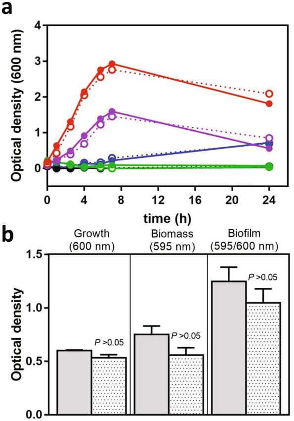
www.nature.com/scientificreports/

                                 Figure 3.  Crystal structure of the F. novicida lysine decarboxylase LdcF. (a) Front (upper panel) and side view
                                 (lower panel) of decameric LdcF, with one highlighted dimer coloured blue and purple, while other dimers
                                 are coloured light and dark grey. The covalently bound pyridoxal phosphate (PLP) cofactor is shown as yellow
                                 spheres. (b) Front (upper panel) and side view (lower panel) of an LdcF dimer extracted from the decamer
                                 shown in (a). In one monomer, different domains are coloured according to a rainbow scheme (WING domain:
                                 blue, linker: green, PLP-binding domain: yellow, AAT-like domain: orange, C-terminal domain: red), with
                                 accompanying annotated amino acid residue ranges. (c) Comparison between the AAT-like domains (termed
                                 ppGpp binding domain in E. coli LdcI) of F. novicida LdcF (left) and E. coli LdcI (right). Residues of E. coli
                                 LdcI involved in ppGpp binding, and the corresponding residues in the AAT-like domain of F. novicida LdcF
                                 are annotated and shown as sticks. Domains are coloured as in (b), but using lighter tints for E. coli LdcI. (d)
                                 Comparison between the RavA-binding site in E. coli LdcI, and the corresponding region in F. novicida LdcF.
                                 Residues of E. coli LdcI involved in RavA binding, and the corresponding residues of F. novicida LdcF are
                                 annotated and shown as sticks.

Scientific Reports |   (2021) 11:972 |               https://doi.org/10.1038/s41598-020-79611-5                                                 5

                                                                                                                                           Vol.:(0123456789)
www.nature.com/scientificreports/
β1 α1 β2 α2
F. novicida LdcF ----MKTVVFVYKD--TLKSYKEKFLLKIEKDLKNHHEYYTLKLDDLSEVVEILEENSRI 54
E. coli LdcI ----MNVI-AILNH--MGVYFKEEPIRELHRALE-RLNFQIVYPNDRDDLLKLIENNARL 52 WING
E. coli LdcC ----MNII-AIMGP--HGVFYKDEPIKELESALV-AQGFQIIWPQNSVDLLKFIEHNPRI 52
P. Aeruginosa LdcA MYKDLKFPVLIVHRDIKADTVAGERVRGIAHELE-QDGFSILSTASSAEGRIVASTHHGL 59 linker
:: : : : : * : : . : . . : :
β3 η1 α3 β4 η2 PLP
F. novicida LdcF CCIVLDRAS--FNI-------EAFHNIAHLNTKLPIFVASDYSQSIK------LNLRDFN
N 99
E. coli LdcI CGVIFDWDK--YNL-------ELCEEISKMNENLPLYAFANTYSTLD------VSLNDLR
R 97 AAT-like
E. coli LdcC CGVIFDWDE--YSL-------DLCSDINQLNEYLPLYAFINTHSTMD------VSVQDMR
MR 97
P. aeruginosa LdcA ACILVAAEGAGENQRLLQDVVELIRVARVRAPQLPIFALGEQVTIENAPAESMADLHQLR
R 119 CTD
. ::. . : **::. : . .:.::.
β5 α4 α5
F. novicida LdcF LNINFLQYDALAGEDSD-FIHKTITNYFNDILPPLTYELFKYSKSFNSAFCTPGHQGGYG 158
E. coli LdcI LQISFFEYALGAAEDIANKIKQTTDEYINTILPPLTKALFKYVREGKYTFCTPGHMGGTA 157
E. coli LdcC MALWFFEYALGQAEDIAIRMRQYTDEYLDNITPPFTKALFTYVKERKYTFCTPGHMGGTA 157
P. aeruginosa LdcA GILYLFEDTV---PFLARQVARAARNYLAGLLPPFFRALVEHTAQSNYSWHTPGHGGGVA 176
: ::: : : :*: : **: *. : . : :: **** ** .
η3 α6 α7 α8 β6
F. novicida LdcF FQRSAVGALFYDFYGENIFKTDLSISMKELGSLLDHSEAHKDAEEYISKVFKSDRSLIVT
KV 218
E. coli LdcI FQKSPVGSLFYDFFGPNTMKSDISISVSELGSLLDHSGPHKEAEQYIARVFNADRSYMVT
ARV
AR 217
E. coli LdcC YQKSPVGCLFYDFFGGNTLKADVSISVTELGSLLDHTGPHLEAEEYIARTFGAEQSYIVT
AR
AR 217
P. aeruginosa LdcA YRKSPVGQAFHQFFGENTLRSDLSVSVPELGSLLDHTGPLAEAEDRAARNFGADHTFFVI
ARN
AR 236
:::* ** *::*:* * :::*:*:*: ********: :**: :: * :::: :*
α9 β7 α10 β8 β9 β10
F. novicida LdcF NGTSTANKIVGMYSVADGDTILVDRNCHKSVTHLMMMVDVNPIYLKPTRNAYGIIGGIPK 278
E. coli LdcI NGTSTANKIVGMYSAPAGSTILIDRNCHKSLTHLMMMSDVTPIYFRPTRNAYGILGGIPQ 277
E. coli LdcC NGTSTSNKIVGMYAAPSGSTLLIDRNCHKSLAHLLMMNDVVPVWLKPTRNALGILGGIPR 277
P. aeruginosa LdcA NGTSTANKIVWHSMVGREDLVLVDRNCHKSILHSIIMTGAIPLYLTPERNELGIIGPIPL 296
*****:**** . . :*:*******: * ::* .. *::: * ** **:* **
η4 α11 β11 β12 α12 β13
F. novicida LdcF KEFKRETIQEKIDNSNIAD---KWPEYAVVTNSTYDGILYNTDTIHRELD--VKKLHFDS 333
E. coli LdcI SEFQHATIAKRVKET-PNA---TWPVHAVITNSTYDGLLYNTDFIKKTLD--VKSIHFDS 331
E. coli LdcC REFTRDSIEEKVAAT-TQA---QWPVHAVITNSTYDGLLYNTDWIKQTLD--VPSIHFDS 331
P. aeruginosa LdcA SEFSKQSIAAKIAASPLARGREPKVKLAVVTNSTYDGLCYNAELIKQTLGDSVEVLHFDE 356
** : :* :: : **:*******: **:: *:: *. * :***.
η5 η6 β14 α13 β15
F. novicida LdcF AWIPYAIFHPIYKHKSAMQIEPR-PEHIIFETQSTHKLLAAFSQSSMLHIKGD----YNE 388
E. coli LdcI AWVPYTNFSPIYEGKCGMSGGRV-EGKVIYETQSTHKLLAAFSQASMIHVKGD----VNE 386
E. coli LdcC AWVPYTHFHPIYQGKSGMSGERV-AGKVIFETQSTHKMLAALSQASLIHIKGE----YDE 386
P. aeruginosa LdcA AWYAYAAFHEFYDGRYGMGTSRSEEGPLVFATHSTHKMLAAFSQASMIHVQDGGTRKLDV 416
** *: * :*. : .* ::: *:****:***:**:*::*::. :
α14 α15 α16
F. novicida LdcF EVLNEAFMLHTSTSPFYPIVASVETAAAMMEGEQGYNLIDKTINLAIDFRRELIKLRS--
MEGE 446
E. coli LdcI ETFNEAYMMHTTTSPHYGIVASTETAAAMMKGNAGKRLINGSIERAIKFRKEIKRLRT--
MKGN 444
E. coli LdcC EAFNEAFMMHTTTSPSYPIVASVETAAAMLRGNPGKRLINRSVERALHFRKEVQRLRE--
LRGN 444
P. aeruginosa LdcA ARFNEAFMMHISTSPQYGIIASLDVASAMMEGPAGRSLIQETFDEALSFRRALANVRQNL
MEG 476
:***:*:* :*** * *:** :.*:**:.* * **: :.: *: **: : .:*
β16 β17 β18 β19
F. novicida LdcF EANGWFFDVWQPDNISNK-----EAWLLRNADKWHGFKNVDGDFLSLDPIKITILTPGIK 501
E. coli LdcI ESDGWFFDVWQPDHIDTT-----ECWPLRSDSTWHGFKNIDNEHMYLDPIKVTLLTPGME 499
E. coli LdcC ESDGWFFDIWQPPQVDEA-----ECWPVAPGEQWHGFNDADADHMFLDPVKVTILTPGMD 499
P. aeruginosa LdcA DRNDWWFGVWQPEQVEGTDQVGTHDWVLEPSADWHGFGDIAEDYVLLDPIKVTLTTPGLS 536
: :.*:*.:*** ::. . * : **** : :.: ***:*:*: ***:.
α17 β20 β21 α18
F. novicida LdcF D-NDVQDWGVPADVVAKFLDEHDIVVEKSGPYSLLFIFSLGTTKAKSVRLISVLNKFKQM
KQM 560
E. coli LdcI KDGTMSDFGIPASIVAKYLDEHGIVVEKTGPYNLLFLFSIGIDKTKALSLLRALTDFKRA
KRA 559
E. coli LdcC EQGNMSEEGIPAALVAKFLDERGIVVEKTGPYNLLFLFSIGIDKTKAMGLLRGLTEFKRS
KR 559
P. aeruginosa LdcA AGGKLSEQGIPAAIVSRFLWERGLVVEKTGLYSFLVLFSMGITKGKWSTLVTELLEFKRC
KR 596
. :.: *:** :*:::* *:.:****:* *.:*.:**:* * * *: * .**:
α19 α20 α21 α22 α23 β22
F. novicida LdcF YDENTLVEKMLPTLYAEDPKFYEDMRIQEVSERLHQYMKEANLPNLMYHAFNVLPEQQLN
DENTLV
V KM MR
MR 620
E. coli LdcI FDLNLRVKNMLPSLYREDPEFYENMRIQELAQNIHKLIVHHNLPDLMYRAFEVLPTMVMT
DLNLRV
VKNM MR
MR 619
E. coli LdcC YDLNLRIKNMLPDLYAEDPDFYRNMRIQDLAQGIHKLIRKHDLPGLMLRAFDTLPEMIMT
DLNLR KNM MR
MR 619
P. aeruginosa LdcA YDANLPLLDVLPSVAQAGGKRYNGVGLRDLSDAMHASYRDNATAKAMKRMYTVLPEVAMR
DA
ANLPL DV VG
VG 656
:* * : .:** : . . *..: :::::: :* . * : : .** :
α24 β23 η7 β24 β25 β26 α25
F. novicida LdcF PHRAFQKLLKGKVKKVPLAELYEHTSAVMILPYPPGIPVIFPGEKITEESKVILDFLLML
KVKKVPL 680
E. coli LdcI PYAAFQKELHGMTEEVYLDEMVGRINANMILPYPPGVPLVMPGEMITEESRPVLEFLQML
MTEEVYL 679
E. coli LdcC PHQAWQRQIKGEVETIALEQLVGRVSANMILPYPPGVPLLMPGEMLTKESRTVLDFLLML
VETIAL 679
P. aeruginosa LdcA YDANLPLLDVLPSVAQAGGKRYNGVGLRDLSDAMHASYRDNATAKAMKRMYTVLPEVAMR
PSVAQAG 656
* *::: ::* .: : : .: : * *::*****:*:::*** :*: :: :*::* :
β27 β28 β29
F. novicida LdcF EKIGSMLPGFDTDIHGPERAKDG---KLYIKVIDDK---
KLYIKVI 713 = ppGpp interac on
E. coli LdcI CEIGAHYPGFETDIHGAYRQADG---RYTVKVLKEESKK
RYTVKVL 715
E. coli LdcC CSVGQHYPGFETDIHGAKQDEDG---VYRVRVLKMAG--
VYRVRVL 713 = RavA interac on
P. aeruginosa LdcA RTFERAFPGFDSDVHGLQHQDGPSGRCYTVECIKE----
CYTVECI 751
. ***::*:** : . :. :.
Figure 4. Alignment of F. novicida LdcF, E. coli LdcI and LdcC, and P. aeruginosa LdcA using Clustal Omega.
Partially and fully conserved residues are annotated with ‘:’ and ‘*’ respectively. Domains are coloured according
to a rainbow scheme (WING domain: blue, linker: green, PLP-binding domain: yellow, AAT-like domain:
orange, C-terminal domain: red), and secondary structure elements are annotated. ppGpp and RavA-interaction
sites are highlighted using red and blue transparent boxes respectively.
Scientific Reports | (2021) 11:972 | https://doi.org/10.1038/s41598-020-79611-5 6
Vol:.(1234567890)www.nature.com/scientificreports/
conservation, only two out of 10 ppGpp-interacting residues are conserved between the two proteins. More
importantly, 7 of the amino acid substitutions in LdcF result either in a change in charge or polarity, or in a
change from hydrophobic to polar or vice versa, revealing that, contrary to LdcI and LdcC but similarly to P.
aeruginosa LdcA35, LdcF is most likely not inhibited by ppGpp.
The C-terminal domain of the E. coli LdcI but not LdcC is known to interact with the MoxR AAA + ATPase
RavA. The molecular determinant of the LdcI-RavA interaction resides in the C-terminal two-standed β-sheet
of LdcI36,38 which was shown to be specifically evolved for RavA binding, contrary to OdcIC, AdcI, LdcA and
even the closer related L dcC35,36. This LdcI-specific interaction leads to the formation of a huge cage-like LdcI-
RavA complex 38–40
proposed to enable enterobacteria, such as E. coli, Salmonella and Vibrio, to withstand acid
stress even under conditions of nutrient deprivation eliciting stringent r esponse41. Indeed, interaction with RavA
was shown to maintain LdcI enzymatic activity upon starvation by preventing ppGpp binding to LdcI particles
engaged in the LdcI-RavA c omplex41. Based on a medium-resolution cryo-EM structure of LdcI cross-linked
with the LdcI-binding domain of R avA36, residues Glutamate 634 (E634), Tyrosine 636 (Y636) and Tyrosine 697
(Y697) are likely to be key players in the LdcI-RavA interaction. These residues are substituted in LdcF by Lysine
(K635), Proline (P637) and Glutamate (E698) residues respectively, resulting in an impairment of a putative
RavA interaction (Fig. 3d). This result is consistent with the absence of an orthologue of RavA in the Francicellae
genome and further highlights the specific evolutionary tailoring of LdcI for RavA binding.
In vitro phenotypic analysis of the ΔldcF mutant. The physiological significance of LdcF was inves-
tigated through the construction of a F. novicida FTN_0504 deletion mutant (ΔldcF). Before proceeding with a
comprehensive phenotypic analysis, we checked whether ldcF deletion affected bacterial fitness. When grown on
PolyViteX-enriched chocolate agar (PVX-CHA) plates, F. novicida wild-type (WT) and ΔldcF displayed similar
colony morphology (Supplementary Fig. S5a). Accordingly, bacterial division and metabolism of both strains
were found unchanged whether the protein was expressed or not (Supplementary Figs. S5b–d, S6). We then
investigated a putative role of LdcF in bacterial tolerance to acidic pH exposure for 1 h but observed no differ-
ence between the WT and the deletion mutant (Supplementary Fig. S7). Bacterial growth was then examined
in liquid Modified Mueller–Hinton (MMH) medium previously adjusted at different pH values ranging from
2.5 to 10 (Fig. 5a). Under all conditions tested, the replication curves for the WT and the deletion mutant were
strictly similar. No growth was observed for the extreme acidic or alkaline pH values tested, while in the range
of pH values from 4 to 8, bacteria grew and reproduced best at pH 6.6. At the 24 h time point the survival of
bacteria was further evaluated by plating serial dilutions of each bacterial suspensions on PVX-CHA plates. For
each pH tested, comparable numbers of colony forming units (cfu) were found for both strains, thus confirming
that bacterial viability was not altered upon ldcF deletion (Supplementary Table S2). The replication rate of both
strains was also found identical at 25 °C or at 37 °C, which correspond to the temperatures in tick and mammal
hosts, respectively (Supplementary Fig. S8).
Besides growth fitness and acid stress response, another physiological process in which polyamine products
of LAOdcs are likely to be involved is biofilm f ormation27,30,42–44. Yet, as assessed by crystal violet staining, no
significant difference between the amount of biofilm produced by F. novicida WT and ΔldcF strains could be
documented (Fig. 5b). We also investigated whether LdcF activity is promoting antibiotic resistance by determin-
ing the minimum inhibitory concentrations (MICs) that were found unchanged for either ciprofloxacin (0.064 µg/
mL ; n = 3) or gentamicin (1 µg/mL ; n = 3). In addition, we examined the rate at which these antibiotics kill bac-
teria – the minimum duration for killing (MDK) metric—as a quantitative indicator of antibiotic tolerance45,46.
Again, the MDK99 values corresponding to the time required to kill 99% of the bacterial populations includ-
ing WT, ΔldcF and ΔldcF-complemented (ΔldcF::ldcF) strains exposed either to ciprofloxacin (Supplementary
Fig. S9a) or to gentamicin (Supplementary Fig. S9b) were very similar, thus discarding the involvement of LdcF
in antibiotic tolerance. This result was confirmed by the Minimal Bactericidal Concentration (MBC)/MIC ratios
found to be below 32, i.e. the value defined by the Clinical Laboratory Standards Institute (CLSI) guidelines as
the tolerance threshold47.
The same set of strains was then tested for susceptibility to oxidative stress. Interestingly, results obtained from
spot plating assays indicated that LdcF significantly contributed to survival of bacteria exposed to hydrogen per-
oxide or to the redox-cycling drugs methyl viologen (MV) and menadione (MD). The ∆ldcF mutant was indeed
found systematically less resistant to ROS exposure than the other strains, whereas complementation restored
the WT phenotype (left panels in Fig. 6). Under such experimental conditions, and while the incubation of ΔldcF
with MD was accompanied with a moderate but reproducible inhibition of growth (Fig. 6c), MV (Fig. 6b) was
found even more efficient than H 2O2 (Fig. 6a). The enhanced sensitivity of the LdcF-deleted strain to H2O2 was
accurately confirmed by a lower number of cfu when ΔldcF was exposed to this reagent as compared to the value
obtained with the WT (Fig. 6d). Because cfu counting is a time-consuming approach not fully appropriate to
evaluate the effect of compounds on short incubation periods, the extent of ∆ldcF susceptibility to MV and MD
was further validated through the disk diffusion assays. Thus, the diameter of inhibition zone, which is related
to the susceptibility of the isolate, was significantly higher for ∆ldcF than for the WT strain when the disks were
impregnated either with both compounds (Fig. 6e,f).
In vivo phenotypic analysis of the ΔldcF mutant. We next evaluated the consequences of ldcF deletion
on bacterial replication in macrophages. The uptake of F. novicida WT, ΔldcF and ΔldcF::ldcF strains into J774
cells, estimated upon macrophage infection with a MOI of 100, was found identical for the three strains which
displayed equivalent intracellular growth profiles over the first 24 h (Fig. 7a). However, at 48 hpi and beyond, the
number of viable intracellular ∆ldcF cells was found significantly lower than observed for the WT strain (∆ldcF:
2.63 × 10⁸ ± 0.47 × 10⁸, n = 8 vs WT: 4.08 × 10⁹ ± 0.64 × 10⁹, n = 8 ; P < 0.0005), and the effect was reversed with
Scientific Reports | (2021) 11:972 | https://doi.org/10.1038/s41598-020-79611-5 7
Vol.:(0123456789)www.nature.com/scientificreports/
Figure 5. Growth and biofilm formation of F. novicida. (a) F. novicida WT (solid lines) and ΔldcF (dotted
lines) were grown under shaking at 37 °C in MMH adjusted at pH 2.5 (green), pH 4 (blue), pH 6.6 (red), pH
8 (purple) or pH 10 (black) and the bacterial growth was monitored by O D600nm measurement. Results are
representative of three independent trials. (b) F. novicida WT (grey columns) and ΔldcF (dotted columns)
were grown for 24 h under static conditions at 37 °C in a 96-wells plates. The bacterial growth was evaluated
by measurement of OD600nm and the biofilm biomass was further determined by OD595nm after Crystal violet
staining. This graph corresponds to mean ± s.e.m. of three independent experiments, with at least 4 technical
replicates each.
the ΔldcF::ldcF (1.27 × 10¹⁰ ± 0.35 × 10¹⁰, n = 4). As assessed by measuring lactate dehydrogenase activity, this
reduced level of recovered viable bacteria was not related with an increased host cell lysis that would result in the
release of bacteria into the extracellular medium (WT: 30.6% ± 4.92% vs ∆ldcF: 25.95% ± 3.29% and ΔldcF::ldcF:
30.79% ± 7.9%; P > 0.05; n = 3, measured at 72 hpi). Together, these data suggest that the deletion mutant displays
a reduced capacity to escape macrophage killing mechanisms. Their failure to survive the antibacterial activities
of host macrophages most probably relies on an intricate overlapping network of signals combining pro-inflam-
matory and immune responses as well as metabolic response of the infected cell. However, considering the role
of macrophage oxidative burst in pathogen clearance, we then evaluated the ROS level in infected J774 cells. Our
results demonstrate that macrophages infected with the ∆ldcF strain, which is less resistant to oxidative stress,
display a higher ROS activity than cells infected either with the WT or the complemented strain (Fig. 7b), an
effect that could result either from an impaired degradation or from an increased production of ROS.
Comparative proteomics reveals proteins impacted by ldcF deletion. A MS-based quantitative
proteomic comparison was performed on whole-cell extracts of F. novicida WT and ΔldcF strains to identify
proteins for which abundance was altered by ldcF deletion. A bioinformatics analysis reliably identified and
quantified 1,263 different proteins from the 1,854 protein-coding ORFs annotated in the F. novicida genome
(Supplementary Table S3; PXD016591). An ensuing statistical analysis revealed that expression levels of 80 pro-
teins were significantly affected by ldcF deletion. Equal numbers of proteins were expressed at lower or higher
abundance in ΔldcF compared to the WT (Table 1). Consistent with the lack of LdcF in the deletion mutant,
the 2,3,4,5-tetrahydropyridine-2,6-carboxylate N-succinyltransferase (FTN_1727, DapD) involved in lysine bio-
synthesis was found to be downregulated. Surprisingly, although all proteins encoded by the FPI (FTN_1309 to
FTN_1326) 5 were detected in the MS-based quantitative proteomic assay (Supplementary Table S3), none of
them showed altered expression levels in the ΔldcF strain. In contrast, the amount of the major transcriptional
Scientific Reports | (2021) 11:972 | https://doi.org/10.1038/s41598-020-79611-5 8
Vol:.(1234567890)www.nature.com/scientificreports/
Figure 6. Sensitivity of F. novicida to oxidative stress. Exponential growth phase bacteria diluted in MMH were
exposed to increasing concentration of (a) H2O2 (b) methyl viologen or (c) menadione for 1 h at 37 °C and 3 µl
of the cell suspensions were spotted on PVX-CHA plates. These pictures are representative of at least 3 distinct
experiments performed in duplicate each. The antibacterial activity of oxidative compounds was also quantified
by (d) cfu counting from a cell suspension containing 1 08 bacteria incubated for 1 h under shaking in presence
of H2O2 or by disk diffusion assays with (e) methyl viologen or menadione (f) as detailled in materials and
methods section. Histograms correspond to the mean ± s.e.m. of at least 3 distinct experiments performed in
duplicate (*P < 0.05, **P < 0.01).
regulator MglA (FTN_1290, MglA), which is described as a FPI gene regulator8,48, was significantly reduced in
the ΔldcF mutant (Table 1).
The KEGG (Kyoto Encyclopedia of Genes and Genomes) d atabase49 includes 99 pathways for F. novicida
(https://www.genome.jp/kegg-bin/show_organism?org=ftn). Interestingly, following KEGG annotation, only
23 of the 80 proteins affected by the ldcF deletion were assigned to functional categories. These 23 proteins can
be roughly grouped into a limited number of distinct functional pathways, including bacterial metabolism,
DNA proofreading and repair, and pathways related to oxidative stress. One group of the KEGG-annotated
differentially expressed proteins (5 out of 23) is associated with DNA proofreading and repair. This group is
composed of two proteins involved in base excision repair pathways (FTN_1486, Ung and FTN_0838, XthA),
one involved in nucleotide excision repair (FTN_1176, UvrB), and two involved in homologous recombination
pathways (FTN_1025, RuvA and FTN_1357, RecB). Two of these proteins (UvrB and Ung), which displayed
reduced expression levels in ΔldcF, are enzymes considered to be prokaryotic defense systems involved in viru-
lence through their protection of bacterial DNA50,51. Other proteins that were downregulated in ΔldcF may
also help bacteria to deal with DNA damage, although they lack functional KEGG assignment. These proteins
included enzymes from type I restriction-modification systems (FTN_1152, HsdM; FTN_0710, HsdR)52 and
Scientific Reports | (2021) 11:972 | https://doi.org/10.1038/s41598-020-79611-5 9
Vol.:(0123456789)www.nature.com/scientificreports/
Figure 7. Replication of F. novicida strains within the J774 macrophage-like cell line. (a) F. novicida WT (black
circles, solid line), ΔldcF (white circles, dotted line) and ΔldcF::ldcF (black triangles, solid line) were inoculated
at a MOI of 100:1 and intracellular bacteria were enumerated by cfu counting at different times post infection
(b) Production of ROS evaluated at 24 h after infection of macrophages with a MOI of 1,000:1 using the redox-
sensitive dye DCFA detected by fluorescence spectroscopy. Data correspond to mean ± s.e.m. of 4 distinct
experiments and after subtraction of background values obtained with uninfected macrophages. *P < 0.05.
the N6-adenine-specific methylase (FTN_0655). In contrast, some other DNA repair proteins were expressed
at higher levels following ldcF deletion. An example is exodeoxyribonuclease III (XthA), a negative regulator of
homologous recombination under log phase growth conditions, of which the overexpression can also result in
unrepaired DNA damage.
The KEGG pathway annotation of proteins for which expression levels were significantly altered in ΔldcF also
revealed several metabolic and transport pathways that could play a role in bacterial replication. Specifically,
the deletion mutant’s reduced capacity to deal with the host immune system and survive within macrophages
may be partly related to the observed decrease in uridine phosphorylase levels (FTN_0652, Udp), as previously
suggested using Drosophila melanogaster as an experimental model53. Similarly, UbiC (FNT_0386) catalyzes the
first step of ubiquinone (or coenzyme Q) biosynthesis involved in electron transport chains and is considered
as a lipid-soluble antioxidant in prokaryotes; its expression was reduced, and could thus impact F. novicida’s
oxidative defense54. These findings are in good agreement with a possible role of LdcF in the activation of the
SOS-response, and are underscored by the increased expression levels measured for RuvA (FTN_1025, Holliday
junction ATP-dependent DNA helicase) and RecB (FTN_1357, ATP-dependent exoDNAse) that could help F.
novicida to cope with oxidative stress53.
Discussion
The phylogenetic analysis and amino acid sequence comparisons presented here indicate that the unique lysine
decarboxylase identified within Francisella proteomes, i.e. LdcF (previously annotated as CadA) is more closely
related to E. coli LdcI and LdcC than to P. aeruginosa LdcA or E. coli AdcI, OdcI and OdcC. However, similarly to
most of the P. aeruginosa strains26 and unlike E. coli, Francisella genomes lack the presence of a RavA orthologue
shown to alleviate inhibition of E. coli LdcI by the alarmone p pGpp32. Consistently, as in L
dcA35, the C-terminal
β-strands of LdcF display different amino acids at locations corresponding to the RavA binding site in E. coli
LdcI. Furthermore, our structural analysis of F. novicida LdcF demonstrates that eight out of the 10 residues
involved in LdcI interaction with ppGpp in E. coli show either a reverse in charge or change in hydrophobicity,
which reveals that, again similarly to L dcA35, it is highly unlikely that LdcF would be inhibited by ppGpp. These
observations underlying major differences between LdcF and LdcI are consistent with the absence of a RavA
orthologue in Francisella genomes.
Scientific Reports | (2021) 11:972 | https://doi.org/10.1038/s41598-020-79611-5 10
Vol:.(1234567890)www.nature.com/scientificreports/
Proteomic data
Gene name Locus Description Log 2FC P value
Downregulated proteins in ΔldcF
cadA FTN_0504 Lysine decarboxylase − 6.135487177 6.5901E−12
udp FTN_0652 Uridine phosphorylase − 2.442473795 0.000355044
uvrB FTN_1176 Excinuclease ABC subunit B − 2.359970258 1.5872E−07
Proton-dependent oligopeptide transporter (POT) family protein, di- or
yhiP FTN_0885 − 2.233739832 1.28816E−08
tripeptide:H + symporter
– FTN_1453 Two-component regulator, sensor histidine kinase − 2.225623368 0.000269144
– FTN_0705 Abortive infection bacteriophage resistance protein − 1.909345488 0.006797082
– FTN_0655 N6-adenine-specific methylase − 1.645819843 0.009836595
– FTN_1348 Acetyltransferase − 1.493386099 3.68028E−07
– FTN_0898 Amino acid permease − 1.424644622 3.5435E−06
panD FTN_1354 Aspartate 1-decarboxylase − 1.405501066 0.006567341
– FTN_0862 Hypothetical protein − 1.276557703 0.000969217
ung FTN_1486 Uracil-DNA glycosylase − 1.261354286 4.40904E−07
– FTN_0308 Membrane protein of unknown function − 1.255628185 0.000806353
Proton-dependent oligopeptide transporter (POT) family protein, di- or
– FTN_1272 − 1.243209089 1.20616E−05
tripeptide:H + symporter
dapD FTN_1727 2,3,4,5-tetrahydropyridine-2,6-carboxylate N-succinyltransferase − 1.185622324 0.000977552
– FTN_1258 Hypothetical protein − 1.11437438 0.000177817
hsdM FTN_1152 Type I restriction-modification system, subunit M (methyltransferase) − 1.098834276 0.000436818
– FTN_1316 Hypothetical protein − 1.075883638 0.005337719
– FTN_1628 LysR family transcriptional regulator − 1.010217775 0.001268379
hdsR FTN_0710 Type I restriction-modification system, subunit R (restriction) − 0.974133834 0.00046316
– FTN_1212 Glycosyl transferases group 1 family protein − 0.967248679 7.07496E−06
– FTN_1397 Hypothetical protein − 0.962399859 6.53792E−05
– FTN_0976 ThiF family protein − 0.962086728 6.76869E−05
waaG FTN_1218 Glycosyl transferase, group 1 − 0.942030987 0.000643833
pilE4 FTN_0389 Type IV pili, pilus assembly protein − 0.94007106 2.01122E−05
– FTN_1440 Hypothetical protein − 0.935671185 0.007635418
ubiC FTN_0386 Chorismate pyruvate lyase − 0.934045712 6.57717E−05
– FTN_0137 Hypothetical protein − 0.928404833 3.73274E−06
mglA FTN_1290 Macrophage growth locus, protein A − 0.926959218 6.24086E−07
– FTN_1697 Galactose mutarotase − 0.901042767 0.008192095
– FTN_1148 Glycoprotease family protein − 0.816650922 0.005012835
galP1 FTN_0687 Major facilitator superfamily galactose-proton symporter − 0.746905316 5.94864E−05
– FTN_1459 Short chain dehydrogenase − 0.723695755 0.001285646
– FTN_1254 Hypothetical protein − 0.716291799 0.00237079
– FTN_1266 ABC transporter membrane protein − 0.66256701 0.001562237
– FTN_0923 Hypothetical protein − 0.654493997 0.002785307
yhbG FTN_0902 ABC transporter, ATP-binding protein − 0.652797305 3.32447E−05
rpsF FTN_0951 30S ribosomal protein S6 − 0.641616628 5.78051E−05
yrbI FTN_0905 3-Deoxy-d-manno-octulosonate 8-phosphate phosphatase − 0.627622272 0.001032099
– FTN_1547 Hypothetical protein − 0.608477024 0.000859667
Upregulated proteins in ΔldcF
rimM FTN_1561 Ribosome maturation factor rimM 0.626513088 0.000555064
apaH FTN_0561 Diadenosine tetraphosphatase 0.629366731 0.000669224
– FTN_0118 S49 family serine peptidase 0.635972422 0.006578849
– FTN_1468 Putative deoxyribonucleotide triphosphate pyrophosphatase 0.640919668 0.000191944
– FTN_0089 Allophanate hydrolase subunit 2 0.672742647 0.000997506
rnc FTN_1463 Ribonuclease 3 0.681039745 0.001289497
– FTN_0789 Putative rhodanese, sulfurtransferase 0.68404703 0.000345296
secF FTN_1094 Preprotein translocase subunit SecF 0.695399198 3.15477E−05
murD FTN_0542 UDP-N-acetylmuramoylalanine–d-glutamate ligase 0.715198106 5.10756E−05
xthA FTN_0838 Exodeoxyribonuclease III 0.726407142 1.90678E−05
sun FTN_1347 tRNA and rRNA cytosine-C5-methylases, sun protein 0.73058117 0.00042405
– FTN_0872 Small conductance mechanosensitive ion channel (MscS) family protein 0.78091602 2.90828E−05
Continued
Scientific Reports | (2021) 11:972 | https://doi.org/10.1038/s41598-020-79611-5 11
Vol.:(0123456789)www.nature.com/scientificreports/
Proteomic data
Gene name Locus Description Log 2FC P value
– FTN_1387 Hypothetical protein 0.812379067 0.00883337
ispD FTN_0623 2-C-methyl-d-erythritol 4-phosphate cytidylyltransferase 0.824072741 5.66929E−06
– FTN_0041 Hypothetical protein 0.828287322 0.002571557
mltA FTN_1286 Membrane-bound lytic murein transglycosylase 0.843953091 0.000666109
– FTN_1080 Phosphosugar binding protein 0.857709173 0.000844453
– FTN_1015 Isochorismatase family protein 0.860701266 0.000470586
pilW FTN_0307 Type IV pilus assembly protein 0.903906623 0.004575204
– FTN_1061 Acid phosphatase, HAD superfamily protein 0.951666713 0.001807004
pilV FTN_0413 Type IV pili, pilus assembly protein 0.975215693 0.001866241
murQ FTN_1504 N-acetylmuramic acid 6-phosphate etherase 0.994710363 1.2722E−06
tdh FTN_0625 l-Threonine 3-dehydrogenase 1.02107175 2.99619E−05
ruvA FTN_1025 Holliday junction ATP-dependent DNA helicase RuvA 1.125358669 5.65196E−07
– FTN_1506 Hypothetical protein 1.20406212 0.000656496
putP FTN_0299 Proline/Na + symporter 1.206589507 0.001401361
– FTN_0452 Hypothetical protein 1.274923951 4.08167E−08
– FTN_0006 Hypothetical protein 1.345660313 1.02878E−06
– FTN_0004 Aspartate/glutamate transporter 1.352713527 0.003023321
– FTN_0829 Hypothetical protein 1.454738617 6.98601E−07
– FTN_1388 Oxidoreductase 1.515433578 0.000565317
– FTN_1267 ABC transporter ATP-binding protein 1.74578111 3.84443E−09
lptC FTN_0904 Lipopolysaccharide export ABC transporter periplasmic protein 1.999604273 1.63282E−05
rnpA FTN_0075 Ribonuclease P protein component 2.366923256 0.000708316
– FTN_0384 Hypothetical protein 2.702678582 0.002040018
– FTN_0987 tRNA-dihydrouridine synthase 2.830069785 5.08771E−06
– FTN_1386 Hypothetical protein 2.845246288 4.58158E−09
– FTN_0722 l-lysine 2,3-aminomutase 3.467288507 1.01829E−10
– FTN_1220 Lipopolysaccharide synthesis sugar transferase 3.613912551 0.000821341
recB FTN_1357 ATP-dependent exoDNAse (exonuclease V) beta subunit 3.825585172 1.16584E−11
Table 1. List of proteins differentially expressed in ΔldcF. The protein deleted in the mutant was labeled in
bold.
Examination of the ldcF genetic environment, which is highly conserved within different Francisella species,
suggests that despite the high sequence identity and strong structural similarity with LdcI, LdcF expression is
differently regulated. Notably, genes encoding both CadB, the putative cadaverine transport protein, and CadC,
the pH sensor and membrane-bound transcriptional regulator of the cadBA operon55 are missing in the ldcF
gene cluster. Upstream of ldcF are lolC and lolD which encode two components of the ABC transporter complex
involved in lipoprotein transport and membrane biogenesis and are described as essential genes in F. tularensis56.
The downstream genes belong to the glycine cleavage system (GCS) and were found significantly upregulated
in the virulent F. tularensis type A Schu S4 strain inside macrophages57. Our observations are therefore in
agreement with the data on the in vivo negative selection of F. novicida transposon mutants that pointed out
the importance of GCS genes together with lolD and ldcF (FTT_0405 to FTT_0409) in the intracellular growth
and/or virulence of F. novicida23.
The combined analysis of the genomic context of Francisella ldcF, the structure of F. novicida LdcF and the
phylogenetic relationships between the new LdcF family and other proteobacterial LAOdcs raised questions
about the LdcF regulation and functional activity. Our experiments showed that the growth rates of F. novicida
WT and ΔldcF are very similar over a broad range of basic to acidic pHs, thus ruling out a strict role of LdcF
in acid tolerance and buffering of the bacterial cytosol upon acid stress. Deletion of ldcF also failed to affect
temperature-dependent bacterial growth. In contrast, in comparison with the WT, ΔldcF displayed a signifi-
cantly lower resistance to oxidative stress. The capacity of cadaverine to scavenge oxygen radicals, thus provid-
ing bacteria with a higher tolerance towards oxidative stress, was previously reported for E. coli58 and Vibrio
vulnificus59,60. Importantly, these results are corroborated by a greater survival of both the WT and ΔldcF::ldcF
strains in infected macrophages which contain a lower amount of ROS than those infected with ΔldcF. Such a
survival strategy could be shared with the bacterium that possesses the closest LdcF relative, i.e. L. fallonii, which
replicates within the protozoan host Acanthamoeba in aquatic environments and must face oxidative and acidic
stress conditions during its stationary phase of g rowth18,61.
To better understand the mechanism by which removal of ldcF and a subsequent defect in cadaverine syn-
thesis altered the oxidative stress resistance, we performed an extensive quantitative comparison of the pro-
tein contents between the F. novicida WT and the ΔldcF strains. This analysis identified 80 proteins for which
expression levels were altered following ldcF deletion. Among them, we were surprised not to observe any ROS
Scientific Reports | (2021) 11:972 | https://doi.org/10.1038/s41598-020-79611-5 12
Vol:.(1234567890)www.nature.com/scientificreports/
scavenging enzymes. Indeed, similarly to several other bacterial species, to cope with oxidative stress, Francisella
utilize enzymes such as SodB, SodC, KatG and the recently identified AhpC16,17,20,62,63 to convert harmful ROS
into innocuous p roducts19. Moreover, expression levels of other factors contributing to ROS defense mecha-
nisms, such as the efflux pump EmrA1 (FTL_0687), involved in SodB and KatG s ecretion64, or the F. tularensis
(FTL_1014) oxidative stress regulator OxyR16,18, also displayed similar expression levels in WT and ΔldcF strains.
However, one of the low-abundance proteins in ΔldcF was an ABC transporter (FTN_0902; FTL_1065, YhbG).
Interestingly, this transporter was recently reported to be down-regulated in a ΔoxyR mutant of F. tularensis LVS
displaying an enhanced sensitivity to oxidative s tress18. Furthermore, the reduced UbiC content compared to
the WT strain could also contribute to the diminished capacity of the F. novicida ΔldcF strain to survive oxida-
tive attack from ROS. Indeed, altered UbiC levels could indirectly promote ROS a ccumulation54. In addition
to ROS-neutralizing enzymes, bacteria can also counteract ROS damage using their DNA damage-responsive
genes. The products of these genes initiate DNA repair pathways to recognize and correct ROS-induced and
other mismatches. An interesting hallmark of the F. novicida ΔldcF proteome is the significant changes in levels
of proteins involved in DNA repair processes potentially reducing the bacteria’s capacity to deal with oxidative
stress. Our results also indicate that MglA expression was significantly reduced in the mutant strain. Interest-
ingly, in addition to ensuring the regulation of Francisella virulence factors – which were unchanged in ΔldcF
compared to the WT strain – MglA has been reported to play a key role in the intracellular growth of F. tularensis
and its adaptation to oxidative s tress8,11.
While the relationships between the proteomic observations and Francisella ROS defense mechanisms are
not straightforward and a thorough understanding of the link between ldcF inactivation and the changes in the
protein expression pattern requires further investigations, our observations provide an important evidence of
the LdcF involvement in F. novicida oxidative stress resistance. By suppressing lysine decarboxylation, the ldcF
deletion promotes the accumulation of lysine and the decrease of cadaverine, which both should have a direct
impact on the bacterial physiology. Lysine harvesting was indeed described as a powerful preventive metabolic
antioxidant strategy displayed by microbial cells65, an effect most probably reverted when this amino acid accu-
mulates. The contribution of polyamines in several bacterial infections has long been described66, and some stud-
ies have specifically emphasized their relevance in F. tularensis virulence. For example, an increased expression
of ornithine decarboxylase was observed in F. tularensis infected mice67. The relevance of spermine within host
cells infected by Francisella, and specifically the capacity of this polyamine to elicit transcriptional changes in
F. tularensis, leading in turn to altered host cell activation, has also been reported68. While never investigated, it
could be hypothesized that cadaverine could also exert transcriptional control on genes implicated in Francisella
resistance against oxidative attack. Taken together, our work provides a biochemical and structural framework to
further explore LdcF as a potential virulence factor and its involvement in Francisella oxidative stress resistance.
While LAOdcs are long-recognised as drug targets, and development of specific mechanism-based Ldc inhibi-
eld69–72, we envision that the LdcF structure and the functional findings presented
tors is an active research fi
in this work will empower further investigations aimed at design of new LdcF-based therapeutic approaches
against tularemia.
Methods
Bioinformatic analyses. Sequences of AAT-fold decarboxylases were retrieved from NCBI: LdcI
(NP_418555.1), LdcC (NP_414728.1), AdcI (NP_418541.1), OdcC (NP_417440.1), and OdcI (NP_415220.1)
from Escherichia coli str. K-12 substr. MG1655 and LdcA (NP_250509.1) from P. aeruginosa PAO1. These
sequences were used as seeds to query a local database containing 4,467 complete proteomes of prokaryotes
(Supplementary Table S4) from the National Center for Biotechnology Information (ftp://ftp.ncbi.nlm.nih.gov)
with the BLASTP 2.2.6 s oftware73 and with HMM-profile based approaches with the HMMER package v3.1b1
(default parameters)74. Finally, searches for unannotated sequences were performed with TBLASTN (default
parameters) on the complete genome sequences corresponding to the 4,467 proteomes using default parameters.
Sequences with an e-value lower than 1 0–4 were retrieved and aligned using MAFFT v.775. The resulting multiple
alignment was visually inspected with AliView 1.2576. Doubtful sequences were systematically verified using
reciprocal best reciprocal blast hit. This led to the identification of 4,091 AAT-fold decarboxylase sequences, 13
of which were unannotated or annotated as pseudogenes (Supplementary Table S5).
A phylogeny of WING-containing AAT-fold decarboxylase sequences was inferred using maximum likeli-
hood. To limit taxonomic redundancy, the phylogenetic analysis was performed on a subset of 1,905 representa-
tive proteomes by selecting randomly one representative strain per species. The 553 WING-containing AAT-fold
decarboxylase sequences contained in these representative proteomes were aligned with MAFFT using the
L-INS-i option and trimmed with BMGE v1.1 with matrix substitution BLOSUM30 (589 amino acid positions
kept after trimming)77. The maximum likelihood tree was inferred with IQ-TREE 1.6.1278. IQ-TREE identified
the LG + R10 as the best suited evolutionary model according to the Bayesian i nformation78. The robustness of
the inferred tree was assessed using the ultrafast bootstrap (1,000 replicates implemented in IQ-TREE). The
genomic context figure has been generated by GeneSpy 1.179 and phylogeny figures by i TOL80.
The percentage of identity between LdcF sequences and LdcI (NP_418555.1), LdcC (NP_414728.1), AdcI
(NP_418541.1), OdcC (NP_417440.1), and OdcI (NP_415220.1) from Escherichia coli str. K-12 substr. MG1655
and LdcA (NP_250509.1) from P. aeruginosa PAO1 has been computed using the Needleman and Wunsch algo-
rithm implemented at the NCBI (default parameters).
Bacterial strains and growth conditions. The strain F. novicida CIP56.12 (Centre de Ressources
Biologiques de l’Institut Pasteur, Paris, France) and the ldc mutants were grown on PVX-CHA plates (bioMé-
rieux, Marcy l’Étoile, France) incubated at 37 °C in a 5% C
O2-enriched atmosphere. Liquid cultures were carried
Scientific Reports | (2021) 11:972 | https://doi.org/10.1038/s41598-020-79611-5 13
Vol.:(0123456789)You can also read