Redox nanomedicine ameliorates chronic kidney disease (CKD) by mitochondrial reconditioning in mice
←
→
Page content transcription
If your browser does not render page correctly, please read the page content below
ARTICLE
                  https://doi.org/10.1038/s42003-021-02546-8                 OPEN

                  Redox nanomedicine ameliorates chronic kidney
                  disease (CKD) by mitochondrial reconditioning
                  in mice
                  Aniruddha Adhikari 1, Susmita Mondal1, Tanima Chatterjee2, Monojit Das3,4, Pritam Biswas5, Ria Ghosh2,
                  Soumendra Darbar6, Hussain Alessa7, Jalal T. Althakafy7, Ali Sayqal7, Saleh A. Ahmed7,8, Anjan Kumar Das9,
                  Maitree Bhattacharyya2 & Samir Kumar Pal 1,3 ✉

                  Targeting reactive oxygen species (ROS) while maintaining cellular redox signaling is crucial
1234567890():,;

                  in the development of redox medicine as the origin of several prevailing diseases including
                  chronic kidney disease (CKD) is linked to ROS imbalance and associated mitochondrial
                  dysfunction. Here, we have shown that a potential nanomedicine comprising of Mn3O4
                  nanoparticles duly functionalized with biocompatible ligand citrate (C-Mn3O4 NPs) can
                  maintain cellular redox balance in an animal model of oxidative injury. We developed a
                  cisplatin-induced CKD model in C57BL/6j mice with severe mitochondrial dysfunction and
                  oxidative distress leading to the pathogenesis. Four weeks of treatment with C-Mn3O4
                  NPs restored renal function, preserved normal kidney architecture, ameliorated over-
                  expression of pro-inflammatory cytokines, and arrested glomerulosclerosis and interstitial
                  fibrosis. A detailed study involving human embryonic kidney (HEK 293) cells and isolated
                  mitochondria from experimental animals revealed that the molecular mechanism behind the
                  pharmacological action of the nanomedicine involves protection of structural and functional
                  integrity of mitochondria from oxidative damage, subsequent reduction in intracellular ROS,
                  and maintenance of cellular redox homeostasis. To the best of our knowledge, such studies
                  that efficiently treated a multifaceted disease like CKD using a biocompatible redox nano-
                  medicine are sparse in the literature. Successful clinical translation of this nanomedicine may
                  open a new avenue in redox-mediated therapeutics of several other diseases (e.g., diabetic
                  nephropathy, neurodegeneration, and cardiovascular disease) where oxidative distress plays
                  a central role in pathogenesis.

                  1 Department of Chemical, Biological and Macromolecular Sciences, S. N. Bose National Centre for Basic Sciences, Kolkata, India. 2 Department of

                  Biochemistry, University of Calcutta, Kolkata, India. 3 Department of Zoology, Uluberia College, University of Calcutta, Uluberia, Howrah, India. 4 Department
                  of Zoology, Vidyasagar University, Rangamati, Midnapore, India. 5 Department of Microbiology, St. Xavier’s College, Kolkata, India. 6 Research &
                  Development Division, Dey’s Medical Stores (Mfg.) Ltd, Kolkata, India. 7 Department of Chemistry, Faculty of Applied Sciences, Umm Al-Qura University,
                  Makkah, Saudi Arabia. 8 Chemistry Department, Faculty of Science, Assiut University, Assiut, Egypt. 9 Department of Pathology, Calcutta National Medical
                  College and Hospital, Kolkata, India. ✉email: skpal@bose.res.in

                  COMMUNICATIONS BIOLOGY | (2021)4:1013 | https://doi.org/10.1038/s42003-021-02546-8 | www.nature.com/commsbio                                                1
ARTICLE                                                              COMMUNICATIONS BIOLOGY | https://doi.org/10.1038/s42003-021-02546-8

R
        eactive oxygen species (ROS) have long been considered as           Results
        an unwanted but inevitable byproduct of aerobic oxygen              Designing aqueous soluble C-Mn3O4 NPs to target kidney cells.
        metabolism1. Excessive generation of ROS may lead to                The size, surface charge, and surface functionalization ligands
tissue damage and numerous undesired physiological con-                     determine the biodistribution of a nanomaterial inside living
sequences. Increased ROS level is linked to inflammation, aging,             organisms. Protein corona (i.e., proteins adsorbed from plasma or
and pathogenesis of diseases like diabetes, cancer, athero-                 intracellular fluids to the nanoparticle surface) is another
sclerosis,      chronic     kidney     disease       (CKD),      and        important factor that critically influences the in vivo biodis-
neurodegeneration2–5. Recent understanding about the pivotal                tribution and cellular internalization of nanoparticles27. Earlier
role of ROS as secondary messengers in cellular signaling to                studies have reported that particles with less than 8 nm diameters
control processes like metabolism, energetics, cell survival, and           having moderate to high surface negative charge tend to accu-
death lead to a paradigm shift to the traditional “oxidants are bad         mulate in the renal system28. Therefore, care was taken at the
—antioxidants are good” based simplistic view of redox                      time of synthesis to control the size of the Mn3O4 nanoparticles
biology6–10. Lack of attention towards the paradox between                  within the range of 6 nm. The transmission electron micrograph
lethality of excessive intracellular ROS (oxidative distress) and           (TEM) of C-Mn3O4 NPs shows the monomodal distribution of
the beneficial role of low concentration ROS (oxidative eustress)            nearly spherical particles with an average diameter of
is the major underlying reason behind the failure of conventional           5.58 ± 2.42 nm (Fig. 1a). High resolution (HR) TEM image of a
antioxidant therapies using natural or synthetic antioxidants               single nanoparticle confirms the crystalline nature with clear
(e.g., α-tocopherol, ascorbic acid, β-carotene, curcumin, and               atomic lattice fringe spacing of 0.311 ± 0.02 nm (Fig. 1a-inset)
numerous dietary polyphenols) that along with stoichiometric                corresponding to the separation between (112) lattice planes of
scavenging of intracellular free radicals, insulate redox                   hausmannite Mn3O4 crystal. All x-ray diffraction (XRD) peaks
signaling10–12. Moreover, meta-analyses of clinical trials show             corresponding to (101), (112), (200), (103), (211), (004), (220),
that the conventional antioxidants are not only ineffective, but            (204), (105), (312), (303), (321), (224), and (400) planes of
also harmful, and even increase mortality12,13. The under-                  C-Mn3O4 NPs (Fig. 1b) exactly reflect the tetragonal hausmannite
standing that proper cell functioning critically requires a                 structure of Mn3O4 with a lattice constant of a = 5.76 Å and
dynamic balance between oxidative eustress and distress (i.e.,              c = 9.47 Å and space group of I41/amd described in the literature
cellular redox homeostasis) forms the conceptual framework of               (JCPDS No. 24-0734). The absence of any additional peak from
redox medicine, a novel therapeutics that passivates the oxidative          other phases indicates the high purity of the synthesized material.
distress while maintaining the normal redox circuitry10,12,14–16.              Surface functionalization with carboxyl rich ligand trisodium
The cellular redox dynamics and its regulations, however, are               citrate not only made the nanoparticles biocompatible and
still largely elusive because of the lack of effective pharmacolo-          aqueous soluble but also helped the surface charge to be negative
gical interventions17. In this regard, biocompatible transition             (i.e., zeta potential, ξ = −12.23 ± 0.6 mV with electrophoretic
metal oxide nanoparticles with potential electron-donating as               mobility −0.96 ± 0.05 μ cm V−1 s). Fourier transformed infrared
well as accepting capability could be a viable option provided              (FTIR) spectroscopy was used to confirm the binding of citrate
they are stable in the biological system, able to assimilate in the         to the surface of the nanomaterial (Fig. 1c). Broadening of the
targeted tissue, and function in the physiological milieu.                  630, 514, and 413 cm−1 bands associated with stretching
   Recently, we have shown that spinel structured citrate func-             vibrations of Mn–O and Mn–O–Mn bonds of Mn3O4 NPs along
tionalized Mn3O4 nanoparticles (C-Mn3O4 NPs) have the unique                with substantial disruption of both symmetric (1410 cm−1)
ability to generate ROS in dark, and when injected into jaundiced           and asymmetric (1619 cm−1) stretching modes of carboxylates
animals can selectively degrade bilirubin (i.e., a toxic byproduct of       (COO−) of citrate indicates a strong covalent interaction
heme metabolism) without showing adverse effects to other blood             between them.
parameters18. At the same time, we found that the nanoparticles                Previously we showed that C-Mn3O4 NPs can selectively
can catalytically scavenge free radicals particularly H2O2 in the           degrade bilirubin without affecting other blood parameters28.
in vitro reaction system. The microenvironment-controlled (i.e.,            Here, initially, we evaluated their potential to scavenge H2O2 in
presence of ROS, and subsequent changes in pH and dissolved                 an in vitro system using Rose Bengal (RB) degradation assay. RB
O2) dynamic equilibrium between disproportionation and com-                 has a distinct absorption peak at 540 nm. In the presence of H2O2,
proportionation involving surface Mn3+, Mn4+, and Mn2+                      degradation of RB takes place causing a decrease in the 540 nm
charge states present in the hausmannite structure of C-Mn3O4               absorbance. When added to the reaction mixture, C-Mn3O4 NPs
NPs is responsible for such contrasting activity19–21. Therefore,           efficiently prevented the RB from H2O2 mediated degradation
depending upon the intracellular redox condition and pH (which              (Supplementary Fig. S1) indicating its strong radical scavenging
can vary between intra- and extra-cellular environments, within             potential towards H2O2.
the organelles and subsequently affect the redox activity of the
nanoparticles), the nanoparticle has the potential to balance the
oxidative distress and eustress, the most important feature of a            C-Mn3O4 NPs maintain redox balance in HEK 293 cells against
redox medicine.                                                             H2O2-induced oxidative distress. In order to test the ability of
   In this study, our major aim was to evaluate the potential of            C-Mn3O4 NPs to combat oxidative stress in the cellular milieu,
C-Mn3O4 NPs as a redox medicine against CKD. CKD, the                       we used a cell-based approach. The HEK 293 cells pretreated with
progressive decline in kidney function, is one of the most serious          different concentrations of nanoparticles (3.75 to 60 μg mL−1)
global public health problem (with 8–16% worldwide prevalence)              were exogenously exposed to H2O2 (100 µM) and cell viability
that originates from redox imbalance due to mitochondrial dys-              was estimated using a well-known 2-(4,5-dimethylthiazol-2-yl)-
function and have no effective medication till date22–26. In order          2,5-diphenyltetrazolium bromide (MTT) assay (Fig. 2a). The
to understand the therapeutic potential of C-Mn3O4 NPs we used              survival rate for H2O2 treated cells was ~35% (p < 0.001 compared
a cisplatin-induced C57BL/6j mice model of CKD. The                         to control, one-way ANOVA, F(11, 48) = 136.7). The C-Mn3O4
mechanistic details of their pharmacological action in the main-            NPs protected the cells from H2O2 induced cell death in a dose-
tenance of redox homeostasis and mitoprotection were further                dependent manner. Cell viability reached a maximum of ~85 and
explored using cellular (human embryonic kidney cell, HEK 293)              ~88% (p < 0.001 compared to H2O2 treated cells, One-way
as well as animal model.                                                    ANOVA, F(11, 48) = 136.7) in H2O2 exposed cells when

2                         COMMUNICATIONS BIOLOGY | (2021)4:1013 | https://doi.org/10.1038/s42003-021-02546-8 | www.nature.com/commsbio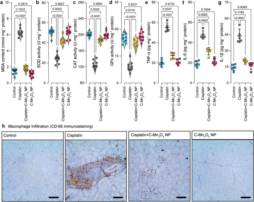
COMMUNICATIONS BIOLOGY | https://doi.org/10.1038/s42003-021-02546-8                                                                      ARTICLE

pretreated with 30 and 60 μg mL−1 NPs, respectively. Pretreat-
ment of the cells with similar concentrations of the NPs alone did
not cause significant cellular mortality except the 60 µg mL−1
(~18%, p < 0.001 compared to control, one-way ANOVA, F(11,
48) = 136.7). Based on the results, we selected the 30 μg mL−1
C-Mn3O4 NPs for further experiments. Identical results were
observed in the lactate dehydrogenase (LDH) assay (Fig. 2b). The
presence of a high concentration of H2O2 inside the cell caused
oxidative damage to the plasma membrane resulting in an
increased release of LDH, a cytosolic enzyme, into the sur-
rounding cell culture medium. Pretreatment with C-Mn3O4 NPs
protected the cells from H2O2 induced oxidative damage resulting
in a ~40% reduction in the LDH release (p < 0.001 compared to
H2O2 treated cells, one-way ANOVA, F(3, 16) = 132.1). In post
hoc analysis, the cells treated with C-Mn3O4 NPs alone also
showed significant difference when compared to untreated con-
trol (p = 0.0057, one-way ANOVA, F(3, 16) = 132.1). However, it
was not reflected in cell mortality. To evaluate the scavenging of
H2O2 by C-Mn3O4 NPs under stress conditions, we monitored
the intracellular oxidative stress using a ROS-sensitive fluores-
cence probe, dihydro dichloro-fluorescein diacetate (DCFH2-
DA). DCFH2-DA is transported across the cell membrane and
hydrolyzed by intracellular esterases to form nonfluorescent 2′,7′-
dichlorofluorescein (DCFH), which is rapidly converted to highly
fluorescent 2′,7′-dichlorofluorescein (DCF) in presence of ROS.
The results illustrate that H2O2 exposure caused a substantial
increase in the cellular ROS level indicated by the enhanced
relative green fluorescence (λem/DCFH2-DA = 520 nm) intensity of
DCFH2-DA (p < 0.001 compared to control, one-way ANOVA,
F(3, 16) = 307.6) when measured using fluorescence microscopy
(Fig. 2c, d) or flow cytometry (Fig. 2e). However, pretreatment
with 30 μg mL−1 C-Mn3O4 NPs significantly lowered intracellular
ROS level which was reflected in decreased fluorescence (~50%
reduction; p < 0.001 compared to H2O2 treated cells, one-way
ANOVA, F(3, 16) = 307.6) of the probe. The C-Mn3O4 NP
treated cells also show a significant amount of ROS (p < 0.001
compared to H2O2 treated cells, one-way ANOVA, F(3,
16) = 307.6), which may be due to the inherent ability of the
nanoparticles to generate ROS. The morphological observations
in differential interference contrast (DIC) microscopy (Fig. 2d)
support the results of cell viability and oxidative damage eva-
luation studies. The cells pretreated with C-Mn3O4 NPs pre-
vented the shrinkage and congregation of the cell body due to
H2O2 overexposure and maintained normal cellular architecture.
   The biological consequences of exposure to nanomaterials can
only be understood in terms of content. Therefore, we evaluated
the amount of nanoparticles internalized by the cells in terms of
cellular manganese content using inductively coupled plasma
atomic emission spectroscopy (ICP-AES). Figure 2f shows the
dose-dependent uptake of C-Mn3O4 NPs in HEK 293 cells. The
amount of intracellular manganese content (i.e., the nanoparticle
content) increased logistically with the increase in the adminis-
tered dose range of 3.75 to 60 µg mL−1, and reached a plateau at               Fig. 1 Characterization of C-Mn3O4 NPs. a TEM image of C-Mn3O4
~50 µg mL−1. Figure 2g depicts the correlation between the                     NPs shows the spherical shape of the nanoparticles with the monomodal
biological impacts (as measured by MTT and ROS assays) with                    distribution. Inset shows an HRTEM image of a single nanoparticle having a
both administered dose and intracellular nanoparticle content.                 high crystalline structure with 0.311 nm interfringe distance corresponding
Both prevention of cell death and quenching of intracellular                   to the (112) plane. The other inset shows the histogram for the size
ROS followed a logistic (Hill equation: y ¼ A1 þ A2ðA           1
                                                                       )       distribution of the nanoparticles having an average diameter of
                                                       1þ10 logx0 xÞp
                                                                               5.58 ± 2.42 nm. b Experimental XRD peaks of the nanoparticle exactly
relationship with administered dose and intracellular nanoparti-
                                                                               match that of Mn3O4 hausmannite defined in the literature (JCPDS
cle content.
                                                                               No. 24-0734). c FTIR spectra of C-Mn3O4 NPs, Mn3O4 NPs, and citrate.
                                                                               Perturbation at Mn–O stretching at 413, 514, 630 cm−1 (shaded gray) of
C-Mn3O4 NPs prevent mitochondria, the master redox reg-                        Mn3O4 NPs and carboxylic groups at 1066, 1112, 1410, 1619 cm−1 (shaded
ulator, from H2O2-induced oxidative damage. Mitochondria                       yellow) of citrate confirms strong covalent binding between citrate and the
despite being the primary source and regulator of intracellular ROS,           nanoparticle.

COMMUNICATIONS BIOLOGY | (2021)4:1013 | https://doi.org/10.1038/s42003-021-02546-8 | www.nature.com/commsbio                                            3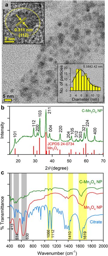
ARTICLE                                                                  COMMUNICATIONS BIOLOGY | https://doi.org/10.1038/s42003-021-02546-8

Fig. 2 Ability of C-Mn3O4 NPs in scavenging of intracellular ROS. a Cell viability was measured using MTT. The gray shaded area represents H2O2
treatment. b LDH release. c Quantification of intracellular ROS as estimated from DCF fluorescence observed under confocal microscopy. d Confocal
fluorescence micrographs of HEK 293 cells stained with DCFH2-DA and counterstained with DAPI. Cells were either left untreated or pretreated with
C-Mn3O4 NPs (30 μg mL−1) prior exposure to H2O2 (100 µM). Scale bar: 30 µm. e Flow cytometry of HEK 293 cells stained with DCFH2-DA. Inset—
intracellular ROS level quantified from flow cytometry analysis. f Dose-dependent internalization of C-Mn3O4 NPs in HEK 293 cells. The intracellular
nanoparticle concentration as measured using inductively coupled plasma atomic emission spectroscopy (ICP-AES). g Correlation between biological
impact (cell death and scavenging of ROS) and administered dose or intracellular nanoparticle content. All four nanoparticle concentration-biological
                                                                A1
response data are fitted with the Hill equation: y ¼ A1 þ Að2logx 0 xÞp
                                                                        . In bar plots data were expressed as Mean ± SD. In box plots, center lines show the
                                                           1þ10
medians; box limits indicate the 25th and 75th percentiles, whiskers extend 1.5 times the interquartile range from the 25th and 75th percentiles. Violins
depict kernel density estimation of the underlying data distribution with the width of each violin scaled by the number of observations at that Y-value.
Three lines (from the bottom to the top) in each violin plot show the location of the lower quartile (25th), the median, and the upper quartile (75th),
respectively. The shaded area indicates the probability distribution of the variable. Individual data points are represented as colored circles (N = 5). One-
way analysis of variance (ANOVA) followed by Tukey’s post hoc multiple comparison test was performed for comparison among the groups. The numbers
inside the plots indicate numerical p values. p < 0.05 is considered significant.

are the most susceptible organelle to oxidative damage leading to               indicated by the increased fluorescence of Mito-sox red (Fig. 3a, c).
redox imbalance and cell death29,30. So, to get further insight into            Pretreatment with 30 μg mL−1 C-Mn3O4 NPs significantly restored
the free radical scavenging activity of C-Mn3O4 NPs we evaluated                the ΔΨm and reduced the mitochondrial ROS. ΔΨm has a causal
their protective effect towards mitochondria. Treatment with H2O2               relationship with mitochondrial permeability transition pore
drastically decreased the mitochondrial membrane potential (ΔΨm),               (mPTP) opening. The results of the Ca2+ induced mitochondrial
as indicated by enhanced rhodamine 123 (Rh123) fluorescence                      swelling assay indicated that the NPs were effective in preventing
(Fig. 3a, b) along with a burst in mitochondrial ROS production, as             the H2O2 induced mPTP opening (Fig. 3d), therefore, maintaining

4                            COMMUNICATIONS BIOLOGY | (2021)4:1013 | https://doi.org/10.1038/s42003-021-02546-8 | www.nature.com/commsbio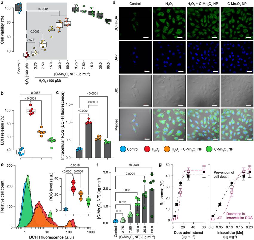
COMMUNICATIONS BIOLOGY | https://doi.org/10.1038/s42003-021-02546-8                                                                       ARTICLE

Fig. 3 Potential of C-Mn3O4 NPs in the regulation of cellular redox condition and protection of mitochondria from oxidative damage. a Confocal
fluorescence micrographs of HEK 293 cells stained with rhodamine 123, MitosoxTM red, and counterstained with DAPI. Cells were either left untreated or
pretreated with C-Mn3O4 NPs (30 μg mL−1) prior exposure to H2O2 (100 µM). Scale bar: 10 µm. b Intensity of rhodamine 123 as a marker of mitochondrial
membrane potential (Δψm). An increase in intensity indicates membrane depolarization. c Mitochondrial ROS level as quantified from MitosoxTM red
fluorescence. d Change in Ca2+-induced mPTP opening. e ATP content. f Cytochrome c oxidase activity. g Superoxide dismutase (SOD) activity. h Catalase
activity. i Glutathione peroxidase (GPx) activity. j Reduced glutathione (GSH) content. k Schematic representation of the redox homeostasis mechanism by
C-Mn3O4 NPs against H2O2 distress through mitochondrial protection. In bar plots data were expressed as Mean ± SD. Violins depict kernel density
estimation of the underlying data distribution with the width of each violin scaled by the number of observations at that Y-value. Three lines (from the
bottom to the top) in each violin plot show the location of the lower quartile (25th), the median, and the upper quartile (75th), respectively. The shaded
area indicates the probability distribution of the variable. Individual data points are represented as colored circles (N = 5). One-way analysis of variance
(ANOVA) followed by Tukey’s post hoc multiple comparison test was performed for comparison among the groups. The numbers inside the plots indicate
numerical p values. p < 0.05 is considered significant.

mitochondrial integrity. The mitochondrial membrane depo-                      mitochondria31–33. The accumulation of highly reactive oxygen
larization and subsequent opening of mPTP led to a significant                  radicals causes damage to biomolecules in cells and alters
fall in the cellular ATP content (Fig. 3e). In C-Mn3O4 NP                      enzyme activities34–36. Hence, we extended our study towards
pretreated cells, such loss in ATP content was not observed. The               evaluating the effect of H2O2 and C-Mn3O4 NPs in the ROS
opening of mPTP, fall in ΔΨm and ATP content cumulatively                      regulatory network. H2O2 exposure significantly reduced the
functions as a proapoptotic signal to initiate the cell death                  activity of SOD, CAT, and GPx resulting in a decrease of the
pathways, also reflected in the increased cytochrome-c oxidase                  reducing pool of cellular thiol constituents (e.g., GSH)
activity (Fig. 3f). Superoxide dismutase (SOD), catalase (CAT),                (Fig. 3g–j). Pretreatment with C-Mn3O4 NPs significantly atte-
and glutathione peroxidase (GPx) constitute the intracellular                  nuated the damage. In cells treated with C-Mn3O4 NPs alone,
antioxidant defense system that works in consort with                          none of the detrimental effects were observed.

COMMUNICATIONS BIOLOGY | (2021)4:1013 | https://doi.org/10.1038/s42003-021-02546-8 | www.nature.com/commsbio                                              5

ARTICLE                                                             COMMUNICATIONS BIOLOGY | https://doi.org/10.1038/s42003-021-02546-8

   Thus, our cellular studies indicate that C-Mn3O4 NPs possess            collapsed Bowman’s space, and glomerular retraction (Fig. 4j).
the distinctive property of scavenging intracellular ROS, inhibit-         Tubular atrophy, dilation of cortical tubules, increased mesangial
ing apoptotic trigger, preventing loss of antioxidant enzymes, and         matrix, obliteration of capillaries, necrosis, vacuolization, and
maintaining high cell viability by acting as a protector of                interstitial mononuclear infiltration were the other features
mitochondria, the master regulator of cellular redox equilibrium           observed in this group. Treatment with C-Mn3O4 NPs notably
(Fig. 3k schematically summarizes the whole sequence).                     reduced focal glomerular necrosis (Fig. 4j). However, sparse
                                                                           tubular changes like vacuolization, dilation, mild mononuclear
                                                                           infiltration, and detachment of epithelial cells were observed in
C-Mn3O4 NPs attenuate glomerular and tubulointerstitial                    this group. Overall, C-Mn3O4 NPs were able to efficiently revert
damage in CKD mice. There is always a gap in the efficacies of a            the marked detrimental changes in the renal architecture of CKD
pharmacological agent tested between cellular and animal model.            animals. The histological observations are quantitatively reflected
Limited bioavailability, nonspecific biodistribution, or unwanted           in the necrosis score (Fig. 4i), glomerular injury score (GIS;
metabolism often restricts the in vivo use of a cytoprotective             Fig. 4k), and tubular injury score (TIS; Fig. 4l).
agent37–39. Thus, we evaluated the potential of C-Mn3O4 NPs in                Previous studies and our histological observations suggested an
the treatment of cisplatin-induced C57BL/6j mice, a well-                  association between renal fibrosis and CKD47–49. So, we
established animal model for testing therapeutic interventions             measured the renal hydroxyproline content, a byproduct of
against CKD40–42. As depicted in Fig. 4a, treatment with                   collagen metabolism, and biochemical marker of fibrosis. The
C-Mn3O4 NPs alone did not cause any mortality during the                   results indicate almost a threefold increase in the hydroxyproline
experimental period. While, chronic administration of cisplatin            content (1.43 ± 0.07 compared to 0.51 ± 0.03 mg gm−1 tissue of
resulted in significant mortality (~40% compared to control),               control; p < 0.001, One-way ANOVA, F(3, 16) = 378.7) in the
administration of C-Mn3O4 NPs in the cisplatin-intoxicated                 cisplatin intoxicated group (Table 1). In accordance with the
group significantly reduced the mortality (Hazards Ratio, HR                histological findings, treatment with C-Mn3O4 NPs markedly
(Log-rank): 2.62; 95% CI of HR: 1.166–4.75; log-rank χ2                    reduced the hydroxyproline content (0.79 ± 0.06 mg gm−1 tissue;
(Mantel–Cox): 5.23; df: 1; P = 0.0222) (Fig. 4a). The fourfold             p < 0.001 compared to cisplatin-treated ones, One-way ANOVA,
higher blood urea nitrogen (BUN) content (Fig. 4b), threefold              F(3, 16) = 378.7), suggesting a decrease in fibrotic damage
higher GFR (Table 1), fourfold higher urinary albumin excretion            (Table 1).
(i.e., albuminuria) (Fig. 4c), and high urine albumin to creatinine
ratio (ACR) (Table 1) along with significantly increased serum
                                                                           C-Mn3O4 NPs augment the intracellular antioxidant defense
urea (Fig. 4d) and creatinine (Fig. 4e) illustrated induction of
                                                                           system. Oxidative stress proved to be one of the major causes of
proteinuria and notable damage to the renal function of mice, the
                                                                           cisplatin-induced nephrotoxicity50–52. Therefore, we tried to
two hallmarks of CKD43–45. Treatment with C-Mn3O4 NPs
                                                                           ascertain whether C-Mn3O4 NPs contributed to nephroprotection
(0.25 mg kg−1 body weight (BW)) to the cisplatin intoxicated
                                                                           by ameliorating oxidative stress. Signs of ROS-mediated damage
animals considerably reduced BUN, GFR, urinary albumin, ACR,
                                                                           including lipid peroxidation (in terms of thiobarbituric acid
serum urea, and creatinine (Fig. 4b–e and Table 1). Treatment
                                                                           reactive substances, TBARS), reduction in cellular GSH pool, and
with citrate (the functionalization group) was unable to reduce
                                                                           inhibition of antioxidant enzyme activities were estimated.
any of the aforementioned parameters (Supplementary Fig. S2),
                                                                           Exposure to cisplatin markedly increased the level of TBARS
confirming the observed effects solely due to the conjugated
                                                                           (Fig. 5a) and oxidative glutathione along with a reduction in GSH
nanomaterial. This may be due to the low dose of citrate (i.e.,
                                                                           concentration. Furthermore, it inhibited the antioxidant actions
0.25 mg kg−1 body weight) used in the study. Such a low dose of
                                                                           of enzymes like SOD, CAT, and GPx (Fig. 5b–d). The results, in
citrate, a small molecule antioxidant having no catalytic activity,
                                                                           consensus with our cellular studies, indicate that C-Mn3O4 NPs
was not enough to prevent the oxidative damage caused by cis-
                                                                           rescued the renal cells from detrimental pleiotropic effects of
platin. These results are well in agreement with the study by
                                                                           increased ROS while maintaining the normal signaling circuitry.
Kondo et. al.,46 where the authors revealed that citrate alone has
no nephroprotective action, but it enhances the protective effect
of bismuth subnitrate against the cis-diamminedichloroplatinum             C-Mn3O4 NPs reduce renal inflammation. Macrophage infil-
induced nephrotoxicity. Cisplatin intoxication caused weight loss          tration in the kidney and subsequent rise in the plasma con-
in mice (Fig. 4f), suggestive of the systemic toxicity that fre-           centrations of pro-inflammatory cytokines like TNF-α are well-
quently arises in individuals receiving this anticancer drug. Ani-         known features of CKD53–55. We found significant increases in
mals treated with C-Mn3O4 NPs were capable of mitigating                   plasma concentrations of TNF-α, IL-1β, and IL-6 in cisplatin-
weight loss. Next, we examined the external morphology of iso-             induced animals (Fig. 5e–g). Treatment with C-Mn3O4 NPs
lated kidneys from each group. The kidneys from the cisplatin              resulted in a notable decrease in cytokine levels. No difference
exposed group deviated from the usual darkish brown to a pale              was observed between the C-Mn3O4 NP treated and the control
brown color with a rough and uneven surface (Fig. 4g). The                 groups. Previous studies have demonstrated that direct adsorp-
kidney to body weight ratio (i.e, kidney index) was also sig-              tion of pro-inflammatory cytokines (e.g., TNF-α, ILs, etc.) by the
nificantly higher (i.e., edema) in cisplatin-treated animals                nanoparticle surface can provide false positive or negative results
(2.1 ± 0.2 compared to 1.5 ± 0.1 mg g−1 of control, p < 0.001, one-        about inflammatory responses56,57. Therefore, in order to further
way ANOVA, F(3, 28) = 31.7; Fig. 4h). Subsequent treatment                 verify the findings of reduction in renal inflammation due to
with C-Mn3O4 NPs overturned the observed changes in mor-                   C-Mn3O4 NP treatment, we performed immunohistochemical
phology and kidney index.                                                  (IHC) staining of the kidney sections with anti-CD68 antibodies,
   Hematoxylin and eosin-stained kidney sections of the control            a well-known macrophage infiltration marker associated with the
and C-Mn3O4 NP treated groups showed normal histologic                     M1 macrophage phenotype. It is evident from Fig. 5h that the
features (Fig. 4j) with negligible necrosis score (Fig. 4i). The           number of CD68 positive area is negligible in cisplatin+C-Mn3O4
kidney sections from cisplatin intoxicated mice displayed several          NP co-treated animals compared to the cisplatin intoxicated
pathological features of CKD like focal segmental as well as global        animals, which showed pronounced staining of macrophage
glomerulosclerosis along with interstitial fibrosis, diffused thick-        infiltrated area (marked in a dotted circle). The animals treated
ening of the capillary walls, glomerular hyalinosis, dilated or            with C-Mn3O4 NPs alone showed characteristics similar to the

6                        COMMUNICATIONS BIOLOGY | (2021)4:1013 | https://doi.org/10.1038/s42003-021-02546-8 | www.nature.com/commsbio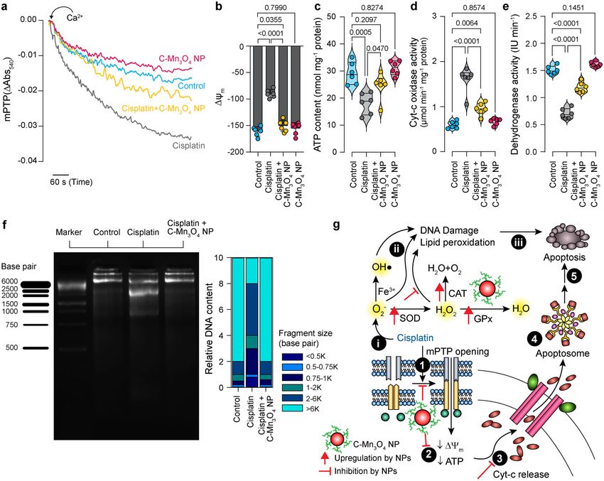
COMMUNICATIONS BIOLOGY | https://doi.org/10.1038/s42003-021-02546-8                                                                            ARTICLE

Fig. 4 Efficacy of C-Mn3O4 NPs in the reversal of CKD in the animal model. a Kaplan–Meier survival analysis curve. The darker shaded area represents
the co-treatment period. b Blood urea nitrogen (BUN) content. c Urinary albumin excretion as an indicator of albuminuria, a hallmark of CKD. d Serum urea
concentration. e Serum creatinine level. f Body weight at the end of the experimental period. g Photographs of kidneys incised after the experimental
period. h Kidney index, defined as a kidney to body weight ratio (mg g−1). i Necrosis score as per the observation of an expert clinical pathologist.
j Hematoxylin and eosin-stained liver sections. Insets show a magnified image of a single glomerulus. Red arrow: segmental glomerulosclerosis; Yellow
arrow: global glomerulosclerosis; Yellow dotted region: mononuclear infiltration. Scale bar: 20 µm. k Glomerular injury score (GIS). l Tubular injury score
(TIS). In bar plots data were expressed as Mean ± SD. Violins depict kernel density estimation of the underlying data distribution with the width of each
violin scaled by the number of observations at that Y-value. Three lines (from the bottom to the top) in each violin plot show the location of the lower
quartile (25th), the median, and the upper quartile (75th), respectively. The shaded area indicates the probability distribution of the variable. Individual data
points are represented as colored circles or squares (N = 10). One-way analysis of variance (ANOVA) followed by Tukey’s post hoc multiple comparison
test was performed for comparison among the groups. The numbers inside the plots indicate numerical p values. p < 0.05 is considered significant.

control ones (Fig. 5h). Therefore, the results together suggest that              efficacy of C-Mn3O4 NPs in animals. Ca2+-induced renal mPTP
the observed reduction in the inflammatory markers by the                          opening is one of the salient features of CKD26,64. Our data clearly
administered nanoparticles happened due to modulation of the                      show that the mitochondria isolated from the cisplatin intoxicated
inflammatory cascade, not by direct adsorption of ILs on the                       group were more sensitive towards Ca2+ manifested into a sharp
surface of the particle.                                                          decrease in 540 nm absorbance (Fig. 6a). Treatment with C-Mn3O4
                                                                                  NPs inhibited mPTP opening and maintained membrane integrity.
                                                                                  ΔΨm and ATP content declined significantly as a result of cisplatin
C-Mn3O4 NPs alleviate mitochondrial damage in CKD mice.                           administration (Fig. 6b, c). These were accompanied by an increase
Considering the inevitable role of mitochondria in the pathogenesis               in cytochrome c oxidase activity (Fig. 6d) and a reduction in
of CKD26,58–63 and the results of our in cellulo observations that                dehydrogenase activity (Fig. 6e). The alterations were abrogated by
C-Mn3O4 NPs protect mitochondria from H2O2 induced oxidative                      C-Mn3O4 NP treatment. Mitochondrial parameters for the animals
damage, we assessed the role of mitoprotection in the therapeutic                 treated with C-Mn3O4 NPs alone showed no signs of toxicity and

COMMUNICATIONS BIOLOGY | (2021)4:1013 | https://doi.org/10.1038/s42003-021-02546-8 | www.nature.com/commsbio                                                   7
ARTICLE COMMUNICATIONS BIOLOGY | https://doi.org/10.1038/s42003-021-02546-8
Table 1 Effect of C-Mn3O4 NPs on nephrotoxic biomarkers.
Group GFR Urine ACR Creatinine clearance Uric acid Hydroxyproline
(μL min−1 g−1 BW) (μmol min−1) (mg dL−1) (mg g−1 tissue)
Control 10.2 ± 1.5 0.34 ± 0.06 1.41 ± 0.08 1.2 ± 0.1 0.51 ± 0.03
Cisplatin 30.4 ± 4.1** 5.62 ± 0.08** 0.35 ± 0.04** 2.6 ± 0.2** 1.43 ± 0.07**
Cisplatin + C-Mn3O4 NPs 14.1 ± 2.3* 1.87 ± 0.09*,** 0.92 ± 0.05*,** 1.6 ± 0.1* 0.79 ± 0.06*,**
C-Mn3O4 NPs 9.8 ± 1.2* 0.41 ± 0.05* 1.38 ± 0.07* 1.3 ± 0.1* 0.42 ± 0.04*
Data expressed as Mean ± SD (N = 6).
Hydroxyproline contents were measured from kidney homogenate.
One-way analysis of variance (ANOVA) followed by Tukey’s post hoc multiple comparison test was performed for comparison among the groups.
GFR glomerular filtration rate, ACR albumin to creatine ratio.
*p < 0.05 compared to Cisplatin treated animals.
**p < 0.05 compared Control animals.
Fig. 5 Effect of C-Mn3O4 NPs in the protection of intracellular redox regulatory network and inhibition of anti-inflammatory response in mice. a Extent
of lipid peroxidation (MDA, malonaldehyde content) was measured in terms of thiobarbituric acid reactive substances (TBARS). b Superoxide dismutase
(SOD) activity. c Catalase activity. d Glutathione peroxidase (GPx) activity. e Tumor necrosis factor-α level. f Interleukin-1β level. g Interleukin-6 level.
h Immunohistochemical analysis of kidney tissues for detection of inflammatory damages. Macrophages are stained with anti-CD-68 antibodies (brown).
Scale bar: 20 µm. The dotted circles indicate the regions with high CD68 positivity (i.e., macrophage infiltration). MDA, SOD, CAT, and GPx were
estimated from kidney homogenate. TNF-β, IL-1β, and IL-6 were measured from serum. Violins depict kernel density estimation of the underlying data
distribution with the width of each violin scaled by the number of observations at that Y-value. Three lines (from the bottom to the top) in each violin plot
show the location of the lower quartile (25th), the median, and the upper quartile (75th), respectively. The shaded area indicates the probability
distribution of the variable. Individual data points are represented as colored circles (N = 10). One-way analysis of variance (ANOVA) followed by Tukey’s
post hoc multiple comparison test was performed for comparison among the groups. The numbers inside the plots indicate numerical p values. p < 0.05 is
considered significant.
8 COMMUNICATIONS BIOLOGY | (2021)4:1013 | https://doi.org/10.1038/s42003-021-02546-8 | www.nature.com/commsbioCOMMUNICATIONS BIOLOGY | https://doi.org/10.1038/s42003-021-02546-8 ARTICLE
Fig. 6 Efficacy of C-Mn3O4 NPs in the protection of mitochondria, the master redox regulator in mice. a Ca2+ induced mPTP opening measured by the
decrease in 540 nm absorbance. b Mitochondrial membrane potential (Δψm) estimated using JC-1 fluorescence. c ATP content. d Cytochrome c oxidase
(complex IV in the electron transport chain, ETC) activity in isolated mitochondria. e Succinate dehydrogenase (SDH, complex II in ETC) activity in isolated
mitochondria. f DNA fragmentation level as a result of oxidative damage measured using agarose gel electrophoresis. In cisplatin-induced CKD animals
DNA ladder formation, indicative of apoptotic DNA fragmentation, is clearly visible. The corresponding stacked bar plot shows the relative abundance of
different-sized fragmented DNA in relation to the total DNA content of the lane. g Schematic overview of the comprehensive molecular mechanism of
action of C-Mn3O4 NPs as a redox medicine against cisplatin-induced CKD. The numbers in the black circles indicate the sequence of events. In bar plots
data were expressed as Mean ± SD. Violins depict kernel density estimation of the underlying data distribution with the width of each violin scaled by the
number of observations at that Y-value. Three lines (from the bottom to the top) in each violin plot show the location of the lower quartile (25th), the
median, and the upper quartile (75th), respectively. The shaded area indicates the probability distribution of the variable. Individual data points are
represented as colored circles (N = 10). One-way analysis of variance (ANOVA) followed by Tukey’s post hoc multiple comparison test was performed for
comparison among the groups. The numbers inside the plots indicate numerical p values. p < 0.05 is considered significant.
were analogous to the control animals. Thus, cisplatin-induced dependent plasma concentration profile of C-Mn3O4 NPs in
renal damage triggered the opening of mPTP, the decline in ΔΨm, order to figure out how the nanoparticles are absorbed into and
and the induction of mitochondrial swelling that resulted in the eliminated from the bloodstream. Intraperitoneal (i.p.) adminis-
release of cytochrome c in the cytosol leading to apoptosis. tration of C-Mn3O4 NPs (0.25 mg kg−1 BW) to mice generated
The ladder-like DNA fragmentation, a hallmark of apoptosis, was the plasma Mn concentration vs. time profile displayed in Fig. 7a.
evident in the case of cisplatin-treated disease groups (Fig. 6f). The PK parameters were calculated using a non-compartmental
Whereas, treatment with C-Mn3O4 NPs notably protected the approach, yielding a maximum plasma concentration (CMAX) of
mitochondria, inhibited cell death, and decreased the extent of 3.1 ± 0.2 µg mL−1 at (tMAX) 2.0 ± 0.1 h. The plasma area under the
DNA fragmentation. Figure 6g schematically illustrates the entire curve (AUC) was 20.5 ± 1.8 µg h mL−1 with a Clearance of
phenomena of redox-mediated nephroprotection by C-Mn3O4 12.2 ± 1.5 L h−1 kg−1. The mean plasma concentration curve for
NPs. C-Mn3O4 NPs (Fig. 7a) presented a two-peak (at ~2.0 and
~12.0 h) absorption phase. The first peak is due to the absorption
Pharmacokinetics and biocompatibility of C-Mn3O4 NPs. Over of NPs from the peritoneal cavity to the blood. While, intestinal
the years, pharmacokinetic (PK) studies have emerged as an absorption, variable gastric emptying, enterohepatic recirculation,
integral part of drug development, especially for identifying the and distribution, and reabsorption are possible interpretations for
in vivo behavior of a drug. Therefore, we evaluated the time- the second peak (at ~12.0 h)65. Next, we evaluated the time-
COMMUNICATIONS BIOLOGY | (2021)4:1013 | https://doi.org/10.1038/s42003-021-02546-8 | www.nature.com/commsbio 9ARTICLE COMMUNICATIONS BIOLOGY | https://doi.org/10.1038/s42003-021-02546-8 Fig. 7 Pharmacokinetics (PK) and biocompatibility of C-Mn3O4 NPs. a Plasma concentration-time profile following intraperitoneal administration of C-Mn3O4 NPs as measured using inductively coupled plasma-atomic emission spectroscopy (ICP-AES). Inset shows the PK parameters calculated using a non-compartmental approach. The dotted line is a guide to the eye. b Time-dependent accumulation and elimination of C-Mn3O4 NPs in the kidney. Data were normalized to wet kidney weight. c Micrographs of hematoxylin and eosin-stained sections of different organs after 1 month of treatment with the therapeutic dose (0.25 mg kg−1 body weight, i.p.) of C-Mn3O4 NPs. Both control and treated animals maintained normal tissue architecture. Scale bars: liver-100 µm, brain-50 µm, ovary-20 µm, pancreas-50 µm, spleen-100 µm, testes-100 µm. Data were expressed as Mean ± SD. Individual data points are represented as white circles (N = 5). dependent uptake and elimination profile of C-Mn3O4 NPs in the histological analysis also revealed normal ovarian structure with kidney up to 24 h (Fig. 7b). The manganese (i.e., C-Mn3O4 NP) follicles at different growth phases and observable luteum follicle. content in the kidney increased exponentially reaching the The brain structure was also typical for C-Mn3O4 NP treated mice maximum concentration of 2.28 ± 0.31 µg g−1 at ~10 h. The sig- with normal cerebellum consisting of the granular and molecular nificantly increased concentration of nanoparticles in the kidney layer, and visible Purkinje cells. upon administration of C-Mn3O4 NPs clearly depicts its ability to enter the kidneys through the glomerular filtration barrier. Discussion Although the elimination phase started after 12 h, a sufficient In this study, we determined whether C-Mn3O4 NPs could be quantity (0.95 ± 0.09 µg g−1) of nanoparticles remained in the used as a redox medicine to treat CKD, an important clinical kidneys even after 24 h post administration. question considering the high prevalence of the disease and the Next, we investigated the effect of C-Mn3O4 NPs in other major nonavailability of effective medication. CKD is defined as the organs in order to evaluate its biocompatibility. The hematoxylin progressive and irreversible loss of renal function characterized by and eosin-stained histopathological sections of the organs from reduced glomerular filtration rate (GFR), increased urinary C-Mn3O4 NP treated animals showed identical features to the albumin excretion (albuminuria), or both43,45,66. Our results organs collected from the control group (Fig. 7c). Livers of the present evidence that treatment with C-Mn3O4 NPs significantly nanoparticle-treated mice showed normal hepatic architecture with improved renal function, glomerular and tubulointerstitial injury, clearly detectable central vein, portal tracts, and hepatocytes cellular antioxidant defense network in line with inhibition of arranged in cords with normal sinusoidal space. In the case of the pro-inflammatory immune response, and attenuation of mito- pancreas, we found a normal pancreatic tissue structure compris- chondrial dysfunction in response to the cisplatin toxicity. Our ing of pancreatic acini, pancreatic ducts, and β-cells islands. cellular and animal studies further enlightened the role of the Nanoparticle treated spleen maintained ideal histological structure unique mitoprotective as well as redox modulatory activity of of white and red pulp. The testes of nanoparticle-treated animals C-Mn3O4 NPs in the therapeutic mechanism. did not show any remarkable structural changes. Their structure is Several underlying factors played role in the selection of comprised of well-organized seminiferous tubules and visible C-Mn3O4 NPs as the material of choice. These include the interstitial space. The maturation of spermatids were normal. The exciting redox modulatory properties (contrasting ability to 10 COMMUNICATIONS BIOLOGY | (2021)4:1013 | https://doi.org/10.1038/s42003-021-02546-8 | www.nature.com/commsbio
COMMUNICATIONS BIOLOGY | https://doi.org/10.1038/s42003-021-02546-8                                                                  ARTICLE

function as prooxidant as well as catalytic antioxidant, the fun-              efficacy of C-Mn3O4 NPs in the treatment of multifaceted
damental feature of a redox medicine), biodegradability, aqueous               diseases like CKD, we propose that both the mechanisms (i.e.,
solubility, low cost of the nanomaterial along with apparent non-              ROS scavenging and mitochondrial protection) simultaneously
toxicity (permissible limit ~12 mg day−1), and abundance of                    take place.
manganese (Mn) as the catalytic metal centers or cofactors in                      The findings that C-Mn3O4 NPs can accelerate the revival of
several enzymes. Furthermore, previous reports67 about the                     proximal tubule epithelium embodies a crucial nephroprotective
nephroprotective action of catalytic antioxidants encouraged us to             function mediated by the nanoparticles. Kidneys show higher
evaluate the therapeutic potential of C-Mn3O4 NPs against CKD.                 regenerative property following tubulointerstitial damage50. The
This study provides prima facie indication that other biode-                   proliferation of a subset of sublethally damaged, yet surviving,
gradable/organic nanomaterials with sufficient mitoprotective                   proximal tubule cells can contribute to the regenerative property of
and redox modulatory activity could be used to treat CKD,                      the kidney80. The acceleration of this process is sufficient to confer
provided they can enter and stay in the kidney for sufficient time              nephroprotection81,82. As revealed in our histological findings, the
to depict therapeutic effect. In this regard, it is worth mentioning           recovery rate of these cells (indicated by the structural integrity of
here that the bioavailability of a nano-compound to the kidney                 the cellular architecture in hematoxylin and eosin-stained sections,
could be enhanced using efficient carrier molecules. For example,               and TIS scores) in C-Mn3O4 NP treated cisplatin exposed animals
the peptide amphiphile micelles (PAM) functionalized with the                  (Fig. 4j, k) was significantly higher and efficient than the auto-
zwitterionic peptide ligand, (KKEEE)3K, developed by Huang et.                 recovery (Fig. 4j, k). Several mechanisms can be proposed for the
al.,68 have the ability to cross the glomerular filtration barrier for          enhanced proliferation by C-Mn3O4 NPs. The restoration of
the efficient delivery of therapeutic agents to kidney.                         structural and functional integrity of mitochondria and recovery of
   Mitochondria have long been recognized for their canonical                  respiratory complexes may contribute towards the increased pro-
roles in cellular respiration and energy production26. Recently,               liferation. It is well known that the mitochondrial ETC has a
they have emerged as the master regulator of a spectrum of                     crucial role in cell proliferation through regulation of ATP gen-
molecular pathways including biosynthesis of macromolecules,                   eration, and supply of energy to proliferative pathways83,84. Earlier
maintenance of cellular redox equilibrium, calcium homeostasis,                studies have demonstrated that mutations in ETC genes or the
inflammation, and cell death69–73. Thus, mitochondria are poised                presence of ETC complex inhibitors cause a reduction in ATP
to play a pivotal role in the functioning of the kidney, an organ              synthesis, thus, obstructs progression through the cell cycle and
with high energy demand, and rich in mitochondria, second only                 proliferation85–87. Henceforth, it is reasonable to assume that the
to the heart60. Our findings that C-Mn3O4 NPs maintain cellular                 mitoprotective activity of C-Mn3O4 NPs have played a significant
redox homeostasis through the prevention of mPTP opening and                   role in the revival of tubulointerstitial epithelial cells, in turn
ATP depletion discloses a key redox-mediated nephroprotective                  protecting the renal architecture. Additionally, ROS scavenging by
mechanism. Virtually, the renal proximal tubules are exclusively               C-Mn3O4 NPs may boost the proliferation because oxidative dis-
dependent on ATP generated by mitochondrial oxidative phos-                    tress in proximal tubules causes cell cycle arrest and impedes cell-
phorylation and are therefore vulnerable to oxidative distress due             cycle progression28.
to mitochondrial damage50,74. Cisplatin accumulates in mito-                       The favorable PK properties and biocompatible nature further
chondria and reduces the activity of all four respiratory com-                 indicates the possibility of using C-Mn3O4 NPs as a nano-drug.
plexes (I–V) involved in the electron transport chain (ETC),                   Beyond size, the localization of a nano-drug in the target organ
thereby a surge in mitochondrial ROS formation takes place                     allows assessment of its therapeutic regimen. The direct evidence
along with mPTP opening, membrane depolarization, and                          that the nanoparticles enter the kidney crossing the glomerular
impairment in ATP production, leading to cell death75,76. The                  filtration barrier and reside there for a sufficient amount of time
cytotoxic mechanism of cisplatin essentially mimics the patho-                 further justifies our claim that the recovery is due to the ther-
genesis of CKD, thus an efficient reversal of damage in this rodent             apeutic action of the nanoparticles. It is worth mentioning here
model is supposed to reflect the possible effects of a compound in              that in virtually all in vivo studies using nanomaterials, the bulk
higher animals. Data from our cellular as well as animal studies               of the material ends up in the spleen and the liver. The same is
provide sufficient evidence that C-Mn3O4 NPs ameliorate mito-                   true for C-Mn3O4 NPs. In one of our recent studies, we revealed
chondrial ROS surge, prevent loss of membrane potential, inhibit               that C-Mn3O4 NPs also have a tendency to accumulate in the
mPTP opening, and stops ATP depletion, thereby prevents                        liver if orally administered for a longer (90 days) period of time88.
mitochondrial dysfunction, cellular redox imbalance, and tubular               Although, identifying the particular cell types that internalizes
or glomerular cell death. As a result, the markers of CKD i.e.,                C-Mn3O4 NPs to the kidney or liver, and understanding the exact
increased BUN, plasma creatinine, serum urea, and GFR returns                  mechanisms of internalization are intriguing, they fall beyond the
to homeostatic condition.                                                      scope of this study and remain an attractive area for further
   This study provides a piece of direct evidence that C-Mn3O4                 investigation.
NPs can scavenge ROS, particularly H2O2 the longest living one                     The role of intracellular redox regulation through mitoprotec-
in the cellular milieu. It also proves the ability of the nanoparticles        tion in the therapeutic action of C-Mn3O4 NPs opens up further
in the prevention of mPTP from opening and subsequent                          avenues for the treatment of several unmet diseases like diabetic
maintenance of mitochondrial structure and function. However,                  nephropathy, neurodegeneration (e.g., Parkinson’s, Huntington’s,
it is not clear whether ROS scavenging protects mitochondria or                Alzheimer’s, multiple sclerosis), cardiovascular disorders, obesity,
protection of mitochondrial integrity results in ROS depletion.                etc. where pathogenesis is very much dependent upon mitochon-
Several studies have shown that a compound having antioxidant                  drial damage and associated redox imbalance89–93. Although in
properties cannot be a sustainable therapeutic solution to                     our study C-Mn3O4 NPs did not show any adverse effect, a detailed
oxidative-stress-related disorders, fundamentally due to its                   study on systemic toxicity, bio-distribution, PKs, and pharmaco-
inability to regenerate after a single reaction77–79. In contrast,             dynamics will greatly enhance the knowledge about its in vivo
redox nanomaterials have the potential to reduce oxidative stress              behavior. Furthermore, a detailed molecular study analyzing the
either by autocatalysis or by protecting cellular machineries that             genome and metabolome of C-Mn3O4 NP-treated animals
control redox homeostasis, until the nanomaterials dissolute in                may enlighten its ability to interfere in other pathogenesis path-
the physiological milieu. Considering the causal relationship                  ways. As an outcome, the nano-drug could be repurposed for other
between mitochondria and cellular redox homeostasis and the                    therapies too.

COMMUNICATIONS BIOLOGY | (2021)4:1013 | https://doi.org/10.1038/s42003-021-02546-8 | www.nature.com/commsbio                                      11
ARTICLE                                                                        COMMUNICATIONS BIOLOGY | https://doi.org/10.1038/s42003-021-02546-8

Conclusion                                                                            treatments were completed. Ten thousand events were analyzed by flow cytometry
There are very few published articles in contemporary literature                      (FACS Verse, Beckton Dickinson, SanJose, USA) and the respective mean fluor-
                                                                                      escence intensity (in FL1 channel, set with a 530/30 nm bandpass filter) values were
that utilize the promising redox regulatory approach for the                          correlated with the ROS levels. For confocal microscopy, 5000 cells were seeded
treatment of chronic diseases like CKD. On the other hand,                            and treated with agents described earlier. Post-treatment cells were stained with
several CKDs are reported to be due to redox imbalance in                             DCFH2-DA (5 μM) at 37 °C in dark and images were acquired with a confocal
mitochondria. Our study suggests that C-Mn3O4 NPs could be an                         microscope (Olympus IX84, Japan). All parameters (pinhole, contrast, gain, and
efficient redox medicine to attenuate renal injury and tubu-                           offset) were held constant for all sections in the same experiment.
leintestinal fibrosis as evidenced by the improved renal functions,
reduction in biochemical markers of nephrotoxicity, reduced                           Mitochondrial superoxide and ΔΨm detection. After treatment with the drug or
                                                                                      vehicle, mitochondrial superoxide production was visualized using MitoSOXTM
fibrotic content, and downregulated proinflammatory cytokines.                          Red (Thermofisher, USA), a mitochondrial superoxide indicator. The ΔΨm in
The molecular mechanism involves regulation of the redox bal-                         intact cells was assessed by confocal microscopy using Rh 123 (Sigma, USA). Cells
ance through synchronization of the causal relationship between                       were loaded with MitoSOXTM Red (0.5 μM) or Rh123 (1.5 μM) for 10 min at 37 °C
mitoprotection and ROS scavenging by C-Mn3O4 NPs. The                                 and imaged using a confocal microscope (Olympus IX84, Japan). All parameters
                                                                                      (pinhole, contrast, gain, and offset) were held constant for all sections in the same
findings highly suggest the translational potential of C-Mn3O4                         experiment. ImageJ (http://imagej.nih.gov/ij/) was used to quantify area normal-
NPs as a redox nanomedicine for treating CKD in the clinic.                           ized fluorescence intensities from the confocal images.

Methods                                                                               Animals and treatment. Healthy nondiabetic C57/6j mice of both sexes
Synthesis of C-Mn3O4 NPs. A template or surfactant-free sol-gel method was            (8–10 weeks old, weighing 27 ± 2.3 g) were used in this work. Animals were
followed for the synthesis of bulk Mn3O4 NPs at room temperature and pressure21.      maintained in standard, clean polypropylene cages (temperature 21 ± 1 °C; relative
To functionalize the nanoparticles with ligand citrate, the as-prepared Mn3O4 NPs     humidity 40–55%; 1:1 light and dark cycle). Water and standard laboratory pellet
(~20 mg mL−1) were mixed extensively with citrate (Sigma, USA) solution (pH 7.0,      diet for mice (Saha Enterprise, Kolkata, India) were available ad libitum throughout
0.5 M) for 15 h in a cyclomixer. The time for mixing was carefully adjusted to have   the experimental period. All mice were allowed to acclimatize for 2 weeks before
a nanomaterial in the size range of 4–6 nm. Non-functionalized larger NPs were        the treatment. The guideline of the Committee for the Purpose of Control and
removed using a syringe filter (0.22 μm).                                              Supervision of Experiments on Animals (CPCSEA), New Delhi, India, was followed
                                                                                      and the study was approved by the Institutional Animal Ethics Committee (Ethical
                                                                                      Clearance No. - 05/S/UC-IAEC/01/2019).
Characterization techniques. TEM and HRTEM images were acquired using an                  The mice were randomly divided into five groups (n = 16 for each group):
FEI TecnaiTF-20 field emission HRTEM (OR, USA) operating at 200 kV. Sample             (1) control; (2) cisplatin; (3) cisplatin + C-Mn3O4 NPs; (4) C-Mn3O4 NPs; and
preparation was done by drop-casting of aqueous C-Mn3O4 NP solution on 300-           (5) cisplatin + citrate. The experimental model of CKD was established according
mesh amorphous carbon-coated copper grids (Sigma, USA) and allowed to dry             to the previous description. In brief, for induction of CKD, we used 8 mg kg−1 BW
overnight at room temperature. XRD patterns were obtained by employing a              cisplatin (i.p.) in each alternative day for 28 days. After induction, we treated
scanning rate of 0.02 s−1 in the 2θ range from 10 to 80 by a PANalytical XPERT        C-Mn3O4 NPs at 0.25 mg kg−1 BW (i.p.) for another 28 days. There was an overlap
PRO diffractometer (Malvern, UK) equipped with Cu-Kα radiation operating at           of 7 days between induction and treatment. Citrate (Sigma, USA) was used at a
40 mA and 40 kV. FTIR (JASCO FTIR-6300, Japan) was used to confirm the                 dose of 0.25 mg kg−1 BW for 28 days. All doses were finalized based on reported
covalent attachment of the citrate molecules with the Mn3O4 NPs. For FTIR stu-        literature and pilot experimentation. As citrate treatment did not improve kidney
dies, powdered samples were blended with KBr powder and pelletized. KBr pellets       function, data were not represented in the main manuscript.
were used as a reference to make the background correction.
                                                                                      Biochemical evaluations. Whole blood samples from treated mice were collected
H2O2 scavenging by C-Mn3O4 NPs. The ability of C-Mn3O4 NPs to prevent H2O2            from retro-orbital sinus plexus and centrifuged at 2000 × g for 20 min to separate
mediated degradation of sodium-containing dye RB (Sigma, USA) was used as an          the serum. Urine samples were collected in metabolic cages during 24-h fasting
indicator of its H2O2 scavenging activity. The addition of H2O2 (10 mM) in the        conditions. Biochemical evaluations were performed using commercially available
aqueous solution of RB (3.5 μM) leads to decolorization of the dye reflected in a      kits (Autospan Liquid Gold, Span Diagnostic Ltd., India) following the protocol
decrease in absorbance (λmax = 540 nm). The presence of C-Mn3O4 NPs (50 μg mL         described by respective manufacturers. GFR was estimated by the determination of
−1) in the reaction mixture reduced degradation. All absorbance measurements          urinary excretion of fluorescein-labeled inulin (FITC–inulin, Sigma, USA).
were performed using Shimadzu UV-Vis 2600 spectrometer (Tokyo, Japan).
                                                                                      Histological examination. After incision, tissues from randomly selected mice were
Culture of human embryonic kidney cells (HEK 293). HEK 293 cells were                 fixed with 4% paraformaldehyde, embedded in paraffin, and cut into a 5 μm thick
maintained at 37 °C in 5% CO2 in RPMI 1640 growth medium (Himedia, India)             section. After de-waxing and gradual hydration with ethanol (Merck, USA), the
that contained 10% fetal bovine serum (Invitrogen, USA), L-glutamine (2 mM),          sections were stained with hematoxylin and eosin (SRL, India). The sections were
penicillin (100 units mL−1), and streptomycin (100 ng mL−1) (Sigma, USA). Before      then observed under an optical microscope (Olympus, Tokyo, Japan). It is note-
experimentation, the cells were washed twice and incubated with RPMI 1640             worthy to mention here that the histopathologist was blinded to the treatment
medium (FBS, 0.5%) for 1 h and then treated as described in the figure legends.        groups while scoring and evaluating the samples. For immunohistochemistry, kidney
    We used H2O2 (Merck, USA) to induce oxidative damage to the cells                 sections were incubated for 60 min with rat anti-mouse CD68 antibody (Santa Cruz
considering the fact that H2O2 is one of the most predominant intracellular ROS       Biotechnology, India) followed by a 30 min incubation with 10 mg mL−1 HRP-
involved in cellular signaling as well as oxidative damage. While, several other      conjugated rabbit anti-rat secondary antibody (Santa Cruz Biotechnology, India).
compounds can endogenously produce of free radicals (i.e., CuSO4 or 3-amino-          After detection of peroxidase activity with 3-amino-9-ethylcarbazole (Sigma, USA),
1,2,4-triazole), we did not use them in this study as none of them is naturally       sections were counterstained with Mayer’s hematoxylin (SRL, India).
produced in the physiological system.
                                                                                      Renal hydroxyproline measurement. For the measurement of renal hydro-
Measurement of cell viability. Cell viability was assessed by MTT and LDH assay.      xyproline content, a previously described method was used32. In brief, snap-frozen
All cell lines were plated in 96-well plates at a density of 1 × 10³ cells/well and   kidney specimens (200 mg) were weighed, hydrolyzed in HCl (6 M; Merck, USA)
cultured overnight at 37 °C. The treatments were performed as described in the        for 12 h at 100 °C. Next, they were oxidized with Chloramine-T (SRL, India). Next,
figure legends. Next, MTT (5 mg mL−1; Himedia, India) was added to each well,          Ehrlich reagent (Sigma, USA) was added which resulted in the formation of a
with a final concentration of ~0.5 mg mL−1, and the cells were cultured for 4 h at     chromophore. Absorbance was measured at 550 nm. Data were normalized to
37 °C in a 5% CO2 atmosphere. The resultant purple formazan was dissolved by the      kidney wet weight.
addition of 10% sodium dodecyl sulfate (Sigma, USA) and the absorbance was read
at 570 and 630 nm using a microplate reader (BioTek, USA). LDH release was            Renal homogenate preparation. Samples of kidney tissue were collected,
analyzed using a colorimetric LDH cytotoxicity assay kit (Himedia, India) fol-        homogenized in cold phosphate buffer (0.1 M; pH 7.4), and centrifuged at 10,000
lowing the manufacturer’s instructions. Five independent experiments were per-        rpm at 4 °C for 15 min. The supernatants were collected for further
formed in each case.                                                                  experimentation.

Measurement of intracellular ROS. The formation of intracellular ROS was              Assessment of lipid peroxidation and hepatic antioxidant status. The super-
measured with the DCFH2-DA method using both FACS and confocal micro-                 natants obtained in the previous stage were used to measure the activity of SOD,
scopy. For FACS, cells were trypsinized, washed with 1X PBS, and stained with         CAT, GPx, and GSH as well as the content of lipid peroxidation (MDA). Lipid
DCFH2-DA (15 μM; Sigma, USA) for 10 min at 30 °C in the dark after the                peroxidation was determined in TBARS formation using a reported procedure31.

12                             COMMUNICATIONS BIOLOGY | (2021)4:1013 | https://doi.org/10.1038/s42003-021-02546-8 | www.nature.com/commsbio
You can also read