Passive transfer of fibromyalgia symptoms from patients to mice
←
→
Page content transcription
If your browser does not render page correctly, please read the page content below
The Journal of Clinical Investigation                                                                                                                                RESEARCH ARTICLE

Passive transfer of fibromyalgia symptoms from
patients to mice
Andreas Goebel,1,2 Emerson Krock,3 Clive Gentry,4 Mathilde R. Israel,4 Alexandra Jurczak,3 Carlos Morado Urbina,3
Katalin Sandor,3 Nisha Vastani,4 Margot Maurer,4 Ulku Cuhadar,4 Serena Sensi,2 Yuki Nomura,3 Joana Menezes,3 Azar Baharpoor,3
Louisa Brieskorn,3 Angelica Sandström,5 Jeanette Tour,5 Diana Kadetoff,5,6 Lisbet Haglund,7 Eva Kosek,5,8 Stuart Bevan,4
Camilla I. Svensson,3 and David A. Andersson4
1
    Walton Centre NHS Foundation Trust, Liverpool, United Kingdom. 2Pain Research Institute, Institute of Life Course and Medical Sciences, University of Liverpool, Liverpool, United Kingdom. 3Department
of Physiology and Pharmacology, Karolinska Institutet, Stockholm, Sweden. 4King’s College London, Wolfson CARD, Institute of Psychiatry, Psychology & Neuroscience, Guy’s Campus, London, United
Kingdom. 5Department of Clinical Neuroscience, Karolinska Institutet, Stockholm, Sweden. 6Stockholm Spine Center, Upplands Väsby, Sweden. 7Department of Surgery, Division of Orthopaedic Surgery,
McGill University, Montreal, Quebec, Canada. 8Department of Surgical Sciences, Uppsala University, Uppsala, Sweden.

                Fibromyalgia syndrome (FMS) is characterized by widespread pain and tenderness, and patients typically experience fatigue
                and emotional distress. The etiology and pathophysiology of fibromyalgia are not fully explained and there are no effective
                drug treatments. Here we show that IgG from FMS patients produced sensory hypersensitivity by sensitizing nociceptive
                neurons. Mice treated with IgG from FMS patients displayed increased sensitivity to noxious mechanical and cold stimulation,
                and nociceptive fibers in skin-nerve preparations from mice treated with FMS IgG displayed an increased responsiveness to
                cold and mechanical stimulation. These mice also displayed reduced locomotor activity, reduced paw grip strength, and a
                loss of intraepidermal innervation. In contrast, transfer of IgG-depleted serum from FMS patients or IgG from healthy control
                subjects had no effect. Patient IgG did not activate naive sensory neurons directly. IgG from FMS patients labeled satellite glial
                cells and neurons in vivo and in vitro, as well as myelinated fiber tracts and a small number of macrophages and endothelial
                cells in mouse dorsal root ganglia (DRG), but no cells in the spinal cord. Furthermore, FMS IgG bound to human DRG. Our
                results demonstrate that IgG from FMS patients produces painful sensory hypersensitivities by sensitizing peripheral
                nociceptive afferents and suggest that therapies reducing patient IgG titers may be effective for fibromyalgia.

Introduction                                                                                               translational relevance and rely on local repeated intramuscu-
Fibromyalgia syndrome (FMS) is a chronic pain condition char-                                              lar injections of acid (14) or systemic depletion of monoamines
acterized by widespread pain, augmented pain sensitivity to                                                by reserpine treatment (15). The development of novel, mecha-
mechanical pressure and cold temperatures (1–4), as well as                                                nism-based therapies has been hampered by the limited under-
fatigue and emotional distress (5–7). The prevalence of FMS is at                                          standing of the basis of FMS.
least 2% (8), and approximately 80% of FMS patients are women.                                                  The increased polymodal pain sensitivity experienced by FMS
The prevalence rises to 10%–30% among patients diagnosed                                                   patients (1, 16) is associated with altered pain processing in the
with autoimmune rheumatological conditions (9, 10), and FMS is                                             central nervous system (11), dysfunctional descending pain mod-
thus one of the most common chronic pain conditions. The etiol-                                            ulation (17, 18), and structural and functional changes in the brain
ogy and pathophysiology of FMS are not completely understood                                               (19–21). FMS is also associated with abnormalities in peripheral
(11) and the current treatment strategies for FMS rely mainly                                              sensory afferents, such as spontaneous activity and sensitization of
on lifestyle changes, physical exercise, and drug therapy with                                             C-fibers, and loss of epidermal innervation (22, 23). Altered levels
antidepressants and anticonvulsants. Unfortunately, the mod-                                               of inflammatory and immunoregulatory cytokines have also been
est efficacy of the available therapies in most patients leaves an                                         reported in FMS patients. Although these alterations do not follow
enormous unmet clinical need (12, 13). The animal models that                                              a consistent pattern (24, 25), they may suggest that immune pro-
have been used for experimental studies of FMS have uncertain                                              cesses are dysregulated in patients. These observations, together
                                                                                                           with the markedly increased prevalence of FMS among patients
                                                                                                           with autoimmune rheumatological conditions (9, 10), and demon-
Authorship note: AG and E Krock are co–first authors. CIS and DAA are                                      strations that less common (26, 27) and very rare chronic pains (28)
co–senior authors.                                                                                         are caused by autoantibodies, led us to hypothesize that FMS may
Conflict of interest: DAA has received research support from Eli Lilly & Co.                               have an autoimmune basis. Here we have investigated the possibil-
Copyright: © 2021, Goebel et al. This is an open access article published under the
                                                                                                           ity that autoreactive IgG is responsible for several key symptoms
terms of the Creative Commons Attribution 4.0 International License.
Submitted: September 11, 2020; Accepted: May 11, 2021; Published: July 1, 2021.
                                                                                                           of FMS, by examining whether these can be transferred to mice by
Reference information: J Clin Invest. 2021;131(13):e144201.                                                administration of IgG purified from FMS patients, and by assessing
https://doi.org/10.1172/JCI144201.                                                                         the tissue localization of IgG after passive transfer.

                                                                                                                                                                                                        1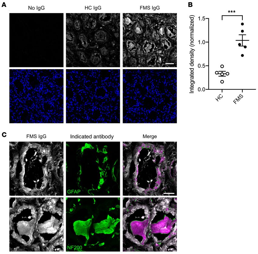
RESEARCH ARTICLE                                                                   The Journal of Clinical Investigation

                                                                               Figure 1. Passive transfer of hypersensitivities
                                                                               from fibromyalgia patients to mice. Adminis-
                                                                               tration of IgG (8 mg on 4 consecutive days) from
                                                                               each of 8 different FMS patients (P1–P8) signifi-
                                                                               cantly reduced the withdrawal threshold in the
                                                                               paw-pressure test (A–F) compared with IgG from
                                                                               healthy control subjects (HC1–HC6). The paw with-
                                                                               drawal latency in the cold-plate test was reduced
                                                                               by IgG from 7 of 8 patients (G–L). Data points are
                                                                               mean ± SEM of n = 6 mice in A, C, E, G, I, and K;
                                                                               n = 5 in D, F, J, and L; and n = 4 in B and H. *P <
                                                                               0.05, **P < 0.01, ***P < 0.001, FMS IgG compared
                                                                               with HC IgG; 2-way repeated measure ANOVA fol-
                                                                               lowed by Sidak’s correction. †P < 0.05, ††P < 0.01,
                                                                               †††
                                                                                  P < 0.001, compared with the naive preinjection
                                                                               value at time zero; 2-way repeated measure
                                                                               ANOVA followed by Dunnett’s test.

                                                                               Results
                                                                               Passive transfer of sensory hypersensitivities.
                                                                               IgG purified from the serum of individual
                                                                               FMS patients and healthy control (HC) sub-
                                                                               jects recruited from the Walton Centre (Liv-
                                                                               erpool, United Kingdom [UK]) was admin-
                                                                               istered to female mice by intraperitoneal
                                                                               injection for 4 consecutive days (8 mg per
                                                                               day). This dose regimen was based on the
                                                                               original studies identifying myasthenia gra-
                                                                               vis as an autoantibody-mediated disorder
                                                                               (29, 30), and more recent studies of complex
                                                                               regional pain syndrome (26, 31). Because
                                                                               FMS is characterized by hypersensitivity
                                                                               to mechanical pressure, we examined paw
                                                                               withdrawal thresholds in mice using the
                                                                               Randall-Selitto paw-pressure test following
                                                                               IgG transfer. IgG from each of the 8 indi-
                                                                               vidual patients, but not from any of the 6
                                                                               HC subjects, rapidly produced mechanical
                                                                               hypersensitivity (Figure 1, A–F). In addition
                                                                               to pressure sensitivity, patients frequently
                                                                               report that pain is exacerbated by cold tem-
                                                                               peratures, and quantitative sensory testing
                                                                               has demonstrated an increased cold pain
                                                                               sensitivity in FMS (1, 2, 4, 16). In good agree-
                                                                               ment with patient observations, administra-
                                                                               tion of IgG from 7 of the 8 FMS patients gave
                                                                               rise to a significantly increased sensitivity to
                                                                               noxious cold in mice (Figure 1, G–L). Both
                                                                               mechanical and cold hypersensitivities were
                                                                               typically established within 24–48 hours
                                                                               after the first injection and were maintained
                                                                               for more than 1 week. The observed hyper-
                                                                               sensitivities produced by IgG preparations
                                                                               from different FMS patients showed sim-
                                                                               ilar amplitudes and time courses (Figure
                                                                               1, A–L, and Supplemental Figure 1, A and
                                                                               B; supplemental material available online
                                                                               with this article; https://doi.org/10.1172/

 2                 J Clin Invest. 2021;131(13):e144201 https://doi.org/10.1172/JCI144201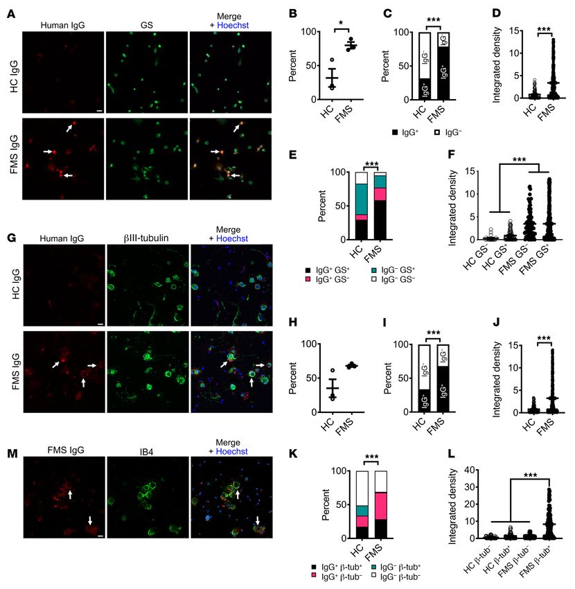
The Journal of Clinical Investigation                                                                                          RESEARCH ARTICLE

Figure 2. FMS IgG produces polymodal abnormalities. FMS IgG increased the sensitivity to punctate stimulation with von Frey filaments (A). The
threshold for leg withdrawal in response to pressure applied to the thigh (using a Randall-Selitto device) was reduced by FMS IgG compared with HC IgG
(B). Female and male mice are affected equally by FMS IgG in the paw-pressure test (C) and the cold-plate test (D). Mechanical hypersensitivity produced
by either 1, 2, or 4 injections of 8 mg FMS IgG (E), and by single injections of 2, 4, or 8 mg (F). The front paw grip strength is reduced by FMS IgG compared
with HC IgG (G). Data points are mean ± SEM or individual measurements. *P < 0.05, **P < 0.01, ***P < 0.001, FMS IgG compared with HC IgG; 2-way
repeated measure ANOVA followed by Sidak’s correction (A, C, D, and G). Data in B were analyzed by unpaired, 2-tailed t test. Data in E and F were com-
pared to the naive preinjection value at time zero by 2-way repeated measure ANOVA followed by Dunnett’s test.

JCI144201DS1), which is reflected in the averaged mechanical and                  2, 4, and 8 mg of FMS IgG, and found that only the highest dose
cold sensitivities produced by IgG from these 8 individual FMS                    increased the mechanical sensitivity in the paw-pressure test (Fig-
patients and 6 HC subjects measured over 10 days (Supplemen-                      ure 2F). These data suggest that a single injection of 8 mg IgG may
tal Figure 1, C and D). FMS IgG also generated hypersensitivity to                be sufficient to transfer mechanical hypersensitivity to mice, and
stimulation with calibrated von Frey filaments, a widely used test                that the effect is saturable (Figure 2E).
of mechanical nociception in mice (Figure 2A). Fibromyalgia pain                       Since FMS is regularly associated with reduced muscular
is characteristically widespread, and studies of the bodily localiza-             strength, and FMS severity is negatively correlated with handgrip
tion of pain have identified the thigh as one of the most commonly                strength (34, 35), we sought to evaluate this experimentally by
affected sites (32, 33). We therefore examined the pressure sen-                  monitoring the front paw grip strength in mice. In this test, FMS IgG
sitivity of the thigh using the Randall-Selitto device (Figure 2B),               significantly reduced grip strength compared with mice treated
to determine whether FMS IgG affects the pressure sensitivity of                  with HC IgG (Figure 2G).
sites other than the hind paw in mice. In this test, FMS IgG pro-                      To determine whether antibodies are responsible for painful
duced significant mechanical hypersensitivity in the thigh com-                   hypersensitivities in a larger and regionally distinct cohort of FMS
pared with treatment with IgG from HCs (Figure 2B).                               patients (recruited at the Karolinska Institute; Supplemental Table
     FMS is more common among women than men, and we                              1), we examined the effects of IgG preparations pooled from mul-
therefore asked whether an increased sensitivity to the effects                   tiple FMS patients and HC subjects. As expected, FMS patients
of IgG contributes to the increased incidence in females. We                      displayed markedly higher pain ratings on a visual analog scale
examined this possibility by comparing the effects of IgG from a                  (VAS) (Figure 3A and Supplemental Table 2) and a significantly
female patient in female and male mice. We observed significant                   increased pressure-pain sensitivity compared with HC subjects
and indistinguishable mechanical and cold hypersensitivities                      (Figure 3B and Supplemental Table 2). Serum IgG isotype levels
in female and male mice (Figure 2, C and D), suggesting that                      of FMS and HC subjects were within published normal concentra-
it is unlikely that the described behavioral findings are due to                  tion ranges (36) despite IgG1 being lower in FMS serum than in
increased female sensitivity to FMS IgG.                                          HC serum (for Swedish subjects; Supplemental Table 3). Admin-
     Hitherto, we had administered 4 injections of 8 mg IgG in all                istration of IgG pooled from 2 separate groups of FMS patients to
experiments. To identify a dose-response relationship for IgG from                mice (8–14 individuals per group, 2 experiments; Supplemental
one patient, we varied the number of daily injections between 1                   Table 2) significantly increased sensitivity in the paw-pressure test
and 4 (Figure 2E). In this experiment, 1, 2, and 4 injections of 8 mg             compared with administration of pooled IgG from groups of HC
produced essentially identical patterns of mechanical hypersensi-                 subjects (Figure 3C). In these experiments, we used a dose regi-
tivity. We additionally examined the effects of single injections of              men of 8 mg IgG on 4 consecutive days. Similar to our observa-

                                           J Clin Invest. 2021;131(13):e144201 https://doi.org/10.1172/JCI144201                                          3
RESEARCH ARTICLE                                                                                          The Journal of Clinical Investigation

                                                                              tion (Figure 3, E and F; see Supplemental Figure 1 for time course
                                                                              with IgG from an individual patient), which is consistent with the
                                                                              time course for the elimination of human IgG from mice (37).
                                                                                   We next compared the effects of FMS patient IgG with the
                                                                              IgG-depleted patient serum from the same individual to assess
                                                                              whether immunoglobulins and serum components other than
                                                                              IgG may contribute to pain and hypersensitivity (Figure 4). As
                                                                              expected, IgG from an FMS patient (8 mg on 4 consecutive days)
                                                                              produced marked cold and mechanical hypersensitivities, but
                                                                              IgG-depleted serum from the same patient and pooled prepara-
                                                                              tions from groups of FMS patients or HC subjects were without
                                                                              any discernable behavioral effect (Figure 4, A and B).
                                                                                   IgG from patients reduces locomotor activity in mice. Fatigue, a
                                                                              feeling of excessive mental or physical weariness and weakness,
                                                                              is one of the key symptoms of FMS (38), but also of a wide range
                                                                              of other chronic diseases (39). FMS patients often display reduced
                                                                              levels of physical activity (40), with a loss of physical fitness as a
                                                                              consequence (41). Encouraged by the demonstration of reduced
                                                                              grip strength following administration of FMS IgG, we next exam-
                                                                              ined locomotor activity of mice injected with FMS IgG or HC IgG
                                                                              over a 24-hour period. In this experiment we used pooled IgG
                                                                              from new groups of FMS patients and HC subjects (Pool 3, Sup-
                                                                              plemental Table 2). Similar to the earlier 2 cohorts (see Figure 3, A
                                                                              and B), patients displayed higher VAS pain ratings and increased
                                                                              pressure-pain sensitivity compared with HC subjects (Figure 5, A
                                                                              and B, and Supplemental Table 2). Since the dose-response stud-
                                                                              ies showed that 2 injections of FMS IgG were sufficient to induce
                                                                              marked mechanical hypersensitivity (see Figure 2), 8 mg of
                                                                              pooled IgG was injected intraperitoneally on 2 consecutive days.
                                                                              As expected, mice were markedly less active during the day than
                                                                              during the dark phase (18:00–6:00 hours), irrespective of whether
                                                                              they received HC or FMS IgG (Figure 5C). During the night
                                                                              (18:00–6:00 hours, when mice are most active), the total number
                                                                              of recorded movements in FMS IgG–treated mice was reduced.
                                                                              When examined in more detail, it was apparent that this reduction
Figure 3. Passive transfer of hypersensitivity by IgG pooled from multi-      was predominantly in the most active phase (22:00–02:00 hours;
ple patients. Visual analog pain scores (VAS, A) and pressure-pain thresh-
                                                                              Figure 5, D–F). These results demonstrate that FMS IgG negatively
olds (PPT, B) in 2 pools of FMS patients and healthy control (HC) subjects.
Pool 1, n = 8 FMS and n = 12 HC; Pool 2, n = 14 FMS and n = 10 HC. **P <      impacts evoked and nonevoked behaviors in mice.
0.01, ***P < 0.001 by Mann-Whitney U test. Administration of IgG pooled            Sensitization of nociceptors. Microneurography of FMS patients
from FMS patients produced mechanical (C) and cold (D) hypersensitivity       has demonstrated sensitization of nociceptive C-fibers (22). To
in mice compared with pooled HC IgG, 4 days after the first injection. Pool   determine whether the sensory abnormalities demonstrated in
1, n = 6 mice per group; Pool 2, n = 12; line and whiskers indicate mean ±
                                                                              mice in vivo following administration of FMS patient IgG could be
SEM. **P < 0.01, ***P < 0.001 by unpaired, 2-tailed t test. Time course
of mechanical (E) and cold (F) hypersensitivity following administration      explained by a sensitization of peripheral nociceptors, we exam-
of pooled IgG (Pool 1). *P < 0.05, FM vs. HC IgG; 2-way repeated measure      ined single afferent fiber units in skin–saphenous nerve prepara-
ANOVA with Sidak’s correction. †P < 0.05, ††P < 0.01, compared with day       tions from IgG-treated mice. Single units were classified according
zero; 2-way repeated measure ANOVA followed by Dunnett’s test. Data in        to their conduction velocity and mechanical response threshold,
E and F are mean ± SEM of 6 mice per group.
                                                                              as described previously (42, 43).
                                                                                   We stimulated the receptive fields of Aδ- and C-mech-
                                                                              anosensitive nociceptors (AM and CM fibers) mechanically
tions with IgG from individual patients (Figure 1), the pooled FMS            and compared the mechanical activation thresholds (the force
IgG also elicited hypersensitivity to cold (Figure 3D). To determine          required to elicit at least 2 action potentials during a 2-second
the duration of the IgG-mediated hypersensitivity in more detail,             challenge) after 4 days of treatment with HC or FMS IgG (Fig-
we studied mechanical thresholds and cold latencies in mice treat-            ure 6, A–C). Both AM and CM fibers in preparations from mice
ed with IgG pooled from patients or HC subjects for 1 month. The              treated with FMS IgG responded to mechanical stimulation at a
onset of hypersensitivities produced by pooled FMS IgG followed               reduced force compared with preparations from HC IgG–treated
the same time course as seen with IgG from individual donors and              mice (Figure 6, A–C). The reduced mechanical response thresh-
resolved fully about 2.5 weeks after cessation of IgG administra-             olds are consistent with the C-fiber sensitization observed in

   4                                      J Clin Invest. 2021;131(13):e144201 https://doi.org/10.1172/JCI144201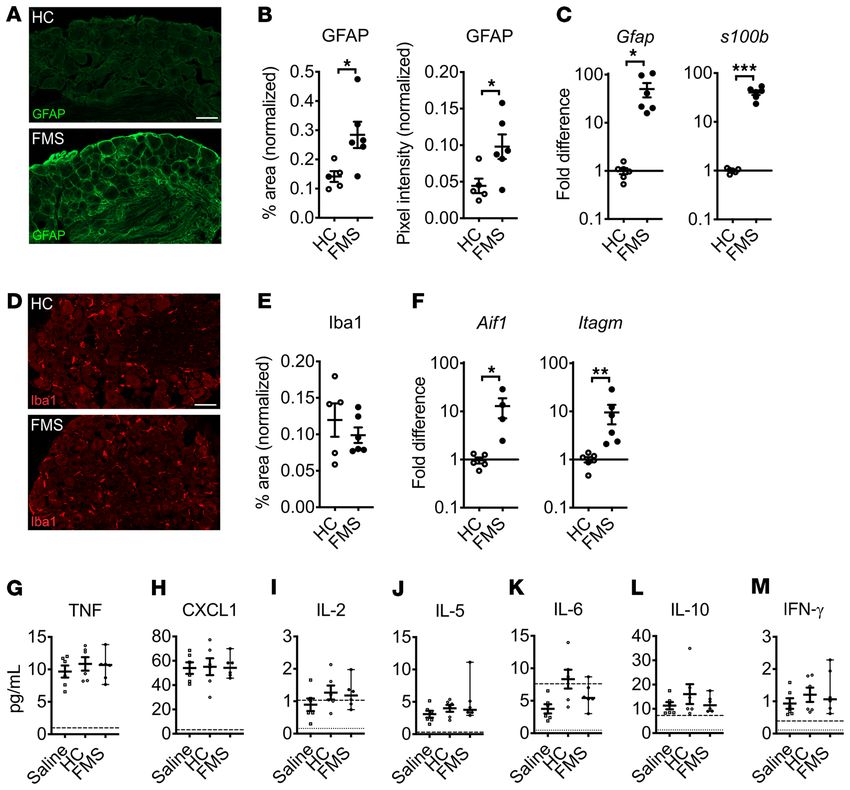
The Journal of Clinical Investigation RESEARCH ARTICLE
pixel intensity were much greater in mice that had been injected
with FMS IgG compared with IgG from HC subjects, and anti–
human IgG generated only minimal reactivity in DRG from saline-
injected mice (Figure 7, B and C).
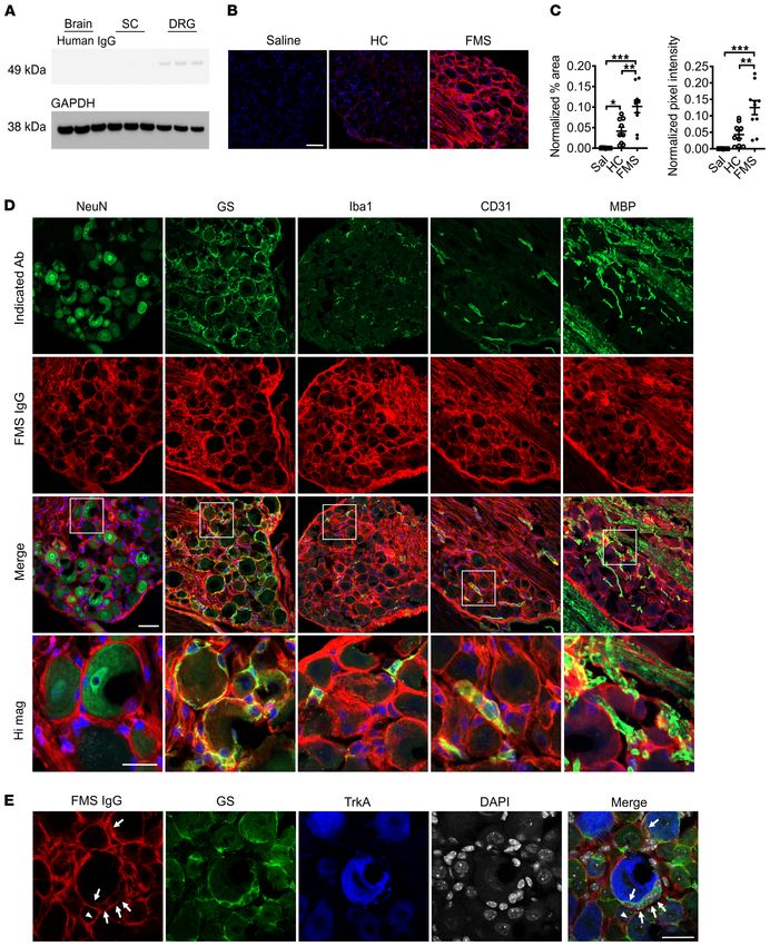
FMS patient IgG staining was primarily localized to satellite
glial cells (SGCs, visualized by glutamine synthase staining) and to
fiber tracts entering the DRG, as well as a small number of Iba1-
positive macrophages and CD31-positive blood vessels (Figure
7D). FMS IgG did not appear to colocalize with the neuronal nucle-
ar marker NeuN, which labels neuronal nuclei and to a lesser extent
cytoplasm. Since NeuN does not label neuronal membranes and
SGCs enwrap neurons with as little as 20 nm separation between
the cells, it was challenging to distinguish neuronal membrane
staining from that of the surrounding SGC (Figure 7D). We further
explored the possibility that FMS IgG also labels the membranes
of sensory neurons by using TrkA as a neuronal surface marker. In
addition to the extensive FMS IgG labeling of glutamine synthase–
Figure 4. IgG-depleted serum is inactive. IgG from an FMS patient, but
not IgG-depleted serum from the same patient or pooled from FMS or HC
expressing SGCs (Figure 7, D and E), FMS IgG also appeared to
cohorts, produced mechanical (A) and cold (B) hypersensitivities. **P < label some, but not all, TrkA-expressing neurons (Figure 7E).
0.01, ***P < 0.01, FM vs. HC IgG; 2-way repeated measure ANOVA followed FMS IgG promotes expression of SGC activity markers. Since
by Sidak’s correction. Data points are mean ± SEM of 6 mice per group. FMS IgG frequently colocalized with SGCs, we investigated
whether this was associated with evidence of changes in SGC
activity. We found that the positive area and pixel intensity of glial
FMS patients using microneurography (22) and demonstrate fibrillary acidic protein (GFAP) immunoreactivity (Figure 8, A and
that patient IgG produces a heightened peripheral nociceptor B) and Gfap and s100b gene expression (Figure 8C) in DRG from
responsiveness that is maintained in the absence of the central mice injected with FMS IgG were increased, compared with HC
nervous system. Since we had observed a robust behavioral cold IgG–injected controls. These changes suggest that the presence of
hypersensitivity in mice treated with FMS IgG, we also exam- FMS IgG in DRG had increased the activity of SGCs. In contrast,
ined whether this phenotype was accompanied by an increased the Iba1 percentage area of immunoreactivity in the neuron-rich
cold responsiveness in mechanosensitive C-fibers (Figure 6, area of the DRG was not different between FMS IgG– and HC
D–F). We challenged CM fibers with a 60-second cooling ramp IgG–injected mice (Figure 8, D and E), suggesting that FMS IgG
(from 32°C to 5°C–9°C) and noted that a larger proportion of CM accumulation in the DRG did not stimulate local macrophage
fibers responded to cold in preparations from mice treated with proliferation or infiltration. However, Aif1 (encoding Iba1) and
FMS IgG compared with HC IgG (Figure 6D). In contrast, FMS Itagm (encoding CD11b) were both elevated in the DRG of FMS
IgG did not influence the cold activation thresholds of cold- IgG–injected mice (Figure 8F), indicating that certain aspects of
sensitive CM (Figure 6E). An increasing number of human dis- macrophage activity were altered by FMS IgG. Importantly, there
orders are recognized as produced by functional autoantibodies were few to no apoptotic cells in the DRG of mice injected with
that alter excitability by binding to neuronal surface proteins FMS or HC IgG (Supplemental Figure 3A), indicating that the
(44). We therefore challenged isolated neurons directly with effects of FMS IgG are not due to neuronal cell death. In contrast
a high concentration of FMS IgG (200 μg/mL), to determine to the DRG, no changes in astrocyte and microglia reactivity were
whether FMS IgG directly excites dorsal root ganglia (DRG) observed in the spinal cord based on GFAP (Supplemental Figure
neurons by such an interaction. We observed no effect of FMS 2, B–D) and Iba1 (Supplemental Figure 2, E–G) immunoreactivity,
IgG on intracellular Ca2+ concentration ([Ca2+]i) in any of the 870 respectively. Together, these results suggest that FMS IgG has a
neurons examined (Figure 6G). local effect in the DRG.
In vivo immunohistochemical localization of human IgG. Since FMS IgG does not induce cytokine production or systemic
CM and AM fibers were hypersensitive in mice treated with FMS inflammation. Although there are inconsistencies between
IgG, we investigated the localization of FMS IgG to better under- published studies, levels of inflammatory and immunomodu-
stand potential sites of action. In mice injected with pooled IgG, latory cytokines and chemokines, such as IL-6, IL-8, and IL-10,
FMS IgG was consistently detected in DRG, but neither in brain have been reported to be elevated in FMS serum or plasma (22,
nor spinal cord tissue by Western blot analysis (Figure 7A; see 23) compared with HCs. Thus, we assessed a panel of factors
complete unedited blots in the supplemental material). Immuno- in serum from mice injected with FMS IgG, HC IgG, or saline.
histochemical analysis of tissues from mice that had been injected IL-1β was below the limit of detection in 5 of 6 samples in each
with pooled FMS IgG, using anti–human IgG antibodies to detect group, indicating that neither FMS IgG nor HC IgG induced
FMS IgG, revealed robust staining in the lumbar DRG (Figure 7, B IL-1β production. Similarly, there was no difference in TNF-α,
and C), while no specific immunoreactivity was observed in the CXCL1 (a murine analog of human IL-8), IL-2, IL-5, IL-6, IL-10,
spinal cord (Figure 7A and Supplemental Figure 2A). The percent- and IFN-γ levels between the groups (Figure 8, G–M). These
age area that was immunoreactive for human IgG as well as the data demonstrate that FMS IgG injection does not alter sys-
J Clin Invest. 2021;131(13):e144201 https://doi.org/10.1172/JCI144201 5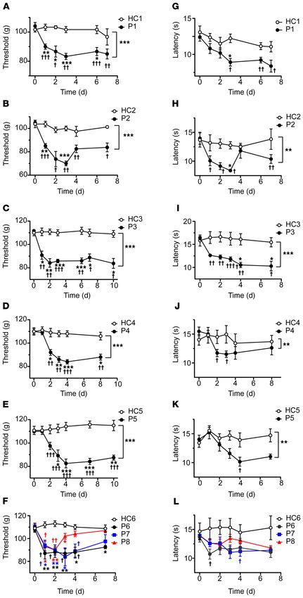
RESEARCH ARTICLE                                                                                         The Journal of Clinical Investigation

                                                                                           neurons. FMS IgG bound to a significantly larger propor-
                                                                                           tion of cells (290 of 368 cells analyzed, 79%) compared
                                                                                           with HC IgG (84 of 264 cells analyzed, 32%) (Figure
                                                                                           9, A–C), and recognized these cells more strongly, as
                                                                                           judged by pixel intensity (Figure 9D). Similarly, FMS IgG
                                                                                           labeled a significantly larger proportion of both gluta-
                                                                                           mine synthase–positive (SGCs) and –negative cells (non-
                                                                                           SGCs) in these cultures and did so with a greater pixel
                                                                                           intensity compared with HC IgG (Figure 9, E and F).
                                                                                                In dissociated DRG neuron cultures approximately
                                                                                           30% of cells were βIII-tubulin–positive neurons (Figure
                                                                                           9G). Similar to our observations with SGC-enriched cul-
                                                                                           tures, FMS IgG bound to a larger proportion of cells from
                                                                                           dissociated DRG (358 of 527 cells analyzed, 68%) com-
                                                                                           pared with HC IgG (158 of 469 cells analyzed, 34%), and
                                                                                           it also labeled these cells more intensely (Figure 9, H–J).
                                                                                           Importantly, FMS IgG labeled almost all βIII-tubulin–
                                                                                           positive neurons, whereas HC IgG labeled far fewer (Fig-
                                                                                           ure 9K). The pixel intensity of FMS IgG binding to neurons
                                                                                           was greater than FMS IgG binding to non-neuronal cells,
                                                                                           and greater than HC IgG binding to neurons and non-neu-
                                                                                           ronal cells (Figure 9L). Neither FMS IgG nor HC IgG
                                                                                           affected cell viability compared with cells that were not
                                                                                           incubated with antibodies (Supplemental Figure 3, B–E).
                                                                                           Finally, incubating live cultures with IB4, a membrane
                                                                                           marker of a subpopulation of nociceptors, demonstrated
                                                                                           that FMS IgG binding colocalized with neuronal mem-
                                                                                           branes (Figure 9M). In conclusion, FMS IgG binds SGCs in
                                                                                           vivo, and while further studies are warranted to determine
                                                                                           if FMS IgG also binds to neuronal membranes in vivo, this
                                                                                           possibility is supported by our data showing that FMS IgG
                                                                                           binds both SGCs and neurons in vitro.
Figure 5. Passive transfer of FMS IgG decreases locomotor activity. Visual analog
pain scores (VAS, A) and pressure-pain thresholds (PPT, B) reported by FMS patients             Reduced epidermal innervation. An involvement of
and healthy control (HC) subjects in Pool 3. Following transfer of HC or FMS IgG to        peripheral   sensory nerves in FMS has previously been
mice, locomotor activity was assessed over a 24-hour period using a Comprehensive          indicated by observations of small-fiber pathology in
Animal Lab Monitoring System. The total number of recorded movements was similar           patients (23, 45). Importantly, the symptom severity of
between HC IgG and FMS IgG mice during the day phase (low activity), but FMS
                                                                                           FMS is correlated with the extent of small-fiber pathol-
IgG–injected mice showed less activity during the night phase (high activity) (C). The
night phase was divided into 3 phases: 18:00–22:00 hours (D), 22:00–02:00 hours (E),       ogy (33). We therefore examined the impact of IgG on
and 02:00–06:00 hours (F), indicated by the dotted lines. Mice injected with FMS IgG       intraepidermal nerve fiber density (IENFD) in the gla-
displayed significantly less locomotor activity during the peak activity phase (E) com-    brous hind paw skin of mice. A significant reduction in
pared with HC IgG–injected mice. Data points are mean ± SEM, FMS n = 14, HC n = 11,        IENFD was apparent 14 days after administration of FMS
n =12 mice per group. *P < 0.05, ***P < 0.001 by Mann-Whitney U test (A and B),
                                                                                           IgG was initiated, compared with skin sections from mice
2-way ANOVA followed by Bonferroni’s correction (C), or unpaired t test (D–F).
                                                                                           similarly treated with HC IgG (Figure 10, A and B).
                                                                                                Screening FMS IgG reactivity with human protein frag-
                                                                                           ments. To investigate whether FMS IgG from different
temic inflammatory and immunomodulatory cytokine levels.                          patients contains a common pattern of autoreactivity, we used
Together, the results suggest that FMS IgG acts by generating a                   a proteome-wide microarray screen (SciLifeLab). This assay
local effect in the DRG.                                                          uses 42,000 human peptides (from >18,000 proteins) from the
     In vitro immunohistochemical binding of FMS IgG. To further                  Human Protein Atlas (46, 47). The peptides in the array are linear,
differentiate between labeling of SGCs and sensory neurons, we                    produced in E. coli, and most are between 50 and 150 amino acids
examined the immunoreactivity of FMS and HC IgG in SGC-                           in length. We examined the reactivity of 4 samples, each prepared
enriched, sensory neuron–depleted cell cultures and dissociated                   from equal parts of 4 FMS patients’ sera (4×4), and noted that all
DRG neuron–rich cell cultures. FMS IgG or HC IgG (pooled IgG,                     4 samples recognized multiple peptides (signal > mean fluores-
100 μg/mL) was added to the culture media prior to cell fixation                  cence intensity + [4 × SD]; Supplemental Table 5). However, our
in order to examine membrane labeling in a nonpermeabilizing                      analysis also revealed that very few peptides were recognized by
condition. In SGC-enriched cultures, approximately 75% of cells                   more than one of the pooled samples, strongly indicating that the
were glutamine synthase–positive SGCs and less than 0.5% were                     antigens included in the array are unlikely to represent a common

   6                                    J Clin Invest. 2021;131(13):e144201 https://doi.org/10.1172/JCI144201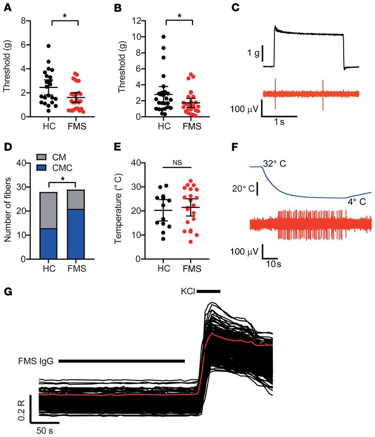
The Journal of Clinical Investigation                                                                                   RESEARCH ARTICLE

                                                                                                      pixel intensity (Figure 11, A and B). Fur-
                                                                                                      thermore, FMS IgG bound GFAP-im-
                                                                                                      munoreactive       cells      and     NF200-
                                                                                                      immunoreactive cells, indicating that FMS
                                                                                                      IgG binds to both human SGCs and human
                                                                                                      sensory neurons (Figure 11C). FMS IgG also
                                                                                                      labeled other cells, but less intensely. Taken
                                                                                                      together, these results suggest that FMS IgG
                                                                                                      contains autoreactive antibodies that bind
                                                                                                      antigens expressed in the DRG.

                                                                                                      Discussion
                                                                                                           Here we demonstrate that several key
                                                                                                           features of FMS can be induced in mice
                                                                                                           by IgG from individuals with FMS. Trans-
                                                                                                           fer of hypersensitivities from patients to
                                                                                                           mice was reproducible across all tested
                                                                                                           FMS subjects, strongly suggesting that
                                                                                                           antibody-dependent processes typically
                                                                                                           underpin the characteristic tenderness
                                                                                                           and thermal hypersensitivities experi-
                                                                                                           enced by patients. The pronociceptive
                                                                                                           effect of pooled IgG shows that samples
                                                                                                           from different patients have additive
                                                                                                           activity, since the IgG from each donor in
                                                                                                           these preparations was diluted to levels
                                                                                                           below those required to generate hyper-
                                                                                                           sensitivity in mice. The behavioral hyper-
                                                                                                           sensitivities produced by FMS IgG were
                                                                                                           reversible in mice and typically resolved
                                                                                                           after 2–3 weeks when the levels of human
                                                                                                           IgG have decreased (37). The reduction in
                                                                                                           grip strength and spontaneous locomotor
Figure 6. Passive transfer of FMS IgG sensitizes nociceptors. The mechanical activation thresholds
of (A) Aδ- (AM) and (B) C-mechanonociceptors (CM) were reduced in preparations from mice treated           activity, and the reduced density of epider-
with FMS compared with HC IgG (n = 22–27 single units). *P < 0.05 by 1-tailed Mann-Whitney U test.         mal innervation observed in mice treated
(C) The example trace illustrates a mechanical threshold response (evoked by the minimum force             with FMS IgG confirm that in addition to
required to elicit at least 2 spikes) in a CM unit. (D) The proportion of cold-sensitive CM units (CMCs)   painful hypersensitivities, FMS IgG also
was increased in preparations from mice treated with FMS IgG (21 of 29 units responded to cold)
                                                                                                           produces several non-evoked symptoms
compared with HC IgG (13 of 28 units responded to cold). *P < 0.05 by 1-sided Fisher’s exact test. (E)
The cold-activation thresholds of CMC fibers did not differ between FMS and HC preparations (n =           and signs of FMS. IgG-depleted serum
13–21). P > 0.05 by 1-tailed t test. (F) The example trace illustrates a cold-evoked response in a CMC     preparations were without any influence
fiber. (G) Application of FMS IgG (200 μg/mL) to isolated DRG neurons loaded with Fura-2 was with-         on mechanical or cold nociception, con-
out effect on [Ca2+]i in all 870 examined neurons (identified by their response to 50 mM KCl).             firming that IgG is the principal pronoci-
The red trace illustrates the average time course of the displayed 230 neurons.
                                                                                                           ceptive, pathological serum component in
                                                                                                           FMS patients. Importantly, FMS IgG binds
                                                                                                           to SGCs and neurons in mouse and human
autoantigen in FMS patients. Overrepresentation analysis (http://                    DRG, strongly suggesting that FMS patients have autoreactive
webgestalt.org/; ref. 48) suggested some degree of enrichment                        antibodies.
for proteins associated with Gene Ontology (GO) terms related to                          Interactions between the nervous and immune systems,
cellular components and microtubules (Supplemental Table 6 and                       and in particular autoimmune mechanisms, have increasingly
Supplemental Figure 4), and a small number of KEGG pathways                          attracted attention in studies of the pathophysiology of chronic
related to metabolism and vesicle cycling (Supplemental Table 7                      pain conditions (49, 50). Pronociceptive effects of autoantibodies
and Supplemental Figure 5).                                                          have been implicated in the uncommon posttraumatic condition
     FMS IgG binds to human DRG. Finally, we assessed whether                        complex regional pain syndrome (31, 51), where passive transfer
FMS IgG binds cells in human DRG. Human DRG sections were                            of IgG additionally requires an experimental trauma to exert its
incubated with either pooled HC IgG or FMS IgG and the intensity                     pronociceptive effects, unlike the observations presented here for
and localization of binding were analyzed. FMS IgG bound cells                       FMS IgG. In the very rare, frequently painful neurological condi-
in human DRG to a greater extent than HC IgG, as assessed by                         tion neuromyotonia, autoantibodies against contactin-associated

                                        J Clin Invest. 2021;131(13):e144201 https://doi.org/10.1172/JCI144201                                     7
RESEARCH ARTICLE The Journal of Clinical Investigation 8 J Clin Invest. 2021;131(13):e144201 https://doi.org/10.1172/JCI144201
The Journal of Clinical Investigation                                                                                    RESEARCH ARTICLE

Figure 7. FMS IgG accumulates in the DRG and binds satellite glial cells.         to hypersensitivity (56–58). Additionally, the accumulation of
Following IgG injection into mice, Western blot analysis detected FMS IgG         SGC- and neuron-bound FMS IgG may stimulate molecular
in the DRG but little to no IgG in spinal cords (SC) or brains (A) (pooled IgG,   changes in DRG-resident macrophages that contribute to neuro-
8 mg per day for 4 consecutive days, tissue collected after last injection).
FMS IgG, but not HC IgG, accumulates in the DRG 14 days after the first
                                                                                  nal hypersensitivity despite a lack of macrophage infiltration or
IgG injection (B and C). Human IgG is red and DAPI is blue. Human IgG             proliferation. Furthermore, the absence of systemic increases in
immunoreactivity in the neuron-rich area was quantified by assessing the          proinflammatory cytokines like TNF and IL-1β further supports
percentage area that was immunoreactive for human IgG and the mean                our hypothesis that FMS IgG acts locally in the DRG, without
pixel intensity of human IgG. Percentage area and pixel intensity were            inducing a systemic inflammatory response. Together, these
normalized to the DAPI signal (n = 9–10; data points are median ± 95% CI).
**P < 0.01, ***P < 0.001 by 1-way ANOVA followed by Tukey’s post hoc
                                                                                  findings point to the DRG as a site of action for FMS IgG and
test. FMS IgG immunoreactivity does not colocalize with neuronal NeuN             future studies will investigate the contributions of IgG acting on
staining but does colocalize with satellite glial cells (SGCs) (glutamate         SGCs and neurons in driving pain-related sensory abnormalities
synthase–expressing [GS-expressing] cells), some macrophages (Iba1-               and neuronal hypersensitivities in mice.
expressing cells) and blood vessels (CD31-expressing cells), and myelinated            Although we did not discover a characteristic pattern of
fiber tracts (myelin basic protein [MBP] staining), but not to myelinated
fibers in the DRG (D). To further delineate between SGCs and neuronal
                                                                                  reactivities in patient sera in the protein microarray, we did note
membranes, FMS IgG immunoreactivity colocalization was compared with              a substantial number of autoreactivities. Our results also indi-
GS and TrkA (a membrane receptor expressed by a subset of nociceptors).           cated some enrichment of reactivities against proteins classi-
FMS IgG colocalizes with GS-expressing SGCs (white arrows) but may also           fied in GO terms for cellular components, and KEGG terms for
infrequently bind to TrkA-positive neuronal cell membranes (white trian-          pathways related to metabolism. Although the potential enrich-
gles) (E). Scale bars indicate 50 μm, except the high-magnification image
scale bar and scale bar in E, which indicate 25 μm.
                                                                                  ment of reactivities may be encouraging, their relevance to the
                                                                                  pathophysiology of FMS is currently unclear, since they were
                                                                                  mostly detected in single samples. Furthermore, most proteins
                                                                                  relevant to the potentially enriched terms are located intracellu-
protein-like 2 (CASPR2) are commonly detected (28). Passive                       larly and are thus unlikely to be recognized by circulating IgG.
transfer of IgG from these patients produces painful hypersensi-                  Genuine autoantigens may also have escaped detection, since
tivities by internalizing voltage-gated K+ channels. Our findings                 the linearized antigenic protein fragments used in the screen are
indicate that similarly nondestructive processes produce hyper-                   unlikely to adopt their native conformation on the chip (47). The
sensitivities, motor symptoms, and loss of epidermal innervation                  peptides and protein fragments were produced in E. coli, and
in the common condition FMS.                                                      therefore lack posttranslational modifications. The prevalence
      The importance of altered central nervous system function                   of reactivities against human peptides in our preliminary assess-
and plasticity in FMS is illustrated by altered brain activity pat-               ment of patient sera is nevertheless consistent with autoreactive
terns (19), glia activation in the brain (52), impaired conditioned               IgG being responsible for our findings. Intriguingly, sera from
pain modulation (53), and multisensory hypersensitivity to a wide                 COVID-19 patients contain a wide range of functional autoanti-
range of stimuli (1, 54, 55). However, it is becoming increasingly                bodies, which have been proposed to influence the symptomatic
clear that peripheral alterations also contribute to the underlying               profile in patients (59).
pathology, as exemplified by identification of small-fiber patholo-                    Small-nerve-fiber pathology and a reduction in skin innerva-
gy in FMS patients (33) and altered cytokine levels (24). Here we                 tion has been observed in FMS patients, and is associated with a
found that mechanosensitive nociceptors in skin-nerve prepara-                    more severe disease burden and may contribute to peripheral sen-
tions from FMS IgG–treated mice displayed an increased respon-                    sitivity (33). Remarkably, we observed that transfer of FMS IgG to
siveness to mechanical and cold stimuli, strongly suggesting that                 mice resulted in a reduction in IENFD in glabrous hind paw skin.
painful hypersensitivity in FMS is produced by sensitization of                   In humans, it is still unclear why reduced skin innervation occurs,
peripheral nociceptors. In contrast, direct application of IgG to                 but our findings indicate that FMS IgG contributes to this fiber
isolated DRG neurons was without effect on [Ca2+]i, consistent                    loss. FMS IgG may drive denervation from the DRG via SGC or
with an indirect action that may involve activity of an additional                neuronal cell body binding but may also act more distally through
peripheral cell type, such as SGCs.                                               other mechanisms.
      In combination with the electrophysiological observations,                       Future studies will examine the underlying cellular and molec-
retention of FMS IgG in the DRG, the lack of IgG in the brain                     ular mechanisms responsible for FMS IgG–mediated peripheral
and spinal cord, and the in vivo binding of FMS IgG to SGCs and                   sensitization. The possibility that the heightened noxious periph-
neurons strongly suggest that the pronociceptive actions of FMS                   eral input produced by FMS IgG may generate altered patterns
IgG were driven by peripheral mechanisms. FMS IgG binding                         of activity in the central nervous system (19) also remains to be
in human DRG and in vitro binding of FMS IgG to the surface                       explored. Similarly, whether FMS IgG drives sensitivity to other
of murine SGCs and neurons further emphasizes that the noci-                      sensory modalities, such as to auditory and olfactory stimuli, still
ceptive effects induced by FMS IgG may stem from recognition                      needs to be investigated. The identification of a pivotal role for
of epitopes in the DRG. Increased signs of in vivo SGC activity                   autoreactive IgG in the pathophysiology of FMS may transform
imply that FMS IgG binding in the DRG has functional effects.                     future research and facilitate development of mechanism-based
It is clear that SGCs and sensory neurons can be electrically cou-                therapeutic interventions. Our results suggest that therapies
pled and that both increased coupling and SGC-derived prono-                      which reduce the total IgG titer, such as plasmapheresis or immu-
ciceptive cytokines can influence neuronal excitability leading                   noadsorption (e.g., with protein A columns), or which specifically

                                            J Clin Invest. 2021;131(13):e144201 https://doi.org/10.1172/JCI144201                                9
RESEARCH ARTICLE The Journal of Clinical Investigation
Figure 8. FMS IgG increases signs of satellite glial cell activity in vivo but does not drive systemic inflammation. DRG from FMS IgG–injected mice have
increased GFAP immunoreactivity (A), which is indicative of increased satellite glial cell activity, compared with HC IgG injected mice when the percentage
area of GFAP immunoreactivity and GFAP mean pixel intensity are quantified and normalized to the DAPI signal (B). Gfap and s100b gene expression is
elevated in the DRG of mice injected with FMS IgG compared with HC IgG (C). The number of Iba1-immunoreactive macrophages was unchanged, as was
the percentage area of Iba1 immunoreactivity, when comparing HC IgG– and FMS IgG–injected mice (D and E). Gene expression of Aif1 (Iba1 gene) and
Itagm (gene for CD11b, another macrophage marker) was elevated in FMS IgG–injected mice compared with HC IgG–injected mice (F). Scale bars: 50 μm.
qPCR data were normalized to Hprt1 expression analyzed using the 2–ΔΔCt method. Serum levels of TNF (G), CXCL1 (H), IL-2 (I), IL-5 (J), IL-6 (K), IL-10 (L), and
IFN-γ (M) were measured and there were no differences between groups. The dashed lines indicate the lower limit of quantification (LLOQ) and the dotted
lines indicate the lower limit of detection (G–M). Line and whiskers indicate mean ± SEM (n = 4–6). Differences between FMS IgG and HC IgG were analyzed
with Mann-Whitney U test (B–F). Cytokine levels were compared between saline, HC, and FMS with 1-way ANOVA followed by Tukey’s post hoc test for
each analyte (G–M) except IL-6, which was not analyzed statistically because most values were below the LLOQ. *P < 0.05, **P < 0.01, ***P < 0.001.
reduce autoreactive IgG (using antigen-specific adsorption) may tigated whether primary sensory afferents were sensitized following
be effective for FMS (60). Alternatively, symptomatic therapies transfer of FMS or HC IgG into mice using an ex vivo skin-nerve prepa-
that interfere with the binding of autoreactive antibodies or pre- ration. Third, we examined FMS IgG localization following transfer
vent their functional consequences may also provide effective and cell-type binding in vivo and in vitro with Western blotting and
treatment approaches. immunofluorescence. Last, we assessed IgG-induced changes in skin
innervation using immunofluorescence. Mechanical hypersensitivity
Methods is a hallmark of FMS and the Randall-Selitto paw-pressure test is a
Study design. Our goal was to determine whether administration of IgG sensitive translational measure of this symptom.
from FMS patients to mice transfers characteristic symptoms of FMS. Power calculations indicated that n = 4 would be sufficient to
FMS patients and age-matched HC individuals were recruited at 2 dif- detect a 15-g reduction of the paw withdrawal threshold with α < 0.025
ferent sites, the University of Liverpool and the Karolinska Institute, and 1 – β > 0.9 (6 repeated measures, 2-sided), but n = 6 to detect a
where serum was collected and IgG was purified. First, we assessed reduction in the cold-plate paw withdrawal latency of 2.5 seconds.
whether IgG from FMS patients drove pain-like behavior in mice by Electrophysiological experiments were analyzed using 1-sided tests
transferring IgG from single individuals into mice as well as transfer- since we examined whether the increased sensitivity observed in vivo
ring IgG pooled from several individuals into mice. Second, we inves- was associated with an increased (not decreased) single unit respon-
10 J Clin Invest. 2021;131(13):e144201 https://doi.org/10.1172/JCI144201The Journal of Clinical Investigation RESEARCH ARTICLE
Figure 9. FMS IgG binds to satellite glial cells and to neurons in vitro. Live cells were incubated with FMS IgG or HC IgG to examine only cell surface
binding. In satellite glial cell (SGC) cultures (A), FMS IgG labeled a greater percentage of cells than HC IgG when analyzed per animal (B) and by the total
number cells (C). The immunoreactivity, analyzed as signal intensity (integrated density), was higher in cells incubated with FMS IgG than HC IgG (D).
FMS IgG labeled SGCs (GS+) and non-SGCs (GS–) to a greater extent (percentage, E) and with a higher intensity (F) than HC IgG. FMS and HC IgG labeling
of neuronal cultures (G) was not different when considering the percentage of cells labeled per animal (H), but a difference was observed when the total
number of cells was considered (I). The signal intensity was higher for cells exposed to FMS IgG than HC IgG (J). FMS IgG labeled neurons (βIII-tubulin+) and
non-neurons (βIII-tubulin–) to a greater extent than HC IgG (K). The signal intensity of FMS IgG binding to neurons was greater than FMS IgG binding to
non-neuronal cells and HC IgG binding to all cells (L). FMS IgG and IB4 colocalization (M) indicates that FMS IgG binds neuronal cell membranes. All scale
bars: 20 μm. Data points are the percentage of cells bound by HC or FMS IgG (B and H). In D, F, J, and L data points are the integrated density of individual
cells across 3 experiments. Bar and whiskers indicate mean ± SEM (n = 3 individual experiments). *P < 0.05; ***P < 0.001 by unpaired t test (B, D, H, and J),
χ2 test (C, E, I, and K), or Kruskal-Wallis test with Dunn’s post hoc test (F and L).
siveness. All other data were analyzed using 2-sided tests. Behavioral tute. Immunofluorescence, Western blotting, and qPCR experiments
(except the Comprehensive Animal Lab Monitoring System [CLAMS]) were performed with tissues from behavioral experiments using IgG
and electrophysiological experiments were performed at King’s Col- pools. Immunocytochemistry experiments were performed using IgG
lege London and CLAMS, immunofluorescence, Western blotting, pools. Sample sizes for each experiment are additionally indicated in
qPCR, and cell culture studies were performed at the Karolinska Insti- the figure legends.
J Clin Invest. 2021;131(13):e144201 https://doi.org/10.1172/JCI144201 11RESEARCH ARTICLE The Journal of Clinical Investigation
10K); concentration measurements were performed
using a Nanodrop 2000 (Thermo Fisher Scientific).
Before eluting the IgG from the protein G column,
the IgG-depleted sera (flow through) was collected
and stored at –80°C for later use. Prior to injection,
the flow through was concentration-adapted using
PBS-prewet concentration columns and the concen-
tration was determined with a Nanodrop 2000.
Animals. Behavioral experiments were per-
formed on female C57BL/6J mice (8–10 weeks old)
obtained from Envigo UK Ltd and housed in a tem-
perature-controlled environment with a 12-hour
light/12-hour dark cycle with access to food and
water ad libitum. Locomotor activity assessment and
immunofluorescence experiments were performed
on 4-month-old female BALB/c mice (Janvier). Mice
Figure 10. FMS IgG transfer decreases intraepidermal nerve fiber density. Intraepidermal were injected intraperitoneally with IgG from HC
nerve fibers (IENFs) were identified in the glabrous hind-paw skin with an anti–PGP 9.5 subjects or FMS patients on 1–4 consecutive days.
antibody (A). The number of IENFs crossing from the dermis to the epidermis was decreased
Behavioral studies. Before any nociceptive test-
following transfer of FMS IgG compared with HC IgG 14 days after the first injection (B) (pooled,
8 mg per day for 4 consecutive days). Scale bar: 20 μm. Data points are mean ± SEM (n = 7). *P ing, mice were kept in their holding cages to acclima-
< 0.05 by unpaired t test. tize (10–15 minutes) to the experimental room. Mice
were randomized between cages and the experi-
menter was blinded to their treatment.
Patient samples. Serum samples for individual testing were The Randall-Selitto paw-pressure test was performed using an
derived from UK patients managed for their fibromyalgia at a depart- Analgesy-Meter (Ugo-Basile). The experimenter lightly restrained
ment of pain medicine, or from age- and sex-matched HCs between the mouse and applied a constantly increasing pressure stimulus to
April 2017 and November 2018. Pooled samples were obtained from the dorsal surface of the hind paw using a blunt conical probe. The
Swedish patients between September 2015 and December 2016 or nociceptive threshold was defined as the force in grams at which the
HCs responding to a study advert between March 2016 and March mouse withdrew its paw (64). A force cutoff value of 150 g was used to
2017. All patients had been examined by a consultant rheumatolo- avoid tissue injury. The thigh-pressure test was also performed using an
gist, and UK patients had additionally been examined by a consul- Analgesy-Meter. A constantly increasing pressure was applied to the
tant in pain medicine. inner thigh muscle of lightly restrained mice, using a blunt, wedge-
We purified serum IgG from 44 FMS patients and 39 HC sub- shaped probe and the force at which the mouse withdrew its leg was
jects. All patients fulfilled both 1990 and 2011 ACR diagnostic cri- recorded as the nociceptive threshold.
teria for fibromyalgia (61, 62). Most patients (42 of 44) and all of the Tactile sensitivity was assessed using von Frey filaments (0.008–2
pooled-sample donors were unaffected by other sensory, autoim- g) according to Chaplan’s up-down method (65). Animals were placed
mune, or rheumatological conditions. Most patient donors (43 of 44) in a Perspex chamber with a metal grid floor allowing access to their
were women. The donors’ demographics and disease characteristics plantar surface and allowed to acclimatize prior to the start of the
are provided in Supplemental Tables 1 and 2. experiment. The von Frey filaments were applied to the plantar surface
Purification of immunoglobulins. IgG for individual testing was of the hind paw with enough force to allow the filament to bend, and
purified as described previously (63), using protein G beads (Sigma- held static for approximately 2–3 seconds. The stimulus was repeated
Aldrich). Serum was diluted 1:3 with Hartmann’s solution, passed up to 5 times at intervals of several seconds, allowing for resolution
through a protein G column, and the bound IgG was eluted using 100 of any behavioral responses to previous stimuli. A positive response
mM glycine pH 2.3; after elution the pH was adjusted to 7.4 using 1 M was noted if the paw was sharply withdrawn in response to filament
Tris pH 8 and then the eluate was dialyzed overnight at 4°C in Hart- application or if the mouse flinched upon removal of the filament.
mann’s using a 10 kDa dialysis membrane (Thermo Fisher Scientific). Any movement of the mouse, such as walking or grooming, was
The concentration of IgG present after dialysis was determined using deemed an unclear response, and in such cases the stimulus was
a modified Lowry assay (DC protein assay, Bio-Rad) and adjusted by repeated. If no response was noted, a higher-force hair was tested and
dilution with Hartmann’s or by concentrating dialysis against a sucrose the filament producing a positive response recorded as the threshold.
solution (Sigma-Aldrich). Finally, the IgG solution was sterile filtered Thermal sensitivity was assessed using a cold plate (Ugo Basile).
using syringe-driven 0.2 μm filter units (Millipore), stored at 4°C, and Paw withdrawal latencies were determined with the plate set at 10°C.
used within 3 months. Sera for pooled sample testing were purified The animals were lightly restrained (scruffed) and the left hind paw
using HiTrap Protein G HP columns (GE Healthcare), eluted with 0.1 M was placed onto the surface of the plate (66, 67). The latency to with-
glycine/HCl pH 2.7, and the pH adjusted to 7.4 with 1 M Tris pH 9; con- drawal of the paw was recorded as the endpoint. A maximum cutoff of
sequently, samples were dialyzed against PBS, concentration adjusted, 30 seconds was used for each paw.
stored at –20°C, and later thawed, pooled, and concentration-adapted Forelimb grip strength performance was assessed using a commer-
using PBS-prewet concentration columns (Pall Corporation, Macrosep cially available grip strength meter (Ugo Basile), incorporating a T-bar
12 J Clin Invest. 2021;131(13):e144201 https://doi.org/10.1172/JCI144201The Journal of Clinical Investigation RESEARCH ARTICLE
Figure 11. FMS IgG binds human DRG. FMS IgG bound human
DRG tissue sections more intensely than HC IgG when assessed
by integrated density normalized to DAPI (A and B). The indicated
IgG is in white in the top row and DAPI is in blue in the bottom row
of A. High-magnification images of FMS IgG (white) demonstrate
colocalization (purple) with GFAP-immunoreactive satellite glial cells
(green) and NF-200–immunoreactive neurons (green) (C). Scale bars:
50 μm (A) and 20 μm (C). Data are mean ± SEM (n = 5 independently
stained slides). ***P < 0.001 by unpaired t test.
and force transducer. The mice were held gently by the base of the tail 26.2 mM NaCO3, 1.65 mM NaH2PO4, 1.53 mM CaCl2, 9.6 mM sodium
and pulled across the horizontal T-bar so that they were able to grip the gluconate, 5.55 mM glucose, and 7.6 mM sucrose. The skin was
bar with their fore paws. A digital readout of the maximum force applied placed inside up (corium side up) and pinned down using insect pins
is given once the grip is released. Mice were acclimatized to the appara- (0.2 mm diameter) in the organ bath to allow access to the receptive
tus by testing twice prior to starting the study. The test was performed at fields. The saphenous nerve was threaded through a small gap from
least twice on each mouse and the mean force value used. the organ bath to an adjacent recording chamber and positioned on a
Locomotor activity was assessed with CLAMS (Columbus Instru- mirror platform. The desheathed saphenous nerve was covered with
ments) cages for 48 hours. Mice were singly housed and habituated to paraffin oil for electrical isolation and the dissected fine nerve fila-
the cages for 24 hours starting during the night 4 days following anti- ments were visualized with a microscope and placed on a gold wire
body injection. Following habituation, locomotor activity was assessed recording electrode (42, 43).
by beam-break recording. The CLAMS cage has a grid of infrared beams Conduction velocity. The saphenous nerve was divided into pro-
in the x-y plane and records infrared beam breaks that result from the gressively thinner filaments until a single unit could be isolated in
mouse moving in the cage. Beam breaks are counted and used to assess response to mechanical stimulation of the receptive field with a
the total movement of the mouse over a 24-hour period. Total move- glass rod. The electrical latency of identified units (Digitimer DS2,
ment is analyzed by the number of beam breaks (counts) in 20-minute Digitimer Ltd) was used to determine the conduction velocity and to
bins over a 24-hour period. The night phase (peak activity phase) was categorize units as Aβ (velocity > 10 m/s), Aδ (1.2 < velocity < 10 m/s),
further subdivided in to 4-hour bins from 18:00–6:00 hours. or C-fibers (0 < velocity < 1.2 m/s).
Skin-nerve recording. Mice were killed by cervical dislocation and Mechanical and cold stimulation. A computer-controlled stim-
the hind paw was shaved prior to dissection of the isolated skin-nerve ulating probe, equipped with a force transducer, was used to deliver
preparation. The saphenous nerve and the shaved skin of the hind mechanical stimuli to the most sensitive point of a receptive field
limb were placed in a recording chamber at 32°C. The chamber was (Avere Solutions UG) of AM and CM fibers in preparations from mice
perfused with a gassed (95% O2 and 5% CO2), prewarmed, synthetic treated with FMS or HC IgG. Recording and analysis were done using
interstitial fluid (SIF): 108 mM NaCl, 3.5 mM KCl, 0.7 mM MgSO4, Spike 2 (Cambridge Electronic Design). Cold sensitivity of CM units
J Clin Invest. 2021;131(13):e144201 https://doi.org/10.1172/JCI144201 13You can also read