NARBreakthroughArticle 2 -phosphotransferase Tpt1 provide insights into NAD+ binding and specificity
←
→
Page content transcription
If your browser does not render page correctly, please read the page content below
Published online 21 April 2021 Nucleic Acids Research, 2021, Vol. 49, No. 17 9607–9624
https://doi.org/10.1093/nar/gkab241
NAR Breakthrough Article
NMR solution structures of Runella slithyformis RNA
2 -phosphotransferase Tpt1 provide insights into
NAD+ binding and specificity
Sébastien Alphonse1 , Ankan Banerjee 2
, Swathi Dantuluri2 , Stewart Shuman 2,*
and
Downloaded from https://academic.oup.com/nar/article/49/17/9607/6240602 by guest on 28 September 2021
Ranajeet Ghose 1,3,4,5,*
1
Department of Chemistry and Biochemistry, The City College of New York, New York, NY 10031, USA, 2 Molecular
Biology Program, Sloan-Kettering Institute, New York, NY 10021, USA, 3 Graduate Program in Chemistry, The
Graduate Center of CUNY, New York, NY 10016, USA, 4 Graduate Program in Biochemistry, The Graduate Center of
CUNY, New York, NY 10016, USA and 5 Graduate Program in Physics, The Graduate Center of CUNY, New York, NY
10016, USA
Received February 15, 2021; Revised March 16, 2021; Editorial Decision March 18, 2021; Accepted March 23, 2021
ABSTRACT transition-state during the first step of the Tpt1-
catalyzed reaction.
Tpt1, an essential component of the fungal and
plant tRNA splicing machinery, catalyzes transfer of
INTRODUCTION
an internal RNA 2 -PO4 to NAD+ yielding RNA 2 -
OH and ADP-ribose-1 ,2 -cyclic phosphate products. In fungal and plant tRNA splicing, ligation of exons by
Here, we report NMR structures of the Tpt1 ortholog tRNA ligase results in a 2 -PO4 , 3 ,5 -phosphodiester splice
from the bacterium Runella slithyformis (RslTpt1), as junction. This internal 2 -PO4 is converted to a 2 -OH by
apoenzyme and bound to NAD+ . RslTpt1 consists of the action of an NAD+ -dependent 2 -phosphotransferase
enzyme, Tpt1 (1,2), via a two-step reaction (Supplementary
N- and C-terminal lobes with substantial inter-lobe
Figure S1). In the first step, a nucleophilic attack on NAD+
dynamics in the free and NAD+ -bound states. ITC by the RNA 2 -PO4 generates an RNA 2 -phospho-(ADP-
measurements of RslTpt1 binding to NAD+ (KD ∼31 ribose) intermediate with the expulsion of nicotinamide. A
M), ADP-ribose (∼96 M) and ADP (∼123 M) indi- subsequent transesterification reaction yields ADP-ribose-
cate that substrate affinity is determined primarily 1 ,2 -cyclic phosphate and a 2 -OH at the splice site (3).
by the ADP moiety; no binding of NMN or nicoti- Tpt1 is an essential component of the tRNA splicing ma-
namide is observed by ITC. NAD+ -induced chemi- chinery in Saccharomyces cerevisiae and its deletion or in-
cal shift perturbations (CSPs) localize exclusively activation by active site mutation is lethal (4,5). Tpt1 ho-
to the RslTpt1 C-lobe. NADP+ , which contains an mologs are found in bacterial, archaeal and metazoan pro-
adenylate 2 -PO4 (mimicking the substrate RNA 2 - teomes, possess NAD+ -dependent 2 -phosphotransferase
PO4 ), binds with lower affinity (KD ∼1 mM) and elicits activity in vitro, and can function in lieu of S. cerevisiae Tpt1
in vivo (6–8). Yet, the role of these enzymes in their native
only N-lobe CSPs. The RslTpt1·NAD+ binary com-
taxa is unclear, insofar as these organisms are not known
plex reveals C-lobe contacts to adenosine ribose to have physiologic RNA transactions that yield internal 2 -
hydroxyls (His99, Thr101), the adenine nucleobase PO4 substrates for Tpt1. Moreover, a mouse Tpt1 knockout
(Asn105, Asp112, Gly113, Met117) and the nicoti- elicits no physiological defects (9). Given their essential role
namide riboside (Ser125, Gln126, Asn163, Val165), in fungal tRNA splicing and the lack of an obvious role in
several of which are essential for RslTpt1 activity in mammalian physiology, Tpt1 enzymes are attractive targets
vivo. Proximity of the NAD+ -phosphate to ribose- for the development of new anti-fungals.
C1 suggests that it may stabilize an oxocarbenium Understanding how Tpt1 recognizes NAD+ and an in-
ternal 2 -PO4 -containing RNA and performs its unique 2 -
* To
whom correspondence should be addressed. Tel: +1 212 650 6049; Email: rghose@sci.ccny.cuny.edu
Correspondence may also be addressed to Stewart Shuman. Tel: +1 212 639 7145; Email: shumans@mskcc.org
C The Author(s) 2021. Published by Oxford University Press on behalf of Nucleic Acids Research.
This is an Open Access article distributed under the terms of the Creative Commons Attribution-NonCommercial License
(http://creativecommons.org/licenses/by-nc/4.0/), which permits non-commercial re-use, distribution, and reproduction in any medium, provided the original work
is properly cited. For commercial re-use, please contact journals.permissions@oup.com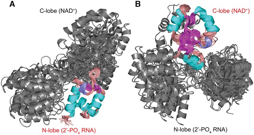
9608 Nucleic Acids Research, 2021, Vol. 49, No. 17
phosphotransferase chemistry hinges on capturing struc- kanamycin (35 mg/l) and grown overnight at 37◦ C. The
tures of the enzyme at each stage along its reaction path- starter culture was then used to inoculate 1 L of LB supple-
way. Initial low-resolution structural probing of yeast and mented with kanamycin (35 mg/l) and grown at 37◦ C until
Escherichia coli Tpt1 enzymes by limited proteolysis indi- the A600 reached 0.8. Isopropyl--D-thiogalactoside (IPTG)
cated that they consist of folded N- and C-terminal do- was then added to a final concentration of 500 M together
mains separated by a protease-sensitive interdomain linker with 2% (v/v) ethanol, and the culture was incubated for 18
(5). A 2.8 Å crystal structure of a Tpt1 homolog from the h at 17◦ C with continuous shaking at 250 rpm. Cells were
hyperthermophilic archaeon Aeropyrum pernix (ApeTpt1) harvested by centrifugation at 4,000 rpm for 30 min using
confirmed its two-lobed fold (10), but did not provide in- a Fiberlite F12–6 × 500 LEX (Thermo Fisher Scientific).
sights into substrate recognition or catalysis. Subsequent Cell pellets were stored at -80◦ C.
biochemical studies verified that ApeTpt1 is a bona fide RslTpt1 samples that were either uniformly 13 C,15 N-
NAD+ -dependent 2 -phosphotransferase (11). Tpt1 from labeled (CN-labeled) or specifically 13 CH3 -labeled at Val-
the mesothermophilic bacterium Clostridium thermocellum ␥ 1/2, Leu-␦1/2, Met-ε, Ile-␦1 positions in an other-
Downloaded from https://academic.oup.com/nar/article/49/17/9607/6240602 by guest on 28 September 2021
(CthTpt1) was recently crystallized and its structure was wise 2 H (except for Met sidechains that were fully
solved at 1.4 Å resolution. The CthTpt1 structure fortu- protonated),12 C,15 N background (ILVM-labeled) were pre-
itously contained co-enzyme A (CoA) and ADP-ribose-1 - pared for the NMR studies. Overnight starter cultures
phosphate engaged to its N- and C-terminal lobes, respec- in M9 medium supplemented with kanamycin (35 mg/l)
tively (12). This structure likely mimics a product state with were used to inoculate either H2 O-based M9 media sup-
the pAp moiety of CoA representing the 2 -OH RNA prod- plemented with 3 g/l of 13 C-glucose (for CN-labeled sam-
uct and the ADP-ribose-1 -phosphate corresponding to the ples or 12 C-glucose for samples that were only uniformly
hydrolyzed remnant of the ADP-ribose-1 ,2 -cyclic phos- 15
N-labeled) and 15 NH4 Cl or D2 O-based M9 media supple-
phate product (12). Whereas this structure represents a sig- mented with 1 g/l 15 NH4 Cl and 2 g/l of 2 H,13 C-glucose (for
nificant advance in understanding the mechanism of the ILVM-labeled samples). For CN (or N) labeling, the cul-
second transesterification step of the Tpt1 pathway, insights tures were grown at 37◦ C under gentle agitation at 250 rpm
on the structural and dynamic interactions in the reactant until the A600 reached 0.7. Protein expression was then in-
states bound to an intact NAD+ and/or an intact 2 -PO4 - duced by the addition of 500 M IPTG and 2% ethanol and
modified RNA prior to step 1 chemistry are still lacking. the cultures were subsequently incubated for 18 h at 17◦ C.
Here we utilize solution NMR methodology to determine For ILVM-labeling, the cells were grown at 37◦ C under gen-
the structures of the substrate-free state of Tpt1 from the tle agitation at 250 rpm until the A600 reached 0.6, at which
bacterium Runella slithyformis (RslTpt1) (6) and that of point 100 mg/l of methyl-13 C, 3,3-D2 ␣-keto-butyric acid
its complex with NAD+ . RslTpt1 displays significant inter- and 250 mg/l of 3–3 -dimethyl-13 C2 , 3-D ␣-keto-isovaleric
lobe mobility in both the apoenzyme and NAD+ -bound acid were added to the medium and growth was continued
states. Biophysical assays and supporting NMR measure- at 37◦ C for an additional hour. The culture was then slowly
ments indicate that a majority of the binding energy for cooled on ice for 30 min, 120 mg/l of uniformly 13 C-labeled
the interaction of RslTpt1 with NAD+ , which localizes ex- methionine was added to the growth medium, and protein
clusively to the C-terminal lobe of the enzyme, is provided expression was induced by the addition of 500 M IPTG.
by the ADP moiety. NMN and nicotinamide do not bind Overexpression continued for 22 h at 17◦ C. The cells were
detectably to RslTpt1 in isolation, and these moieties con- harvested as before and stored at -80◦ C.
tribute modestly to the affinity for NAD+ . NADP+ , in con- Cell pellets were thawed and resuspended in 45 ml of ly-
trast to NAD+ , binds exclusively to the N-terminal lobe of sis buffer (50 mM Tris pH 7.5, 1 M NaCl, 10% glycerol,
RslTpt1, in effect behaving as a low-affinity mimetic of the 0.5% Triton X100, 0.05% NaN3 ) and supplemented with
nucleoside 2 -PO4 at the splice junction. The NMR struc- one tablet of protease inhibitor (EDTA-free, Pierce) and 1
ture of the binary RslTpt1·NAD+ complex provides the first mg/ml of lysozyme. The samples were kept at 4◦ C under
detailed insight into the recognition of NAD+ prior to step gentle rotation for 1 h, then lysed by sonication (7 min, pulse
1 chemistry and identifies enzymic contacts to NAD+ by on for 0.3 s, pulse off for 0.7 s, power amplitude at 35%) us-
C-lobe amino acid residues conserved among Tpt1 fam- ing a Sonic Dismembrator Model 500 (Thermo Fisher Sci-
ily members. Structure-guided alanine mutagenesis identi- entific). The cell debris was separated by centrifugation at
fies stacking interactions with the adenine nucleobase and 15 000 rpm for 30 min (using a Fiberlite F21–8 × 50y ro-
nicotinamide moieties of NAD+ as essential for RslTpt1 tor). The supernatant was then incubated with nickel NTA
function in vivo, as gauged by yeast tpt1 complementa- agarose beads (GoldBio), which were pre-washed with lysis
tion. buffer, for 1 h at 4◦ C under gentle rotation and the slurry
was then poured into an Econo-Pac chromatography col-
MATERIALS AND METHODS umn (Bio-Rad). The column flow-through was discarded
and beads were washed, first with 9 column volumes of ly-
Protein expression and purification sis buffer, then with 9 column volumes of washing buffer 1
The plasmid pET28a-His6 -Tpt1, encoding residues Val5 (50 mM Tris-HCl, pH 7.5, 3 M KCl, 0.05% NaN3 ) and fi-
to Val178 of RslTpt1, was transformed into E. coli One nally with 9 column volumes of washing buffer 2 (50 mM
Shot BL21 Star (DE3) cells (Thermofisher). For the ex- Tris-HCl, pH 7.5, 500 mM NaCl, 10% glycerol, 20 mM
pression of unlabeled RslTpt1, colonies were inoculated imidazole, 0.05% NaN3 ). The bound protein was eluted
into starter Luria-Bertani (LB) medium supplemented with with 50 mM Tris-HCl, pH 7.5, 500 mM NaCl, 10% glyc-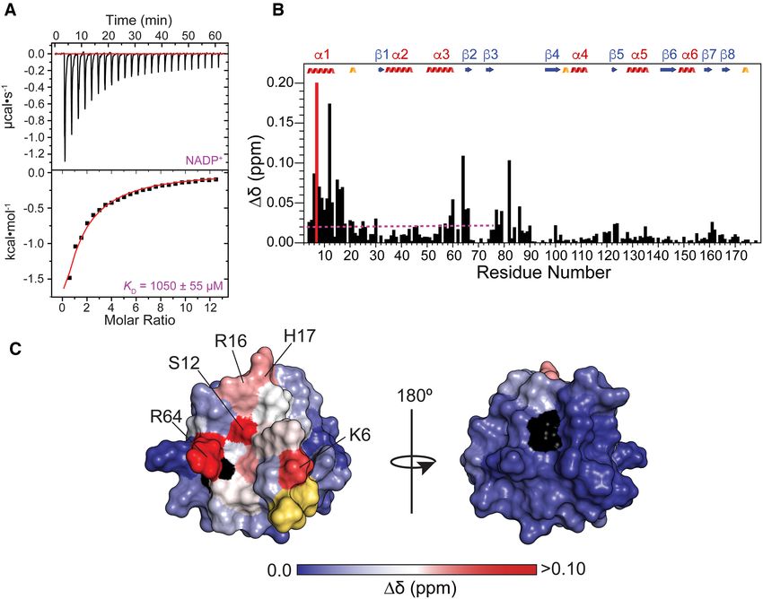
Nucleic Acids Research, 2021, Vol. 49, No. 17 9609
erol, 300 mM imidazole, 0.05% NaN3 and subsequently di- NMR resonance assignments
alyzed against a buffer containing 20 mM Tris-HCl, pH
All NMR experiments were performed at 17◦ C on Bruker
8.0, 200 mM NaCl, 1.5 mM CaCl2 , 5% glycerol, 5 mM
Avance spectrometers operating at 600, 700, 800 or 900
-mercaptoethanol under gentle stirring at 4◦ C. One unit
MHz, all equipped with cryogenic probes capable of ap-
of human ␣-thrombin (Enzyme Research Laboratory) per
plying pulsed-field gradients along the z-axis. Sample con-
mg of protein was added after 1 h and dialysis was then
centrations ranged from 220–320 M for apo-RslTpt1
continued for 16 h at 4◦ C. Complete cleavage of the N-
(unliganded RslTpt1 was unstable in solution at higher
terminal His6 -tag was confirmed by SDS-PAGE. The result-
concentrations) and 650–720 M for the RslTpt1·NAD+
ing RslTpt1 protein contained a 4-amino acid tag (Gly-Ser-
complex in 4 mm Shigemi tubes. All NMR data were
His-Met) at its N-terminus.
processed using NMRpipe (13) and analyzed using NM-
The cleaved RslTpt1 samples were supplemented with 10
RviewJ (14). 1 H, 13 C, 15 N resonance assignments for
mM each of DTT and EDTA, concentrated using Millipore
both apo-RslTpt1 and the RslTpt1·NAD+ complex were
3 kDa MWCO concentrators, and injected into a HiLoad
Downloaded from https://academic.oup.com/nar/article/49/17/9607/6240602 by guest on 28 September 2021
achieved using standard gradient-enhanced, backbone-
16/60 Superdex 75 PG (GE Healthcare) pre-equilibrated
directed triple-resonance strategies and included HNCO,
with NMR buffer (20 mM HEPES pH 7.0, 200 mM NaCl,
HN(CA)CO, HNCA, HN(CO)CA, CBCA(CO)NH, HN-
5% glycerol, 2 mM EDTA, 2 mM DTT, 0.04% NaN3 ). All
CACB experiments (15). Aliphatic sidechain resonances
NMR samples were prepared in NMR buffer containing
were assigned using H(CCO)NH, (H)C(CO)NH, HC(C)H-
5% D2 O for field locking and 1 mM of 4-(2-aminoethyl)
TOCSY and (H)CCH-TOCSY experiments (16). Reso-
benzenesulfonyl fluoride hydrochloride (AEBSF, Sigma
nance assignments for the aromatic sidechains were ob-
Aldrich) to reduce protein degradation over time. Sam-
tained using a 13 C-edited NOESY-HSQC optimized for the
ples used to record 13 C-edited NOESY and methyl-based
aromatic region, while Met-ε resonances were assigned us-
experiments were exchanged (using spin columns) against
ing 13 C-NOESY-HMQC optimized for methyl resonances
NMR buffer in which glycerol and EDTA were replaced by
2 using ILVM-labeled apo-RslTpt1 or RslTpt1·NAD+ sam-
H8 -glycerol and 2 H16 -EDTA (Cambridge Isotope Labora-
ples. Non-uniform sampling was used for some experi-
tories), respectively. NMR samples of NAD+ -bound com-
ments and the corresponding sampling schemes were gener-
plexes were prepared by direct addition of the ligand (the
ated using the Poisson gap sampling method (17). Parame-
NAD+ stock solution was prepared in NMR buffer ap-
ters for all experiments used in the resonance assignment
propriate for the experiment type followed by the adjust-
procedure are provided in Supplementary Table S1 (apo-
ment of pH as needed) in 4 molar excess into the NMR
RslTpt1) and Supplementary Table S2 (RslTpt1·NAD+
sample.
complex). The resonance assignments for apo-RslTpt1 and
the RslTpt1·NAD+ complex have been deposited into the
Isothermal titration calorimetry measurements BMRB with accession codes 30819 and 30820, respectively.
The protocol used to prepare unlabeled RslTpt1 samples for
isothermal titration calorimetry (ITC) measurements was Measurement of ligand-induced chemical shift perturbations
the same as that for NMR, described above, except that no
DTT was added after the overnight dialysis step. For the fi- Backbone chemical shift perturbations (900 MHz) induced
nal purification step, a HiLoad 16/60 Superdex 75 PG col- on RslTpt1 by NAD+ , NADP+ , ADP, ADPR, NMN or
umn was pre-equilibrated with ITC buffer (20 mM HEPES nicotinamide were derived from 15 N, 1 H HSQC experi-
pH 7.0, 200 mM NaCl, 5% glycerol, 2 mM EDTA, 0.04% ments (using 512 and 128 complex points for the direct and
NaN3 ). The same buffer was used to dissolve each of the six indirect dimensions, respectively, with corresponding spec-
ligands used in the titrations: NAD+ , ADPR (ADP-ribose), tral windows of 12.5 ppm and 34 ppm). The chemical shift
ADP, NADP+ , nicotinamide and NMN (-nicotinamide perturbations induced on a 75 M sample of RslTpt1 by 4
mononucleotide). Stock solution were 40 mM for NAD+ , molar equivalents each of NAD+ and NADP+ , or 12 molar
ADP and NADP+ , and 20 mM for the other ligands. The equivalents of each of ADP and ADPR, or 10 molar equiv-
pH in each case was adjusted to within ±0.1 units after dis- alents of each of NMN and nicotinamide, were calculated
solution of the ligand in ITC buffer. All ITC measurements using Equation (1) below. Perturbations (800 MHz) on the
utilized 100 M of RslTpt1 in the cell and 3 mM of each methyl resonances on Ile (␦1), Leu, Val and Met residues
of the ligands in the syringe with the exception of ADPR were obtained from 13 C, 1 H SOFAST-HMQC experiments
and NADP, for which concentrations of 4.5 and 6 mM re- (18) (using 512 complex points and 64 complex points for
spectively, were used. All experiments were performed on an the direct and indirect dimensions, respectively, with corre-
ITC200 (Malvern) at 17◦ C using a 400 rpm stirring speed, a sponding spectral windows of 12.5 ppm and 18 ppm, uti-
reference power of 10 Cal/s, and the following injection lizing a 0.2 s recycling delay). Measurements utilized a 70
parameters: 20 injections of 2 l with 4.0 s injection times, M sample of ILVM-labeled RslTpt1 and 4 molar equiva-
210 s injection delays and filter periods of 5 s. All titrations lents of NAD+ , or 8 molar equivalents of each of ADP or
were recorded at least in duplicate to confirm reproducibil- ADPR, or 10 molar equivalents of each of NMN or nicoti-
ity of the parameters obtained. Reference isotherms using namide. 4-mm Shigemi tubes were used for all experiments.
the same experimental parameters, in which the same con- In all cases, the chemical shift perturbations values (␦, in
centration of ligand was injected into buffer alone, were ppm) were calculated using Equation (1):
obtained for each case. The thermograms were analyzed
in Origin 5.0 (OriginLab) and thermodynamic parameters 2 δ j,r e f − δ j 2
δ = δi,r e f − δi + (1)
were obtained by fits to a one-site binding model. σ j,type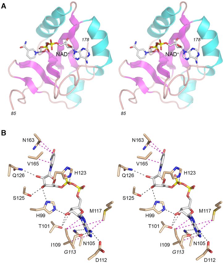
9610 Nucleic Acids Research, 2021, Vol. 49, No. 17
where i and j index the 1 H (amide or methyl) and the corre- Measurement of structural restraints
sponding heteronucleus (15 N or 13 C), respectively. The refer- Distance restraints used to calculate the solution structures
ence shifts for apo-RslTpt1 in each case are indexed by ‘ref’ of apo-RslTpt1 and of the RslTpt1·NAD+ complex were
and the heteronuclear shift is normalized by the standard obtained from a set of 3D 15 N-edited NOESY-HSQC and
deviation ( j,type ) obtained from the BMRB for the specific 3D 13 C-edited NOESY-HSQC experiments, the latter ex-
position (amide or methyl). In addition, a full titration set periments were separately optimized either for aliphatic or
using both amide and methyl-based experiments was col- aromatic resonances (see Supplementary Tables S1 and S2
lected for NAD+ , using 0, 0.1, 0.25, 0.5, 1, 2, 3 and 4 molar for experimental details). Additional distance restraints in-
equivalents of the ligand. volving methyl groups were derived from 3D 13 C-NOESY-
HMQC and 3D 13 C-HMQC-NOESY-HMQC experiments.
Measurement of hydrogen/deuterium exchange Mixing times of 150 ms were used in all NOESY exper-
iments except for those optimized for methyl groups for
Amide-based hydrogen/deuterium exchange NMR exper-
Downloaded from https://academic.oup.com/nar/article/49/17/9607/6240602 by guest on 28 September 2021
which mixing times of 300 ms were used. Additional intra-
iments were performed at 700 MHz, using 4-mm Shigemi molecular distance restraints for NAD+ were obtained from
tubes at 17◦ C. Two 160 l samples for each of apo-RslTpt1 a set of 2D-NOESY spectra recorded using mixing times
or the RslTpt1·NAD+ complex at initial concentrations of of either 150 or a 300 ms, performed on a 15 N, 2 H, ILVM-
280 and 680 M, respectively, were lyophilized and dis- labeled sample of the RslTpt1·NAD+ complex in NMR
solved in either 160 l of ddH2 O (as reference) or 160 l of buffer (see above) prepared in 100% D2 O. Experimental pa-
100% D2 O. Then, a series of 10 1 H,15 N SOFAST HMQC rameters utilized for these experiments are provided in Sup-
spectra (using 512 complex points and 128 complex points plementary Tables S1 and S2. The distance restraints were
for the direct and indirect dimensions, respectively, with cor- supplemented with backbone dihedral angle restraints ob-
responding spectral windows of 12.5 ppm and 34 ppm, uti- tained from chemical shifts utilizing the TALOS-N suite
lizing a 0.2 second recycling delay) were collected over 3 h to (21).
monitor the loss of signal due to the exchange of the amide
1
H with the solvent D2 O. Structure calculations
Structure calculations for apo-RslTpt1 were performed us-
{1 H}-15 N steady-state NOE measurements
ing the ARIA2.3 suite (22,23) utilizing experimentally ob-
Steady-state {1 H}-15 N NOE experiments (19) were per- tained distance and dihedral angle restraints. The PAR-
formed at 800 MHz using uniformly 15 N-labeled samples ALLHDG force field with PROLSQ (24) for non-bonded
of apo-RslTpt1 (350 M) or the RslTpt1·NAD+ complex parameters was used. A simulated annealing protocol com-
(600 M). 512 and 128 complex points with sweep-widths prising of 20 000 steps (27 fs integration time) was carried
of 13 and 34 ppm for the 1 H 15 N dimensions, respectively. A out at high temperature (10 000 K) followed by two Carte-
set of two experiments were acquired with or without (ref- sian cooling phases of 1000 and 50 K of 40 000 steps each
erence) 5 s of 1 H saturation in a total pre-delay period of (3 fs integration times). A network anchoring protocol was
7 s. introduced for the first three iterations of the protocol using
default parameters, and floating chirality was implemented
for prochiral moieties. Starting with the fourth iteration, hy-
Methyl relaxation dispersion measurements drogen bonding restraints were added for residues predicted
13
C-1 H multiple-quantum dispersion CPMG experiments by TALOS-N to be part of defined secondary structure ele-
(20) were performed at 17◦ C (800 MHz) for both apo- ments and for which amide resonances persisted in 15 N, 1 H
RslTpt1 and the RslTpt1·NAD+ complex using 300 M HSQC spectra 3 h after dissolution in pure D2 O.
of uniformly 15 N, 2 H, ILVM-labeled sample of RslTpt1 in For calculation of the structure of the RslTpt1·NAD+
NMR buffer (see above) prepared in 100% D2 O without or complex, the topology and parameter files for NAD+
with 1.2 mM of NAD+ . Experiments utilized sweep-widths were extracted from the PHENIX ligand database (25)
of 12.5 and 18 ppm with 512 and 64 complex points in the and incorporated into the ARIA2.3 force-field, which was
1
H and 13 C dimensions, respectively, using a recovery delay also manually modified to enable automatic assignment of
of 3 s and the relaxation delay, TCPMG set to 32 ms. The fol- NAD+ resonances. Structure calculations of the complex
lowing RF fields (υC PMG ): 0, 40, 80, 120, 200, 320, 400, 480, were performed using same strategy as described above with
600, 720, 800, 920, 1000 Hz were used. The effective relax- activation of the non-crystallographic symmetry and with a
ation rate for a given RF field (R2,eff ) was obtained using: slight change of the simulated annealing protocol where the
integration time was reduced from 3 to 2 fs during the two
1 Iν C PMG Cartesian cooling phases of the calculation. To ensure cor-
R2,e f f (νC PMG ) = − ln (2)
TC PMG I0 rect positioning of NAD+ a set of intermolecular distance
restraints based on unambiguous assignments were used
Where Iν C PMG and I0 are the peak intensities at a field of
at the beginning of the structure calculations using broad
CPMG or in the absence of a CPMG block, respectively. The
upper and lower bounds (1.8 to 5.5 Å) to minimize pack-
curves were visually inspected and Rex values for those res-
ing artifacts. These intermolecular restraints were obtained
onances that displayed clear dispersion behavior were ob-
from manually assigned cross-peaks of the methyl-based 3D
tained using the following expression: 13
C-NOESY-HMQC recorded on a uniformly 15 N, ILVM-
Rex = R2,e f f (40) − R2,e f f (1000) (3) labeled sample of the RslTpt1·NAD+ complex, therefore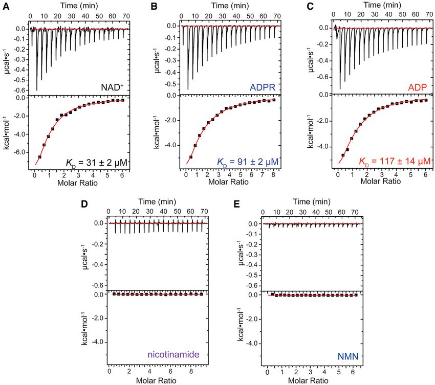
Nucleic Acids Research, 2021, Vol. 49, No. 17 9611
the only detectable 1 H-1 H cross-peaks involved protons of determined for each lobe using:
methyl groups from RslTpt1 or protons from NAD+ . Af-
ter few runs, the ARIA protocol was able to assign a suf- 2
ϑ N−lobe/C−lobe = S2 (5)
ficient number of intermolecular NOEs, and therefore the 3 j =x,y,z N−lobe/C−lobe, j j
loose manual distance restraints were no longer necessary
and were removed in the final production run. Where Sjj are the eigenvalues of the alignment tensor for
For the final production run, a similar simulated anneal- each of the N- or C-lobe. These GDO values were then used
ing protocol was used as described above except that 1000 to define an order parameter given by (35):
structures were generated for each iteration. At the final
step, the 100 lowest energies structures were submitted to a ϑ N−lobe 2
S2 = (6)
short restrained molecular dynamics simulation in explicit ϑC−lobe
solvent using XPLOR-NIH (26). For the final NMR ensem-
bles corresponding to apo-RslTpt1 or the RslTpt1·NAD+
Downloaded from https://academic.oup.com/nar/article/49/17/9607/6240602 by guest on 28 September 2021
Complementation of Saccharomyces cerevisiae tpt1 by
complex, 20 lowest energies structures displaying the low- RslTpt1 mutants
est RDC Q values (see below) that showed no distance re-
straint violations larger than 0.5 Å and no dihedral angle The S. cerevisiae tpt1 haploid strain YBS501 (MATa
violations larger than 5◦ were analyzed using PROCHECK- ura3–1 ade2–1 trp1–1 his3–11,15 leu2–3,11–2 can1–100
NMR (27), WHATIF (28) and PSVS (29). The final struc- tpt1::LEU2 p360-TPT1), in which the TPT1 ORF was
tural ensembles, comprising 20 structures each for apo- deleted and replaced by LEU2, is dependent for viability
RslTpt1 and the RslTpt1·NAD+ complex, were deposited on the p360-TPT1 plasmid (CEN URA3 SceTPT1) (5).
in the PDB with accession codes 7KW8 and 7KW9, respec- YBS501 was transformed with: (i) a p413-SceTPT1 plas-
tively. The regions of specific secondary structure (␣-helix, mid (CEN HIS3 SceTPT1) plasmid as a positive control;
-strand and 310 helix) were determined using STRIDE (30) (ii) the empty CEN HIS3 vector as negative control; and
for consistency and the consensus regions were defined as (iii) p413 (CEN HIS3) plasmids expressing wild-type and
those that were maintained in at least 80% of the structures mutated RslTPT1 open reading frames under the control
in the final NMR ensemble (see Supplementary Table S3). of a constitutive yeast TPI1 promoter. Transformants were
selected at 30◦ C on His– agar medium. Three individual
His+ colonies from each transformation were patched to
Measurement of 15 N-1 H residual dipolar couplings His– agar medium and cells from each isolate were then
streaked on agar medium containing 0.75 mg/ml 5-FOA
Residual dipolar couplings (RDCs) were measured utilizing
(5-fluoroorotic acid). The plates were incubated at 30◦ C.
aligned media generated by the direct addition of Pf1 phage
The SceTPT1, RslTPT1, RslTPT1-(S125A-Q126A) and
(Asla Biotech) into the NMR samples of apo-RslTpt1 (250
RslTPT1-(N105A-D112A) plasmids supported the forma-
M) or the RslTpt1·NAD+ complex (400 M RslTpt1 con-
tion of FOA-resistant colonies within 3 d. By contrast, the
taining a 4-fold excess of NAD+ ) to final phage concen-
vector and the RslTPT1-(I109A-M117A) plasmids did not
trations of 16 and 15 mg/ml, respectively. In order to op-
allow formation of FOA-resistant colonies after 8 d; accord-
timize the degree of alignment for the apo-RslTpt1 or the
ingly, the I109A-M117A mutation was judged to be lethal.
RslTpt1·NAD+ complex samples, the corresponding NaCl
The RslTPT1-(N163A-V165A) plasmid sustained the for-
concentrations were adjusted by the addition of 5.4 and 5.8
mation of sporadic tiny FOA-resistant colonies after 6 d. Vi-
l of 5 M NaCl, respectively. Amide 15 N,1 H RDC values
able FOA-resistant tpt1 p413-RslTPT1 wild-type, S125A-
were extracted from a set of 2D HSQC-IPAP (31) experi-
Q126A and N105A-D112A strains were grown in YPD-Ad
ments (512 and 300 complex points using sweep-widths of
(yeast extract, peptone, 2% dextrose, 0.1 mg/ml adenine)
12.5 ppm and 34 ppm in the 1 H and 15 N dimensions re-
liquid medium at 30◦ C to mid-log phase (A600 0.4–0.6), then
spectively in both cases). The alignment tensor values were
diluted to attain A600 of 0.1, and aliquots (3 l) of serial 10-
obtained using PALES (32). Only regions with well-defined
fold dilutions were spotted on YPD agar plates and incu-
secondary structure values and those with {1 H}-15 N steady-
bated at 20◦ C for 4 d, 25◦ C for 3 d, and 30, 34, and 37◦ C for
state NOE values > 0.75 (800 MHz) were used in the anal-
2 d. FOA-resistant tpt1 p413-RslTPT1-(N163A-V165A)
ysis. Analyses were performed with the intact individual
colonies were patched to YPD agar and, after 2 d incuba-
structures and separately for the N-lobe and C-lobe. The
tion at 30◦ C, transferred to YPD-Ad liquid medium; serial
Q values were defined as (33):
10-fold dilutions were spot-tested for growth on YPD agar
r ms (Dobs − Dcalc ) at 20, 25, 30, 34 and 37◦ C as above. No growth was detected
Q= (4a) at any temperature, from which we surmise that the N163A-
r ms (Dobs )
V165A mutation was effectively lethal in vivo.
2Da2 4 + 3R2 RESULTS
r ms (Dobs ) = (4b)
5 Structure of apo-RslTpt1
Where Dobs and Dcalc are the observed and calculated val- Resonance assignments for 94% of the backbone and 86%
ues of the 15 N,1 H RDC values; Da and R are the anisotropy of the sidechains of RslTpt1 were obtained using a vari-
and rhombicity of the alignment tensor. In order to deter- ety of labeling schemes (Supplementary Figure S2), as de-
mine the relative flexibility between the N- and C-terminal scribed in the ‘Materials and Methods’ section. These as-
lobes, a generalized degree of order (GDO, ϑ N/C ) (34) was signments were subsequently utilized to generate a sufficient
9612 Nucleic Acids Research, 2021, Vol. 49, No. 17
number of NOE-based distance restraints that were supple- Tpt1 orthologs (5,6). The catalytic tetrad directly engages
mented with chemical shift-derived backbone dihedral an- the RNA 2 -PO4 to promote the transesterification step of
gle constraints (21) to obtain the solution structure of apo- the Tpt1 pathway (12). Arg16, His17 and Arg64 present a
RslTpt1. Experimental constraints and structure statistics positively charged solvent-exposed surface at the interface
are shown in Table 1. RslTpt1 consists of two globular ␣- between the two lobes of the apo-RslTpt1 structure. These
domains, the N-lobe (Val5-Gln78) and the C-lobe (Pro93- sidechains show significant disorder in the NMR ensemble
Val178) that are connected by a flexible linker (Figure 1A). (Supplementary Figure S4A) in the absence of the RNA
The disorder in the linker, defined by the Gly79-Val92 seg- substrate. Arg119 is housed in the C-lobe and its sidechain
ment, is reflected by the relatively low steady-state {1 H}- is solvent-exposed and disordered in the NMR ensemble in
15
N NOE values (average ± standard deviation over the the absence of NAD+ (Supplementary Figure S4B).
segment: 0.63 ± 0.12 at 800 MHz; 0.56 ± 0.05 over the As mentioned above, crystal structures of two Tpt1 en-
conserved Gly79-Val82 segment; Supplementary Figure S3) zymes have been reported: a 1.4 Å structure of CthTpt1
compared to the values for the N-lobe (0.80 ± 0.10) and the in complex with CoA/ADP-ribose-1 -phosphate (12) and
Downloaded from https://academic.oup.com/nar/article/49/17/9607/6240602 by guest on 28 September 2021
C-lobe (0.81 ± 0.11). To independently validate the struc- a 2.8 Å structure of ApeTpt1 in the unliganded state (10).
ture of apo-RslTpt1, we measured a set of one-bond 15 N- There is a high degree of similarity between the N-lobe of
1 RslTpt1 and the N-lobes of CthTpt1 (PDB: 6E3A, 6EDE)
H residual dipolar couplings (RDCs) utilizing a Pf1 phage
alignment medium (36). While good quality factors (Q) (33) and ApeTpt1 (PDB: 1WFX) with RMSDs of 1.5 ± 0.1 Å
are obtained for the individual lobes (N-lobe: 0.17 ± 0.03, and 1.4 ± 0.1 Å, respectively (Supplementary Figure S5),
C-lobe: 0.28 ± 0.04; Table 1), the corresponding values for determined over regions of well-defined secondary struc-
the intact structures are significantly worse (0.38 ± 0.04). ture. The corresponding C-lobes align with RMSDs of 1.9
The lack of inter-lobe NOEs and low RDC-based S2 values ± 0.1 Å (Rsl versus Cth) and 2.2 ± 0.2 Å (Rsl versus Ape),
(35) (0.36 ± 0.03) for the structures comprising the NMR respectively (Supplementary Figure S6), with the regions
ensemble suggests the absence of a well-defined orientation comprising 5 and ␣6 being the largest sources of these de-
between the two lobes. As illustrated in Figure 2, superposi- viations. 5 in RslTpt1 is shorter than in both CthTpt1 and
tion of the constituent structures of the ensemble on either ApeTpt1; ␣6 displays a closed conformation in ApeTpt1
their N-lobes or their C-lobes reveals a high degree of posi- and a more open conformation in RslTpt1, with the ␣6 he-
tional variation of the respective non-superimposed lobes. lix in CthTpt1 lying between these two extremes (Supple-
The RslTpt1 N-terminal lobe (Figure 1A), which has mentary Figure S6). The degree of similarity between the
been termed the RNA lobe (12), has a winged helix fold C-lobes of RslTpt1, CthTpt1 and ApeTpt1 improves with
comprising three ␣-helices (see Supplementary Table S3 the exclusion of these two structural elements, lowering the
for consensus secondary structures), ␣1 (5-14), ␣2 (34- RMSDs to 1.4 ± 0.1 Å in both cases; these values are com-
44), and ␣3 (50–60) and a three-stranded anti-parallel - parable to those obtained for the corresponding N-lobes.
sheet (1: 32–33, 2: 65–67 and 3: 73–76). The 20–22 seg-
ment forms a 310 helix. The C-terminal lobe, also termed
the NAD+ lobe, forms a mixed ␣-fold (Figure 1A) resem- Interaction of RslTpt1 with nucleotide ligands
bling that of NAD+ -utilizing toxins that catalyze ADP ri- We used isothermal titration calorimetry (ITC) measure-
bosylation of proteins and DNA (12). The C-lobe is com- ments to gauge the affinity of RslTpt1 for NAD+ (Table 2,
posed of three ␣-helices (see Supplementary Table S3), ␣4 see Figure 3 for representative traces). NAD+ binds RslTpt1
(106–112), ␣5 (128–136), and ␣6 (148–154), and two 310 with an apparent KD of 31 ± 1 M (Table 2; Figure 3A
helices (103–105 and 173–175). A 310 helix also forms in shows a representative plot). It is notable that the intracel-
the 135–137 segment in about 40% of the structures in the lular concentrations of NAD+ of ∼1.0–1.6 mM in S. cere-
NMR ensemble. The C-lobe also contains five -strands - visiae (37) and 2.6 mM in E. coli (38) are well in excess
4 (96–102), 5 (122–124), 6 (141–147), 7 (158–161) and of this apparent KD . RslTpt1 is also able to bind ADP-
8 (165–168), that are organized as an antiparallel -sheet. ribose (ADPR, Figure 3B shows a representative plot) and
The C-terminus is well ordered, as indicated by high {1 H}- ADP (Figure 3C shows a representative plot) with apparent
15
N NOE values (0.88 ± 0.07 for Ile176 and 0.74 ± 0.09 for KD values of 96 ± 6 M and 123 ± 10 M (Table 2), re-
Lys177) and forms a sixth -strand (9) in about 40% of the spectively. These observations suggest that the ADP moiety
structures in the NMR ensemble. This last -strand is also provides most of the binding energy for the interaction be-
present in the structure of CthTpt1 (12). tween RslTpt1 and NAD+ ; the nicotinamide riboside moi-
Figure 1B shows an alignment of the RslTpt1 primary ety makes only a small contribution. Indeed, no binding
structure to those of biochemically validated Tpt1 or- of nicotinamide (Figure 3D) or -nicotinamide mononu-
thologs from the bacterium Clostridium thermocellum and cleotide (NMN, Figure 3E) to RslTpt1 can be detected by
three species of fungi: the model yeast Saccharomyces cere- ITC.
visiae (Sce) and the human fungal pathogens Candida albi- Next, we utilized solution NMR to determine the nature
cans (Cal) and Candida auris (Cau). The alignment high- of the interaction between NAD+ and RslTpt1 in residue-
lights 55 positions of sidechain identity/similarity in all five specific detail. As shown in Figure 4A, significant chemi-
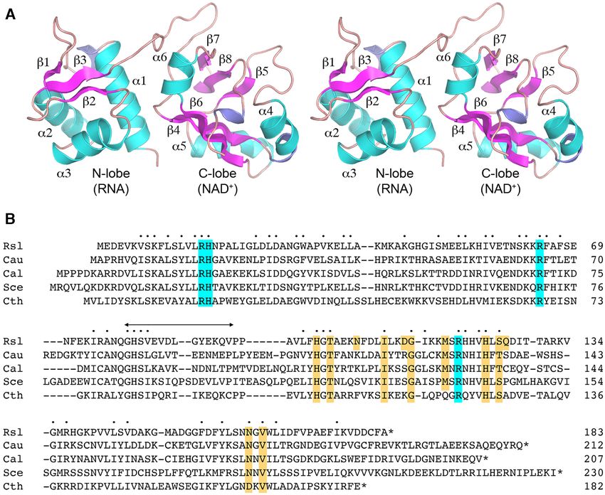
Tpt1 proteins. RslTpt1 residues Arg16, His17, Arg64, and cal shift perturbations (CSPs) are observed for backbone
15
Arg119 comprise an active site catalytic tetrad (highlighted N, 1 H resonances in the presence of NAD+ and are local-
in cyan in Figure 1B) (5) that is essential for the activity ized exclusively on the C-lobe (␦ = 0.14 ± 0.12 ppm, aver-
of RslTpt1, SceTpt1, and EcoTpt1 and is conserved in all aged over the 72 C-lobe amide resonances for which numer-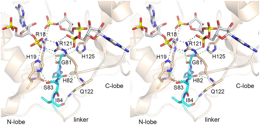
Nucleic Acids Research, 2021, Vol. 49, No. 17 9613
Table 1. Constraints, refinement and structure statistics for apo-RslTpt1 and the RslTpt1·NAD+ complex
Pairwise Cartesian RMS deviation (Å)
Apo-RslTpt1 RslTpt1·NAD+
PDB ID: 7KW8 7KW9
N-lobe C-lobe N-lobe C-lobe
Global heavy atoms 1.61 ± 0.16 2.18 ± 0.58 1.73 ± 0.17 2.14 ± 0.55
Global backbone atomsa 0.75 ± 0.12 1.15 ± 0.35 0.67 ± 0.13 1.06 ± 0.30
Ordered heavy atoms 1.37 ± 0.12 1.70 ± 0.44 1.37 ± 0.35 1.76 ± 0.17
Ordered backbone atomsb 0.56 ± 0.10 0.79 ± 0.22 0.41 ± 0.13 0.74 ± 0.14
Restraint Information
NOE-derived distance restraints
Intra-residue 895 886
Downloaded from https://academic.oup.com/nar/article/49/17/9607/6240602 by guest on 28 September 2021
Inter-residue 1180 1547
Sequential 441 552
Medium 292 362
Long 447 633
Inter-molecular - 29
Ambiguous 512 906
Dihedral angle restraints 278 269
Hydrogen bond restraints 132 136
Energies (kcal/mol)
Total −5811.7 ± 78.1 −5702.2 ± 110.9
NOE 3.8 ± 1.0 4.5 ± 1.2
Dihedral angles 97.6 ± 8.8 235.7 ± 11.6
Ramachandran statistics (%)c
Most favored regions 85.1 ± 1.3 81.4 ± 1.5
Additional allowed regions 13.4 ± 1.3 16.6 ± 1.5
Generously allowed regions 1.1 ± 0.6 2.0 ± 1.2
Disallowed regions 0.4 ± 0.5 0.0 ± 0.0
Restraint violations
Distance restraints (>0.5 Å) 0 0
Dihedral angle restraints (>5◦ ) 0 0
Average RMS deviation from experimental restraints
Distance restraints (Å) 0.027 ± 0.001 0.043 ± 0.001
Dihedral angle restraints (◦ ) 0.471 ± 0.060 0.517 ± 0.072
Average RMS deviation from idealized geometries
Distance restraints (Å) 0.017 ± 0.001 0.019 ± 0.001
Dihedral angle restraints (◦ ) 1.528 ± 0.038 1.656 ± 0.032
Average RMS Z-scores for deviation from current reliable structuresd
Bond lengths 0.813 ± 0.014 0.910 ± 0.020
Bond angles 0.875 ± 0.024 0.914 ± 0.020
Omega angle 0.822 ± 0.035 0.903 ± 0.043
Side-chain planarity 0.782 ± 0.126 0.880 ± 0.106
Improper dihedral distribution 1.022 ± 0.032 1.165 ± 0.027
Inside/Outside distribution 0.980 ± 0.015 0.961 ± 0.013
Average Z-scores for deviation from current reliable structuresd
First-generation packing quality −1.2 ± 0.2 −1.1 ± 0.2
Second-generation packing quality −2.2 ± 0.3 −2.4 ± 0.3
1 / 2 rotamer normality −3.9 ± 0.4 −4.3 ± 0.5
Backbone conformation −1.2 ± 0.3 −1.7 ± 0.4
Residual Dipolar Couplingse
N-lobe C-lobe N-lobe C-lobe
Q for individual lobes 0.17 ± 0.03 0.28 ± 0.04 0.25 ± 0.03 0.28 ± 0.04
Pearson correlation co-efficient 0.97 ± 0.01 0.93 ± 0.18 0.95 ± 0.01 0.92 ± 0.02
S2 0.36 ± 0.03 0.40 ± 0.06
a The global RMSD calculations include residues 3–77 for the N-lobe and residues 93–178 for the C-lobe for both apo-RslTpt1 and the RslTpt1·NAD+
complex.
b The ordered RMSD calculations include residues 4–14, 20–21, 32–33, 34–44, 50–59, 66–67, 73–75 for the N-lobe; residues 100–102, 103–105, 106–112,
122–124, 128–135, 136–137, 141–146, 148–153, 158–161, 165–168, 173–175, 176–177 for the C-lobe.
c Based on PROCHECK analysis.
d Values based on WHATIF.
e The 15 N-1 H RDCs were only used to validate the structural ensembles and not utilized for structure refinement.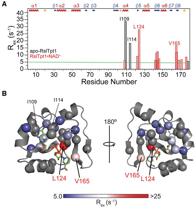
9614 Nucleic Acids Research, 2021, Vol. 49, No. 17
Downloaded from https://academic.oup.com/nar/article/49/17/9607/6240602 by guest on 28 September 2021
Figure 1. Solution structure of unliganded RslTpt1. (A) Stereo view of the structure of apo-RslTpt1 as illustrated by a representative member selected from
the NMR ensemble. The secondary structure elements are labeled; -strands are colored magenta; ␣-helices are colored cyan; 310 helices are colored blue.
The N-lobe (Val5-Gln78) and the C-lobe (Pro93-Val178) are connected by a flexible linker. The structures of the individual lobes are well defined in solution,
but their relative orientation is not (see Supplementary Figure S3). (B) Alignment of the amino acid sequences of Tpt1 proteins from Runella slithyformis
(Rsl, Genbank: WP 013927919.1), Candida auris (Cau, Genbank: PIS54536.1), Candida albicans (Cal, Genbank: AOW28085.1), Saccharomyces cerevisiae
(Sce, Genbank: NP 014539.1) and Clostridium thermocellum (Cth, Genbank: ABN54255.1). Positions of amino acid identity/similarity in all five proteins
are indicated by dots above the alignment. Gaps in the alignment are indicated by dashes. The peptide linker between the N-terminal RNA lobe and
the C-terminal NAD+ lobe of RslTpt1 is indicated by the bidirectional arrow above the alignment. The conserved Arg-His-Arg-Arg catalytic tetrad is
highlighted in cyan shading. Conserved amino acids that contact NAD+ in the RslTpt1·NAD+ structure reported here are highlighted in gold shading.
Figure 2. Structures of the NMR ensemble of apo-RslTpt1 overlaid on the N-lobe (that engages 2 -PO4 RNA, panel A) and the C-lobe (that engages
NAD+ , panel B). The lobe on which the structures are aligned is labeled in red font. The inability to align the structures on both lobes simultaneously
illustrates the fact that while the structures of the individual lobes are well-defined in solution, their relative orientation is not, due to significant inter-lobe
flexibility. Given that there are no experimental inter-lobe constraints, the apparent parsing into two sub-families for the unaligned lobe is likely an artifact
of the force-field and the limited number of structures (20) selected to represent the NMR ensemble. Elements of secondary structure are indicated with
␣-helices, -strands and 310 helices colored cyan, magenta and blue, respectively. Loops are colored salmon.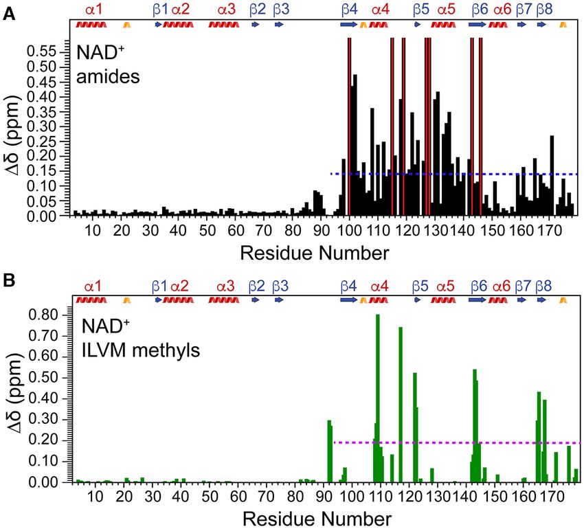
Nucleic Acids Research, 2021, Vol. 49, No. 17 9615
Table 2. ITC-determined affinities of NAD+ and variants for RslTpt1 (except H1D, ␦ = 7 Hz; see Supplementary Figure S10)
is complicated by extensive line-broadening in an already
Ligand KD (M)1
crowded region, measurable CSPs (>5 Hz) are noted for
NAD+ 31 ± 1
1 H nuclei of both the adenosine (H2A, 12 Hz) and nicoti-
ADPR 96 ± 6 namide moieties (H4N, 10 Hz; H5N, 6 Hz). Some represen-
ADP 123 ± 10
Nicotinamide NB2
tative examples of these CSPs are shown in Supplementary
NMN NB2 Figure S10.
NADP+ 1021 ± 53 In line with the large CSPs described above, increased
protection from solvent is seen for the RslTpt1 C-lobe in the
1 Averageand standard deviations obtained from duplicate measurements presence of NAD+ . Indeed, the backbone amides for apo-
made at various times. RslTpt1 show rapid exchange with solvent and few amide
2 NB implies no observable binding.
resonances from either the N- or the C-lobe are observable
after ∼3 h following dissolution in D2 O. While the pro-
Downloaded from https://academic.oup.com/nar/article/49/17/9607/6240602 by guest on 28 September 2021
tection of resonances corresponding to the N-lobe remains
ical values could be obtained from 2D correlation spectra) largely unchanged in the presence of an excess of NAD+ , a
while no significant CSPs are noted in the N-lobe, indicat- substantial increase in the protection of C-lobe amide reso-
ing that NAD+ partitions exclusively to the C-lobe. Further, nances is noted (Supplementary Figure S11).
the lack of CSPs on the N-lobe in the presence of NAD+ The recognition of the internal 2 -PO4 of an RNA sub-
is indicative of the fact that this ligand is unable to induce strate by Tpt1 is proposed to occur through the N-lobe (12).
stable closure between the two lobes. A closer inspection of We measured CSPs induced by NADP+ that contains a 2 -
the C-lobe CSPs shows that the most significant ␦ values PO4 and which we suspected might bind to the RNA sub-
(>0.25 ppm) are centered on 4 (Thr101: 0.44 ppm, Ala102: strate site. ITC assays show that RslTpt1 binds NADP+
0.47 ppm), ␣4 (Leu108: 0.36 ppm), the ␣4-5 loop (Ser118: with an apparent KD of ∼1 mM (Table 2; Figure 5A), that is,
0.39 ppm), 5 (Val122: 0.35 ppm), ␣5 (Thr130: 0.39 ppm, with ∼30-fold less affinity than NAD+ . The observed back-
Ala131: 0.42 ppm, Lys133: 0.27 ppm, Val134: 0.31 ppm, bone amide CSPs indicate that NADP+ interacts exclusively
Gly135: 0.35 ppm) and near the C-terminus (Val171: 0.27 with the N-lobe (␦ = 0.02 ± 0.03 ppm) of RslTpt1 (Figure
ppm). In addition, specific resonances on 4 (Gly100), 5B). No significant NADP+ -induced CSPs are seen in the
on the ␣4-5 loop (Lys115, Arg119), on the 5-␣5 loop C-lobe. The largest CSPs (>0.08 ppm) are seen for N-lobe
(Asp127), on ␣5 (Ile128), and on 6 (Val143, Val146) are residues Lys6 (0.09 ppm), Ser12 (0.17 ppm) and Arg64 (0.11
significantly shifted (or fully broadened out), their NAD+ - ppm). A large CSP is also noted for Val82 (0.10 ppm) in the
bound states are not available in the 2D correlation spectra inter-lobe linker peptide. It is notable that the N-lobe con-
for analysis. Most residues that show large CSPs appear to stituents of the RslTpt1 catalytic tetrad all show significant
be in slow exchange on the chemical shift timescale (see Sup- perturbations, as follows: Arg16 (0.07 ppm), His17 (0.07
plementary Figure S7 for representative examples). This is, ppm) and Arg64 (0.11 ppm). Arg16 and Arg64 have been
however, not surprising given the extremely large chemical proposed to engage the 2 -PO4 of the RNA-2 -phospho-
shift differences between the free and NAD+ -bound states ADPR intermediate and catalyze step 2 transesterification
of RslTpt1 that enable the slow exchange condition (39) to (Supplementary Figure S1) to form the RNA 2 -OH prod-
be maintained despite the modest affinity. CSPs in the pres- uct. Indeed, in the structure of CthTpt1, the 1 -PO4 prod-
ence of ADPR or ADP are also limited to the C-lobe with uct (corresponding to a 2 -PO4 in the reactant) is encased
no significant perturbations seen on the N-lobe (Supple- within a mesh of hydrogen bonds involving the correspond-
mentary Figure S8). As expected in light of the ITC data, ing residues Arg18 and Arg68 (12). In the case of RslTpt1,
no significant CSPs are observed in the presence of nicoti- alanine mutations of Arg16 and Arg64 were found to reduce
namide or NMN (not shown). the step 2 transesterification rate constant by 710-fold and
Next, we analyzed the CSPs induced on the 13 C,1 H reso- 210-fold, respectively (6).
nances of Ile (␦1 only), Leu, Val and Met methyl residues, As described above, NAD+ and NADP+ partition exclu-
given that such residues are candidates to take part in hy- sively to the C- and N-lobes of RslTpt1, respectively, with
drophobic interactions with NAD+ . As in the case of the the ligand binding to a given lobe having no observable
backbone resonances, the largest methyl CSPs in the pres- effect on the other. To further probe this effect, we mea-
ence of NAD+ (Figure 4B; methyl CSPs in the presence sured by ITC the binding affinity of NAD+ for RslTpt1
of ADPR and ADP are shown in Supplementary Figure pre-saturated with NADP+ . Only a small change in the
S9) are seen for the C-lobe (␦=0.19 ± 0.21 ppm over 37 NAD+ binding affinity KD = 47 ± 3 M (compared to
Ile-␦1, Leu, Val and Met methyl groups) with no signifi- 31 ± 1 M, Table 2) was noted (Supplementary Figure
cant perturbations seen in the N-lobe (0.39 ppm) are noted for Ile109, ␦1 (0.80 ally the same with (951 ± 33 M, see Supplementary Figure
ppm), Met117, ε (0.74 ppm), Val122, ␥ 1/2 (0.52/0.36 ppm), S12B) or without (1021 ± 53 M, Table 2) pre-saturation
Val143, ␥ 1/2 (0.54/0.49 ppm), Val165, ␥ 1/2 (0.43/0.29 with NAD+ . CSPs induced by each ligand, NAD+ on the
ppm) and Leu167, ␦1/2 (0.39/0.15 ppm). C-lobe and NADP+ on the N-lobe, are also independent
The interaction of NAD+ with RslTpt1 can also be of the presence of the other (Supplementary Figure S13).
gauged by the observed perturbations induced on the 1 H These observations confirm that binding of NADP+ and/or
spectrum of NAD+ by the presence of RslTpt1. While anal- NAD+ is insufficient to induce stable interaction between
yses of the perturbations of the 1 H nuclei of ribose moieties the two lobes of RslTpt1 or influence the affinities for the9616 Nucleic Acids Research, 2021, Vol. 49, No. 17
Downloaded from https://academic.oup.com/nar/article/49/17/9607/6240602 by guest on 28 September 2021
Figure 3. Interaction of RslTpt1 with a variety of nucleotide ligands. Representative thermograms from isothermal titration calorimetry (ITC) measure-
ments for the interaction of RslTpt1 with (A) NAD+ , (B) ADP-ribose (ADPR), (C) ADP, (D) nicotinamide and (E) -nicotinamide mononucleotide
(NMN), are shown. In each case, the top panel shows the heat corresponding to each injection and the bottom panel shows the corresponding fit of the
integrated intensity to a one-site binding model. The KD values and errors indicated on the thermograms correspond to those obtained from the fits for
the specific traces that are shown. Averages (and corresponding standard deviations) over multiple measurements are listed in Table 2.
corresponding ligands. It is, however, possible that a more complete assignments of backbone (92%) and sidechain
substantial engagement of the RNA-binding groove on the (83%) resonances of RslTpt1 bound to NAD+ (Supple-
N-lobe, beyond simple low affinity 2 -PO4 recognition (as mentary Figure S2) could then be leveraged to yield a
in NADP+ ), is required to elicit cross-talk between the two sufficient number of intra-molecular and inter-molecular
lobes. NOE-based distance restraints (see Supplementary Figure
S14 for some representative examples) that were combined
with chemical shift based dihedral angle restraints (Table
Structure of the RslTpt1·NAD+ complex
1) to obtain a structure of the RslTpt1·NAD+ complex
Our NMR studies on apo-RslTpt1 gave us confidence (Figure 6A).
that we would be able to solve the structure of the A comparison of secondary structure elements between
RslTpt1·NAD+ complex using solution NMR methodol- the apo and NAD+ -bound states of RslTpt1 shows a few
ogy. However, significant changes in resonance positions ev- changes (Supplementary Table S3). Strand 6 appears to
ident from the amide and methyl CSPs in the presence of be slightly distorted at Leu144 in some of the structures;
NAD+ (Figure 4) necessitated extensive reassignment of the the Glu103-Asn105 segment that forms a 310 -helix in apo-
backbone and sidechain resonances of RslTpt1. The near RslTpt1 integrates into ␣4 in the complex that now en-Nucleic Acids Research, 2021, Vol. 49, No. 17 9617
Downloaded from https://academic.oup.com/nar/article/49/17/9607/6240602 by guest on 28 September 2021
Figure 4. Spectral perturbations induced by NAD+ on RslTpt1. Chemical shift perturbations induced on (A) amide 15 N, 1 H resonances or (B) methyl
13 C, 1 H resonances of Ile (␦1 only), Leu, Met and Val residues in the presence of 4 molar equivalents of NAD+ , are shown. The red bars indicate residues
for which the corresponding resonances were perturbed but the chemical shifts for their final NAD+ -bound states could not be analyzed due to vanishing
resonances, missing assignments, or spectral crowding. The blue and magenta dashed lines represent the average ␦ values for the C-lobe amide (0.14 ppm)
and ILVM methyl (0.19 ppm) resonances, respectively. Secondary structural elements (definitions are as per apo-RslTpt1, see Supplementary Table S3) are
indicated; ␣-helices, -strands and 310 -helices are colored red, blue and gold, respectively.
compasses residues Glu103-Asp112 in the complex. Ad- dividual lobes (N-lobe: 0.25 ± 0.03, C-lobe: 0.28 ± 0.04)
ditionally, the terminal 9-strand (Ile176-Lys177) appears suggest good agreement between the experimental and pre-
to be better defined in the structural ensemble represent- dicted values. In contrast, the Q-factors for the intact struc-
ing the complex, where it is formed in all of the structures tures are significantly higher (0.69 ± 0.23) suggesting poor
compared to only 40% of the structures for apo-RslTpt1. correlation between the predicted and experimentally deter-
Other changes are restricted to single-residue variations at mined RDCs. Additionally, the RDC-based S2 values also
the termini of the secondary structural elements (Supple- confirm substantial inter-lobe mobility (S2 = 0.40 ± 0.06).
mentary Table S3). While their positions remain largely un-
changed, the relative orientations of these secondary struc-
Recognition of NAD+
ture elements appear to be altered upon engaging NAD+ .
The RMSD over the secondary structural elements for the The NAD+ moiety makes several contacts with the C-lobe
C-lobe comparing the NMR ensembles of apo-RslTpt1 and of RslTpt1 (Figure 6B); no stable contacts with the N-lobe
the RslTpt1·NAD+ complex is found to be significant (Sup- are evident. The adenine ring stacks against the hydropho-
plementary Figure S15A) at 1.9 ± 0.2 Å in line with the bic residues Ile109 and Met117 (the methyl groups of both
large CSPs induced by the ligand. A smaller but significant hydrophobic residues show substantial CSPs; see Supple-
RMSD of 1.4 ± 0.2 Å is also noted for the N-lobe (Sup- mentary Figure S17) and engages in a pair of hydrogen
plementary Figure S15B). Given the limited CSPs induced bonds to enzymic sidechains: adenine-N3 with Asn105-N␦2
on the N-lobe by NAD+ (9618 Nucleic Acids Research, 2021, Vol. 49, No. 17
Downloaded from https://academic.oup.com/nar/article/49/17/9607/6240602 by guest on 28 September 2021
Figure 5. Interaction of RslTpt1 with NADP+ . (A) Representative ITC thermogram and corresponding fit of the integrated intensity to a one-site binding
model are shown in the top and bottom panels, respectively. (B) Chemical shift perturbations induced on the amide 15 N, 1 H resonances of RslTpt1 in
the presence of 4 molar equivalents of NADP+ . The magenta dashed line indicates the average ␦ value for the N-lobe amides (0.02 ppm). The red
bar represents the Val7 resonance that is fully broadened out in the presence of NADP+ . (C) NADP+ -induced chemical shift perturbations for amide
resonances mapped onto a surface representation of the RslTpt1 N-lobe. Residues that display the most significant perturbations are labeled. Residues for
which resonances are missing and those corresponding to the 4-residue N-terminal tag are colored black and yellow, respectively.
His99-Nε2 and Thr101-O␥ 1. This latter interaction would Conformational dynamics in the RslTpt1·NAD+ complex
be disrupted by the presence of a 2 -PO4 , which would also
elicit numerous steric clashes, most notably with His99 and As seen in Supplementary Figure S18, the NMR structural
Gly100, explaining the inability of the C-lobe to engage ensemble of the RslTpt1·NAD+ complex suggests some dis-
NADP+ . The pyrophosphate that links the adenosine and order in the orientation of NAD+ . The adenine moiety ap-
the nicotinamide riboside moieties is relatively disordered in pears to be better ordered (RMSD: 0.89 ± 0.44 Å) com-
the NMR ensemble (Supplementary Figure S18) either due pared to the nicotinamide moiety (RMSD: 1.33 ± 0.68 Å).
to the absence of specific constraints resulting from a lack of The relatively stable interaction of the RslTpt1 C-lobe with
1
H nuclei within this segment, or dynamics within the com- the adenosine moiety of ADP is in line with the ITC results
plex, or both. However, His123 or His138, both located in that indicate the dominant role of the latter in the interac-
close proximity to the phosphates, could potentially serve as tion compared to nicotinamide. In order to test whether
hydrogen-bond donors to stabilize this segment. The nicoti- this disorder reflects, at least in part, conformational dy-
namide moiety of NMN stacks against Val165 (the methyl namics within the solution ensemble, we conducted relax-
groups of which show substantial CSPs, see Supplementary ation dispersion measurements on multiple-quantum co-
Figure S17) and Asn163. The NMN ribose is stabilized by a herences involving the methyl groups of Ile (␦1), Leu, Val
pair of hydrogen-bonds of the 2 -OH with Gln126-Nε2 and and Met residues. For apo-RslTpt1, only three residues,
the 3 -OH with Ser125-O␥ . Ile109, Ile114 and the C-terminal Val178, display signifi-
cant exchange contributions (Rex > 5 s−1 ; Figure 7A) sug-You can also read