E.coliRep helicase and RecA recombinase unwind G4 DNA and are important for resistance to G4-stabilizing ligands
←
→
Page content transcription
If your browser does not render page correctly, please read the page content below
6640–6653 Nucleic Acids Research, 2020, Vol. 48, No. 12                                                              Published online 25 May 2020
doi: 10.1093/nar/gkaa442

E. coli Rep helicase and RecA recombinase unwind
G4 DNA and are important for resistance to
G4-stabilizing ligands
Tapas Paul1 , Andrew F. Voter2 , Rachel R. Cueny2 , Momčilo Gavrilov1 , Taekjip Ha1,3,4 ,
James L. Keck 2 and Sua Myong 1,3,*
1
 Department of Biophysics, Johns Hopkins University, Baltimore, MD 21218, USA, 2 Department of Biomolecular
Chemistry, University of Wisconsin School of Medicine and Public Health, Madison, WI 53706, USA, 3 Physics

                                                                                                                                                              Downloaded from https://academic.oup.com/nar/article/48/12/6640/5843820 by guest on 27 September 2020
Frontier Center (Center for Physics of Living Cells), University of Illinois, 1110 W. Green St., Urbana, IL 61801, USA
and 4 Howard Hughes Medical Institute, Johns Hopkins University, USA

Received March 12, 2020; Revised April 24, 2020; Editorial Decision May 11, 2020; Accepted May 21, 2020

ABSTRACT                                                                        riched at important genomic regions including replication
                                                                                origins, oncogene promoters, telomeres, and immunoglobu-
G-quadruplex (G4) DNA structures can form physical                              lin switch regions (5–7). G4 enrichment has also been found
barriers within the genome that must be unwound                                 in Escherichia coli at regulatory regions of genes involved in
to ensure cellular genomic integrity. Here, we report                           transcription, secondary metabolite biosynthesis, and sig-
unanticipated roles for the Escherichia coli Rep heli-                          nal transduction (8–10). Inefficient regulation of G4 struc-
case and RecA recombinase in tolerating toxicity in-                            tures has been linked to genome instability (11). Stable G4
duced by G4-stabilizing ligands in vivo. We demon-                              structures can act as roadblocks to numerous cellular pro-
strate that Rep and Rep-X (an enhanced version of                               cesses such as replication, transcription, and translation
Rep) display G4 unwinding activities in vitro that are                          (12–14). In addition, G4 ligands (e.g. BRACO-19 and N-
significantly higher than the closely related UvrD he-                          methyl mesoporphyrin IX (NMM)) can further enhance the
licase. G4 unwinding mediated by Rep involves repet-                            stability of G4 structures, disrupting critical cellular path-
                                                                                ways and thereby inducing toxicity in cells (13,15). For ex-
itive cycles of G4 unfolding and refolding fueled by
                                                                                ample, BRACO-19 has been shown to inhibit DNA replica-
ATP hydrolysis. Rep-X and Rep also dislodge G4-                                 tion, transcription, and telomerase activity (15). Therefore,
stabilizing ligands, in agreement with our in vivo G4-                          dedicated cellular machineries have evolved to resolve G4
ligand sensitivity result. We further demonstrate that                          structures.
RecA filaments disrupt G4 structures and remove G4                                 Helicases are DNA unwinding motor proteins that play
ligands in vitro, consistent with its role in countering                        important roles in preserving genome integrity. Many heli-
cellular toxicity of G4-stabilizing ligands. Together,                          cases are capable of unwinding G4 DNA structures (16,17),
our study reveals novel genome caretaking functions                             including RecQ family helicases (bacterial RecQ, yeast Sgs1
for Rep and RecA in resolving deleterious G4 struc-                             and human BLM, WRN), XPD family enzymes such as
tures.                                                                          FANCJ, and Pif1 family and DEAH box family including
                                                                                DHX36 (12,18–22). A recent study reported that a bacte-
                                                                                rial RecQ helicase, which possesses a guanine-specific bind-
INTRODUCTION                                                                    ing pocket that is essential for G4 unwinding, resolves G4
                                                                                through repetitive cycles of unwinding and refolding (23).
Guanine-rich nucleic acid sequences have strong propen-                         In addition, several reports have shown that Pif1, BLM and
sities to form four-stranded G-quadruplex (G4) structures                       DHX36 exhibit similar repetitive unfolding of G4 and can
under physiological conditions (1). In these structures, four                   successfully dislodge G4-stabilizing ligands (20,24,25). In
guanine bases are cyclically coordinated through Hoog-                          some cases, helicase activity was limited by G4 ligand bind-
steen hydrogen bonds to form a G-quartet or tetrad ring,                        ing (18,26).
which is further stabilized by stacking interaction with other                     The superfamily I helicases, UvrD and Rep are similar
ring layers in the presence of monovalent cation (1–3). G4s                     in structure and exhibit 3 -5 direction of translocation, but
can fold into various conformations in vitro and evidence                       do not overlap in vivo activity (27–30). UvrD plays an im-
has confirmed that G4 structures are present in living cells                    portant role in nucleotide excision repair, mismatch repair
(3,4). In the human genome, potential G4 clusters are en-

* To   whom correspondence should be addressed. Tel: +1 410 516 5122; Fax: +1 410 516 4118; Email: smyong@jhu.edu


C The Author(s) 2020. Published by Oxford University Press on behalf of Nucleic Acids Research.
This is an Open Access article distributed under the terms of the Creative Commons Attribution License (http://creativecommons.org/licenses/by/4.0/), which
permits unrestricted reuse, distribution, and reproduction in any medium, provided the original work is properly cited.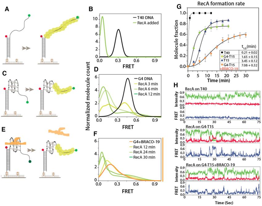
Nucleic Acids Research, 2020, Vol. 48, No. 12 6641
and in the regulation of homologous recombination (31). tions were purchased from IDT. Amine-modified oligonu-
Additionally, like the RecQ helicase, E. coli UvrD unwinds cleotides were labelled with NHS ester-conjugated fluores-
intermolecular and intramolecular G4 structures (31). Rep cent dyes following an established protocol (38). Briefly, 30
was the first helicase discovered in E. coli and unwinds DNA l of 100 M amine modified ssDNA was mixed and incu-
with a limited processivity of ≤400 bp (28,30,32). Rep shows bated overnight with 0.2 mg of NHS ester-conjugated Cy3
low unwinding activity as a monomer in vitro, but multi- dye in 100 mM NaHCO3 , pH 8.5. The excess dye was re-
merizes upon binding to DNA to show robust helicase ac- moved by ethanol precipitation and repeated twice. Each
tivity (33). Intramolecular crosslinking of Rep monomer to partial duplex DNA construct (10 M) was prepared by
Rep-X enhances the unwinding activity and makes Rep-X mixing the biotin-conjugated DNA strand with its comple-
a processive superhelicase capable of continuous unwind- mentary strand at molar ratio of 1:1.2 (biotinylated:non-
ing of more than 6000 base pairs without dissociation (34). biotinylated) and annealed in T50 Buffer (10 mM Tris–HCl,
Though Rep can unwind DNA, it is unclear whether Rep pH 7.5 and 50 mM NaCl) in a thermocycler with the follow-
helicase can participate in the resolution of G4 structures. ing protocol: 95◦ C for 2 min; gradual cooling to 40◦ C at the
Downloaded from https://academic.oup.com/nar/article/48/12/6640/5843820 by guest on 27 September 2020
In replication, recombinases assist in strand exchange re- rate of 2◦ C/min; further cooling by 5◦ C/min until 4◦ C. G4-
pair for reloading of the accessory proteins. In fact, there duplex (i.e. 42mer with T15) annealed in 10 mM Tris–HCl,
is a significant interplay between accessory helicases and pH 7.5 and 5 mM MgCl2 containing buffer following the
recombinases in both bacteria and lower eukaryotes (35). same protocol as described above.
Δrep cells are still viable because UvrD partially compen-
sates for the absence of Rep. However, the double deletion
Protein purification
Δrep and ΔuvrD causes lethality (29,31,36). Furthermore,
RecA and RecBCD can sustain viability in Δrep and ΔuvrD Escherichia coli UvrD protein was purified as described
but only in the presence of an RNA polymerase mutation previously (39). Briefly, UvrD was transformed into
that alleviates transcriptional barriers to replication (35). BL21(DE3) pLysS using kanamycin and chloramphenicol
Escherichia coli ΔrecA cells induce conflicts between repli- as selection markers. UvrD was overexpressed by adding 1
cation and transcription, similar to the case of Δrep cells. mM IPTG when the culture had an OD600 of 0.6; cells were
Therefore, both RecA and Rep help mitigate the conflict then grown for an additional 4 hours at 37◦ C and subse-
between transcription and replication (35). In vitro study quently pelleted and stored at -80◦ C. Cells were resuspended
showed that E. coli RecA binds monomeric G4 from pilin into Lysis Buffer (50 mM Tris pH 8.3, 10% sucrose, 200 mM
expression locus (pilE) of N. gonorrhoeae with similar affin- NaCl, 5 mM ethylenediaminetetraacetic acid (EDTA), 0.5
ity to ss-DNA but does not bind other G4 structures (37). mM ethylene glycol-bis (-aminoethyl ether)-N,N,N’,N’-
However, it still remains unclear whether RecA can resolve tetraacetic acid (EGTA), 15 mM 2-mercaptoethanol, 0.1
G4 structure. mM phenylmethylsulfonyl fluoride, 100 ug/ml lysozyme,
To better define the mechanisms underlying G4 home- and one Roche protease inhibitor tablet), lysed by sonica-
ostasis in bacteria, we have identified genome maintenance tion, and centrifuged at 34 864 × g. Nucleic acid contami-
genes in E. coli that are important for growth in the pres- nants were removed from the supernatant by the addition of
ence of G4-stabilizing ligands and show that each encodes Polymin P to a final concentration of 0.3% (v/v) followed by
a protein that is able to unwind G4 DNA structures in vitro. centrifugation at 34 864 × g. UvrD was precipitated from
rep and recA E. coli strains are found to be sensitive to the supernatant by adding ammonium sulfate to a final con-
G4-stabilizing ligands whereas strains deficient in several centration of 176 g/l and centrifuged again at 34 864 ×
other key genome maintenance genes are resistant to the g. The resulting pellet was resuspended into Resuspension
compounds. Rep and RecA display robust G4 DNA un- Buffer (20 mM Tris pH 8.3, 20% glycerol, 1 mM EDTA,
folding and G4 ligand displacement activities in vitro. In 0.5 mM EGTA, 15 mM 2-mercaptoethanol, 300 mM NaCl)
contrast, UvrD, a helicase that shares significant structural followed by another centrifugation step at 34 864 × g. The
similarity with Rep, demonstrated substantially weaker G4 resulting supernatant was mixed 1:2 with Buffer A (20 mM
unwinding activity. Rep translocates on single-stranded (ss) Tris pH 8.3, 20% glycerol, 1 mM EDTA, 0.5 mM EGTA,
DNA in an ATP-hydrolysis dependent fashion and resolves 15 mM 2-mercaptoethanol) and loaded onto a HiPrep Hep-
G4 structures by repetitive cycles of unfolding activity. Rep- arin FF affinity column (GE Healthcare). The column was
X, an enhanced version of Rep (34), displayed accelerated washed thoroughly with Buffer A followed by protein elu-
G4 unwinding activity via a similar mechanism. RecA dis- tion using a gradient of 0.1 to 0.45 M NaCl in Buffer A.
rupts G4 structures and dislodges G4 ligands by forming UvrD-containing fractions were collected and concentrated
filaments along ssDNA. Together, the results suggest novel into ∼2 ml and loaded onto a HiPrep 16/60 Sephacryl S-
activities for Rep and RecA in resolving G4 structures that 300 column (GE Healthcare) that was equilibrated previ-
are important for protecting cells against the threat of G4 ously with Buffer B (20 mM Tris pH 8.3, 20% glycerol, 1
genomic roadblocks. mM EDTA, 0.5 mM EGTA, 15 mM 2-mercaptoethanol,
500 mM NaCl). UvrD-containing fractions were concen-
MATERIALS AND METHODS trated, dialyzed into storage buffer (20 mM Tris pH 8.3,
50% glycerol, 1 mM EDTA, 0.5 mM EGTA, 25 mM 2-
Preparation of DNA constructs
mercaptoethanol, 200 mM NaCl), and stored at –20◦ C.
The HPLC-purified DNA oligonucleotides (tabulated in Escherichia coli Rep was purified as described previously
Supplementary Table S1) containing both biotin for im- (40). Briefly, pET28a(+) vector containing Rep-DM4 was
mobilization and either Cy3, Cy5 or amine modifica- transformed into E. coli. BL21(DE3), and cells were in-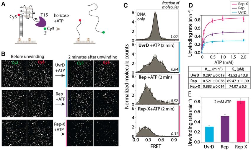
6642 Nucleic Acids Research, 2020, Vol. 48, No. 12
duced at OD600 of 0.6 with 0.5 mM IPTG and harvested (m-PEG-5000; Laysan Bio) and 2% biotin PEG (biotin-
after an overnight incubation at 18◦ C. Cell pellets were re- PEG-5000; Laysan Bio). The microfluidic sample chamber
suspended in lysis buffer and sonicated followed by centrifu- was created between the plasma-cleaned slide and coverslip
gation at 34 864 × g. N-terminally 6xHis-tagged Rep pro- coated with PEG and biotin-PEG.
tein was purified using Ni-NTA column and eluted with 150 Stocks of annealed partial duplex DNA (in T50 buffer)
mM imidazole containing buffer. The protein concentration labelled with biotin, Cy3, and Cy5 were diluted to 15–20
was always kept below 4 mg/ml (50 M) to avoid aggrega- pM using buffer consisting of 10 mM Tris–HCl, pH 7.5 and
tion, and the final Rep protein was stored at –20◦ C with 50% 100 mM KCl (to ensure stable G-quadruplex formation).
glycerol. Diluted DNAs were immobilized on the PEG-passivated
Rep crosslinking (Rep-X) was performed using 10 mM surface via the biotin–neutravidin (50 g/ml) interaction
BMOE (bismaleimidoethane) crosslinker solution in DMF and unbound molecules were washed out by flowing excess
(34). Optimal crosslinking was achieved at concentration of buffer. All smFRET measurements were carried out in an
20–25 M and the final molar ratio of Rep and BMOE imaging buffer containing 10 mM Tris–HCl, pH 7.5, 50 mM
Downloaded from https://academic.oup.com/nar/article/48/12/6640/5843820 by guest on 27 September 2020
was 1:5. Excess imidazole and crosslinker were removed KCl, 3 mM MgCl2 , 10% glycerol, and an oxygen scavenging
by overnight dialysis in 600 mM NaCl. Rep-X was finally system (10 mM Trolox, 0.5% (w/v) glucose, 1 mg/ml glu-
stored at –20◦ C in storage buffer (50% glycerol, 600 mM cose oxidase and 4 g/ml catalase) to avoid blinking and
NaCl, 50 mM Tris, pH 7.6). improve dye stability. Milli-Q water was used to prepare all
buffers and then filtered through 0.22 m membrane filters.
All experimental data were recorded at room temperature
E. coli strain construction
(∼23◦ C ± 2◦ C).
Escherichia coli knockout strains were generated using P1 A solid-state 532 nm diode laser (Compass 315M, Coher-
transductions as described previously (41). Briefly, P1 phage ent) was used to generate an evanescent field of illumination
lysate was grown on each donor knockout strain and used to excite the Cy3 dye (donor) and the fluorescence from Cy3
to transduce the kanamycin-sensitive parent MG1655 or and Cy5 (acceptor) were simultaneously collected using a
imp4213 strains. Individual knockout strains were gifts from water immersion objective. Emission signals were divided
Michael Cox. Kanamycin resistant colonies were isolated by a dichroic mirror (cut off = 630 nm) and projected onto
and insertion of the kan cassette was confirmed by PCR. the EMCCD camera (Andor). Data were recorded with 100
ms frame integration time and then processed by IDL script
and analyzed by Matlab scripts.
Sensitization to G4 stabilizing ligands
NMM and BRACO-19 were resuspended in 18 M ultra-
smFRET data analysis
pure water, the concentration of NMM was measured using
molar extinction coefficient as 145 000 M−1 cm−1 at 379 nm To generate the FRET histogram, 21 frames of 20 short
as described previously (42), and the resuspended solutions movies were collected at different imaging locations, yield-
were stored at 4◦ C. The G4 stabilizing ligand was added ing >6000 molecules. Alternating lasers (green and red)
to molten LB-agar to the specified concentration during were used to excite sequentially both Cy3 and Cy5 (10
plating and plates were stored in dark at 4◦ C. Cultures of frames for Cy3, 1 frame dark and 10 frames for Cy5) to ex-
each knockout strain were grown overnight at 37◦ C in 5 clude the donor-only molecules from the histogram at the
ml of LB supplemented with 50 ug/ml kanamycin. Next, low FRET region. Furthermore, the donor leakage was cor-
overnight cultures were diluted to OD600 ∼ 1. The cultures rected based on FRET values of donor-only molecules. Ori-
were then serially diluted from 10−1 to 10−6 in LB, and 10 gin 2018 was used to fit the Gaussian distributions with an
l of the indicated dilution was plated onto prewarmed LB- unrestrained peak centre position of the individually cor-
agar plates with or without G4 stabilizer. These plates were rected and normalized histogram. The restrained peak cen-
grown overnight at 37◦ C and imaged. tre position was used for RecA bound histogram. All the
results and standard deviations shown in histogram fittings
were calculated by incorporating more than three indepen-
Single-molecule FRET assays and data acquisition
dent experiments.
Single-molecule FRET (smFRET) data were acquired us-
ing a custom-built prism-type total internal reflection
smFRET real time experiment
(PTIR) inverted fluorescence microscope (Olympus IX 71)
as described previously (25,43,44). All experiments were The smFRET real-time G4 unwinding assays using UvrD,
carried out on quartz slides and coated with polyethylene Rep and Rep-X were carried out with a flow chamber and
glycol (PEG) to avoid non-specific interactions of excess the same micro-fluidic imaging channel described above. A
DNA and protein. First, the predrilled quartz slides and small piece of the plastic reservoir was placed above the one
glass coverslips were washed thoroughly with methanol, hole at the one end of the chamber and corresponding other
acetone, and etched by sonication in 1 M potassium hy- holes at the opposite end connected with a silicone tube with
droxide. Then slides were burned for 2–3 min, and cover- a syringe pump (Harvard Apparatus, MA, USA). Protein
slips were quickly sterilized by passing through the flame 4– (100 nM) and ATP (2 mM) suspended in imaging buffer was
5 times to remove all sources of fluorescence. Subsequently, loaded into the reservoir. The real-time FRET images were
both slides and coverslips were treated with aminosilane for collected by passing solution through the imaging chamber
30 min and finally coated with a mixture of 98% mPEG that contained dual-labeled (Cy3 and Cy5) partial duplex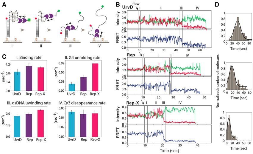
Nucleic Acids Research, 2020, Vol. 48, No. 12 6643
DNA with G4 via a silicone tubing at a flow rate of 20 L/s. in vivo (2). However, it remains unclear which of these pro-
The smFRET time trajectories were analyzed using Matlab teins are required for G4 tolerance. To explore the roles that
scripts. Using the individual single-molecule real-time flow genome maintenance proteins play in tolerating G4s, strains
traces, the binding kinetics were calculated from moment of lacking selected set of proteins were grown in the presence of
flow to the moment of first irreversible FRET decline, and ligand stabilized-G4s. We tested the ability of mutant strains
the G4 unwinding kinetics were measured from the fluctua- with deletions in rep::kan, uvrD::kan, recA::kan, recQ::kan
tion in FRET signal. In all three helicase experiments, more to grow on LB plates containing two structurally distinct
than 150 molecules were quantified for all kinetic calcula- G4 stabilizers, N-methyl mesoporphyrin IX (NMM) or
tions. Additionally, the DNA construct annealed either in BRACO-19. To determine the G4 stabilizer concentrations
50 mM NaCl and diluted at 100 mM KCl or directly 100 for these experiments, the sensitivities of E. coli MG1655
mM KCl resulted in the same unwinding activity of all pro- and of an imp-4213 strain were tested using a range of
teins tested here. NMM and BRACO-19 concentrations. Minimal concen-
trations at which the strains had unaffected colony-forming
Downloaded from https://academic.oup.com/nar/article/48/12/6640/5843820 by guest on 27 September 2020
smFRET unwinding assays units in spot plating experiments were 20 M NMM for
MG1655 and 75 M BRACO-19 for the imp-4213 strain.
FRET spots disappeared as the helicase uncoiled G4, al- While the uvrD and recQ deletion strains tolerated the G4
lowing the duplex to unwind. To calculate the unwinding ligand toxicity, strains lacking either rep or recA were sensi-
rate, short movies (2 s) were recorded just after the injection tized to NMM (Figure 1A). None of the deletion strains
of the respective helicase with or without ATP in the imag- tested were susceptible to BRACO-19. We hypothesized
ing buffer. The short movies were continuously recorded at that BRACO-19 was unable to reach high enough intra-
different imaging areas and continued for almost 10 min. cellular concentration to have an effect. When the rep and
The number of FRET spots (300–400 molecules per view) recA deletions were transferred to an imp-4213 background
counted over time. Consecutive three counted spot numbers (known to have a hyperpermeable membrane) both strains
and also time were averaged and plotted using Origin. Us- were sensitized to BRACO-19 (45). We therefore used the
ing the single exponential decay fitting curve, the unwinding imp-4213 background for all BRACO-19 experiments.
rate was determined. These unwinding experiments were Surprisingly, single gene knockout strains uvrD::kan and
performed with different ATP concentrations. recQ::kan, which both encode known G4 helicases (23,31),
RecA (1 M; from NEB) and 2 mM ATP were used for did not decrease cell viability in the presence of NMM or
RecA assembly assays using the same imaging buffer as used BRACO-19 (Figure 1A, B). This indicates that these heli-
in the helicase experiment. Short movies were collected at cases may not be as important for the tolerance of unre-
different imaging areas over time, and FRET histograms solved G4 structures in vivo. Another group of genes, in-
were generated. The kinetic rate of RecA filament forma- cluding uvrA, the G4-interacting mutS (46), and the G4 he-
tion was calculated from fitting histogram by Gaussian dis- licase dinG (47), were found to be important for NMM tol-
tribution at different times after RecA addition. erance, but not BRACO-19 (Supplementary Figure S1).
G4 ligand dislodge assay Rep-X and Rep unwind G-quadruplex proficiently
The ligand displacement assay was carried out by apply- The G4-ligand sensitivity shown above strongly suggests
ing 10 M ligand to the immobilized G4-containing sample that Rep, but not UvrD, is critical in overcoming G4 ligand-
chamber. Unbound ligands were washed away, and100 nM induced toxicity. This observation is puzzling in two aspects.
of respective helicase with 2 mM of ATP was flowed in the First, the stark contrast between Rep and UvrD is not ex-
same imaging buffer. Short movies were recorded and the pected because they share structural and functional sim-
spots were counted over time as described above. To moni- ilarities as closely related members of Superfamily I heli-
tor the real-time ligand dislodging from G4, ligand was ap- case (48,49). Second, Rep has not been shown to unfold G4
plied followed by washing out the free ligand and finally whereas G4 DNA unwinding has been demonstrated for E.
adding the respective helicase with ATP while recording one coli UvrD (31). Nevertheless, based on the in vivo G4 ligand
continuous long movie. sensitivity results, we hypothesized that Rep may be capable
After washing the excess ligands, RecA (1 M) supple- of unwinding G4 better than UvrD.
mented with ATP (2 mM) was applied and short movies To test this hypothesis, we compared G4 unwinding by
(∼2 s) were recorded at different time intervals to generate Rep, UvrD and Rep-X, a Rep variant that displays height-
histograms for kinetic analysis. Subsequently, long movies ened duplex DNA unwinding activity (34). To investigate
were recorded for 2 min to observe the molecular behaviour. the G4 resolving activity of three different helicases by
single-molecule (sm) FRET, we prepared a substrate con-
RESULTS taining a duplex DNA (18 bp) and ssDNA composed of a
G4 (four repeat of TTAGGG which folds into G4) and a
Cells lacking rep or recA are sensitive to G4 stabilizing lig-
T15 tail for helicase loading. The donor (Cy3) and acceptor
ands
(Cy5) dyes were situated across G4-T15 to probe the heli-
Unresolved G4 structures have been shown to disrupt DNA case binding to the ssDNA tail followed by G4 and duplex
replication and repair with potentially lethal effects (12). (18 bp) unwinding (Figure 2A). The complete unwinding
Genome maintenance proteins are implicated in regulating of the duplex leads to disappearance of Cy3 signals and
the formation and unwinding of G4 structures in vitro and concomitant loss of FRET. Representative fields of view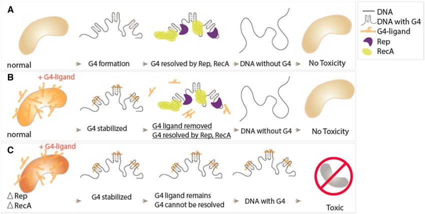
6644 Nucleic Acids Research, 2020, Vol. 48, No. 12
Downloaded from https://academic.oup.com/nar/article/48/12/6640/5843820 by guest on 27 September 2020
Figure 1. G4 ligand sensitivity assay. (A) Deletion strains, rep::kan, uvrD::kan, recA::kan, recQ::kan grown on LB (left) and with 30 M NMM (right). (B)
Deletion strains, rep::kan, uvrD::kan, recA::kan, recQ::kan grown on LB (left) and with 75 M BRACO-19 (right) in imp-4213 background to increase
cell permeability.
recorded before and after the addition of individual heli- indicate that Rep-X and Rep unwind G4 more proficiently
cases (100 nM) and ATP (2 mM) show reduction in number than UvrD.
of both Cy3 and Cy5 molecules over time in all three cases To measure the extent to which G4 acts as a barrier in
(Figure 2B). unwinding, we performed the same Rep induced unwind-
FRET histograms were built by collecting FRET values ing assay using T15, T40 tail without G4 and G4-duplex
from >4000 molecules obtained from 20 different fields of with T15 tail (Supplementary Figure S3). T15 is the same
view. The G4 DNA exhibits the FRET peak at ∼0.5 due to tail length used in the G4 construct used above whereas
the distance between Cy3 and Cy5 separated by the G4 and T40 is the length sum of G4 and T15 (24+15 = 39), keeping
T15 tail (Figure 2C). After addition of UvrD, Rep, or Rep- the same 18bp duplex. G4-duplex contain the same length
X with ATP, the total number of molecules decreased over and identical nucleotides composition as of (TTAGGG)4 -
time, as expected from G4 unwinding. Approximately 75%, T15 construct that use here for unwinding experiment. This
50% and 25% molecules disappeared in two minutes for control can directly reflects the G4 versus G4-duplex un-
Rep-X, Rep and UvrD, respectively (Figure 2B, C). In ad- winding. Surprisingly, the Vmax for T15 and G4-duplex are
dition, the FRET histogram peaks shifted to lower FRET almost similar (∼1.37 ± 0.03/min and ∼1.32 ± 0.05/min)
∼0.2, especially for Rep and Rep-X, signifying faster un- and less from T40 (∼2.86 ± 0.03/min). Those rates reflect
folding of G4. The widely distributed histogram with addi- ∼2.5–5 times faster unwinding than that of G4 containing
tional peak ∼0.6 for UvrD arises from slower G4 unwind- unwinding, clearly indicating a delay due to G4 unfolding.
ing, which is evidenced by the extended period of repetitive The Km of T15 and G4-duplex remains comparable to the
FRET fluctuation (see Figure 3B). To test the ATP depen- G4-containing substrate, but ∼4 times less for T40 (Supple-
dence in unwinding, we titrated ATP concentration from mentary Figure S3). All the unwinding experiments were
1 M to 2 mM while keeping the same protein concentra- carried out by applying protein and ATP together. Much
tion (100 nM). For kinetic analysis, we counted the num- slower unwinding was observed when protein was loaded
ber of Cy3 molecules by capturing short movies (∼2 s) se- before the ATP addition, indicating a requirement of mul-
quentially at different fields of view to avoid photobleaching tiple or successive protein loading for efficient unwinding
(Supplementary Figure S2). The unwinding rates from three (Supplementary Figure S4).
proteins were then fitted to the Michaelis-Menten plot from
which we obtained Vmax and Km for Rep-X, Rep and UvrD
(Figure 2D). The unwinding rate at 2 mM ATP and Vmax Repetitive G4 unfolding is the rate limiting step
for Rep and Rep-X was two and three times higher than To gain mechanistic insight into the G4 resolving activ-
UvrD, respectively (Figure 2E). Taken together, these data ity of three helicases, we examined the smFRET traces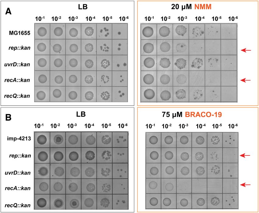
Nucleic Acids Research, 2020, Vol. 48, No. 12 6645

                                                                                                                                                            Downloaded from https://academic.oup.com/nar/article/48/12/6640/5843820 by guest on 27 September 2020
Figure 2. Rep-X and Rep unwind G4 more proficiently than UvrD. (A) Schematic diagram of DNA construct with G4 and T15 tail at the 3 end and
expected loss of Cy3 strand upon completion of unwinding. (B) Representative field of view before and after unwinding. Each white dot represents fluores-
cent DNA molecules. The FRET efficiency and the number of molecules decreases over time of unwinding for all three proteins (100 nM) added with ATP
(2 mM). (C) FRET histograms taken before unwinding (top, DNA only) and after 2 min of unwinding for all three cases. (D) Unwinding rate calculated by
the molecule count over time and the Vmax and Km deduced from Michaelis–Menten fit. (E) The unwinding rate of three proteins at 2 mM ATP condition.

taken in real-time flow measurements in which data were                           The completion of G4 unwinding is followed by the du-
acquired while the protein and ATP were added to the G4-                       plex unwinding, which is denoted by a decrease in FRET
containing DNA substrate. This experiment provides in-                         (Figure 3B, III). Compared to the G4 unwinding, the rate
sights into all stages of the protein activity in one contigu-                 of duplex unwinding is up to two orders of magnitude faster.
ous movie (25,50,51). The four stages include (I) helicase                     Interestingly, the duplex unwinding rate was similar for all
loading, (II) G4 unfolding, (III) duplex unwinding and (IV)                    three proteins, in the range of approximately 3 s−1 (Figure
completion of unwinding (Figure 3A). The representative                        3C, III). The last stage is the ejection of the Cy3 strand,
smFRET traces of UvrD, Rep and Rep-X display all four                          which is not part of G4 or duplex unwinding (Figure 3B,
stages of activity (Figure 3B). The flow of protein (100 nM)                   IV). The delay of Cy3 strand departure can be due to the
and ATP (2 mM) started at 10 s (Figure 3B, arrows) in all                      helicase holding onto the tracking strand before releasing it
cases. Immediately after flow, we observed a spike in Cy3                      into solution (more traces in Supplementary Figure S5A).
signal as expected from the protein loading at the 3 end,                     The repetitive fluctuation was not present but delay of Cy3
which exhibits protein induced fluorescence enhancement                        strand departure observed when the same helicase activity
(PIFE) (Figure 3B, I) (52,53). The binding rate calculated                     was probed on T15 partial duplex (lack of G4) (Supplemen-
from over 150 traces is similar for all three proteins (Fig-                   tary Figure S5B). Taken together, we demonstrate for the
ure 3C, I). Helicase binding is followed by a long period of                   first time that Rep-X, Rep and UvrD are capable of unfold-
FRET fluctuations, which is similar to the repetitive G4 re-                   ing G4 powered by ATP hydrolysis, albeit at different rates.
solving activity seen in other G4 helicases such as RHAU,                      Interestingly, the G4 unwinding mechanism of three pro-
BLM, and WRN (Figure 3B, II) (19,20,25). We interpret                          teins follow the similar pattern of repetitive unfolding and
this pattern as emerging from repetitive cycles of unwind-                     refolding cycles.
ing and refolding of G4 which acts as a physical barrier.
Interestingly, such G4 unwinding takes 35–45 s for UvrD,
but only 10 and 20 seconds for Rep-X and Rep, respectively                     Role of tail length and G4 conformation.
(Figure 3D). Therefore, unlike the binding rate, the G4 un-                    To further probe the unwinding abilities of the three heli-
winding rate shows a significant difference between the heli-                  cases, we tested the G4 substrate with a shorter 3 ssDNA
cases with the rank order of Rep-X > Rep > UvrD (Figure                        tail, T9 (Supplementary Figure S6A). In all three cases, only
3C, II).                                                                       40% of unwinding (loss of Cy3 molecule) was observed in
6646 Nucleic Acids Research, 2020, Vol. 48, No. 12
Downloaded from https://academic.oup.com/nar/article/48/12/6640/5843820 by guest on 27 September 2020
Figure 3. G4 unwinding mechanism involves multiple unfolding and refolding cycles. (A) Schematic diagram of four stages of unwinding activity: (I)
binding, (II) G4 unfolding, (III) duplex unwinding and (IV) Cy3 departure. (B) The representative real-time smFRET unwinding traces of UvrD, Rep and
Rep-X (100 nM protein and 2 mM ATP) and four stages as stated in (A). (C) Rates of I, II, III and IV calculated from smFRET traces plotted with SEM
(>150 traces per condition). (D) Gaussian fit of the dwell time histogram of G4 unwinding for the corresponding proteins.
10 minutes (Supplementary Figure S6B). The rest 60% of viously (Supplementary Figure S7D, E). Interestingly, Rep-
molecules remained protein bound, indicated by the over- X doesn’t show unwinding of parallel c-Myc G4 with T9
all FRET shift represented in FRET histogram. The ki- tail (Supplementary Figure S7F), although T9 is sufficient
netics analysis showed that the rate of unwinding is al- for Rep-X to unwind non-parallel G4. This likely reflecting
most equal of three helicases (0.15671 ± 0.02581 min−1 for that the cross-linked conformation of Rep-X may require
UvrD, 0.16269 ± 0.02472 min−1 for Rep and 0.18364 ± longer tail for efficient unwinding of a tightly folded paral-
0.019 min−1 for Rep-X), which are substantially lower than lel G4.
that of T15 tailed substrate (Supplementary Figure S6C).
Such difference between T9 and T15 might be due to the
tail length required for proper loading and G4 unfolding Rep and Rep-X dislodge G4 ligand and unfold G4
which involves iterative cycles of unfolding and refolding. We showed above that UvrD, Rep and Rep-X helicase ac-
So far, we used non-parallel G4 as unwinding substrates. tivity is sufficient to unwind G4 DNA structures. Based on
Next, we tested the parallel G4 unwinding by preparing a the G4 ligand sensitivity result (Figure 1), we next asked
DNA construct bearing c-Myc sequence with T15 tail at if the G4 unfolding activity can lead to dislodging of the
the 3 end (Supplementary Figure S7A). Parallel G4s are G4 bound ligand. To test this, we chose BRACO-19, a po-
generally more stable than antiparallel G4s, making them tent G4 ligand which was also tested in the sensitivity assay
more challenging to unwind. Upon addition of individual (Figure 4A, BRACO-19 drawn in orange). The addition of
helicase (100 nM with 2 mM ATP) only about ∼10% Cy3 BRACO-19 to G4 and washing out excess ligand shifted the
signal for both UvrD and Rep disappeared whereas ∼75% FRET peak from 0.5 to 0.2 primarily due to quenching of
for Rep-X was lost in 10 minutes (Supplementary Figure both FRET dyes (Figure 4B). When helicase (100 nM) and
S7B), signifying the efficient unwinding activity of Rep-X ATP (2 mM) were added, 25%, 65% and 85% of molecules
for parallel G4. The negligible and much slower unwind- disappeared for UvrD, Rep and Rep-X respectively after
ing rate indicates that UvrD and Rep are not capable of re- 10 min, signifying the removal of BRACO-19 and subse-
solving the parallel G4 (Supplementary Figure S7C). The quent resolving of G4 and completion of unwinding (Fig-
Vmax is ∼0.44 min−1 with Km is ∼215 M from Michaelis– ure 4B). For kinetic analysis, we counted the number of Cy3
Menten fitting is approximately two times lower Vmax and molecules over time after the addition of helicase and ATP
three times higher Km than the non-parallel G4 shown pre- in the G4 ligand-bound condition (Figure 4C). The unwind-Nucleic Acids Research, 2020, Vol. 48, No. 12 6647
Downloaded from https://academic.oup.com/nar/article/48/12/6640/5843820 by guest on 27 September 2020
Figure 4. Rep and Rep-X dislodge G4 bound ligand. (A) Chemical structure of BRACO-19 and a pictorial depiction of BRACO-19 in orange. (B) Nor-
malized smFRET histogram of G4 DNA (top), with BRACO-19 after buffer wash of excess ligand (second) and then helicases with ATP taken after 10
minutes (bottom three). (C) Single exponential fitting of unwinding kinetics for UvrD, Rep and Rep-X in ligand-bound condition. (D) Bar graph of the
overall unwinding rate (Cy3 departure rate) in presence (filled) and absence (blank with dotted line) of G4-ligand. (E) Representative smFRET flow traces
with demarcations for sequential steps including BRACO-19 addition, buffer wash, protein and ATP addition, G4 resolution, duplex unwinding and Cy3
strand departure with each step marked I to VII. (F) Schematic diagram of G4 DNA (I), with excess ligand (II), after buffer wash of free ligand (III),
helicase added (IV), helicase dislodging ligand and resolving G4 (V), duplex unwinding (VI) and Cy3 departure (VII). (G) Rep and Rep-X induced G4
unwinding rate in presence (filled) and absence (blank, dotted line) of bound ligand.
ing rate obtained from the fitted decay curve is substantially (step V). The subsequent stages of VI and VII represent du-
lower than that of G4 free of ligand, albeit in the same order plex unwinding and departure of Cy3 strand, respectively
of Rep-X > Rep > UvrD (Figure 4D and Supplementary as seen before. Interestingly, the repetitive unwinding and
Table S2). refolding cycles corresponding to the G4 resolving activ-
To capture the sequence of events from the ligand bind- ity (step V) were observed for Rep and Rep-X but not for
ing, displacement, G4 resolution and duplex unwinding, we UvrD. The lack of G4 resolving activity by UvrD likely
performed real-time flow measurement in which ligand and indicates that UvrD cannot remove BRACO-19 bound to
helicase were added in succession. In the representative sin- G4 (more traces in Supplementary Figure S8). This result
gle molecule traces (Figure 4E), we define seven sequential is in agreement with the G4 ligand sensitivity result which
steps (I- VII) depicting distinguishable stages of helicase ac- showed that deletion of uvrD had no effect whereas dele-
tivity (Figure 4F). First, the flow of BRACO-19 to G4 at tion of Rep reduced the tolerance for G4 ligand-mediated
∼5 s induced an immediate quenching of Cy3 and Cy5 sig- toxicity. However, we calculated the rate of protein binding,
nals (step II, 5–15 s). Second, the buffer wash of free lig- G4 unwinding and duplex unwinding for Rep and Rep-X.
and enhanced the Cy3 signal (step III, at ∼15 s) likely due While rate of protein binding (also for UvrD) and duplex
to dequenching of Cy3 by removal of excess BRACO-19. unwinding are almost similar to the rate measured for Rep
Third, the helicase loading to ssDNA tail makes Cy3 signal and Rep-X in the absence of BRACO-19, there was a sig-
brighter, exhibiting PIFE effect (step IV, at ∼25 s) (52,53). nificant difference in the rate of G4 unwinding, strongly in-
Fourth and fifth, the helicase dislodges the G4 ligand and dicating the delay due to dislodging of BRACO-19. The G4
resolves the G4 structure represented by a lag period of low unwinding rate was approximately two-fold lower than that
FRET state and subsequent FRET fluctuation respectively obtained without G4 ligand due to the extra time it took6648 Nucleic Acids Research, 2020, Vol. 48, No. 12
for the ligand removal prior to G4 unwinding (Figure 4G fluctuation observed for G4-T15 likely indicates partial un-
and Supplementary Table S2). Therefore, the data indicate folding and refolding of G4 in dynamic exchange. This pat-
that Rep and Rep-X are able to dislodge the G4 ligand pro- tern indicates that RecA filament cannot unfold G4 com-
ficiently whereas UvrD cannot. pletely in this condition. If the filament completely unfolded
G4, smFRET traces would show a stable low FRET as seen
RecA recombinase resolves G4 and dislodges G4 ligand by in the case of T40 since the total length of G4-T15 is 39 nu-
filament formation cleotides. Nevertheless, the dynamic unfolding and refold-
ing from the G4 state mediated by RecA is sufficient to re-
The hypersensitivity of the recA strains to G4 ligands led move the G4 ligand. Interestingly, in the presence of sodium
us to examine its activity on G4 DNA as well. RecA is es- buffer, which is less stabilizing for G4 folding, we observed a
sential for mediating homologous recombination required stable low FRET on G4-T15, indicating complete unfolding
for maintaining genomic integrity (54). RecA binds ssDNA induced by RecA (Supplementary Figure S9C, D). There-
and forms a helical filament that becomes stable when at fore, we conclude that RecA dislodges the G4 ligand via
Downloaded from https://academic.oup.com/nar/article/48/12/6640/5843820 by guest on 27 September 2020
least six RecA monomers bind ssDNA of 18 nucleotides forming filament on single stranded tail and invading into
or longer (55,56). To establish the working condition, we G4, which is consistent with our in vivo observation (Figure
used partial duplex FRET construct with a 3 -T40 tail to 1).
test RecA filament formation (Figure 5A). Addition of 1
M RecA with 2 mM ATP immediately shifted the FRET
peak from 0.3 (T40 ssDNA) to ∼0.05, which is consistent DISCUSSION
with stretching of the ssDNA due to stable RecA filament Rep is a newly identified G4 resolving helicase
formation (Figure 5B). Next, we asked whether RecA can
resolve a G4 structure through such filament formation. It has been estimated that human genome contains over
To test this, we applied RecA (1 M with 2 mM ATP) 350 000 potential G4 forming sequences (PQS) whereas
to the non-parallel G4 with T15 tail (Figure 5C). Consis- E. coli genome has over 3000 (9). The PQS is distributed
tent with RecA-mediated G4 unwinding, the FRET peak unevenly with high enrichment near sites of replication of
shifted gradually from ∼0.5 to ∼0.05, with ∼75% molecules origin, transcription, translation and telomerase mainte-
shifting within 12 minutes (Figure 5D). By contrast, when nance, strongly suggesting that G4 plays important regu-
RecA is added to G4 DNA without tail, the FRET peak latory roles (5–10). Nevertheless, unresolved G4 structures
remained unchanged, consistent with a requirement for a have been shown to cause genome instability (2). Therefore,
ssDNA tail for RecA loading (data not shown). As a con- cells have evolved specialized helicases to recognize and un-
trol, RecA was added to a T15 DNA without a G4, and wind G4 structures and thereby prevent genomic instability
∼80% molecules showed FRET shift from ∼0.75 to ∼0.2 (31). In vitro, bacterial RecQ helicases can unwind G4 DNA
after 12 minutes (Supplementary Figure S9A, B). Compar- and an X-ray crystal structure identified the presence of a
ing the RecA bound FRET peak of T15 at ∼0.2 with G4- guanine-specific binding pocket on the surface of the heli-
T15 at ∼0.05 suggest that RecA disrupt the G4 structure as case that can sequester guanine bases from unwound G4
almost same length T40 show RecA bound FRET at ∼0.05. DNA (23). However, it remains unclear which helicases are
The similar RecA experiment performed on parallel c-Myc- responsible for G4 tolerance in cells. Here, we report an un-
T15 construct reveals that RecA cannot disrupt the tightly expected role of Rep, but not RecQ or UvrD, in tolerating
folded parallel G4 structure (Supplementary Figure S9E, G4 ligand toxicity in cells. Strikingly, we show that Rep is
F). Therefore, the RecA filaments can resolve non-parallel a robust G4 resolvase whereas UvrD has much weaker ac-
G4 provided that a ssDNA tail is present. tivity. We also show that Rep’s strong G4 unwinding activ-
Next, we sought to test whether RecA can dislodge ity dislodges G4 ligand whereas UvrD’s weaker G4 unfold-
BRACO-19 and resolve ligand-bound G4 DNA (Figure ing is insufficient to remove G4 ligands. Rep and UvrD are
5E). Upon the addition of RecA (1 M with 2 mM ATP), both 3 to 5 helicases that share 40% homology (27) and
the FRET peak gradually shifted to ∼0.05, with ∼60% of have been hypothesized to be redundant in function due to
the population shifting in 30 min (Figure 5F). the lethality caused by rep, uvrD double mutants (59). Later
Considering half-time (t1/2 ) i.e. 50% of RecA assembled studies, however showed that UvrD is functionally distinct
on the respective construct, we compared the rate of RecA from Rep (49,60). The difference between Rep and UvrD in
formation. The highest rate of RecA formation was ob- G4 unwinding may be due to the different binding prefer-
tained for T40, followed by G4-T15, T15, and the ligand ences i.e Rep has high affinity to 3 tailed ssDNA whereas
bound G4-T15 (Figure 5G). We noticed that the fraction UvrD prefers associating at junctions between ssDNA and
of RecA formation reached 100% for T40, but plateaued at duplexes (61). Efficient loading of Rep at 3 tails may facil-
70–80% for G4-T15 and T15 and 60% for the BRACO-19 itate its translocation on ssDNA, powering it forward for
bound G4-T15. The single molecule traces taken after fila- G4 resolution.
ment formation revealed a stable low FRET for T40, sug-
gesting that T40 allows stable filament formation. By con-
Repetitive G4 unfolding is a shared mechanism
trast, the traces taken for G4-T15 displayed dynamic FRET
fluctuations, likely representing binding and dissociation of Interestingly, each of the helicases studies here shared a
RecA on the G4-T15 substrate (Figure 5H). The similar pat- common mechanism of G4 in which repetitive cycles of G4
tern of FRET fluctuation was observed for T15, in agree- unwinding and refolding continued until the G4 was com-
ment with 18 nt required for a stable nucleation (more traces pletely unfolded and the helicase made its way into the du-
in Supplementary Figure S10) (56–58). Hence, the FRET plex DNA. Although the dye position in this substrate onlyNucleic Acids Research, 2020, Vol. 48, No. 12 6649
Downloaded from https://academic.oup.com/nar/article/48/12/6640/5843820 by guest on 27 September 2020
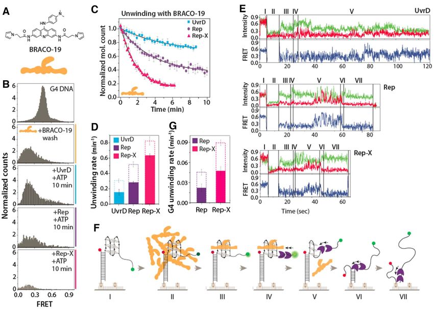
Figure 5. RecA assemble on G4 and dislodge ligand. (A, C, E) Schematic smFRET model of partial duplex with T40 tail, G4-T15 and ligand-bound G4
(left panel) and the corresponding RecA filament formation (right panel). (B, D, F) The smFRET histogram of DNA only and ligand-bound G4 and
with RecA (1 M with 2 mM ATP) filament formation of corresponding DNAs over time. (G) Rate of RecA filament formation on T40, G4-T15, T15
and G4-T15 in ligand bound condition. (H) After RecA filament formation, the representative smFRET traces of T40 (top), G4-T15 (middle) and after
dislodging G4 ligand (bottom).
indirectly reports on G4 unwinding, we chose the terminal ing of G4 which eventually leads to complete G4 unfold-
position of Cy3 based on our previous study in which Cy3 ing followed by duplex unwinding for all three helicases. In-
at the entry of G4 i.e. junction between G4 and ssDNA terestingly, the duration of the repetitive unfolding activity
tail prevented G4 helicases from properly loading on the scaled with the unwinding strength: UvrD, the weakest G4
substrate (25). This position of Cy3 provides an advantage resolvase, spent more time in the repetitive cycles of unfold-
of measuring both loading of the protein and subsequent ing motion than Rep or Rep-X. Such motion is also shared
G4 unfolding activity in repetitive manner. Such repetitive by many other G4 unwinding enzymes (22,25,62,63), sug-
fluctuation was not present when the same helicase activ- gesting that repetitive unfolding may be a conserved mech-
ity was probed on a substrate which is devoid of G4 struc- anism used by many helicases to overcome G4 barriers. The
ture (Supplementary Figure S5B). However, the repetitive four- to five-fold slower unwinding rate observed with G4
FRET fluctuations we observe for all three helicases here compared to the duplex DNA reflects that G4 is a highly
are reminiscent of the unwinding mechanism observed for stable structure that represents a high energetic and physi-
an unrelated G4 resolvase, DHX36. While the DNA-G4 un- cal barrier.
folding by DHX36 was somewhat similar to the pattern seen
here, it never resulted in subsequent duplex unwinding (25),
Rep unwinding of G4 requires non-parallel G4 conformation
likely due to a limited G4 disrupting activity (62). In addi-
and sufficient tail length
tion, DHX36 exhibits distinct stepwise unfolding of RNA-
G4 which occurs at ∼20-fold slower rate (25) than that ac- Our results point out several characteristics of Rep’s G4 un-
quired for Rep, Rep-X and UvrD. We interpret the FRET folding activity. First, Rep can only unwind a non-parallel
pattern shown in Figure 3B as repetitive and partial unfold- G4 structure which is thermally less stable than the parallel6650 Nucleic Acids Research, 2020, Vol. 48, No. 12
G4. By contrast, Rep-X is able to unwind even the paral- G4-bound ligands can be efficiently dislodged by Rep and
lel G4, suggesting a stronger G4 resolving power of Rep- RepX but not by UvrD. This finding is correlated with our
X. Second, all three helicases, Rep, Rep-X and UvrD re- in vivo observations in which the Δrep cells were sensitized
quire a ssDNA tail of sufficient length for unwinding ac- to both NMM and BRACO-19 (Figure 1). Surprisingly,
tivity because T15 led to efficient unwinding whereas T9 ΔrecQ and ΔuvrD did not show a decrease in cell viabil-
did not. Based on the footprint of Rep of approximately ity in the presence of G4 ligands, indicating these helicases
eight nucleotides (28), the requirement for T15 suggest that may not be important for the G4 tolerance. Although RecQ
more than one helicase may be required for unwinding of was previously shown to unwind G4 by a similar repetitive
G4. This is consistent with the DNA duplex unwinding by unfolding mechanism, the G4 unwinding rate of RecQ was
UvrD and Rep, which also require dimer loading on ssDNA 2–3 fold slower than that of Rep and Rep-X (23). In addi-
(28). The T15 requirement seen for Rep-X may arise from tion, RecQ mediated unwinding of G4 was inhibited by G4
the crosslinked conformation of Rep-X that requires longer ligand, an NMM derivative (18). Both findings are consis-
tail even for a monomer loading for unwinding. In addi- tent with our in vivo result presented here, i.e. RecQ’s ability
Downloaded from https://academic.oup.com/nar/article/48/12/6640/5843820 by guest on 27 September 2020
tion, a longer ssDNA may be required to provide a dock- to unwind G4 and dislodge G4 ligand is weaker than that of
ing site for retaining the helicase as it moves back and forth Rep. Consistently, previous biochemical study showed that
in order to effectively resolve the bulky G4 structure dur- UvrD can unwind inter- and intra-molecular G4 structures,
ing repetitive cycles of G4 unwinding and reformation. This but the unwinding activity was impeded by G4 ligand bind-
was evidenced in the case of T9 where all the helicases re- ing (31). In vivo, Rep is known to function to rescue stalled
mained bound to the tail without being able to resolve the replication fork. Previously, we demonstrated that the repet-
G4 (Supplementary Figure S6). By contrast, DHX36, a G4 itive translocation activity of Rep on ssDNA occurs in such
resolvase specific for parallel G4 unwinding only required 9 context i.e. on the lagging strand downstream of Okazaki
bases of ssDNA for G4 resolving, displaying the same type fragment (66). We envision that the same may be true in
of repetitive FRET fluctuations as a monomer (25,62). The the context of G4 bearing DNA, i.e. the repetitive cycles
DHX36 structure revealed an architecture for the protein of translocation of Rep is useful to resolve the blockade
that is highly specialized binding to parallel G4, making an of thermally stable G4 structure, thus removing the phys-
intimate and extensive contact with all sides of the parallel ical barrier. Together, our results suggest that Rep may be
G4 structure (62). Due to this strong interaction, DHX36 specialized in G4 removal activity, which may stem from its
may not require a long tail for resolving G4. inherent ability to repetitively shuttle on ssDNA (66).
If both Rep and RecA are capable of resolving G4 struc-
ture and removing G4 ligand, why did Δrep and ΔrecA
RecA mediates G4 resolution and G4 ligand removal
show sensitivity toward BRACO-19 and NMM? Rep and
It is interesting that RecA, a non-helicase protein, un- RecA could not compensate for the absence of each other
folded G4 DNA. RecA is a well-known recombinase in E. (Figure 1). This may arise from the different pathways in
coli. In homologous recombination double-stranded DNA which Rep and RecA participate. Rep functions to facilitate
break repair, the broken DNA ends undergo resection restarting of stalled replication forks (67) whereas RecA is
in which RecBCD creates the 3 -terminated ssDNA onto responsible for forming a filament on single strand DNA
which RecA is loaded, forming a filament on the DNA that during homologous recombination (68).
executes a homology search to form a D-loop (64). In light
of such function of RecA in homologous recombination,
Compare the activity of helicases and RecA in this study
our results demonstrate that RecA filamentation promotes
unwinding of G4 structures that might emerge within the The smFRET experiment does not allow us to measure the
resected ssDNA. RecA activity was also dependent on the level of tension on single-strand DNA generated by the two
length of ssDNA. Intriguingly, RecA could eject BRACO- proteins, our result reveals that helicases are more efficient
19 and resolve G4s even on short DNAs that do not sup- at resolving G4 structure and removing G4 bound ligand in
port RecA filament formation. Together, our result reveals the context of the DNA constructs we tested here. The G4
that RecA as a recombinase is capable of resolving G4 struc- unfolding rate by helicases is more than two orders of mag-
ture on ssDNAs which can facilitate nucleoprotein filament nitude higher than RecA (Figures 4D vs. 5G). Likewise, the
formation needed for the homology search. This may be G4 ligand removal rate is approximately 5–7 fold higher in
important for RecA activity in cells grown in the presence helicases than in RecA (Figures 4D versus 5G). In terms of
of G4-stabilizing ligands, as observed in experiments with RecA activity, longer filament of RecA formed on a longer
recA cells (Figure 1 and Supplementary Figure S1). Hy- ssDNA tail can lead to more robust G4 and ligand removal
persensitivity of recA cells to the G4 ligands may also be activity by generating higher tension.
due RecA’s essential roles in repair of double-strand DNA
breaks induced by the G4 ligands.
G4 unwinding activity contributes to genome integrity
Our study has led to identification of two key G4 resolvases,
Dislodging of G4 ligand by Rep and Rep-X
Rep and RecA that are critical for tolerance of ligand-
G4-interacting small molecules enhance G4 stability, stabilized G4 structures in E. coli. They are both impor-
thereby inhibiting cellular pathways including telomerase tant for overcoming cellular toxicity that arises from sta-
activity (20,65). G4 ligand binding to G4 structure can also bly folded G4, such as G4s bound to G4 ligands (Figure 6).
downregulate gene expression (65). We demonstrate that the Despite their functional differences, both Rep and RecA areNucleic Acids Research, 2020, Vol. 48, No. 12 6651
Downloaded from https://academic.oup.com/nar/article/48/12/6640/5843820 by guest on 27 September 2020
Figure 6. Model summary of possible causes of nontoxic and toxic cell. (A) Schematic of a normal cell containing G4-forming sequences. The G4 is
resolved by either Rep or RecA. (B) The G4 ligand stabilized the G4 structure, and the G4 ligand can be dislodged by Rep or RecA. (C) When Rep or
RecA are deleted, the cell is unable to dislodge the G4 ligand or resolve the G4 structure, which may lead to cellular toxicity.
fueled by ATP hydrolysis and operate on ssDNA in a direc- FUNDING
tional manner. G-rich sequences in ssDNA can easily fold
National Institute of Health General Medicine
into a thermally stable G4 structures, presenting blockades
[1R01GM115631-01A1 to T.P. and S.M.]; National
for proteins such as polymerases, helicases, and ssDNA-
Science Foundation Physics Frontiers Center Program
binding proteins of many types. In particular, the ssDNA
[0822613] through the Center for the Physics of Living
targeted by Rep and RecA are high-risk spots that can lead
Cells [to S.M., R01 GM098885 to J.L.K., R.R.C., A.F.V.].
genomic instability. For example, if G4 forms in ssDNA
Funding for open access charge: National Institute of
within a stalled replication fork, reversal or recovery of the
Health General Medicine [1R01GM115631-01A1].
stalled fork structure will be impeded. Therefore, Rep’s abil-
Conflict of interest statement. None declared.
ity to resolve G4 in this important junction is critical for
rescuing replication forks and thereby preserving genome
integrity. Next, if G4 formed in resected DNA cannot be
REFERENCES
removed, homologous recombination would fail, resulting
in the prolonged double strand break without proper repair, 1. Burge,S., Parkinson,G.N., Hazel,P., Todd,A.K. and Neidle,S. (2006)
Quadruplex DNA: sequence, topology and structure. Nucleic Acids
which increases the risk of cell death. Therefore, the G4 re- Res., 34, 5402–5415.
solving power of RecA is essential for ironing out the G4 2. Bochman,M.L., Paeschke,K. and Zakian,V.A. (2012) DNA
structure and enabling RecA recombinase activity. secondary structures: stability and function of G-quadruplex
structures. Nat. Rev. Genet., 13, 770.
3. Tippana,R., Xiao,W. and Myong,S. (2014) G-quadruplex
conformation and dynamics are determined by loop length and
SUPPLEMENTARY DATA sequence. Nucleic Acids Res., 42, 8106–8114.
4. Biffi,G., Tannahill,D., McCafferty,J. and Balasubramanian,S. (2013)
Supplementary Data are available at NAR Online. Quantitative visualization of DNA G-quadruplex structures in
human cells. Nat. Chem., 5, 182.
5. Koirala,D., Mashimo,T., Sannohe,Y., Yu,Z., Mao,H. and
Sugiyama,H. (2012) Intramolecular folding in three tandem guanine
repeats of human telomeric DNA. Chem. Commun., 48, 2006–2008.
ACKNOWLEDGEMENTS 6. Besnard,E., Babled,A., Lapasset,L., Milhavet,O., Parrinello,H.,
Dantec,C., Marin,J.-M. and Lemaitre,J.-M. (2012) Unraveling cell
We thank Kevin Rhine and Manindra Bera for careful type–specific and reprogrammable human replication origin
proofreading of the manuscript and members of the S.M. signatures associated with G-quadruplex consensus motifs. Nat.
laboratory and the T.H. laboratory for helpful discussions. Struct. Mol. Biol., 19, 837.
Authors contributions: T.P., A.F.V., J.L.K. and S.M. de- 7. Fry,M. (2012) In: Guanine Quartets, pp. 223–236.
signed research; T.P., A.F.V., R.R.C. and M.G. performed 8. Yadav,V.K., Abraham,J.K., Mani,P., Kulshrestha,R. and
Chowdhury,S. (2007) QuadBase: genome-wide database of G4
experiments; T.P., A.F.V., T.H., J.L.K. and S.M. analyzed DNA––occurrence and conservation in human, chimpanzee, mouse
data; and T.P. and S.M. wrote the manuscript and J.L.K, and rat promoters and 146 microbes. Nucleic Acids Res., 36,
A.F.V. and R.R.C. edited the manuscript. D381–D385.You can also read