A multidisciplinary approach remains the best strategy to improve and strengthen the management of ovarian cancer (Review)
←
→
Page content transcription
If your browser does not render page correctly, please read the page content below
INTERNATIONAL JOURNAL OF ONCOLOGY 59: 53, 2021

A multidisciplinary approach remains the best strategy to improve
   and strengthen the management of ovarian cancer (Review)
            LUCA FALZONE1*, GIUSEPPA SCANDURRA2*, VALENTINA LOMBARDO2, GIUSEPPE GATTUSO3,
        ALESSANDRO LAVORO3, ANDREA BENEDETTO DISTEFANO4, GIUSEPPE SCIBILIA4 and PAOLO SCOLLO4

           1
          Epidemiology and Biostatistics Unit, National Cancer Institute‑IRCCS Fondazione G. Pascale, I‑80131 Naples;
    2
     Medical Oncology Unit, Cannizzaro Hospital, I‑95126 Catania; 3Department of Biomedical and Biotechnological Sciences,
     University of Catania, I‑95123 Catania; 4Unit of Obstetrics and Gynecology, Cannizzaro Hospital, I‑95126 Catania, Italy

                                         Received March 7, 2021; Accepted March 31, 2021

                                                     DOI: 10.3892/ijo.2021.5233

Abstract. Ovarian cancer represents one of the most                   1. Introduction
aggressive female tumors worldwide. Over the decades, the
therapeutic options for the treatment of ovarian cancer have          Ovarian cancer represents the eighth most frequently diagnosed
been improved significantly through the advancement of                tumor and the seventh most lethal cancer in women leading
surgical techniques as well as the availability of novel effective    to almost 185,000 deaths annually worldwide (1). Despite the
drugs able to extend the life expectancy of patients. However,        improvement of screening strategies and the advancement of
due to its clinical, biological and molecular complexity, ovarian     anticancer surgical and pharmacological treatments, ovarian
cancer is still considered one of the most difficult tumors to        cancer is still considered one of the most commonly diagnosed
manage. In this context, several studies have highlighted how         and aggressive urogenital female tumors, with a 5‑year relative
a multidisciplinary approach to this pathology improves the           survival rate of 93% and 5‑year cause‑specific survival rates
prognosis and survival of patients with ovarian cancer. On            of 82, 71, 66 and 43% for endometrioid, mucinous, clear cell
these bases, the aim of the present review is to present recent       carcinoma and serous ovary carcinoma, respectively (2,3). The
advantages in the diagnosis, staging and treatment of ovarian         majority of ovarian cancer cases are epithelial, which accounts
cancer highlighting the benefits of a patient‑centered care           for 85‑90% of all diagnosed ovarian tumors. This type of
approach and on the importance of a multidisciplinary team            tumor usually affects women aged between 55 and 65 years
for the management of ovarian cancer.                                 old (4); contrariwise, germ cell ovarian cancer accounts for
                                                                      ~5% of all diagnosed tumors with an average age of onset of
                                                                      20 years old (4).
Contents
                                                                      2. Risk and protective factors for ovarian cancer
1. Introduction
2. Risk and protective factors for ovarian cancer                     Several risk factors have been recognized for ovarian cancer.
3. Ovarian cancer symptoms, diagnosis and staging                     It was demonstrated that the risk of developing ovarian cancer
4. Ovarian cancer surgical treatments                                 increases significantly with age and in particular after meno‑
5. Ovarian cancer pharmacological treatments                          pause, probably due to hormonal imbalance (5). In this regard,
6.	Ovarian cancer management: From linear to multidisci‑             it was observed that post‑menopause hormone therapies,
    plinary patient‑centered care approach                            based on the administration of estrogens alone or in combi‑
7. Conclusions                                                        nation with progesterone, significantly increased the risk
                                                                      of developing ovarian cancer (relative risk, 1.53; confidence
                                                                      interval, 1.40‑1.66) (6). Strictly associated with menopause
                                                                      and hormone imbalance risk factors, weight gain and obesity
                                                                      have also been associated with an increased risk of ovarian
Correspondence to: Dr Luca Falzone, Epidemiology and                  cancer (7). Of note, obesity represents one of the main impor‑
Biostatistics Unit, National Cancer Institute‑IRCCS Fondazione        tant modifiable risk factors for different tumors. In patients
G. Pascale, Via Mariano Semmola 53, I‑80131 Naples, Italy
                                                                      suffering from ovarian cancer, it was also demonstrated that
E‑mail: l.falzone@istitutotumori.na.it
                                                                      obesity negatively affects the prognosis of patients leading
*
    Contributed equally                                               to therapeutic failure and worse overall survival time (7). As
                                                                      widely described for other tumors such as breast and prostate
Key words: ovarian cancer, patient‑centered care, multidisciplinary   cancer (8,9), besides these physiological variations of hormone
team, multidisciplinary approach, poly (ADP‑ribose) polymerase        levels, occupational and environmental risk factors as well as
inhibitors, diagnosis, staging, therapy                               endocrine disruptors and other chemical substances have been
                                                                      associated with the development of ovarian cancer (10‑13).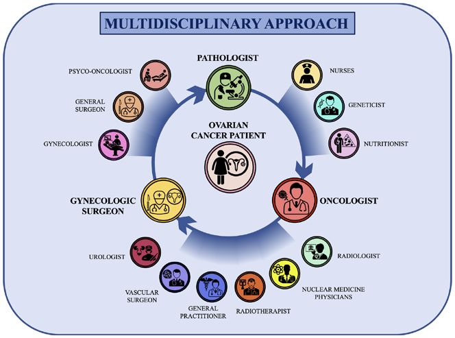
2                             FALZONE et al: MULTIDISCIPLINARY MANAGEMENT OF OVARIAN CANCER

    Other well‑recognized risk factors are gene mutations and         most frequently observed are pelvic distension, abdominal
hereditary syndromes that represent the most notable predis‑          and pelvic pain and urgent or frequent urination (34). Other
posing causes for the development of ovarian cancer (14).             symptoms may include pain during sex, back pain, constipa‑
A growing body of literature has demonstrated that indi‑              tion, altered menstruation, fatigue and weight loss (35). The
viduals harboring germline mutations affecting BRCA1 and              correct self‑assessment of these symptoms by the patients may
BRCA2 genes have an increased risk of breast and ovarian              improve the timing of diagnosis allowing the gynecological
cancer (15‑18). Overall, ~25% of ovarian cancer tumors are            surgeon and oncologist to intervene promptly by increasing
positive for BRCA1 or BRCA2 mutations (19). As explained              the patient response to treatments (36).
in the following sections, the evaluation of such mutations               Regarding ovarian cancer staging, two main staging
is important for the choice of anticancer pharmacological             systems are used worldwide for ovarian cancer, which are
treatments (20,21). Other hereditary syndromes related to             the International Federation of Gynecology and Obstetrics
ovarian cancer include hereditary non‑polyposis colon cancer          (FIGO 2018) system and the American Joint Committee
syndrome, Peutz‑Jeghers syndrome and adenine DNA glyco‑               on Cancer (AJCC 8th edition) system both based on
sylase (MUTYH)‑associated polyposis syndrome affecting                the Tumor‑Node‑Metastasis (TNM) parameters (37,38).
several mismatch repair genes (including MSH2, MSH6 and               Table I shows both FIGO and AJCC staging in terms of the
MLH1), STK11 and MUTYH (22‑24), respectively.                         pathological characteristics of tumors: Tumor dimension (T),
    Other controversial and not yet ascertained risk factors          lymph node involvement (N) and presence of distant metas‑
are represented by tobacco smoking, androgens, diet and talc          tasis (M) (Table I).
powder. For all these risk factors, observational, case‑control,          At present, several diagnostic strategies are available
retro‑ and prospective studies have generated conflicting results     to make a correct and timely diagnosis of ovarian cancer
thus limiting the awareness about the causative effects of these      when recurrent symptoms are observed. The first step for a
factors (25). It was demonstrated that tobacco smoking is             correct diagnosis of ovarian cancer is based on the collection
associated with the development of mucinous ovarian cancer,           of patient's medical history and on a correct physical exam
but it does not increase susceptibility to other types of ovarian     performed by a gynecologist with expertise in gynecological
tumors (26,27). Unconvincing data have been obtained for the          oncology (39,40). The aim of these procedures is the collection
association between powder use in the genital area and the risk       of all relevant data about the presence of pre‑existing condi‑
of ovarian cancer. In this context, some studies highlighted a        tions or risk factors that could increase the risk of developing
slightly increased risk of ovarian cancer in women using talc         ovarian cancer. In particular, as previously mentioned, the
powder in the genital area (28‑30). However, a recent obser‑          presence of a family member with ovarian cancer or the pres‑
vational study on 250,000 women observed for 11 years has             ence of hereditary syndromes and genetic mutations may lead
demonstrated that the use of powder does not significantly            the clinician to make a diagnosis of suspected ovarian cancer
increase the incidence of ovarian cancer (28‑30).                     in the presence of specific abdominal symptoms. In the same
    Besides these risk factors, numerous studies have also            manner, the physical examination of the abdomen and pelvis
identified some protective factors able to reduce the incidence       is of fundamental importance to observe pelvic mass, ascites
of ovarian cancer. Among these factors, pregnancy and breast‑         or abdominal distension suggestive of ovarian cancer (41).
feeding are both associated with a reduced risk of developing         The physical examination could include a rectovaginal exam
this tumor. In particular, a significantly reduced risk for ovarian   performed with empty bladder to evaluate the presence of
cancer has been observed in women carrying full‑term pregnan‑         abdominal or pelvic masses. However, although important and
cies before 26 years old (31). In addition, the increased number      easy to perform, physical investigations have a low sensitivity
of full‑term pregnancies, together with the time of breast‑           and a low specificity, especially in overweight patients or in
feeding, is associated with a lower risk of ovarian cancer (31).      presence of small tumors, as abdominal or pelvic distention
Finally, the use of oral contraceptives for birth control seems       may be caused by other benign pathologies (42,43).
to play an important protective role against ovarian cancer               After the physical examination, patients with suspected
with higher protective effects the longer the treatments are          ovarian cancer are subjected to various laboratory and
administered (32). In this context, other birth control strategies,   imaging tests useful to detect the presence of the tumor, its
including intrauterine devices and tubal ligation, have also been     severity and extent (41). Among the most used laboratory tests
associated with a reduced risk of ovarian cancer (33).                both for preventive and diagnostic purposes is the evaluation
                                                                      of blood tumor markers, namely cancer antigen (CA) 125 and
3. Ovarian cancer symptoms, diagnosis and staging                     human epididymis protein 4 (HE4), alongside the normal
                                                                      hematochemical parameters (red and white blood cells count,
During the early stages, ovarian cancer is not associated with        platelets and hemoglobin). In particular, CA 125 is considered
clinical symptoms, therefore the diagnosis of this tumor is           the main predictive serum biomarker for ovarian cancer as it
often delayed. Mild ovarian cancer symptoms may be often              is elevated in 50% of patients with early‑stage ovarian cancer
confused with other benign pathologies, including gastroin‑           and in over 80% of all patients with this tumor (44).
testinal disorders, urogenital infections and benign ovarian              Regarding HE4, this marker is evaluated together with
lesions (including ovarian cysts, teratomas and fibromas) (34).       CA 125 as it appears to be elevated in a significant fraction
However, unlike benign diseases, ovarian cancer symptoms are          of patients with ovarian cancer negative for CA 125 (45,46).
persistent and worsen over time (34). Generally, moderate or          Therefore, the use of HE4 is of fundamental importance in
severe symptoms are associated with the spread of the disease         screening strategies to intercept all those ovarian carcinomas
in adjacent anatomical regions. Among these symptoms, the             negative for other tumor biomarkers. The evaluation of these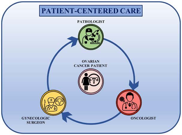
INTERNATIONAL JOURNAL OF ONCOLOGY 59: 53, 2021                                                    3

Table I. Ovarian cancer staging and pathological features.

FIGO      AJCC           TNM
stage     stage      characteristics                                            Description of tumor

I         I  T1                          The tumor is limited to the inner part of one ovary (T1) and there is no involvement of
		           N0                          neighboring lymph nodes (N0). There are no metastases (M0).
		M0
IA       IA T1a                          The tumor is limited to the inner part of one ovary without the involvement of
		           N0                          the outer surface (T1a) and there is no involvement of neighboring
		          M0                           lymph nodes (N0). There are no metastases (M0).
IB       IB T1b                          The tumor is limited to the inner part of both ovaries and there are no cancer cells in ascites
		           N0                          or in the abdominal and pelvic cavities (T1b) and there is no involvement of
		          M0                           neighboring lymph nodes (N0). There are no metastases (M0).
IC       IC T1c                          The tumor is in one or both ovaries and the tumor capsule is broken during surgery (IC1);
		           N0                          the tumor capsule is broken before surgery or the tumor is on the outer surface of
		          M0                           the ovary(ies) (IC2); tumor cells are present in the ascitic fluid or in the washing liquid
			                                      obtained from the abdomen and pelvis (IC3). There is no involvement
			                                      of neighboring lymph nodes (N0). There are no metastases (M0).
II       II  T2                          The tumor is in one or both ovaries and has spread to other adjacent pelvic organs or
		           N0                          to the peritoneum (T2). There is no involvement of neighboring
		          M0                            lymph nodes (N0). There are no metastases (M0).
IIA     IIA T2a                          The tumor has invaded or grown into the uterus or the fallopian tubes (T2a).
		           N0                           It has not invaded lymph nodes (N0) or distant sites (M0).
		M0
IIB     IIB T2b                          The tumor has invaded the outer and inner surface of pelvic organs including, uterus,
		           N0                          fallopian tubes, bladder and sigmoid colon (T2b). There is no involvement of neighboring
		          M0                           lymph nodes (N0). There are no metastases (M0).
IIIA1 IIIA1 T1-2                         The tumor has invaded ovaries, the peritoneum and other pelvic organs (T1-2).
		           N1                          The tumor has spread to the retroperitoneal (pelvic and/or para-aortic)
		          M0                            lymph nodes (N1) without forming distant metastasis (M0).
IIIA2 IIIA2 T3a                          The tumor affects one or both ovaries and has invaded the peritoneal cavity
		          N0-1                         and organs outside the pelvis; however, it is not visible during
		          M0                           surgery (T3a). The tumor is present or not on the retroperitoneal
			                                      lymph nodes (N0-1). There are no metastases (M0).
IIIB   IIIB T3b                          The tumor affects one or both ovaries and has invaded the peritoneal cavity and organs outside
		          N0-1                         the pelvis. During surgery the tumor is visible but is 2 cm (T3c). The tumor is present or not
		                        M0             on retroperitoneal lymph nodes (N0-1). There are no metastases (M0).
IVA  IVA                  T1-4           The tumor is present or not on retroperitoneal lymph nodes (N0-3). Tumor cells have invaded
		                        N0-3           the bloodstream leading to malignant pleural effusion. However, cancer cells have not invaded
		                        M1a            the spleen, intestine, liver neither lymph nodes outside the abdominal cavity (M1a).
IVB  IVB                  T1-4           The tumor is present or not on retroperitoneal lymph nodes (N0-3).
		                        N0-3           The tumor has spread to the liver or spleen, to extra abdominal lymph nodes
		                        M1b            and/or to other extra peritoneal organs or tissues, such as the lungs and bones (M1b).

FIGO, International Federation of Gynecology and Obstetrics; AJCC, American Joint Committee on Cancer; T, tumor; N, node; M, metastasis.

two markers, together with the evaluation of six symptoms                improvement in diagnostic accuracy from 83.8 to 98.5% (46).
predicting the presence of ovarian cancer (pelvic pain, abdom‑           Other tumor biomarkers, such as serum α‑fetoprotein and quan‑
inal pain, urinary urgency/frequency, increased abdominal size,          titative β‑human chorionic gonadotropin (β‑hCG), are less used
bloating and difficulty eating/feeling full) showed a significant        and are used for the diagnosis of germ cell ovarian cancer (47).

4                            FALZONE et al: MULTIDISCIPLINARY MANAGEMENT OF OVARIAN CANCER

    After these preliminary assessments, women who present           ovarian cancer, thus being curative in such tumors limited to
symptoms and biomarkers predictive for ovarian cancer                the ovaries (66,67). Ovarian cancer surgery can be performed
undergo imaging tests, including ultrasound, computed                by open surgery with midline incision or by minimally inva‑
tomography (CT) scan, magnetic resonance imaging (MRI)               sive surgery (MIS). MIS, performed by laparoscopic surgery,
scan and positron emission tomography (PET) scan (48).               is generally performed for newly diagnosed tumors limited to
    Generally, the first imaging test to be performed is a           one or both ovaries and to the pelvic cavity without metastatic
transvaginal ultrasonography. Several studies have shown how         dissemination (68). However, MIS is generally used only in
transvaginal ultrasonography is able to distinguish benign           structured centers equipped with experienced gynecological
lesions from tumors with an excellent rate of accuracy (pooled       surgeons (68). In the case of advanced tumors (stages II,
sensitivity and specificity of 92 and 88%, respectively) thus        III and IV), open surgery is always used in order to perform
allowing the clinician to evaluate the structure and vascular‑       extended cytoreduction (debulking) aimed at eliminating all
ization of the ovarian parenchyma, the presence of cysts or          cancer lesions with a thickness of >1 cm (69,70). Briefly, in
masses and any ascitic effusions (49‑52).                            both MIS and open surgery, the first steps consist in the collec‑
    Both CT, MRI and PET are not widely used for the diag‑           tion of ascitic fluid and in the execution of peritoneal lavage
nosis of ovarian cancer but for the evaluation of the extent of      used for immunocytochemistry evaluations useful to estab‑
the tumor and the possible presence of distant metastases.           lish the presence of tumor cells in the peritoneal cavity (71).
Specifically, CT scan can be used to perform biopsies of             Subsequently, surgeons check the entire peritoneal cavity
suspected metastases in a procedure called CT‑guided needle          to assess the absence of any suspicious extra ovary lesions.
biopsy (53,54). Meanwhile, PET and MRI are mostly used to            In the case of no suspicious masses, biopsies from different
evaluate the spread of diseases in neighboring lymph node            parts of the peritoneal cavity (paracolic gutters, pelvis and
stations and in distant organs, such as the medulla and brain,       diaphragm) should be obtained to exclude cancer dissemina‑
through the use of radiotracers or contrast agents (for example      tion (71). After these preliminary steps, surgeons can remove
gadolinium) (55).                                                    the primary tumor through bilateral salpingo‑oophorectomy,
    Finally, after tumor diagnosis, it is essential to perform       hysterectomy, omentectomy and lymph node dissection (both
molecular tests and genetic counseling to determine the              pelvic and paraaortic nodes). To avoid post‑surgery cancer
presence of relevant mutations in tumor specimens useful for         dissemination, the tumor has to be removed encapsulated. In
prognostic and therapeutic purposes (56‑60). As aforemen‑            the case of young patients (20‑45 years old) with monolateral
tioned, the most frequent mutations observed in ovarian cancer       stage IA and IC ovarian cancer that would maintain fertility,
are those affecting the BRCA1 and BRCA2 genes as well as             the surgeon could opt for unilateral ovariectomy and adnexec‑
other mutations within STK11, MSH2, MSH6, MLH1, PMS6                 tomy, thus preserving the contralateral ovary and uterus (71).
and MUTYH (56,57). Besides molecular evaluations, previous               Overall, the main objectives of ovarian cancer surgery are
studies have demonstrated that immunohistochemical inves‑            the removal of the primary tumor and the maximal debulking
tigations are fundamental both for diagnostic and prognostic         of pelvic and peritoneal masses. In presence of advanced
purposes for different abdominal tumors, including that of           tumors, the clinicians can opt for neoadjuvant chemo‑
ovaries (58‑60).                                                     therapy (NAC) followed by debulking surgery. If the NAC
                                                                     plus surgery approach is chosen, tumor biopsies are collected
4. Ovarian cancer surgical treatments                                before chemotherapy to assess the molecular features of
                                                                     the tumors (through immunohistochemistry or molecular
Over the decades, the therapeutic options for the treatment          tests) thus allowing administration of appropriate anticancer
of advanced ovarian cancer have been improved significantly          drugs (72). In those patients undergoing NAC and debulking
through the development of more precise and less invasive            surgery, a whole‑abdominal radiation treatment should be
surgical techniques as well as the availability of novel effective   applied if residual disease is still observed after a second‑look
drugs able to extend the life expectancy of patients, especially     laparotomy; however, this approach needs to be carefully
for metastatic ovarian cancer (61,62). Different studies have        evaluated to avoid bowel toxicity (73,74).
demonstrated that in the last 20‑35 years there was a signifi‑
cant improvement in the survival rates of patients with ovarian      5. Ovarian cancer pharmacological treatments
cancer; however, some reports have shown that the advance‑
ments of the anticancer treatment have not ameliorated the           Besides surgery, anticancer pharmacological treatments are
long‑term survival and the cure rate of ovarian cancer (63‑65).      the best therapeutic option for the management of ovarian
In particular, a recent study showed that both incidence and         cancer. Over the years, several chemotherapeutic agents
5‑year survival rates have improved in the last 30 years.            have been used for the treatment of ovarian cancer. Thanks
Indeed, the 5‑year survival rate increased from 39.3% in the         to the evolution of anticancer pharmacological treatments, it
80s to 45.4% observed in 2012; similarly, the survival time          is now possible to effectively treat the different histological
was also improved passing from 34 months observed in 1983            and molecular subtypes of ovarian cancer, contributing to the
to 52 months observed in 2012, highlighting how the latest           improvement of the quality of life and life expectancy of these
treatments have improved the survival time of patients with          patients (2,75).
ovarian cancer (62).
    At present, surgery represents the gold standard for the         Ovarian cancer chemotherapy. After surgery, chemotherapy
treatment of ovarian cancer. Ovariectomy and adnexectomy             can be optionally administered in patients with low‑grade
are used for both staging, debulking and treatment of early          tumors (stage IA or IB), while the first‑line treatment for
INTERNATIONAL JOURNAL OF ONCOLOGY 59: 53, 2021 5
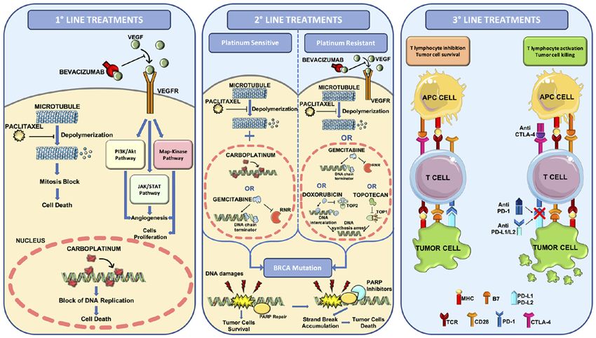
Figure 1. Overview of the pharmacological treatments for the management of ovarian cancer. First‑line treatments based on the administration of plat‑
inum/taxane regimen plus anti‑VEGF mAb bevacizumab. Second‑line treatments based on the administration of paclitaxel plus carboplatin or gemcitabine in
the case of platinum sensitive tumors or bevacizumab plus paclitaxel, gemcitabine, doxorubicin or topotecan in the case of platinum resistant tumors. For BRCA
mutated ovarian cancer, PARP inhibitors can be used for first‑line or second‑line treatments. Third‑line treatments consist in the administration of the first‑line
and second‑line drugs plus immune checkpoint inhibitors. MHC, major histocompatibility complex; RNR, ribonucleotide reductase; TCR, T‑cell receptor;
TOP1, type 1 topoisomerase; TOP2, type 2 topoisomerase.
ovarian cancer with more advanced stages is based on the demonstrated that the prolonged administration of 15 mg/kg
administration of platinum‑based chemotherapy. Indeed, the 3‑weekly of bevacizumab up to 15 months together with stan‑
first‑line regimen consists of the administration of intravenous dard carboplatin/paclitaxel chemotherapy is associated with
platinum/taxane every three weeks for six cycles (76). The a prolonged PFS time; however, due to the expensive cost of
same compounds are usually administered also in patients treatments and the related gastrointestinal and vascular toxici‑
with stage III/IV ovarian cancer undergoing NAC protocols for ties, novel protocols based on a low dose of bevacizumab for
three cycles followed by debulking surgery plus six additional 30 months is still under evaluation and it is awaiting approval
cycles of platinum/taxane (76,77). as a therapeutic standard for this tumor (76,85,86) (Fig. 1).
Thus, for >20 years, the first‑line treatment for ovarian After the first‑line chemo‑ and targeted therapy, the
cancer has been based on the administration of carboplatin patients can completely respond to treatments or develop a
(used instead of cisplatin because it is less toxic and equally relapse. In the case of a partial or complete response, patients
effective) and paclitaxel administered every three weeks in a can undergo maintenance chemotherapy with the same drugs
six‑cycle schedule. The preferred route of administration is the used in the first‑line treatment to improve PFS (87,88).
intravenous systemic one, although several studies have also In the case of tumor recurrence, patients are treated with a
proposed intraperitoneal administration, which has not given second‑line treatment that is different depending on whether the
improved results in terms of improvement of progression‑free tumor is resistant or sensitive to platinum compounds (89,90).
survival (PFS) (78,79). Similarly, several trials have investi‑ Tumor recurrence can be observed through biochemical
gated the beneficial effects of paclitaxel weekly administration (increased expression of CA125 and other biomarkers) or
compared with the conventional 3‑week schedule; however, clinical (imaging techniques) examinations (91) after which
this therapeutic option is not widely used as conflicting patients are assigned to standard treatment for recurrent
data have been generated in three different clinical studies disease or to experimental clinical trials using novel drugs or
(JGOG 3016, GOG 262 and MITO 7) (80‑82) (Fig. 1). different drug combinations (92,93).
More recently, the introduction of anticancer targeted For patients with ovarian cancer developing a plat‑
therapies has improved the efficacy of first‑line treatments inum‑resistant disease, the second‑line treatments consist of
for patients with ovarian cancer who can benefit from treat‑ single non‑platinum‑based therapies using different agents,
ments based on the administration of carboplatin, paclitaxel including docetaxel, paclitaxel, topotecan and gemcitabine,
and bevacizumab (83,84). Bevacizumab is a monoclonal with a therapeutic efficacy ranging from 19 to 27% of the
antibody against the pro‑angiogenetic factor VEGF‑A that treated patients (71). Similar percentages of response have been
has prolonged the PFS and OS time of patients, especially of obtained treating ovarian cancer relapse with bevacizumab
those patients with advanced tumors (83). In particular, it was (therapeutic response observed in ~20% of patients) (94). More6 FALZONE et al: MULTIDISCIPLINARY MANAGEMENT OF OVARIAN CANCER
recently, in absence of severe adverse events, combined thera‑ maintenance treatment with olaparib significantly improves
pies with bevacizumab plus one agent among doxorubicin, overall survival time (SOLO2 and E19 trials) (106‑108).
topotecan and paclitaxel have shown a significant improve‑ The therapeutic efficacy of PARP inhibitors has been
ment of OS in patients with platinum‑resistant recurrent demonstrated also in patients without BRCA mutations. The
disease (95) (Fig. 1). NOVA trial based on the administration of niraparib in patients
In the case of platinum‑sensitive recurrence, there are with ovarian cancer demonstrated that all patients can benefit
different therapeutic options based on the administration of from this treatment, although improved results were obtained
several drug combinations including carboplatin plus paclitaxel for patients with homologous recombination deficiency (HRD)
(or docetaxel with weakly or 3‑weeks administration), carbo‑ compared with patients without mutations affecting the HR
platin plus gemcitabine and bevacizumab, carboplatin plus system (105).
liposomal doxorubicin and cisplatin plus gemcitabine (96). In In December 2016, the Food and Drug Administration
addition, patients with ovarian cancer who are platinum‑sensi‑ launched an accelerated approval process for the use of
tive are often eligible for novel clinical trials assessing the rucaparib as a single agent for the treatment of patients with
efficacy of novel agents or combined therapies. Among these ovarian cancer at an advanced stage and with a BRCA muta‑
trials, recent evidence has demonstrated the therapeutic tion (germline or somatic) who had been previously treated
efficacy of poly (ADP‑ribose) polymerase (PARP) inhibi‑ with two or more lines of chemotherapy (109). In addition,
tors in both platinum‑sensitive and ‑resistant ovarian cancer rucaparib is also used as second‑line maintenance therapy in
harboring BRCA1 or BRCA2 mutations. In particular, patients patients with platinum‑sensitivity with or without BRCA1/2
with platinum‑sensitivity with complete or partial response mutations as reported in the Ariel 3 Trial (100).
to at least two lines of treatments can benefit from olaparib
single‑agent maintenance therapy improving their PFS from Novel therapeutic approaches for the treatment of ovarian
5.5 to 19.1 months (97). Similarly, olaparib single‑therapy can cancer. Besides these conventional therapies, novel approaches
be used also in patients with platinum‑resistant BRCA mutated for the treatment of advanced or metastatic ovarian cancer
ovarian cancer who failed three or more lines of chemo‑ are being developed and studied. Modest results have been
therapy (98) (Fig. 1). obtained in several clinical trials assessing the efficacy of
immune checkpoint inhibitors (ICIs) already used for the
Novel first‑line and second‑line treatments with PARP treatment of several advanced and metastatic tumors (110‑112).
inhibitors. As aforementioned, some patients with ovarian In particular, the administration of anti‑PD‑1 (nivolumab or
cancer can benefit from novel first‑line and second‑line treat‑ pembrolizumab) or anti‑PD‑L1 (atezolizumab) in advanced
ments based on the administration of selective inhibitors of ovarian cancer has a good response only in 10‑15% of
PARP. PARP proteins are a family of 17 enzymes involved patients (113‑115). Similar results have been obtained with
in numerous cellular processes, and in particular PARP‑1 and the single administration of the anti‑CTLA‑4 ICI ipilimumab,
PARP‑2 play a crucial role in DNA damage repair (99,100). which is effective only in a small fraction of patients who has
The development of PARP inhibitors has repre‑ previously received an anticancer therapeutic vaccine (116).
sented the turning point in the treatment of ovarian cancer, Overall, single‑agent ICI administration shows limited efficacy
both in the first‑line and in case of tumor recurrence, high‑ in advanced ovarian cancer, therefore novel protocols assessing
lighting the importance of studying the molecular profile of the concomitant administration of ipilimumab and nivolumab
tumors to improve the selection of patients eligible for these have been proposed (117). Such studies have demonstrated an
innovative treatments. Indeed, these drugs, including olaparib, improved and longer response rate in patients treated with two
rucaparib, niraparib and talazoparib, find application in tumors ICIs compared with patients treated with nivolumab alone,
with germline mutations affecting BRCA1 and BRCA2 or in thus replacing the single‑agent ICI regimens (117). A recent
advanced ovarian cancer refractory to three or more lines of review of the literature collected all the completed and ongoing
treatment (101). clinical trials using different combinations of ICIs, selective
Recent evidence has demonstrated that patients with ovarian inhibitors or chemotherapeutic agents showing encouraging
cancer harboring BRCA1 or BRCA2 mutations could benefit and conflicting results based on the clinical and molecular
from a first‑line or a second‑line treatment with olaparib, which features of the patients enrolled (118) (Fig. 1).
reduces the risk of progression or death by 70% compared Other investigated therapeutic options for advanced
with placebo in patients who achieved a complete or partial ovarian cancers are represented by therapeutic vaccines,
response to the first platinum‑based line. In particular, 60.4% adoptive cellular therapy, T cell transfer and chimeric
of patients treated with olaparib showed a progression‑free antigen receptor T‑cell therapy; however, further clinical
survival of 36 months compared with 26.9% of women in the studies are needed to assess the efficacy and safety of these
placebo arm (102). These data are further corroborated by the further treatments (119).
results presented during the European Society for Medical Finally, several treatments are available as maintenance
Oncology 2019 and European Society of Gynecological or palliative therapy for disseminated and metastatic ovarian
Oncology 2019 conferences regarding two clinical trials using cancer. Similarly, VEGFR inhibitors, including pazopanib,
niraparib and olaparib plus bevacizumab (PRIMA and PAOLA nintedanib and cediranib, are often used for the treatment of
1 trials, respectively) as first‑line treatments (103‑105). The recurrent platinum‑resistant ovarian cancer (100). In line with
therapeutic efficacy of PARP inhibitors has been demonstrated these treatments, VEGF inhibitors such as aflibercept are used
mainly in patients with BRCA1/2‑positive ovarian cancer in case of malignant ascites showing an improvement of time
who develop platinum‑sensitive relapse. In these patients, to next paracentesis but not of OS (120).INTERNATIONAL JOURNAL OF ONCOLOGY 59: 53, 2021 7
Figure 2. Linear management of ovarian cancer. Main players of ovarian cancer management are the surgeon who removes the tumor, the pathologist who
assesses the tumor histotype and the medical oncologist who selects and starts the pharmacological treatments. There is no interaction between these three
main specialists in the management of the patient with ovarian cancer.
Despite the availability of all these surgical and pharma‑ and predictive markers to make the best treatment decisions
cological treatments, the prognosis of patients with ovarian for the management of recurrence (121).
cancer is often poor. To improve the quality of life and
life expectancy of these patients, it is necessary to opt for 6. Ovarian cancer management: From linear to
therapeutic choices that take into account the patient's comor‑ multidisciplinary patient‑centered care approach
bidities, the adverse effects of therapies and the patient's age.
Therefore, the management of the patient with ovarian cancer As aforementioned, the symptomatology, diagnosis, staging
is extremely complex and requires the convergence of different and treatments of ovarian cancer are extremely complex and
professional skills to ensure high standards of care. require the convergence of various specialists able to provide
the gynecological oncologist with a clinical picture as detailed
5th ovarian cancer consensus conference of Tokyo. The 5th as possible, useful for designing the appropriate therapeutic
ovarian cancer consensus conferences held in Tokyo, Japan, protocol for each patient. Therefore, at present, the approach to
in 2015 (121), established that platinum‑based regimens are the patient with advanced ovarian cancer should be multidisci‑
doubtless the standard of care in the first‑line treatment of plinary. This includes a team of experts who follow the patient
ovarian cancer. However, for the first time, besides highlighting step by step during the diagnosis, surgical therapy, pharma‑
the importance of the platinum‑free interval (PFI) as a strati‑ cological therapy, rehabilitation and follow‑up, creating a
fication factor or to define patient eligibility for clinical trials, collaborative network where the patient is at the center and
great importance was given to treatment‑free interval useful can benefit of high standards of care in the perspective of
to improve selection of successive chemotherapy regimens for personalized medicine and patient‑centered care (122).
patients with recurrent disease. In this context, over the decades, great advancements in the
The main decision criterion for second‑line treatments is management of patients with ovarian cancer have occurred,
the definition of platinum‑sensitivity or resistance. Sensitivity passing from a linear approach to care, where the patient is
to platinum‑based treatments must be assessed after a period treated by individual specialists without communication
of at least 6 months; however, there is a linear relationship between them, to a multidisciplinary and integrated approach
between PFI and platinum sensitivity, therefore the evaluation where different specialists share clinical information and
of PFI is of primary importance in future therapeutic choices chose the best therapeutic options together (123,124).
and must be considered as a continuous variable in the deci‑ Until 30 years ago, the therapeutic approach followed a
sion‑making process leading to the new therapy. Furthermore, linear trend where the main stakeholders of cancer manage‑
PFI will be used as a parameter for the eligibility of patients ment were the surgeon, who operated the surgical resection
in novel clinical trials, therefore the evaluation of PFI cannot of the tumor, the pathologist, who made the histological
be limited to a fixed 6‑month window but should be evaluated diagnosis, and the medical oncologist, who dealt with the
periodically. therapeutic schedule to be administered (Fig. 2). Although
Overall, platinum‑based therapy remains the most effective other professionals participated in the clinical management
therapy in the management of epithelial ovarian cancer, and of ovarian cancer (including gynecologists, radiologists and
primary PFI provides relevant prognostic and predictive infor‑ laboratory technicians), they did not actively take part in the
mation. A significant fraction of patients receives different clinical‑therapeutic decisions. In addition, the interactions
lines of platinum‑based therapy, thus evaluating the interval between the patient, the surgeon, the pathologist and the
of time after the most recent line can provide prognostic infor‑ oncologist rarely occurred and each of these three professional
mation about acquired resistance and clonal evolution of the figures made therapeutic choices without first discussing with
tumor due to intervening non‑platinum treatments. Over the colleagues (125,126).
years different non‑platinum agents have been integrated into Since the late 80s, some studies have highlighted the
conventional therapy; this led to the need of new prognostic benefits of the multidisciplinary management of patients with8 FALZONE et al: MULTIDISCIPLINARY MANAGEMENT OF OVARIAN CANCER
cancer in terms of diagnosis, therapeutic response, survival As surgery represents the gold standard for the treatment
and quality of life, suggesting that an integrated approach to of ovarian cancer, it is well established that patients treated
cancer could lead to improved outcomes for patients (127‑129). in experienced centers benefit from maximum cytoreductive
With regards ovarian cancer, it was demonstrated that a surgical resection which positively correlates with the overall
collegial discussion can lead all the specialists to evaluate survival of patients (135,138). Besides the importance of
the diagnostic‑therapeutic areas beyond those of their own surgery, the discussion of clinical cases in the multidisciplinary
competence, leading to an increase of awareness in the number team does not end with the diagnosis and surgical resection
of potential treatments available and expected pitfalls thus of tumor masses, but it takes place at every decision‑making
improving the effectiveness of treatments (130,131). point, especially in case of recurrent diseases (139). In these
The development of multidisciplinary teams has changed cases, the interaction of the various specialists can lead to the
the previous linear approach to patients into a circular one. design of novel and effective therapeutic strategies tailored
Indeed, the main players of ovarian cancer management are to each patient (139). Thus, such strategies may involve new
now working together, comparing their clinical findings each surgical interventions in the peritoneal cavity or other body
other in a patient‑centered care approach where the patients districts, which requires the expertise of different types of
are in the middle of a circular decision‑making pathway surgeons, including urologists, vascular surgeons and general
receiving information and therapeutic options shared between surgeons, or could lead to novel anticancer treatments using
the gynecological surgeon, the pathologist and the medical both chemo‑ and radiation therapies when distant metastases
oncologist (Fig. 3). This circular approach to ovarian cancer are observed (140,141).
treatment has introduced different therapeutic opportunities It is important to note that each specialist within the multi‑
that are continuously evaluated and re‑elaborated according disciplinary team has a fundamental role in the diagnostic or
to the clinical information received from the different special‑ therapeutic process. Indeed, the use of imaging techniques
ists involved in the collaborative care network. Therefore, this performed by the radiologist is fundamental to formulate a
approach results in a improved management of ovarian cancer diagnosis of suspected ovarian cancer and to establish the local‑
and patient awareness about the status of the disease, as well ization of lesions (142,143). In the same manner, the precise
as greater confidence in the therapeutic options that they will histological and biomolecular evaluation of ovarian cancer is
undergo (132). now essential for modern cancer treatments (144,145). In this
Although patient‑centered care has significantly improved context, the pathologist, geneticist and molecular biologist are
the standard of ovarian cancer care, several studies have fundamental for the assessment of grading, histotyping and
demonstrated that patients treated in specialized structures molecular typing of ovarian cancer (146,147). In addition, the
where multidisciplinary teams operate have an improved multidisciplinary network of specialists is further enriched
prognosis compared with patients treated in non‑specialized by the inclusion of breast specialists and nutritionists. In
centers (133‑135). A possible explanation of this trend could particular, breast specialists intervene in the case of BRCA1‑
be related to the well‑organized approach to treatment in and BRCA2‑positive ovarian cancer who could develop a
specialized hospitals with a high volume of patients with secondary neoplasm affecting the breast (148,149); while nutri‑
ovarian cancer per year where the components of the multi‑ tionists are now a key professional figure in medical oncology
disciplinary team meet together weekly to discuss the periodic departments. In fact, several studies have demonstrated that
clinical, laboratory and instrumental findings useful to take nutrition represents an important protective factor against the
appropriated and shared clinical decision. Of note, despite the development of tumors (150) but also represents an effective
undoubted advantages of a multidisciplinary team in terms of therapeutic intervention for patients with cancer (151,152). In
the quality of the assistance provided, this type of interdis‑ this context, several studies have demonstrated that dietary and
ciplinary display requires appropriate organization, time for lifestyle interventions during cancer treatments can ameliorate
periodic meetings, willingness to collaborate and adequate the adherence to treatment as well as patient quality of life
IT support; it is useful to share medical records and clinical and prognosis [hazard ratio (HR) for physical activity, 0.60;
data, favoring a continuous constructive debate for the better 95% CI, 0.39‑0.92; P=0.02; HR for highest vs. lowest tertile
management of each patient (136). of quality diet, 0.73; 95% CI, 0.55 to 0.97; P=0.03] (153‑156).
As shown in Fig. 3, the gynecologist (or gynecological These data, together with the clinical features of patients allow
surgeon), the pathologist and the oncologist are the key clinicians to determine the best therapeutic approach as well
nodes of this circular multidisciplinary network. However, as to predict the prognosis and outcomes of patients (157).
as aforementioned, due to the current complexity of ovarian The importance of a multidisciplinary team for the
cancer diagnosis, staging and treatments other professionals, management of ovarian cancer has emerged during the
including the general surgeon, urologist, vascular surgeon, COVID‑19 pandemic where patients with ovarian cancer have
radiologist, nuclear medicine physicians, geneticist, molecular experienced difficulties in accessing medical treatment (158).
biologist and psycho‑oncologist are fundamental in the Indeed, due to the spread of infection, patients with cancer
circular patient‑centered model (Fig. 4). have experienced delays in treatment or missed some thera‑
Only a structured center can offer well‑structured multi‑ pies with a negative impact on the treatment response (159).
disciplinary teams able to address all of the patient needs. In In addition, patients with cancer are considered vulnerable
this context, the Mercado et al (137) study shows that patients individuals with an increased risk of COVID‑19 infection and
treated in referral centers and treated by expert physicians severe symptomatology (160). In this context, the multidisci‑
have a 40% higher survival compared with patients treated in plinary team involved in the management of ovarian cancer
a peripheral center. has improved novel telemedicine strategies useful to monitorINTERNATIONAL JOURNAL OF ONCOLOGY 59: 53, 2021 9
Figure 3. Circular approach to the management of patients with ovarian cancer patients. Main specialists involved in ovarian cancer care interact with each
other sharing all of the relevant information and all clinical decisions are patient‑centered.
Figure 4. Circular and multidisciplinary network for the management of patients with ovarian cancer. The gynecological surgeon, pathologist and oncologist
are the key nodes of the patient‑centered circular multidisciplinary network. Other specialists, including nurses, geneticists, nutritionists, radiologists, nuclear
medicine physicians, radiotherapists, general practitioners, vascular surgeons, urologists, gynecologists, general surgeons and psycho‑oncologists, actively
participate in all the decision‑making steps of ovarian cancer management.
patients with ovarian cancer at a distance, thus following the pharmacological treatments and mini‑invasive surgical
progression of the disease and patient health status during techniques. Besides these advancements, a multidisciplinary
the treatment. In addition, thanks to the patient‑centered approach for the treatment of ovarian cancer has significantly
circular multidisciplinary network the information is easily improved the quality of life and prognosis of patients. Overall,
transferred among specialists, thus increasing the speed of a multidisciplinary team is able to face clinical, molecular,
the implementation of therapeutic strategies and follow‑up pathological and psychological issues of patients with
visits (161‑163). ovarian cancer, ensuring a high standard of care supporting
the process of personalized medicine. The importance of a
7. Conclusions multidisciplinary team and periodic meetings lays also on
the constant improvement of molecular, biological and thera‑
In recent years, the management of ovarian cancer has been peutic knowledge in the field of ovarian cancer care. Indeed,
significantly improved through the introduction of novel the active discussion performed within a multidisciplinary10 FALZONE et al: MULTIDISCIPLINARY MANAGEMENT OF OVARIAN CANCER
team improves the adoption of the best therapies for patients 10. Amir S, Shah STA, Mamoulakis C, Docea AO, Kalantzi OI,
Zachariou A, Calina D, Carvalho F, Sofikitis N, Makrigiannakis A
as well as the efficacy of treatments. and Tsatsakis A: Endocrine disruptors acting on estrogen and
androgen pathways cause reproductive disorders through multiple
Acknowledgements mechanisms: A review. Int J Environ Res Public Health 18: 1464,
2021.
11. Ianoşi S, Ianoşi G, Neagoe D, Ionescu O, Zlatian O, Docea AO,
Not applicable. Badiu C, Sifaki M, Tsoukalas D, Tsatsakis AM, et al:
Age‑dependent endocrine disorders involved in the pathogenesis
of refractory acne in women. Mol Med Rep 14: 5501‑5506, 2016.
Funding 12. Del Pup L, Mantovani A, Cavaliere C, Facchini G, Luce A,
Sperlongano P, Caraglia M and Berretta M: Carcinogenetic
No funding was received. mechanisms of endocrine disruptors in female cancers (Review).
Oncol Rep 36: 603‑612, 2016.
13. Rachoń D: Endocrine disrupting chemicals (EDCs) and female
Availability of data and materials cancer: Informing the patients. Rev Endocr Metab Disord 16:
359‑364, 2015.
14. Shulman LP and Dungan JS: Cancer genetics: Risks and
Not applicable. mechanisms of cancer in women with inherited susceptibility to
epithelial ovarian cancer. Cancer Treat Res 156: 69‑85, 2010.
Authors' contributions 15. Gomes R, Spinola PD, Brant AC, Matta BP, Nascimento CM,
de Aquino Paes SM, Bonvicino CR, Dos Santos AC and
Moreira MA: Prevalence of germline variants in consensus
LF, GSca and PS conceived the manuscript, performed moderate‑to‑high‑risk predisposition genes to hereditary breast
bibliographic research and wrote the article. VL, GG and AL and ovarian cancer in BRCA1/2‑negative Brazilian patients.
Breast Cancer Res Treat 185: 851‑861, 2021.
performed the bibliographic research and prepared the table 16. Hodgson A and Turashvili G: Pathology of hereditary breast and
and figures. GSci and ABD provided critical revisions. All ovarian cancer. Front Oncol 10: 531790, 2020.
authors provided critical revisions and read and approved the 17. Jara L, Morales S, de Mayo T, Gonzalez‑Hormazabal P,
Carrasco V and Godoy R: Mutations in BRCA1, BRCA2 and
final manuscript. Data authentication is not applicable. other breast and ovarian cancer susceptibility genes in Central
and South American populations. Biol Res 50: 35, 2017.
Ethics approval and consent to participate 18. Tedaldi G, Tebaldi M, Zampiga V, Danesi R, Arcangeli V,
Ravegnani M, Cangini I, Pirini F, Petracci E, Rocca A, et al:
Multiple‑gene panel analysis in a case series of 255 women with
Not applicable. hereditary breast and ovarian cancer. Oncotarget 8: 47064‑47075,
2017.
19. Hanley GE, McAlpine JN, Miller D, Huntsman D, Schrader KA,
Patient consent for publication Blake Gilks C and Mitchell G: A population‑based analysis of
germline BRCA1 and BRCA2 testing among ovarian cancer
Not applicable. patients in an era of histotype‑specific approaches to ovarian
cancer prevention. BMC Cancer 18: 254, 2018.
20. Madariaga A, Lheureux S and Oza AM: Tailoring ovarian
Competing interests cancer treatment: Implications of BRCA1/2 mutations. Cancers
(Basel) 11: 416, 2019.
21. Ashour M and Ezzat Shafik H: Frequency of germline mutations
The authors declare that they have no competing interests. in BRCA1 and BRCA2 in ovarian cancer patients and their effect
on treatment outcome. Cancer Manag Res 11: 6275‑6284, 2019.
22. Tsaousis GN, Papadopoulou E, Apessos A, Agiannitopoulos K,
References Pepe G, Kampouri S, Diamantopoulos N, Floros T, Iosifidou R,
Katopodi O, et al: Analysis of hereditary cancer syndromes by
1. Bray F, Ferlay J, Soerjomataram I, Siegel RL, Torre LA and using a panel of genes: Novel and multiple pathogenic mutations.
Jemal A: Global cancer statistics 2018: GLOBOCAN estimates BMC Cancer 19: 535, 2019.
of incidence and mortality worldwide for 36 cancers in 185 coun‑ 23. Nakonechny QB and Gilks CB: Ovarian cancer in hereditary
tries. CA Cancer J Clin 68: 394‑424, 2018. cancer susceptibility syndromes. Surg Pathol Clin 9: 189‑199,
2. Falzone L, Salomone S and Libra M: Evolution of cancer phar‑ 2016.
macological treatments at the turn of the third millennium. Front 24. Nishioka Y, Kobayashi K, Sagae S, Sugimura M, Ishioka S,
Pharmacol 9: 1300, 2018. Nagata M, Terasawa K, Tokino T and Kudo R: Mutational
3. Torre LA, Trabert B, DeSantis CE, Miller KD, Samimi G, analysis of STK11 gene in ovarian carcinomas. Jpn J Cancer
Runowicz CD, Gaudet MM, Jemal A and Siegel RL: Ovarian Res 90: 629‑632, 1999.
cancer statistics, 2018. CA Cancer J Clin 68: 284‑296, 2018. 25. McLemore MR, Miaskowski C, Aouizerat BE, Chen LM and
4. Reid BM, Permuth JB and Sellers TA: Epidemiology of ovarian Dodd MJ: Epidemiological and genetic factors associated with
cancer: A review. Cancer Biol Med 14: 9‑32, 2017. ovarian cancer. Cancer Nurs 32: 281‑288, 2009.
5. Shafrir AL, Rice MS, Gupta M, Terry KL, Rosner BA, 26. Faber MT, Kjær SK, Dehlendorff C, Chang‑Claude J,
Tamimi RM, Hecht JL and Tworoger SS: The association Andersen KK, Høgdall E, Webb PM, Jordan SJ; Australian
between reproductive and hormonal factors and ovarian cancer Cancer Study (Ovarian Cancer); Australian Ovarian Cancer
by estrogen‑ α and progesterone receptor status. Gynecol Study Group, et al: Cigarette smoking and risk of ovarian cancer:
Oncol 143: 628‑635, 2016. A pooled analysis of 21 case‑control studies. Cancer Causes
6. D'Alonzo M, Bounous VE, Villa M and Biglia N: Current Control 24: 989‑1004, 2013.
evidence of the oncological benefit‑risk profile of hormone 27. Modugno F, Ness RB and Cottreau CM: Cigarette smoking and
replacement therapy. Medicina (Kaunas) 55: 573, 2019. the risk of mucinous and nonmucinous epithelial ovarian cancer.
7. Craig ER, Londoño AI, Norian LA and Arend RC: Metabolic Epidemiology 13: 467‑471, 2002.
risk factors and mechanisms of disease in epithelial ovarian 28. O'Brien KM, Tworoger SS, Harris HR, Anderson GL,
cancer: A review. Gynecol Oncol 143: 674‑683, 2016. Weinberg CR, Trabert B, Kaunitz AM, D'Aloisio AA, Sandler DP
8. Fenga C, Gangemi S, Di Salvatore V, Falzone L and Libra M: and Wentzensen N: Association of powder use in the genital area
Immunological effects of occupational exposure to lead (Review). with risk of ovarian cancer. JAMA 323: 49‑59, 2020.
Mol Med Rep 15: 3355‑3360, 2017. 29. Fletcher NM, Harper AK, Memaj I, Fan R, Morris RT and
9. Falzone L, Marconi A, Loreto C, Franco S, Spandidos DA and Saed GM: Molecular basis supporting the association of talcum
Libra M: Occupational exposure to carcinogens: Benzene, pesti‑ powder use with increased risk of ovarian cancer. Reprod Sci 26:
cides and fibers (Review). Mol Med Rep 14: 4467‑4474, 2016. 1603‑1612, 2019.INTERNATIONAL JOURNAL OF ONCOLOGY 59: 53, 2021 11
30. Berge W, Mundt K, Luu H and Boffetta P: Genital use of talc and 54. Thabet A, Somarouthu B, Oliva E, Gervais DA, Hahn PF and
risk of ovarian cancer: A meta‑analysis. Eur J Cancer Prev 27: Lee SI: Image‑guided ovarian mass biopsy: Efficacy and safety.
248‑257, 2018. J Vasc Interv Radiol 25: 1922‑1927.e1, 2014.
31. Troisi R, Bjørge T, Gissler M, Grotmol T, Kitahara CM, 55. Khiewvan B, Torigian DA, Emamzadehfard S, Paydary K,
Myrtveit Saether SM, Ording AG, Sköld C, Sørensen HT, Salavati A, Houshmand S, Werner TJ and Alavi A: An update on
Trabert B and Glimelius I: The role of pregnancy, perinatal the role of PET/CT and PET/MRI in ovarian cancer. Eur J Nucl
factors and hormones in maternal cancer risk: A review of the Med Mol Imaging 44: 1079‑1091, 2017.
evidence. J Intern Med 283: 430‑445, 2018. 56. Zhao C, Li S, Zhao M, Zhu H and Zhu X: Prognostic values of
32. Havrilesky LJ, Moorman PG, Lowery WJ, Gierisch JM, DNA mismatch repair genes in ovarian cancer patients treated
Coeytaux RR, Urrutia RP, Dinan M, McBroom AJ, Hasselblad V, with platinum‑based chemotherapy. Arch Gynecol Obstet 297:
Sanders GD and Myers ER: Oral contraceptive pills as primary 153‑159, 2018.
prevention for ovarian cancer: A systematic review and 57. Hirotsu Y, Nakagomi H, Sakamoto I, Amemiya K, Oyama T,
meta‑analysis. Obstet Gynecol 122: 139‑147, 2013. Mochizuki H and Omata M: Multigene panel analysis identified
33. Huang Z, Gao Y, Wen W, Li H, Zheng W, Shu XO and germline mutations of DNA repair genes in breast and ovarian
Beeghly‑Fadiel A: Contraceptive methods and ovarian cancer cancer. Mol Genet Genomic Med 3: 459‑466, 2015.
risk among Chinese women: A report from the shanghai women's 58. Missaoui N, Salhi S, Bdioui A, Mestiri S, Abdessayed N,
health study. Int J Cancer 137: 607‑614, 2015. Mokni M and Yacoubi MT: Immunohistochemical characteriza‑
34. Goff B: Symptoms associated with ovarian cancer. Clin Obstet tion improves the reproducibility of the histological diagnosis of
Gynecol 55: 36‑42, 2012. ovarian carcinoma. Asian Pac J Cancer Prev 19: 2545‑2551, 2018.
35. Bankhead CR, Kehoe ST and Austoker J: Symptoms associ‑ 59. Zlatian OM, Comănescu MV, Roşu AF, Roşu L, Cruce M,
ated with diagnosis of ovarian cancer: A systematic review. Găman AE, Călina CD and Sfredel V: Histochemical and immu‑
BJOG 112: 857‑865, 2005. nohistochemical evidence of tumor heterogeneity in colorectal
36. Rossing MA, Wicklund KG, Cushing‑Haugen KL and Weiss NS: cancer. Rom J Morphol Embryol 56: 175‑181, 2015.
Predictive value of symptoms for early detection of ovarian 60. Docea AO, Mitruţ P, Grigore D, Pirici D, Călina DC and Gofiţă E:
cancer. J Natl Cancer Inst 102: 222‑229, 2010. Immunohistochemical expression of TGF beta (TGF‑ β), TGF
37. Kehoe S: FIGO staging in ovarian carcinoma and histological beta receptor 1 (TGFBR1), and Ki67 in intestinal variant of
subtypes. J Gynecol Oncol 31: e70, 2020. gastric adenocarcinomas. Rom J Morphol Embryol 53 (Suppl 3):
38. Tokunaga H, Shimada M, Ishikawa M and Yaegashi N: TNM S683‑S692, 2012.
classification of gynaecological malignant tumours, eighth 61. Lee JY, Kim S, Kim YT, Lim MC, Lee B, Jung KW, Kim JW,
edition: Changes between the seventh and eighth editions. Jpn Park SY and Won YJ: Changes in ovarian cancer survival during
J Clin Oncol 49: 311‑320, 2019. the 20 years before the era of targeted therapy. BMC Cancer 18:
39. Chua KJC, Patel RD, Trivedi R, Greenberg P, Beiter K, 601, 2018.
Magliaro T, Patel U and Varughese J: Accuracy in referrals 62. Wu J, Sun H, Yang L, Deng Y, Yan Y, Wang S, Yang G and Ma H:
to gynecologic oncologists based on clinical presentation for Improved survival in ovarian cancer, with widening survival gaps
ovarian mass. Diagnostics (Basel) 10: 106, 2020. of races and socioeconomic status: A period analysis, 1983‑2012.
40. Barnes D, Rivera R, Gibson S, Craig C, Cragun J, Monk B and J Cancer 9: 3548‑3556, 2018.
Chase D: The utility of patient reported data in a gynecologic 63. Timmermans M, Sonke GS, Van de Vijver KK, van der Aa MA
oncology clinic. Gynecol Oncol Res Pract 5: 4, 2018. and Kruitwagen RF: No improvement in long‑term survival for
41. Funston G, Van Melle M, Baun ML, Jensen H, Helsper C, epithelial ovarian cancer patients: A population‑based study
Emery J, Crosbie EJ, Thompson M, Hamilton W and Walter FM: between 1989 and 2014 in the Netherlands. Eur J Cancer 88:
Variation in the initial assessment and investigation for ovarian 31‑37, 2018.
cancer in symptomatic women: A systematic review of interna‑ 64. Nagle CM, Francis JE, Nelson AE, Zorbas H, Luxford K,
tional guidelines. BMC Cancer 19: 1028, 2019. de Fazio A, Fereday S, Bowtell DD, Green AC and Webb PM:
42. Liu JH and Zanotti KM: Management of the adnexal mass. Reducing time to diagnosis does not improve outcomes for women
Obstet Gynecol 117: 1413‑1428, 2011. with symptomatic ovarian cancer: A report from the Australian
43. Chan KK, Tam KF, Tse KY and Ngan HY: The role of regular ovarian cancer study group. J Clin Oncol 29: 2253‑2258, 2011.
physical examination in the detection of ovarian cancer recur‑ 65. Engel J, Eckel R, Schubert‑Fritschle G, Kerr J, Kuhn W, Diebold J,
rence. Gynecol Oncol 110: 158‑161, 2008. Kimmig R, Rehbock J and Hölzel D: Moderate progress for
44. Scholler N and Urban N: CA125 in ovarian cancer. Biomark ovarian cancer in the last 20 years: Prolongation of survival, but
Med 1: 513‑523, 2007. no improvement in the cure rate. Eur J Cancer 38: 2435‑2445,
45. Dochez V, Caillon H, Vaucel E, Dimet J, Winer N and Ducarme G: 2002.
Biomarkers and algorithms for diagnosis of ovarian cancer: CA125, 66. Martín‑Cameán M, Delgado‑Sánchez E, Piñera A, Diestro MD,
HE4, RMI and ROMA, a review. J Ovarian Res 12: 28, 2019. De Santiago J and Zapardiel I: The role of surgery in advanced
46. Andersen MR, Goff BA, Lowe KA, Scholler N, Bergan L, epithelial ovarian cancer. Ecancermedicalscience 10: 666, 2016.
Drescher CW, Paley P and Urban N: Use of a symptom index, 67. Vinotha T, Anitha T, Ajit S, Rachel C and Abraham P: The role
CA125, and HE4 to predict ovarian cancer. Gynecol Oncol 116: of completion surgery in ovarian cancer. J Obstet Gynaecol
378‑383, 2010. India 66 (Suppl 1): S435‑S440, 2016.
47. Bastani A, Asghary A, Heidari MH and Karimi‑Busheri F: 68. Fagotti A, Perelli F, Pedone L and Scambia G: Current recom‑
Evaluation of the sensitivity and specificity of serum level of mendations for minimally invasive surgical staging in ovarian
prostasin, CA125, LDH, AFP, and hCG+β in epithelial ovarian cancer. Curr Treat Options Oncol 17: 3, 2016.
cancer patients. Eur J Gynaecol Oncol 38: 418‑424, 2017. 69. Hall M, Savvatis K, Nixon K, Kyrgiou M, Hariharan K,
48. Fischerova D and Burgetova A: Imaging techniques for the evalu‑ Padwick M, Owens O, Cunnea P, Campbell J, Farthing A, et al:
ation of ovarian cancer. Best Pract Res Clin Obstet Gynaecol 28: Maximal‑effort cytoreductive surgery for ovarian cancer patients
697‑720, 2014. with a high tumor burden: Variations in practice and impact on
49. van Nagell JR Jr and Hoff JT: Transvaginal ultrasonography in outcome. Ann Surg Oncol 26: 2943‑2951, 2019.
ovarian cancer screening: Current perspectives. Int J Womens 70. Shih KK and Chi DS: Maximal cytoreductive effort in epithelial
Health 6: 25‑33, 2013. ovarian cancer surgery. J Gynecol Oncol 21: 75‑80, 2010.
50. Zhang X, Meng X, Dou T and Sun H: Diagnostic accuracy of 71. Suh DH, Chang SJ, Song T, Lee S, Kang WD, Lee SJ, Roh JW,
transvaginal ultrasound examination for assigning a specific Joo WD, Yoon JH, Jeong DH, et al: Practice guidelines for
diagnosis to adnexal masses: A meta‑analysis. Exp Ther Med 20: management of ovarian cancer in Korea: A Korean society of
265, 2020. gynecologic oncology consensus statement. J Gynecol Oncol 29:
51. Meng XF, Zhu SC, Sun SJ, Guo JC and Wang X: Diffusion e56, 2018.
weighted imaging for the differential diagnosis of benign vs. 72. Kim SR, Kotsopoulos J, Sun P, Bernardini M, Laframboise S,
malignant ovarian neoplasms. Oncol Lett 11: 3795‑3802, 2016. Ferguson S, Rosen B, Narod SA and May T: The impacts of
52. Qiao JJ, Yu J, Yu Z, Li N, Song C and Li M: Contrast‑enhanced neoadjuvant chemotherapy and of cytoreductive surgery on
ultrasonography in differential diagnosis of benign and malig‑ ten‑year survival from advanced ovarian cancer. Int J Gynaecol
nant ovarian tumors. PLoS One 10: e0118872, 2015. Obstet 2017 (Epub ahead of print).
53. Sahdev A: CT in ovarian cancer staging: How to review and 73. Choi N, Chang JH, Kim S and Kim HJ: Radiation for persistent
report with emphasis on abdominal and pelvic disease for or recurrent epithelial ovarian cancer: A need for reassessment.
surgical planning. Cancer Imaging 16: 19, 2016. Radiat Oncol J 35: 144‑152, 2017.You can also read