NOGG 2017: Clinical guideline for the prevention and treatment of osteoporosis
←
→
Page content transcription
If your browser does not render page correctly, please read the page content below
NOGG 2017:
Clinical guideline for the
prevention and treatment
of osteoporosis

National Osteoporosis Guideline Group on behalf of:
Bone Research Society
British Geriatrics Society
British Orthopaedic Association
British Orthopaedic Research Society
International Osteoporosis Foundation
National Osteoporosis Society
Osteoporosis 2000
Osteoporosis Dorset
Primary Care Rheumatology Society
Royal College of General Practitioners
Royal Pharmaceutical Society
Society for Endocrinology

Updated March 2017

www.shef.ac.uk/NOGG

NOGG 2017: Clinical guideline for the prevention and treatment of osteoporosis

Contents
Summary of main recommendations
Guideline document
Section 1:   Introduction and scope

Section 2:   Background

Section 3:   Definition and diagnosis of osteoporosis

Section 4:   Fracture risk assessment

Section 5:   Lifestyle measures in the management of osteoporosis

Section 6:   Pharmacological interventions

Section 7:   Duration and monitoring of bisphosphonate therapy

Section 8:   Glucocorticoid-induced osteoporosis

Section 9:   Osteoporosis in men

Section 10: Post-fracture care and Fracture Liaison Services

Section 11: Case finding and intervention thresholds

Section 12: Recommendations for training

Section 13: Recommendations for commissioners of healthcare and the Department of Health

Section 14: Review criteria for audit

Appendix I Guideline Development Writing Group

Appendix II List of stakeholders

Appendix III Grading of recommendations

Appendix IV AMSTAR grading of systematic surveys and meta-analyses

                                          NICE has accredited the process used by the National
                                          Osteoporosis Guideline Group to produce Clinical guideline for
                                          the prevention and treatment of osteoporosis. Accreditation is
                                          valid for 5 years from 7 March 2017.

The NOGG Guideline has been approved by the IOF Committee
of Scientific Advisors and formally endorsed by IOF as a
position paper.

This document is published with permission from Springer Science and Business Media. The guideline
has been published in Archives of Osteoporosis (2017; http://link.springer.com/article/10.1007/
s11657-017-0324-5)
                                                                                                           1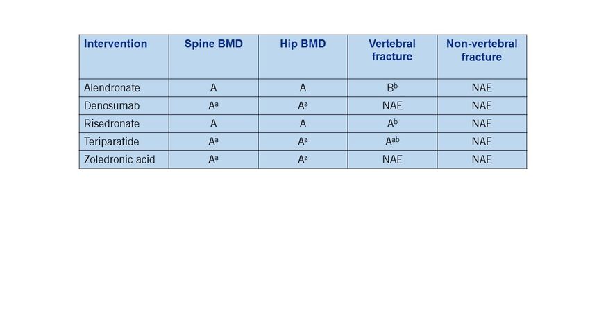
NOGG 2017: Clinical guideline for the prevention and treatment of osteoporosis

    Summary of main recommendations
    Assessment of fracture risk
    1. Fracture probability should be assessed in postmenopausal women, and men age 50 years or more, who have
       risk factors for fracture, using FRAX. In individuals at intermediate risk, bone mineral density (BMD) measurement
       should be performed using dual-energy X-ray absorptiometry and fracture probability re-estimated using FRAX.

    2. Vertebral fracture assessment should be considered in postmenopausal women and men age >50 years if there is
       a history of ≥4cm height loss, kyphosis, recent or current long-term oral glucocorticoid therapy, or a BMD T-score
       ≤ -2.5.

    Lifestyle and dietary measures
    1. A daily calcium intake of between 700 and 1200mg should be advised, if possible achieved through dietary
       intake, with use of supplements if necessary.

    2. In postmenopausal women and older men (≥50 years) at increased risk of fracture a daily dose of 800IU
       cholecalciferol should be advised.

    3. In postmenopausal women and older men receiving bone protective therapy for osteoporosis, calcium
       supplementation should be given if the dietary intake is below 700 mg/day, and vitamin D supplementation
       considered in those at risk of, or with evidence of, vitamin D insufficiency.

    4. Regular weight-bearing exercise should be advised, tailored according to the needs and abilities of the individual
       patient.

    5. Falls history should be obtained in individuals at increased risk of fracture and further assessment and appropriate
       measures undertaken in those at risk.

    Pharmacological intervention in postmenopausal women
    1. Alendronate or risedronate are first line treatments in the majority of cases. In women who are intolerant of oral
       bisphosphonates or in whom they are contraindicated, intravenous bisphosphonates or denosumab provide the
       most appropriate alternatives, with raloxifene or hormone replacement therapy as additional options. The high cost
       of teriparatide restricts its use to those at very high risk, particularly for vertebral fractures.

    2. Treatment review should be performed after 3 years of zoledronic acid therapy and 5 years of oral bisphosphonate
       treatment. Continuation of bisphosphonate treatment beyond 3-5 years can generally be recommended in
       individuals age ≥75 years, those with a history of hip or vertebral fracture, those who sustain a fracture while on
       treatment, and those taking oral glucocorticoids.

    3. If treatment is discontinued, fracture risk should be reassessed after a new fracture, regardless of when this occurs.
       If no new fracture occurs, assessment of fracture risk should be performed again after 18 months to 3 years.

    4. There is no evidence to guide decisions beyond 10 years of treatment and management options in such patients
       should be considered on an individual basis.

2
NOGG 2017: Clinical guideline for the prevention and treatment of osteoporosis
Glucocorticoid-induced osteoporosis
1. Women and men age ≥70 years with a previous fragility fracture, or taking high doses of glucocorticoids
(≥7.5 mg/day prednisolone), should be considered for bone protective therapy.
2. In other individuals fracture probability should be estimated using FRAX with adjustment for glucocorticoid
dose.
3. Bone-protective treatment should be started at the onset of glucocorticoid therapy in individuals at high risk of
fracture.
4. Alendronate and risedronate are first line treatment options. Where these are contraindicated or not tolerated,
zoledronic acid, denosumab or teriparatide are alternative options.
5. Bone protective therapy may be appropriate in some premenopausal women and younger men, particularly in
individuals with a previous history of fracture or receiving high doses of glucocorticoids.
Osteoporosis in men
1. Alendronate and risedronate are first line treatments in men. Where these are contraindicated or not tolerated,
zoledronic acid or denosumab provide the most appropriate alternatives, with teriparatide as an additional option.
2. For estimation of fracture probability, femoral neck BMD T-scores in men should be based on the NHANES female
reference database. When using the online version of FRAX for the estimation of fracture probability, femoral neck
BMD values (g/cm2) should be entered and the manufacturer of the densitometer specified.
Intervention thresholds for pharmacological intervention
1. The thresholds recommended for decision-making are based on probabilities of major osteoporotic and hip fracture
derived from FRAX and can be similarly applied to men and women.
2. Women with a prior fragility fracture can be considered for treatment without the need for further assessment,
although BMD measurement may be appropriate, particularly in younger postmenopausal women.
3. Age-dependent intervention thresholds up to 70 years and fixed thresholds thereafter provide clinically appropriate
and equitable access to treatment.
Systems of care
1. Coordinator-based Fracture Liaison Services (FLS) should be used to systematically identify men and women with
fragility fracture.
3NOGG 2017: Clinical guideline for the prevention and treatment of osteoporosis
Guideline document
Section 1: Introduction and scope
1. This updated guideline has been prepared with the support of the societies listed to provide guidance on
prevention and treatment of osteoporosis. This guideline updates those previously developed by the Royal College
of Physicians [RCP 1999, 2000] and the National Osteoporosis Guideline Group [Compston et al 2009, Compston
et al 2013].
2. The scope of the guideline is to review the assessment and diagnosis of osteoporosis, the therapeutic interventions
available and the manner in which these can be used to develop management strategies for the prevention of
osteoporotic fracture in postmenopausal women and in men age 50 years or over.
3. The guideline has been prepared by a writing group (Appendix I) and has been approved after consultation with
stakeholders (see Appendix II).
4. The guideline is intended for all healthcare professionals involved in the management of osteoporosis. This includes
primary care practitioners and relevant specialists in secondary care including rheumatologists, gerontologists,
gynaecologists, endocrinologists and orthopaedic surgeons.
5. The conclusions and recommendations in the document are systematically graded, according to the quality
of information available, to indicate the level of evidence on which recommendations are based. The grading
methodology is summarised in Appendix III. Where available, systematic reviews, meta-analyses and randomized
controlled trials have been used to provide the evidence base. The evidence base was updated using PubMed
to identify systematic reviews and meta-analyses from January 2009 to June 2016. The quality of systematic
reviews and meta-analyses used in the formulation of recommendations was assessed using AMSTAR
(amstar.ca) (Appendix IV). The recommendations in this guideline were agreed unanimously by the National
Osteoporosis Guideline Development Group.
6. It is recommended that the guideline is reviewed at an interval of not more than 5 years. Earlier revision may be
necessary if new drugs become approved or there is a major change to the evidence base. Minor changes, for
example extension of an indication, new safety data or changes to the Summary of Product Characteristics (SPC)
of an intervention, will be made on the website when and if appropriate.
7. This guideline provides a framework from which local management protocols should be developed to provide
advice for healthcare professionals. Implementation of the guidelines should be audited at a local level.
8. The recommendations in the guideline should be used to aid management decisions but do not replace the need
for clinical judgment in the care of individual patients in clinical practice.
References
Royal College of Physicians. Osteoporosis: clinical guidelines for the prevention and treatment. London: Royal College of Physicians; 1999.
Royal College of Physicians and Bone and Tooth Society of Great Britain. Update on pharmacological interventions and an algorithm for management. London,
UK: Royal College of Physicians; 2000.
Compston J, Cooper A, Cooper C et al; National Osteoporosis Guideline Group (NOGG). Guidelines for the diagnosis and management of osteoporosis in
postmenopausal women and men from the age of 50 years in the UK. Maturitas 2009;62:105-8.
Compston J, Bowring C, Cooper A et al; National Osteoporosis Guideline Group. Diagnosis and management of osteoporosis in postmenopausal women and older
men in the UK: National Osteoporosis Guideline Group (NOGG) update 2013. Maturitas 2013;75:392-6.
4NOGG 2017: Clinical guideline for the prevention and treatment of osteoporosis
Section 2: Background
1. Osteoporosis is described by the World Health Organization (WHO) as a “progressive systemic skeletal disease
characterized by low bone mass and microarchitectural deterioration of bone tissue, with a consequent increase
in bone fragility and susceptibility to fracture” [Kanis et al 1994].
2. The clinical significance of osteoporosis lies in the fractures that arise. In the UK, approximately 536,000
new fragility fractures occur each year, comprising 79,000 hip fractures, 66,000 clinically diagnosed vertebral
fractures, 69,000 forearm fractures and 322,000 other fractures (i.e. fractures of the pelvis, rib, humerus, tibia,
fibula, clavicle, scapula, sternum and other femoral fractures) [Svedbom et al 2013]. Such fractures cause severe
pain and disability to individual sufferers, at an annual cost to the National Health Service (NHS) of over £4.4
billion, estimated for 2010. First year costs, subsequent year costs and pharmacological fracture prevention costs
amounted to £3.2 billion, £1.1 billion and £84 million, respectively [Svedbom et al 2013]. More than one-third of
adult women and one in five men will sustain one or more fragility fractures in their lifetime [van Staa et al 2001].
3. Common sites of fragility fracture include the vertebral bodies, distal radius, proximal humerus, pelvis and proximal
femur. Hip fractures account for occupation of over 4,000 beds at any one time across England, Wales and
Northern Ireland and an average hospital length of stay of around 20 days (http://www.nhfd.co.uk/2016report).
Hip fractures account for around 50% of the total cost of fractures to the UK annually [Svedbom et al 2013].
Approximately 53% of patients suffering a hip fracture can no longer live independently and 28.7% die within 12
months of the fracture. Only 54% of individuals admitted from home with a hip fracture return there within 30
days [http://www.nhfd.co.uk/2016report; Neuburger et al 2015]. Furthermore, most major osteoporotic fractures
are associated with reduced relative survival, with an impact persisting more than five years after the index event
[Bliuc et al 2009; Harvey et al 2010].
4. In the UK, fracture rates vary by geographic location, socioeconomic status and ethnicity [Moon et al 2016,
Curtis et al 2016] and changes in age- and sex-adjusted fracture rates have been observed in recent decades,
with increases in hip fractures amongst men, and vertebral fracture amongst women [van de Velde et al 2016].
Furthermore, the ageing of the UK population will give rise to a doubling in the number of osteoporotic fractures
over the next 50 years if changes are not made to current practice [Gullberg et al 1997, Svedbom et al 2013].
5. In Europe, osteoporosis accounts for more disability-adjusted life years than many non-communicable diseases
including rheumatoid arthritis, Parkinson’s disease, breast cancer and prostate cancer [Johnell & Kanis 2006].
6. Fall-related risk factors add significantly to the risk of fracture and often overlap with risk factors for osteoporosis.
Identification of older people at risk of fracture should therefore involve an integrated approach [Blain et al 2016].
References
Kanis JA, Melton 3rd LJ, Christiansen C, Johnston CC, Khaltaev N. The diagnosis of osteoporosis. J Bone Miner Res 1994; 9: 1137–41.
Svedbom A, Hernlund E, Ivergård M et al and the EU review panel of the IOF. Osteoporosis in the European Union: A compendium of country-specific reports. Arch
Osteoporos 2013; 8: 137. DOI 10.1007/s11657-013-0137-0.
van Staa TP, Dennison EM, Leufkens HG, Cooper C. Epidemiology of fractures in England and Wales. Bone 2001; 29:517-22.
National Hip Fracture Database 2016 Annual Report. www.nhfd.co.uk/2016report
Neuburger J, Currie C, Wakeman R et al. The impact of a national clinician-led audit initiative on care and mortality after hip fracture in England: an external
evaluation using time trends in non-audit data. Med Care 2015;53:686-91.
Bliuc D, Nguyen ND, Milch VE, Nguyen TV, Eisman JA, Center JR. Mortality risk associated with low-trauma osteoporotic fracture and subsequent fracture in men
and women. JAMA 2009;301:513-21.
Harvey N, Dennison E, Cooper C. Osteoporosis: impact on health and economics. Nat Rev Rheumatol 2010; 6:99-105.
Moon RJ, Harvey NC, Curtis EM, de Vries F, van Staa T, Cooper C. Ethnic and geographic variations in the epidemiology of childhood fractures in the United
Kingdom. Bone 2016; 85:9-14.
Curtis EM, van der Velde R, Moon RJ et al. Epidemiology of fractures in the United Kingdom 1988-2012: Variation with age, sex, geography, ethnicity and
socioeconomic status. Bone 2016; 87:19-26.
van der Velde RY, Wyers CE, Curtis EM et al. Secular trends in fracture incidence in the UK between 1990 and 2012. Osteoporos Int 2016;27:3197-206.
Gullberg B, Johnell O, Kanis JA. World-wide projections for hip fracture. Osteoporos Int 1997;7:407-13.
Johnell O, Kanis JA. An estimate of the worldwide prevalence and disability associated with osteoporotic fractures. Osteoporos Int 2006; 17:1726-33.
Blain H, Masud T, Dargent-Molina P et al; EUGMS Falls and Fracture Interest Group; European Society for Clinical and Economic Aspects of Osteoporosis and
Osteoarthritis (ESCEO), Osteoporosis Research and Information Group (GRIO), and International Osteoporosis Foundation (IOF). A comprehensive fracture prevention
strategy in older adults: The European Union Geriatric Medicine Society (EUGMS) Statement. J Nutr Health Aging 2016;20:647-52.
5NOGG 2017: Clinical guideline for the prevention and treatment of osteoporosis
Section 3: Definition and diagnosis of osteoporosis
1. Prospective studies have shown that the risk of fracture increases progressively with decreasing bone
mineral density (BMD). Systematic review and meta-analysis of observational population-based studies using
absorptiometric techniques indicate that the risk of fracture increases approximately twofold for each standard
deviation (SD) decrease in BMD [Marshall et al 1996, Johnell et al 2005]; (Evidence level Ia). The predictive
value of BMD for hip fracture is at least as good as that of blood pressure for stroke.
2. Osteoporosis is defined operationally on the level of bone mass, measured as BMD. Two thresholds of BMD
have been defined by the World Health Organization, on the basis of the relationship of fracture risk to BMD.
‘Osteoporosis’ denotes a value for BMD that is 2.5 SDs or more below the young adult mean value for women
(T-score equal to or less than –2.5). ‘Severe’ or ‘established’ osteoporosis denotes osteoporosis as defined above
in the presence of one or more documented fragility fractures [Kanis et al 1994].
3. The World Health Organization and the International Osteoporosis Foundation recommend that the reference
technology for the diagnosis of osteoporosis is dual-energy X-ray absorptiometry (DXA) applied to the femoral neck.
The femoral neck is the preferred site because of its higher predictive value for fracture risk [Kanis & Gluer 2000,
Kanis et al 2008]; (Evidence level 1a). The spine is not a suitable site for diagnosis in older people because of the
high prevalence of degenerative changes, which artefactually increase the BMD value; however, it is the preferred
site for assessing response to treatment [ISCD 2015]. The normal reference range in men and women is that
derived from the NHANES survey for Caucasian women age 20-29 years [Kanis et al 2008]. The writing group
endorses these recommendations (Grade C recommendation). Other sites and validated technologies may be used
in clinical practice but it should be recognised that the significance of a given T-score differs between sites and
technologies [Faulkner et al 1999]; (Grade B recommendation).
4. Femoral neck and total hip T-scores calculated from two-dimensional projections of quantitative computed
tomography (QCT) data are equivalent to the corresponding DXA-derived T-scores used for the diagnosis of
osteoporosis [Cann et al 2014, ISCD 2015].
5. On GE Healthcare bone densitometers there is an option for T-scores for men to be given relative to either the male
or female reference range in DXA readouts. The same diagnostic cut-off values for BMD can be applied to men as
for women since there is evidence that the risk of fracture for any given femoral neck BMD and age is similar in
men to that in women [De Laet et al 1998, Binkley et al 2014]; (Grade B recommendation).
6. Some guidelines favour the concurrent use of BMD at the proximal femur and at the lumbar spine for patient
assessment. Patients are defined as having osteoporosis on the basis of the lower of the two T-scores. The
prediction of fracture is, however, not improved by the use of multiple sites [Kanis et al 2006, Leslie et al 2007];
(Evidence level II) and the use of multiple sites for diagnosis is not recommended (Grade B recommendation).
However, where hip measurement is not possible for technical reasons or in younger postmenopausal women and
men in whom the spine is differentially affected, spine BMD measurements may be used. If neither hip nor spine
measurements are possible, BMD measurements at the distal radius may be considered.
7. Additional techniques for assessing skeletal status have been less well validated than absorptiometric techniques.
The writing group does not recommend the use of other techniques, including quantitative ultrasound, for the
diagnosis of osteoporosis. This does not preclude the use of these or other validated techniques in risk assessment.
References
Marshall D, Johnell O, Wedel H. Meta-analysis of how well measures of bone mineral density predict occurrence of osteoporotic fractures. BMJ 1996;312:1254–9.
Johnell O, Kanis JA, Oden A et al. Predictive value of bone mineral density for hip and other fractures. J Bone Miner Res 2005;20:1185–94.
Kanis JA, Melton 3rd LJ, Christiansen C, Johnston CC, Khaltaev N. The diagnosis of osteoporosis. J Bone Miner Res 1994; 9: 1137–41.
Kanis JA, Gluer CC. An update on the diagnosis and assessment of osteoporosis with densitometry. Committee of Scientific Advisors, International Osteoporosis
Foundation. Osteoporos Int 2000;11:192-202.
Kanis JA, McCloskey EV, Johansson H, Oden A, Melton LJ, 3rd, Khaltaev N. A reference standard for the description of osteoporosis. Bone 2008;42:467-47.
International Society for Clinical Densitometry. http://www.iscd.org/documents/2015/06/2015-iscd-adult-official-positions.pdf
Faulkner KG, von SE, Miller P. Discordance in patient classification using T-scores. J Clin Densitom 1999;2:343–50.
De Laet CEDH, Van Hout BA, Burger H et al. Hip fracture prediction in elderly men and women: validation in the Rotterdam study. J Bone Miner Res
1998;13:1587–93.
Cann CE, Adams JE, Brown JK, Brett AD. CTXA hip - an extension of classical DXA measurements using quantitative CT. PLoS One. 2014;9:e91904.
Binkley N, Adler R, Bilezikian JP. Osteoporosis diagnosis in men: the T-score controversy revisited. Curr Osteoporos Rep 2014;12:403-9.
Kanis JA, Johnell O, Oden A et al. The use of multiple sites for the diagnosis of osteoporosis. Osteoporos Int 2006;17:527-34.
Leslie WD, Tsang JF, Caetano PA, Lix LM; Manitoba Bone Density Program. Number of osteoporotic sites and fracture risk assessment: a cohort study from the
Manitoba Bone Density Program. J Bone Miner Res 2007;22:476-83.
6NOGG 2017: Clinical guideline for the prevention and treatment of osteoporosis
Section 4: Fracture risk assessment
1. In addition to its diagnostic use, the assessment of BMD provides information on the likelihood of future fractures.
The risk of fracture increases approximately twofold for each SD decrease in BMD, but the gradient of risk
(relative risk/standard deviation; RR/SD) varies according to the site and technique used, the patient’s age and
the fracture outcome [Johnell et al 2005]; (Evidence level Ia).
2. The use of BMD alone to assess fracture risk has a high specificity but low sensitivity, meaning that most fragility
fractures will occur in women who do not have osteoporosis as defined by a T-score ≤-2.5 [Siris et al 2001];
(Evidence level Ia). The working group does not recommend the use of BMD testing alone for population screening
[NICE 2012]; (Grade B recommendation).
3. Techniques of clinical value include DXA at the hip regions, lumbar spine and forearm. DXA measurements of
femoral neck BMD are used in FRAX. Other non-invasive techniques include quantitative ultrasound and computed
axial tomography. No one technique subserves all the functions of skeletal assessment (diagnosis, prognosis and
monitoring of treatment).
4. The performance characteristics of BMD assessment can be improved by the concurrent consideration of
risk factors that operate independently of BMD. Of particular importance is age, which contributes to risk
independently of BMD [Kanis et al 2007, Kanis et al 2008]; (Evidence level Ia).
5. Several additional clinical risk factors have been identified that provide information on fracture risk independently
of both age and BMD (Evidence level Ia).
(a) Low body mass index (BMI). Low BMI is a significant risk factor for hip fracture, but the value of BMI
in predicting other fractures is very much diminished when adjusted for BMD [De Laet et al 2005];
(Evidence level 1a).
(b) A history of a prior fracture at a site characteristic for osteoporosis is an important risk factor for further
fracture. Fracture risk is approximately doubled in the presence of a prior fracture, including morphometric
vertebral fractures. The increase in risk is even more marked for more than one vertebral fracture. The risks
are in part independent of BMD [Kanis et al 2004a]; (Evidence level 1a).
(c) A parental history of hip fracture is a significant risk factor that is largely independent of BMD [Kanis et al
2004b]; (Evidence level 1a).
(d) Smoking is a risk factor that is in part dependent on BMD [Kanis et al 2005a]; (Evidence level 1a).
(e) Glucocorticoids increase fracture risk in a dose-dependent manner. The fracture risk conferred by the use
of glucocorticoids is, however, not solely dependent upon bone loss and BMD-independent risks have been
identified [van Staa et al 2000, Kanis et al 2004c]; (Evidence level 1a).
(f) Alcohol. The relationship between alcohol intake and fracture risk is dose-dependent. Where alcohol intake is
on average two units or less daily, no increase in risk has been identified. Intakes of 3 or more units daily are
associated with a dose-dependent increase in fracture risk [Kanis et al 2005b]; (Evidence level 1a).
(g) Rheumatoid arthritis. There are many secondary causes of osteoporosis (e.g. inflammatory bowel disease,
endocrine disorders), but in most instances it is uncertain to what extent this is dependent on low BMD
or other factors such as the use of glucocorticoids. By contrast, rheumatoid arthritis increases fracture risk
independently of BMD and the use of glucocorticoids [Kanis et al 2004c]; (Evidence level 1a). Recent
information suggests that diabetes (particularly type 2) may also exert BMD-independent effects on fracture
risk [Leslie et al 2012, Giangregorio et al 2012].
6. The consideration of these risk factors improves the sensitivity of testing without sacrificing specificity, and the
writing group recommend their inclusion in case finding algorithms (Grade B recommendation). Indeed, the use of
combined clinical risk factors alone performs very similarly to that of BMD alone [Johansson et al 2009]; the use
of clinical risk factors with the addition of BMD is optimal, but the latter can be included in targeted groups
(see below).
7. There are many additional risk factors for fracture that act solely by reducing BMD and others that have been
less well validated or identify a risk that may not be amenable to particular treatments. Liability to falls is an
appropriate example where the risk of fracture is high, but treatment with agents affecting bone metabolism have
an uncertain effect on fracture risk in such patients. The writing group recommend the identification and validation
of additional clinical risk factors as an important area for further research.
7NOGG 2017: Clinical guideline for the prevention and treatment of osteoporosis
8. Biochemical indices of skeletal turnover have the potential to aid risk assessment but probably play a more
immediate role in the monitoring of treatment [Johansson et al 2014a]; (Evidence level Ia). Further research in
this field is recommended so that their utility in clinical practice can be evaluated for use in diagnosis, prognosis
and monitoring of treatment [Vasikaran et al 2011].
9. The International Osteoporosis Foundation and the World Health Organization (WHO) recommend that risk of
fracture should be expressed as an absolute risk, i.e. probability over a ten-year interval. The absolute risk of
fracture depends upon age and life expectancy as well as the current relative risk. The period of 10 years covers
the likely initial duration of treatment and the benefits that may continue if treatment is stopped. The writing group
endorses these recommendations (Grade C recommendation).
10. Algorithms that integrate the weight of clinical risk factors for fracture risk, with or without information on
BMD, have been developed by the WHO Collaborating Centre for Metabolic Bone Diseases at Sheffield. The FRAX
tool (www.shef.ac.uk/FRAX) computes the 10-year probability of hip fracture or a major osteoporotic fracture. A
major osteoporotic fracture is a clinical spine, hip, forearm or humerus fracture. The tool has been externally
validated in independent cohorts [Kanis et al 2007]; (Evidence level Ia). QFracture is based on a UK prospective
open cohort study of routinely collected data from general practices that takes into account numerous risk factors
and estimates the 1-10 year cumulative incidence of hip or major osteoporotic fracture [Hippisley-Cox & Coupland
2009; http://www.qfracture.org]. The National Institute for Health and Care Excellence (NICE) has recommended
the use of fracture risk assessment tools (FRAX or QFracture) in the assessment of patients, including the proposal
that their use should be considered in all women age 65 years or older and men age 75 years or older [NICE
2012]. In the Scottish Intercollegiate Guidelines Network guideline (SIGN 142), QFracture is preferred and is used
to provide a threshold for BMD assessment [SIGN 2015]. Since FRAX and QFracture yield different outputs
(probability of fracture accounting for mortality risk in the case of FRAX, and a cumulative risk of fracture in the
case of QFracture), the two calculators cannot be used interchangeably. In addition, BMD cannot be incorporated
into QFracture estimations. Finally, the NOGG intervention thresholds are based on FRAX probability and thus
cannot be used with fracture risk derived from QFracture or other calculators [Kanis et al 2016]. The use of FRAX
for fracture risk assessment is therefore preferred (Grade B recommendation).
11. The FRAX assessment takes no account of prior treatment or of dose responses for several risk factors. For
example, two prior fractures carry a much higher risk than a single prior fracture. Dose responses are also evident
for glucocorticoid use and are partially addressed in the NOGG guideline. A prior clinical vertebral fracture carries
an approximately two-fold higher risk than other prior fractures. Since it is not possible to model all such scenarios
with the FRAX algorithm, these limitations should temper clinical judgement.
12. Diagnostic assessment of individuals with osteoporosis should include not only the assessment of BMD where
indicated but also the exclusion of diseases that mimic osteoporosis, elucidation of the cause of the osteoporosis
and the management of any associated morbidity. Recommendations for the routine investigation of patients with
osteoporosis are shown in Table 1.
13. The majority of vertebral fractures do not come to medical attention and thus remain undiagnosed [Fink et al
2005]. Moderate or severe vertebral fractures, even when asymptomatic, are strong risk factors for subsequent
fracture at the spine and other skeletal sites [Melton et al 1999, Lindsay et al 2001, Johansson et al 2014b].
Vertebral fracture assessment should therefore be considered in high risk individuals, using either lateral lumbar
and thoracic spine radiographs or lateral spine DXA imaging. The latter delivers a significantly lower radiation dose
but performs comparably to traditional radiographs [Lewiecki et al 2010].
14. Vertebral fracture assessment should be considered in postmenopausal women and older men if there is a
history of ≥4cm height loss, kyphosis, recent or current long-term oral glucocorticoid therapy, or a BMD T-score
≤-2.5 (Grade C recommendation). It should also be considered in individuals with a history of non-vertebral fracture
after the age of 50 years [Gallacher et al, 2007].
8NOGG 2017: Clinical guideline for the prevention and treatment of osteoporosis
Table 1 Procedures proposed in the investigation of osteoporosis
Routine Other procedures, if indicated
• History and physical examination • Lateral radiographs of lumbar and thoracic spine
or DXA-based lateral vertebral imaging
• Blood cell count, sedimentation rate or C-reactive
protein. Serum calcium, albumin, creatinine, • Serum protein immunoelectrophoresis and urinary
phosphate, alkaline phosphatase and liver Bence Jones proteins
transaminases • Serum 25-hydroxyvitamin D
• Thyroid function tests • Plasma parathyroid hormone
• Bone densitometry (DXA) • Serum testosterone, sex hormone binding globulin,
follicle stimulating hormone, luteinizing hormone
• Serum prolactin
• 24 hour urinary free cortisol/overnight
dexamethasone suppression test
• Endomysial and/or tissue transglutaminase
antibodies
• Isotope bone scan
• Markers of bone turnover
• Urinary calcium excretion
Other investigations, for example, bone biopsy and genetic testing for osteogenesis imperfecta, are largely
restricted to specialist centres.
References
Johnell O, Kanis JA, Oden A et al. Predictive value of BMD for hip and other fractures. J Bone Miner Res 2005;20:1185-94.
Siris ES, Miller PD, Barrett-Connor E et al. Identification and fracture outcomes of undiagnosed low bone mineral density in postmenopausal women: results from the National
Osteoporosis Risk Assessment. JAMA 2001; 286:2815-22.
National Institute for Health and Care Excellence. NICE Clinical Guideline 146. Osteoporosis: assessing the risk of fragility fracture. 2012
Kanis JA, Oden A, Johnell O et al. The use of clinical risk factors enhances the performance of BMD in the prediction of hip and osteoporotic fractures in men and women.
Osteoporos Int 2007;18:1033-46.
Kanis JA on behalf of the WHO Scientific Group. Assessment of osteoporosis at the primary health-care level. Technical Report. WHO Collaborating Centre, University of Sheffield,
UK, Sheffield 2008.
De Laet C, Kanis JA, Oden A et al. Body mass index as a predictor of fracture risk: a meta-analysis. Osteoporos Int 2005;16:1330-8.
Kanis JA, Johnell O, De Laet C et al. A meta-analysis of previous fracture and subsequent fracture risk. Bone 2004a;35:375-82.
Kanis JA, Johansson H, Oden A et al. A family history of fracture and fracture risk: a meta-analysis. Bone 2004b;35:1029-37.
Kanis JA, Johnell O, Oden A et al. Smoking and fracture risk: a meta-analysis. Osteoporos Int 2005a;16:155-62.
van Staa TP, Leufkens HG, Abenhaim L, Zhang B, Cooper C. Oral corticosteroids and fracture risk: relationship to daily and cumulative doses. Rheumatology 2000; 39:1383-9.
Kanis JA, Johansson H, Oden A et al. A meta-analysis of prior corticosteroid use and fracture risk. J Bone Miner Res 2004c;19:893-9.
Kanis JA, Johansson H, Johnell O et al. Alcohol intake as a risk factor for fracture. Osteoporos Int 2005b;16:737-42.
Leslie WD, Rubin MR, Schwartz AV, Kanis JA. Type 2 diabetes and bone. J Bone Miner Res 2012; 27:2231-7.
Giangregorio LM, Leslie WD, Lix LM et al. FRAX underestimates fracture risk in patients with diabetes. J Bone Miner Res 2012;27:301-8.
Johansson H, Kanis JA, Oden A, Johnell O, McCloskey E. BMD, clinical risk factors and their combination for hip fracture prevention. Osteoporos Int 2009;20:1675-82.
Johansson H, Odén A, Kanis JA et al and the IFCC-IOF Joint Working Group on standardisation of biochemical markers of bone turnover. A meta-analysis of markers of bone
turnover for prediction of fracture. Calcif Tissue Int 2014a; 94: 560-7.
Vasikaran S, Cooper C, Eastell R et al. International Osteoporosis Foundation and International Federation of Clinical Chemistry and Laboratory Medicine position on bone marker
standards in osteoporosis. Clin Chem Lab Med 2011; 49:1271-4.
Hippisley-Cox J, Coupland C. Predicting risk of osteoporotic fracture in men and women in England and Wales: prospective derivation and validation of QFracture Scores. BMJ
2009; 339:b4229.
Scottish Intercollegiate Guidelines Network (SIGN) (2015) Management of osteoporosis and the prevention of fragility fractures. Edinburgh: SIGN; 2015. (SIGN publication no.
142). http://www.sign.ac.uk.
Kanis JA, Compston J, Cooper C et al. SIGN Guidelines for Scotland: BMD Versus FRAX Versus QFracture. Calcif Tissue Int 2016;98:417-25.
Fink HA, Milavetz DL, Palermo L, Nevitt MC, Cauley JA, Genant HK. What proportion of incident radiographic vertebral deformities is clinically diagnosed and vice versa? J Bone
Miner Res 2005;20:1216-22.
Melton LJ 3rd, Atkinson EJ, Cooper C, O’Fallon WM, Riggs BL. Vertebral fractures predict subsequent fractures. Osteoporos Int 1999;10:214-21.
Lindsay R, Silverman SL, Cooper C et al. Risk of new vertebral fracture in the year following a fracture. JAMA 2001;285:320-3.
Johansson H, Oden A, McCloskey EV, Kanis JA. Mild morphometric vertebral fractures predict vertebral fractures but not non-vertebral fractures. Osteoporos Int 2014b; 25:235-41.
Lewiecki EM. Bone densitometry and vertebral fracture assessment. Curr Osteoporos Rep 2010; 8:123-30.
Gallacher SJ, Gallagher AP, McQuillian C, Mitchell PJ, Dixon T. The prevalence of vertebral fracture amongst patients presenting with non-vertebral fractures. Osteoporos Int
2007;18:185-92.
9NOGG 2017: Clinical guideline for the prevention and treatment of osteoporosis
Section 5: Lifestyle measures in the management
of osteoporosis
1. Lifestyle measures to improve bone health include increasing the level of physical activity , stopping smoking,
reducing alcohol intake to ≤2 units/day, reducing the risk of falls and ensuring adequate dietary calcium intake and
vitamin D status.
2. Increasing calcium intake, either through the diet or in the form of supplements, has been shown to result in small
increases in BMD [Tai 2015]; (Evidence level 1a) but convincing evidence that calcium alone reduces fracture risk
is lacking [Shea et al 2002, Bolland et al 2015]; (Evidence level 1a). Calcium supplements are associated with an
increased risk of nephrolithiasis [Candelas et al 2012] and gastrointestinal side-effects. Concerns have also been
raised that calcium supplements increase the risk of cardiovascular disease, but in a recent meta-analysis little
evidence was found for a significant association. [Lewis et al 2015]; (Evidence level 1a). It is recommended that a
daily calcium intake of between 700 and 1200 mg should be advised, if possible achieved through dietary intake
[https://www.gov.uk/government/uploads/system/uploads/attachment_data/file/384775/familyfood-method-
rni-11dec14.pdf] (Grade B recommendation). A simple dietary calcium intake calculator is available at http://
www.cgem.ed.ac.uk/research/rheumatological/calcium-calculator
3. The Scientific Advisory Committee on Nutrition (SACN) has recently recommended a reference nutrient intake
(RNI) of 400 IU daily for adults of all ages [SACN 2016]. However, in postmenopausal women and older men at
increased risk of fracture, the available evidence supports the use of higher doses. Vitamin D alone is ineffective in
reducing fracture risk but when combined with calcium supplements results in a small reduction in hip and non-
vertebral fractures, and possibly also in vertebral fractures [Tang et al 2007, Avenell et al 2014]; (Evidence level
1a). In another meta-analysis, a protective effect of vitamin D on fractures was only seen at daily doses≥800 IU (20
µg) [Bischoff-Ferrari et al 2009a] (Evidence level 1a). This dose of vitamin D may also reduce falls [Bischoff-Ferrari
et al 2009b]; (Evidence level 1a). It is recommended that in postmenopausal women and men ≥50
years who are at increased risk of fracture, a daily dose of 800 IU of cholecalciferol should be advised(Grade A
recommendation). Intermittent administration of large doses of vitamin D e.g. ≥100,000 IU is not advised, based on
recent reports of an associated increased risk of fracture and falls [Sanders et al 2010, Bischoff-Ferrari et al2016].
4. Supplementation with calcium and vitamin D is often advocated as an adjunct to other treatments for osteoporosis,
as the clinical trials of these agents were performed in patients who were calcium and vitamin D replete. In
postmenopausal women and older men receiving bone protective therapy for osteoporosis it is recommended
that calcium supplementation should also be given if the dietary intake is below 700 mg/day, and vitamin D
supplementation with 800 IU/day of cholecalciferol considered in those at risk of/with evidence for vitamin D
insufficiency (Grade B recommendation).
5. Weight-bearing exercise has beneficial effects on BMD [Howe et al 2011]; (Evidence level 1a) but has not been
shown to reduce fracture risk [Kemmler et al 2012]; (Evidence level 1a). Regular weight-bearing exercise should
be advised, tailored according to the individual patient (Grade B recommendation). Physiotherapy is an important
component of rehabilitation after fracture. Muscle strengthening and balance training exercise interventions may
reduce falls by improving confidence and coordination as well as maintaining bone mass.
6. The majority of fractures are preceded by a fall. Multi-component group and home-based exercise programmes, Tai
Chi and home safety interventions have been shown to reduce the risk of falls in people living in the community
[Gillespie et al 2012]; (Evidence level 1a). Falls prevention exercise programmes in community dwelling adults age
>60 years may reduce falls resulting in fracture [El-Khoury et al 2013]; (Evidence level 1a) although in individuals
at higher risk of falling this benefit has not been shown. Falls history should be obtained in patients with
osteoporosis and further assessment and appropriate measures undertaken in those at risk (Grade B
recommendation).
7. Hip protectors may reduce the risk of hip fractures in older people in nursing care or residential care settings.
[Santesso et al 2014]; (Evidence level 1a). However, poor acceptance and adherence by older people offered hip
protectors are barriers to their use.
8. Sufficient protein intake is necessary to maintain the function of the musculoskeletal system and also decreases the
complications that occur after hip fracture. Protein supplementation in patients with a recent hip fracture has been
shown to improve the subsequent clinical course by significantly lowering the rate of infection and duration of
hospital stay. [Myint et al 2013]; (Evidence level Ib).
10NOGG 2017: Clinical guideline for the prevention and treatment of osteoporosis
References
Tai V, Leung W, Grey A, Reid IR, Bolland MJ. Calcium intake and bone mineral density: systematic review and meta-analysis. BMJ 2015 Sep 29;351:h4183.
Shea B, Wells G, Cranney A et al. Meta-analyses of therapies for postmenopausal osteoporosis. VII. Meta-analysis of calcium supplementation for the prevention of
postmenopausal osteoporosis. Endocr Rev 2002; 23, 552-9.
Bolland MJ, Leung W, Tai V et al. Calcium intake and risk of fracture: systematic review. BMJ 2015 Sep 29;351:h4580. doi: 10.1136/bmj.h4580.
Candelas G, Martinez-Lopez JA, Rosario MP, Carmona L, Loza E. Calcium supplementation and kidney stone risk in osteoporosis: a systematic literature review.
Clin Exp Rheumatol 2012;30:954-61.
Lewis JR, Radavelli-Bagatini S, Rejnmark L et al. The effects of calcium supplementation on verified coronary heart disease hospitalization and death in
postmenopausal women: a collaborative meta-analysis of randomized controlled trials. J Bone Miner Res 2015;30:165-75.
https://www.gov.uk/government/uploads/system/uploads/attachment_data/file/384775/familyfood-method-rni-11dec14.pdf
http://www.cgem.ed.ac.uk/research/rheumatological/calcium-calculator
Scientific Advisory Council on Nutrition Vitamin D and Health Report. https://www.gov.uk/government/publications/sacn-vitamin-d-and-health-report
Tang BM, Eslick GD, Nowson C, Smith C, Bensoussan A. Use of calcium or calcium
in combination with vitamin D supplementation to prevent fractures and bone loss in people aged 50 years and older: a meta-analysis. Lancet 2007 Aug
25;370(9588):657-66.
Avenell A, Mak JCS, O’Connell D. Vitamin D and vitamin D analogues for preventing fractures in post-menopausal women and older men. The Cochrane database of
systematic reviews 2014;4, CD000227-CD000227.
Bischoff-Ferrari HA, Willett WC, Wong JB et al. Prevention of nonvertebral fractures with oral vitamin D and dose dependency: a meta-analysis of randomized
controlled trials. Arch Intern Med 2009a;169:551-61.
Bischoff-Ferrari HA, Dawson-Hughes B, Staehelin HB et al. Fall prevention with supplemental and active forms of vitamin D: a meta-analysis of randomised controlled
trials. BMJ 2009b; 339, b3692-b3692.
Sanders KM, Stuart AL, Williamson EJ et al. Annual high-dose oral vitamin D and falls and fractures in older women: a randomized controlled trial. JAMA 2010;
303:1815-22.
Bischoff-Ferrari HA, Dawson-Hughes B, Orav EJ et al. Monthly high-dose vitamin D treatment for the prevention of functional decline: a randomized clinical trial.
JAMA Intern Med 2016;176:175-83.
Howe TE, Shea B, Dawson LJ et al. Exercise for preventing and treating osteoporosis in postmenopausal women. Cochrane Database Syst Rev 2011 Jul
6;(7):CD000333. doi: 10.1002/14651858.CD000333.pub2.
Kemmler W, Häberle L, von Stengel S. Effects of exercise on fracture reduction in older adults. A systematic review and meta-analysis. Osteoporos Int 2013;
24:1937–50.
Gillespie LD, Robertson MC, Gillespie WJ et al. Interventions for preventing falls in older people living in the community. Cochrane Database Syst Rev 2012 Sep
12;(9):CD007146.
El-Khoury F, Cassou B, Charles MA, Dargent-Molina P. The effect of fall prevention exercise programmes on fall induced injuries in community dwelling older adults:
systematic review and meta-analysis of randomised controlled trials. BMJ 2013; 347:f6234.
Santesso N, Carrasco-Labra A, Brignardello-Petersen R. Hip protectors for preventing hip fractures in older people. Cochrane Database Syst Rev 2014 Mar
31;(3):CD001255. doi: 10.1002/14651858.CD001255.pub5.
Myint MW, Wu J, Wong E et al. Clinical benefits of oral nutritional supplementation for elderly hip fracture patients: a single blind randomised controlled trial.
Age Ageing 2013;42:39-45.
11NOGG 2017: Clinical guideline for the prevention and treatment of osteoporosis
Section 6: Pharmacological interventions
1. In the context of strategies for treating individuals at high risk of fracture, no distinction is made between
prevention and treatment. A range of pharmaceutical interventions has been shown to be effective in reducing
fracture risk in postmenopausal women with osteoporosis [Crandall et al 2014]. Recommendations concerning the
major interventions for osteoporosis are based on high levels of evidence (Evidence level 1a and Ib), and the grade
of these recommendations is summarised in Table 2.
Table 2. Anti-fracture efficacy of approved treatments for postmenopausal women
with osteoporosis when given with calcium and vitamin D.
Intervention Vertebral fracture Non-vertebral fracture Hip fracture
Alendronate A A A
Ibandronate A A* NAE
Risedronate A A A
Zoledronic acid A A A
Calcitriol A NAE NAE
Denosumab A A A
HRT A A A
Raloxifene A NAE NAE
Teriparatide A A NAE
A; grade A recommendation
NAE: not adequately evaluated
* in subsets of patients only (post-hoc analysis)
HRT: hormone replacement therapy
2. Bisphosphonates are analogues of inorganic pyrophosphate that inhibit bone resorption.
a) Alendronate is approved for the treatment of postmenopausal osteoporosis (10 mg daily or 70 mg once weekly
by mouth) and osteoporosis in men (10 mg daily). It is also approved for the prevention of postmenopausal
osteoporosis (5 mg daily) and for prevention and treatment of glucocorticoid-induced osteoporosis (5 mg daily
or, in postmenopausal women not receiving hormone replacement therapy 10 mg daily).
In postmenopausal women with osteoporosis, alendronate 10 mg daily has been shown to reduce vertebral,
non-vertebral and hip fractures [Black et al 1996]. Approval for the use of alendronate in men with
osteoporosis and in men and women taking glucocorticoids was granted on the basis of BMD bridging studies
[Orwoll et al 2000, Saag et al 1998].
Side-effects include upper gastrointestinal symptoms, bowel disturbance, headaches and musculoskeletal pain.
Alendronate should be taken after an overnight fast and at least 30 minutes before the first food or drink (other
than water) of the day or any other oral medicinal products or supplementation (including calcium). Tablets
should be swallowed whole with a glass of plain water (~ 200 ml) while the patient is sitting or standing in
an upright position. Patients should not lie down for 30 minutes after taking the tablet. Alendronic acid is also
available as 70 mg effervescent or soluble tablets, to be dissolved in half a glass of plain water (≥120 ml).
b). Ibandronate 150 mg once monthly by mouth or 3 mg as an intravenous injection every 3 months is approved
for the treatment of osteoporosis in postmenopausal women at increased risk of fracture.
In a dose of 2.5 mg daily by mouth a significant reduction in vertebral fractures was demonstrated [Delmas
et al 2004]. In a post hoc analysis of high fracture risk women (femoral neck BMD T-score below -3.0),
a significant reduction in non-vertebral fractures was shown [Chesnut et al 2004]. No data are available for
hip fracture. Approval for the oral 150 mg once monthly and 3 mg intravenously every 3 months formulations
was granted on the basis of BMD bridging studies.
12NOGG 2017: Clinical guideline for the prevention and treatment of osteoporosis
Side-effects with the oral preparation include upper gastrointestinal side-effects and bowel disturbance.
Intravenous administration may be associated with an acute phase reaction, characterised by an influenza-like
illness; this is generally short-lived and typically occurs only after the first injection.
Oral ibandronate should be taken after an overnight fast and 1 hour before the first food or drink (other than
water) of the day or any other oral medicinal products or supplementation (including calcium). Tablets should
be swallowed whole with a glass of plain water (180 to 240 ml) while the patient is sitting or standing in an
upright position. Patients should not lie down for 1 hour after taking the tablet.
c). Risedronate 5 mg daily or 35 mg once weekly by mouth is approved for the treatment of postmenopausal
osteoporosis, to reduce the risk of vertebral fracture and for the treatment of established postmenopausal
osteoporosis, to reduce the risk of hip fractures. It is also indicated for the treatment of osteoporosis in men
at high risk of fractures. Risedronate 5 mg daily is approved for the prevention of glucocorticoid-induced
osteoporosis in postmenopausal women.
In postmenopausal women with osteoporosis risedronate 5 mg daily has been shown to reduce vertebral
and non-vertebral fractures [Harris et al 1999, Reginster et al 2000]. In a large population of older women,
risedronate significantly decreased the risk of hip fractures, an effect that was greater in osteoporotic women
[McClung et al 2001]. Approval for use of risedronate in men with osteoporosis and in postmenopausal women
taking glucocorticoids was granted on the basis of BMD bridging studies [Boonen et al 2009, Wallach et al
2000, Reid et al 2000].
Side-effects include upper gastrointestinal symptoms, bowel disturbance, headache and musculoskeletal pain.
Risedronate should be taken after an overnight fast and at least 30 minutes before the first food or drink (other
than water) of the day or any other oral medicinal products or supplementation (including calcium). Tablets
should be swallowed whole with a glass of plain water (~120 ml) while the patient is sitting or standing in an
upright position. Patients should not lie down for 30 minutes after taking the tablet.
d). Zoledronic acid 5 mg intravenously once yearly is approved for the treatment of osteoporosis in
postmenopausal women and men at increased risk of fracture, including those with a recent low trauma
fracture, and for the treatment of osteoporosis associated with long-term systemic glucocorticoid therapy in
postmenopausal women and men.
Zoledronic acid has been shown to reduce the incidence of vertebral, non-vertebral and hip fractures in
postmenopausal women with osteoporosis [Black et al 2007] and to reduce the risk of clinical fracture and
attendant mortality when given to patients shortly after their first hip fracture [Lyles et al 2007]. Approval for
its use in men with osteoporosis and postmenopausal women and men taking glucocorticoids was granted on
the basis of BMD bridging studies [Boonen et al 2012, Reid et al 2009].
Side-effects include an acute phase reaction (see above), usually only after the first infusion, and
gastrointestinal symptoms. Creatinine clearance should be calculated (e.g. using the Cockroft-Gault formula
[140-age (years) x weight (kg) x f /serum creatinine (µmol/l) where f = 1.23 for men and 1.04 for women]
prior to initiation of treatment and serum creatinine monitored in high-risk patients. An increase in atrial
fibrillation, reported as a serious adverse event, was seen in the main phase III trial although this finding has
not been replicated in other trials involving zoledronic acid. Zoledronic acid is given as an intravenous infusion
over a minimum period of 15 minutes.
e). Contraindications and special precautions for the use of bisphosphonates
Oral and intravenous bisphosphonates are contraindicated in patients with hypocalcaemia, hypersensitivity to
bisphosphonates, and severe renal impairment (GFR ≤ 35 ml/min for alendronate and zoledronic acid and ≤30
ml/min for other bisphosphonates). Pregnancy and lactation are also contraindications. Oral bisphosphonates
are contraindicated in people with abnormalities of the oesophagus that delay oesophageal emptying such as
stricture or achalasia, and inability to stand or sit upright for at least 30-60 minutes. They should be used with
caution in patients with other upper gastrointestinal disorders. Pre-existing hypocalcaemia must be investigated
and, where due to vitamin D deficiency, treated with vitamin D (e.g. 50,000 to 100,000 IU orally as a loading
dose) before treatment is initiated.
Rare adverse effects, in particular osteonecrosis of the jaw and atypical femoral fractures, have led to
additional precautions. In patients with dental disease or other risk factors (e.g. glucocorticoids, tobacco
use), dental examination with preventive dentistry is recommended prior to treatment with oral or intravenous
bisphosphonates. While on treatment, patients should avoid invasive dental procedures if possible. For patients
requiring dental procedures, there are no data available to indicate whether discontinuation of treatment
reduces the risk of osteonecrosis of the jaw. Clinical judgment of the treating physician should guide the
management plan of each patient based on individual benefit/risk assessment. During treatment, all patients
should be encouraged to maintain good oral hygiene, receive routine dental check-ups, and report any oral
symptoms such as dental mobility, pain, or swelling.
13NOGG 2017: Clinical guideline for the prevention and treatment of osteoporosis
The possibility of osteonecrosis of the external auditory canal should be considered in patients who present with
ear symptoms including chronic ear infections. Possible risk factors for osteonecrosis of the external auditory
canal include steroid use and chemotherapy and/or local risk factors such as infection or trauma.
During treatment patients should be advised to report any thigh, hip or groin pain and any patient presenting
with such symptoms should be evaluated for possible atypical femur fracture.
3. Denosumab is a fully humanised monoclonal antibody against Receptor Activator of Nuclear factor Kappa B Ligand
(RANKL), a major regulator of osteoclast development and activity. It is approved for the treatment of osteoporosis in
postmenopausal women and men at increased risk of fractures, and for the treatment of bone loss associated with
hormone ablation in men with prostate cancer at increased risk of fractures. It is also approved for the treatment of
bone loss associated with long term systemic glucocorticoid therapy in adults at increased risk of fracture [Saag et al
2018]. It is given as a subcutaneous injection of 60 mg once every 6 months.
Denosumab has been shown to reduce the incidence of vertebral, non-vertebral and hip fractures in postmenopausal
women with osteoporosis [Cummings et al 2009]. Approval for its use in men with osteoporosis was granted on the
basis of a BMD bridging study [Langdahl et al 2015].
Contraindications and special precautions
Denosumab is contraindicated in patients with hypocalcaemia or with hypersensitivity to any of the constitu ents of the
formulation. Its use is not recommended in pregnancy or in the paediatric population (age≤18 years). Side-effects
include skin infection, predominantly cellulitis, and hypocalcaemia.
Hypocalcaemia is an identified risk in patients treated with denosumab, which increases with the degree of renal
impairment. Pre-existing hypocalcaemia must be investigated and, where due to vitamin D deficiency, treated with
vitamin D (e.g. 50,000 to 100,000 IU orally as a loading dose) before treatment is initiated. Adequate intake of
calcium and vitamin D is important in all patients, especially in those with severe renal impairment.
Monitoring of calcium levels should be conducted prior to each dose of denosumab and within two weeks after the
initial dose in patients predisposed to hypocalcaemia (e.g. patients with severe renal impairment, creatinine clearance
30 ml/min) or if suspected symptoms of hypocalcaemia occur or if otherwise indicated. Patients should be advised to
report symptoms of hypocalcaemia.
The rare occurrence of osteonecrosis of the jaw and atypical femoral fractures in patients treated with denosumab has
led to additional precautions. In postmenopausal women with osteoporosis, the incidence of ONJ was 0.04% at 3
years, 0.06% at 5 years and 0.44% at 10 years of treatment, suggesting that the risk of ONJ increases with duration
of treatment. In patients with dental disease or other risk factors (e.g. glucocorticoid therapy, tobacco use), dental
examination with preventive dentistry is recommended prior to treatment. While on treatment, patients should avoid
invasive dental procedures if possible. For patients requiring dental procedures, there are no data available to indicate
whether discontinuation of treatment reduces the risk of osteonecrosis of the jaw. Clinical judgment of the treating
physician should guide the management plan of each patient based on individual benefit/risk assessment. During
treatment, all patients should be encouraged to maintain good oral hygiene, receive routine dental check-ups, and
report any oral symptoms such as dental mobility, pain, or swelling.
During treatment patients should be advised to report any thigh, hip or groin pain and any patient presenting with
such symptoms should be evaluated for an atypical femur fracture.
Following cessation of denosumab therapy rapid bone loss occurs [Bone et al 2011]. Whether this results in an
increase in fracture risk is unclear but there are case reports of vertebral fractures, often multiple, occurring within 18
months after stopping treatment [Popp et al 2016, Aubry-Rozier et al 2016, Anastasilakis & Makras 2016]. Although
further studies are required, in patients who stop denosumab, switching to an alternative therapy such as a
bisphosphonate should be considered (Grade C recommendation).
4. Raloxifene is a selective oestrogen receptor modulator and inhibits bone resorption. It is approved for the treatment
and prevention of osteoporosis in postmenopausal women.
Raloxifene has been shown to reduce vertebral fracture risk [Ettinger et al 1999] but reduction in non-vertebral and
hip fractures has not been demonstrated.
Raloxifene is contraindicated in women with child-bearing potential, a history of venous thromboembolism or
unexplained uterine bleeding. Hepatic impairment and severe renal impairment are also contraindications. It should
be used with caution in women with a history of stroke or with risk factors for stroke. Side-effects include leg cramps,
oedema and vasomotor symptoms. There is a small increase in the risk of venous thromboembolism, mostly within
the first few months of treatment and a small increase in the risk of fatal stroke has been reported. In the phase III
trials, women treated with raloxifene had a significantly decreased risk of developing breast cancer. Raloxifene is
taken orally as a single daily dose (60 mg) and may be taken at any time without regard to meals.
14You can also read