New light shed on the early evolution of limb-bone growth plate and bone marrow
←
→
Page content transcription
If your browser does not render page correctly, please read the page content below
RESEARCH ARTICLE

                                    New light shed on the early evolution of
                                    limb-bone growth plate and bone marrow
                                    Jordi Estefa1*, Paul Tafforeau2, Alice M Clement3, Jozef Klembara4,
                                    Grzegorz Niedźwiedzki1, Camille Berruyer2, Sophie Sanchez1,2*
                                    1
                                     Department of Organismal Biology, Evolution and Development, Uppsala
                                    University, Uppsala, Sweden; 2European Synchrotron Radiation Facility, Grenoble,
                                    France; 3Flinders University, College of Science and Engineering, Adelaide,
                                    Australia; 4Comenius University in Bratislava, Faculty of Natural Sciences,
                                    Department of Ecology, Bratislava, Slovakia

                                    Abstract The production of blood cells (haematopoiesis) occurs in the limb bones of most
                                    tetrapods but is absent in the fin bones of ray-finned fish. When did long bones start producing
                                    blood cells? Recent hypotheses suggested that haematopoiesis migrated into long bones prior to
                                    the water-to-land transition and protected newly-produced blood cells from harsher environmental
                                    conditions. However, little fossil evidence to support these hypotheses has been provided so far.
                                    Observations of the humeral microarchitecture of stem-tetrapods, batrachians, and amniotes were
                                    performed using classical sectioning and three-dimensional synchrotron virtual histology. They
                                    show that Permian tetrapods seem to be among the first to exhibit a centralised marrow
                                    organisation, which allows haematopoiesis as in extant amniotes. Not only does our study
                                    demonstrate that long-bone haematopoiesis was probably not an exaptation to the water-to-land
                                    transition but it sheds light on the early evolution of limb-bone development and the sequence of
                                    bone-marrow functional acquisitions.

*For correspondence:
jordi.estefa@gmail.com (JE);
sophie.sanchez@ebc.uu.se (SS)       Introduction
                                    Tetrapod long bones are among the most studied skeletal elements in the field of bone biology as
Competing interests: The
                                    they constitute a unit of reference for understanding the development and biomechanics of the
authors declare that no
                                    appendicular skeleton (e.g. Duboule, 1994; Fröbisch, 2008; Hall, 2008; Shubin et al., 1997). The
competing interests exist.
                                    recent discovery of fossil tetrapod trackways (Ahlberg, 2018; Niedźwiedzki et al., 2010) suggested
Funding: See page 24                that limbs supported weight and engaged substrate locomotion earlier than previously thought in
Received: 04 September 2019         early tetrapod evolution. Not only crucial for their biomechanical properties, long bones also host
Accepted: 21 December 2020          bone marrow including stem-cell niches for the production of blood cells, that is haematopoiesis
Published: 02 March 2021            (Orkin and Zon, 2008). After birth, bone marrow is the definitive haematopoietic system in mostly
                                    terrestrial mammals and many other tetrapods (Akiyoshi and Inoue, 2012; Kapp et al., 2018;
Reviewing editor: Diethard
Tautz, Max-Planck Institute for
                                    Orkin and Zon, 2008) but not in fish or some aquatic tetrapods (Akiyoshi and Inoue, 2012;
Evolutionary Biology, Germany       Avagyan and Zon, 2016; Kapp et al., 2018). Indeed, red blood cells are produced in the supraspi-
                                    nal organ in the lamprey, the kidney and liver in actinopterygians (ray-finned fish) and some amphib-
   Copyright Estefa et al. This
                                    ians (tadpoles and aquatic adults, Akiyoshi and Inoue, 2012), and the kidney in lungfish
article is distributed under the
                                    (Amemiya et al., 2007; Kapp et al., 2018). Several studies proposed that the skeleton would have
terms of the Creative Commons
Attribution License, which          played a major role in hosting blood-cell production over the water-to-land transition and (1) pro-
permits unrestricted use and        tecting it against temperature changes (Weiss and Wislocki, 1956), (2) protecting it against poten-
redistribution provided that the    tial DNA mutations induced by UV exposure on land (Horton, 1980; Kapp et al., 2018) or (3)
original author and source are      providing a better efficiency in red-blood-cell production necessary for metabolically-demanding ter-
credited.                           restrial locomotion and aerial respiration (Tanaka, 1976). Our study focusses on characterising the

Estefa et al. eLife 2021;10:e51581. DOI: https://doi.org/10.7554/eLife.51581                                                      1 of 30
Research article                                                                                                         Evolutionary Biology

                                    eLife digest For many aquatic creatures, the red blood cells that rush through their bodies are
                                    created in organs such as the liver or the kidney. In most land vertebrates however, blood-cell
                                    production occurs in the bone marrow. There, the process is shielded from the ultraviolet light or
                                    starker temperature changes experienced out of the water.
                                        It is possible that this difference evolved long before the first animal with a backbone crawled out
                                    of the aquatic environment and faced new, harsher conditions: yet very little fossil evidence exists to
                                    support this idea. A definitive answer demands a close examination of fossils from the water-to-land
                                    transition including lobe-finned fish and early limbed vertebrates. To support the production of red
                                    blood cells, their fin and limb bones would have needed an internal cavity that can house a specific
                                    niche that opens onto a complex network of blood vessels.
                                        To investigate this question, Estefa et al. harnessed the powerful x-ray beam produced by the
                                    European Synchrotron Radiation Facility and imaged the fin and limb bones from fossil lobe-finned
                                    fish and early limbed vertebrates. The resulting three-dimensional structures revealed spongy long
                                    bones with closed internal cavities where the bone marrow cells were probably entrapped. These
                                    could not have housed the blood vessels needed to create an environment that produces red blood
                                    cells.
                                        In fact, the earliest four-legged land animals Estefa et al. found with an open marrow cavity lived
                                    60 million years after vertebrates had first emerged from the aquatic environment, suggesting that
                                    blood cells only began to be created in bone marrow after the water-to-land transition. Future work
                                    could help to pinpoint exactly when the change in blood cell production occurred, helping
                                    researchers to identify the environmental and biological factors that drove this change.

                                    early evolution of the bone marrow and long-bone growth in fossils to contextualise these
                                    hypotheses.
                                       Tetrapod long bones are regionalised in three parts mirrored from midshaft (Figure 1): (1) the
                                    middle of the shaft is called diaphysis; (2) the metaphyses are located at each extremity of the shaft

                                    Figure 1. Schematic drawing of the long-bone epiphyses of extant amniotes (A) and amphibians (B). Four
                                    conditions are figured here. They are separated by yellow dashed lines: A1, condition in crocodiles (interpreted
                                    from Haines, 1938); A2, condition in mammals at an early developmental stage before the appearance of the
                                    secondary ossification centre (Anderson and Shapiro, 2010; Tanaka, 1976); B1, condition in Triturus (Cynops)
                                    pyrrhogaster (Quilhac et al., 2014; Tanaka, 1976); B2, condition in Rana catesbeiana (Francillon, 1981;
                                    Tanaka, 1976). Abbreviations: c., cortex; Dia., diaphysis; e., endosteal bone; Epi., epiphysis; h.c., hypertrophied
                                    chondrocytes; Meta., metaphysis; m.p., marrow process; s., sinusoids; sept., septum; trab., trabeculae.

Estefa et al. eLife 2021;10:e51581. DOI: https://doi.org/10.7554/eLife.51581                                                                       2 of 30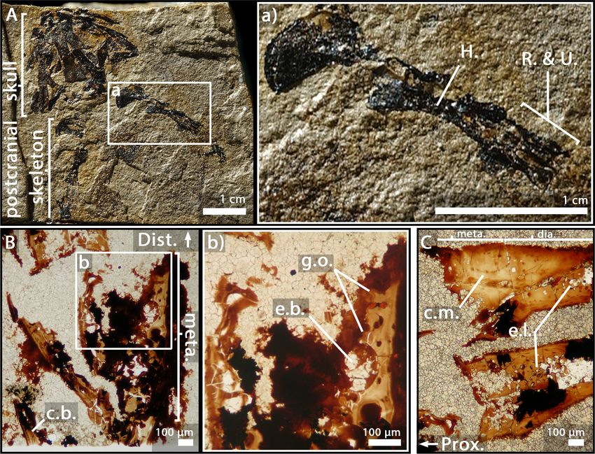
Research article                                                                                             Evolutionary Biology

                                    and (3) the epiphyses start above the ossification notch (i.e. where the cortical bone stops forming),
                                    extend beyond the metaphyses, and comprise one or more condyles in the case of concave articula-
                                    tions (Francillon-Vieillot et al., 1990). Long bones elongate from the growth plate, which is located
                                    in the metaphysis (Figure 1). In this region, the cartilage is progressively substituted with bone: this
                                    process is called endochondral ossification (Francillon-Vieillot et al., 1990; Hall, 2005).
                                        In extant amniotes, long-bone elongation results from the proliferation of longitudinal columns of
                                    hypertrophic cartilage cells, called hypertrophic chondrocytes (Francillon-Vieillot et al., 1990;
                                    Haines, 1942; Xie et al., 2020; Figure 1A). The latter express collagen type X which facilitates the
                                    calcification of the surrounding matrix (Gudmann and Karsdal, 2016; Lüllmann-Rauch, 2015). To do
                                    so, the hypertrophic chondrocytes secrete matrix vesicles containing calcium phosphate crystals
                                    (Amizuka, 2012; Anderson and Shapiro, 2010). The vesicles align longitudinally along the septa.
                                    The crystals penetrate the vesicle membranes to form stellate clusters of needle-shaped apatite in
                                    the extra cellular matrix (Amizuka, 2012). The mineralisation thus propagates following the longitu-
                                    dinal organisation of the septa (Amizuka, 2012; Anderson and Shapiro, 2010; Figure 1A). Blood
                                    vessels and marrow processes invade the growth plate along these columns of hypertrophic carti-
                                    lage (Lüllmann-Rauch, 2015; Figure 1A). Lytic enzymes secreted by the bone-marrow cells degrade
                                    the cartilage matrix, which is progressively substituted by bone deposition (Lüllmann-Rauch, 2015;
                                    Suzuki et al., 1981). Growth factors, such as the vascular endothelial growth factor (VEGF), trigger
                                    cartilage calcification and regulate endochondral ossification through stimulation of blood-vessel
                                    ingrowth into the diaphysis (Gerber et al., 1999). The lines of calcifying stellate clusters of crystals
                                    therefore form a scaffold for future trabecular bone deposition (Amizuka, 2012). This results in the
                                    formation of a bony mesh of longitudinal trabeculae (Figure 1A), which is progressively incorporated
                                    into the metaphysis where haematopoietic stem cell (HSC) niches (Figure 1A) are located
                                    (Calvi et al., 2003; Zhang et al., 2003) in the close vicinity of trabecular/endothelial surfaces
                                    (Gong, 1978; Nilsson et al., 2001; Wilson and Trumpp, 2006). HSC form localised niches whose
                                    environment is greatly controlled and regulated (Orkin and Zon, 2008; Sipkins et al., 2005;
                                    Zhang et al., 2003). Often in mature animals the growth plate disappears, causing the senescence
                                    of long-bone elongation (Kilborn et al., 2002). In most amniotes, the trabecular mesh in the meta-
                                    physis can be vastly remodelled (Haines, 1975). HSC can thereafter be observed adjacent to epiphy-
                                    seal trabeculae (Askenasy and Farkas, 2002).
                                        In extant urodeles (e.g. Pleurodeles waltl, De Ricqlès, 1964; De Ricqlès, 1965), the elongation of
                                    limb bones differs from the process in amniotes (Francillon-Vieillot et al., 1990; Haines, 1938;
                                    Figure 1B). Unlike mammals, endochondral ossification starts at a later stage in urodeles
                                    (De Ricqlès, 1964). The diaphyseal cartilaginous matrix is first hollowed by the formation of lacunae
                                    that are subsequently filled in with bone marrow, far before endochondral ossification starts
                                    (De Ricqlès, 1964). In mammals and birds (e.g. mouse, Zelzer et al., 2002; chicken,
                                    Carlevaro et al., 2000), VEGF initiates vascular ingrowth before endochondral ossification starts. In
                                    the amphibian Bufo gargarizan, a peak of VEGF expression is present in the hindlimb at metamor-
                                    phic climax (Gao et al., 2018) paralleling an increase of endochondral ossification activity (Bo et al.,
                                    2018). VEGF would therefore seem to play a major role in amphibian long-bone endochondral ossifi-
                                    cation as well, but this role still needs to be characterised. The growth plate in the metaphysis of
                                    urodeles exhibits no aligned columns of hypertrophic cartilage cells or drastically reduced alignment
                                    of a few cells at most (De Ricqlès, 1965; Dickson, 1982; Felisbino and Carvalho, 1999;
                                    Felisbino and Carvalho, 2001; Figure 1B). Contrary to amniotes, when present, these aligned col-
                                    umns of hypertrophic cartilage do not constitute the location where the ossification takes place
                                    (Figure 1B). Instead, the mineralisation front is located in the underlying areas of the growth plate
                                    (i.e. in a layer of non-oriented hypertrophic cartilage or stratified non-oriented hypertrophic carti-
                                    lage, De Ricqlès, 1965). There, after erosion of the cartilage, mineralisation occurs in urodeles via
                                    the formation of globular structures called globuli ossei (Figure 1B; De Ricqlès, 1965;
                                    Quilhac et al., 2014) and spherical mineralisation around them (forming Liesegang’s rings, Francil-
                                    lon-Vieillot et al., 1990). Globuli ossei are either (1) opened hypertrophic cartilaginous cells which
                                    died and were subsequently invaded by a cell from the blood/marrow system to initiate mineralisa-
                                    tion or (2) uneroded hypertrophic cartilaginous cells modified into active cells which synthesise
                                    bone-like collagen fibrils (of intermediate size between type II of the cartilage and type I of the
                                    bone, Quilhac et al., 2014). The endochondral ossification therefore does not produce a longitudi-
                                    nally-oriented trabecular network (Figure 1B), but forms instead a light reticular mesh (De Ricqlès,

Estefa et al. eLife 2021;10:e51581. DOI: https://doi.org/10.7554/eLife.51581                                                         3 of 30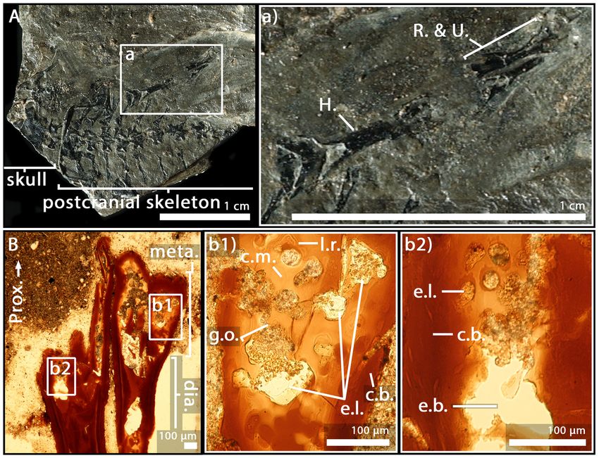
Research article                                                                                            Evolutionary Biology

                                    1965; Quilhac et al., 2014; Sanchez et al., 2008), rich in globuli ossei (De Ricqlès, 1964;
                                    Haines, 1938; Quilhac et al., 2014; Sanchez et al., 2010a). The epiphyses of urodeles remain carti-
                                    laginous while they often ossify in anurans (Castanet et al., 2003; Francillon, 1981; Sanchez et al.,
                                    2008). This is probably an adaptation to a demanding jumping locomotion and/or heterochronic
                                    mechanisms relevant to this clade (Francillon, 1981). In the medullary cavity of their long bones
                                    however, the cartilaginous cells hypertrophy with no preferential orientation as in urodeles (Dick-
                                    son, 1982; Felisbino and Carvalho, 1999; Felisbino and Carvalho, 2001; Miura et al., 2008;
                                    Rozenblut and Ogielska, 2005). The resulting spongiosa is largely reduced (even quite often
                                    absent, Francillon, 1981). The epiphyseal cartilage hangs over the ossification notch and the shaft
                                    (Francillon, 1981) to ossify straight after the metamorphosis (Miura et al., 2008; Rozenblut and
                                    Ogielska, 2005). The function of bone marrow in amphibian long bones also differs from the func-
                                    tion in extant amniotes. Indeed, in amphibians, the sites for haematopoiesis almost exclusively com-
                                    prise the thymus, spleen and liver (Akiyoshi and Inoue, 2012; Hightower and Pierre, 1971). Bone
                                    marrow only plays a role of haematopoiesis in a few amphibian species (e.g. Xenopus laevis, Rana
                                    catesbeiana, Tanaka, 1976; Phillobates terribilis, Dendrobates tinctorius, Kapp et al., 2018). In
                                    these cases, haematopoiesis occurs in endosteal regions of the diaphysis between sinusoids (i.e. fen-
                                    estrated capillaries; Figure 1B2) and endosteum (Tanaka, 1976). No HSC has been observed so far
                                    in the epiphysis of frogs.
                                        Is the urodele model the plesiomorphic or the derived condition for tetrapod long-bone elonga-
                                    tion and bone-marrow function? Very little attention has been given to these aspects of limb-bone
                                    evolution. On the one hand, some authors suggest that the amniote-like elongation process may
                                    have been the primitive state (Haines, 1942) but no fossil evidence was provided. On the other
                                    hand, early tetrapods had cartilaginous epiphyses like extant urodeles. Could that be an indication
                                    for a urodele-like primitive condition (e.g. Sanchez et al., 2008; Sanchez et al., 2010a)? This debate
                                    relied on the absence of evidence from stem-tetrapod data. Recently, palaeohistological studies
                                    revealed a fan-like longitudinal trabecular arrangement in the long-bone metaphysis of the 380-mil-
                                    lion-year-old lobe-finned fish Eusthenopteron (Sanchez et al., 2014), and in the 365-million-year-old
                                    limbed stem-tetrapod Acanthostega (Sanchez et al., 2016). These patterns would result from the
                                    same elongation process as in amniotes and would rather suggest that amphibians exhibit a derived
                                    condition. Using three-dimensional (3D) virtual histology based on propagation phase-contrast X-ray
                                    synchrotron radiation micro-computed tomography (PPC-SRmCT), as well as classical thin-section his-
                                    tology, we herein investigate several stem amphibians and stem amniotes to provide the first glimp-
                                    ses for characterising the early evolution of long-bone elongation and bone-marrow roles.

                                    Results
                                    The diaphyseal and metaphyseal microarchitectures of the stem tetrapods Eusthenopteron and
                                    Hyneria were described by Sanchez et al., 2014 and Kamska et al., 2018, respectively. The diaphy-
                                    sis of Discosauriscus (Sanchez et al., 2008), Apateon (Sanchez et al., 2010a; Sanchez et al.,
                                    2010b), Metoposaurus (Konietzko-Meier and Sander, 2013) and Seymouria (Estefa et al., 2020)
                                    were thoroughly described, but the metaphyseal organisation of their humeri was only succinctly
                                    mentioned in the cited articles. Here, we provide a detailed description of them (Table 1) in 3D
                                    when possible.

                                    Apateon caducus, juvenile specimen GPIM-N 1297, humerus
                                    As the humerus was crushed (Figure 2A), only a small region of the metaphysis could be sectioned
                                    and visualised (Figure 2B). Nevertheless, a relatively complete sequence of calcification (extending
                                    over 600 mm) can be described here. The upper part of the section reflects the irregular surface of
                                    the calcification front (separating the unpreserved eroded non-calcified cartilage from the preserved
                                    calcified cartilage) (Figure 2B). Under this region, obvious figures of globuli ossei are entrapped in
                                    Liesegang’s rings (g.o. and l.r., Figure 2Bb1-2). They are numerous and unevenly arranged. Their
                                    sizes (ranging from 9 to 15 mm in diameter) seem as well unevenly distributed. The trabeculae are
                                    very few in this thin section (t., Figure 2Bb2).

Estefa et al. eLife 2021;10:e51581. DOI: https://doi.org/10.7554/eLife.51581                                                        4 of 30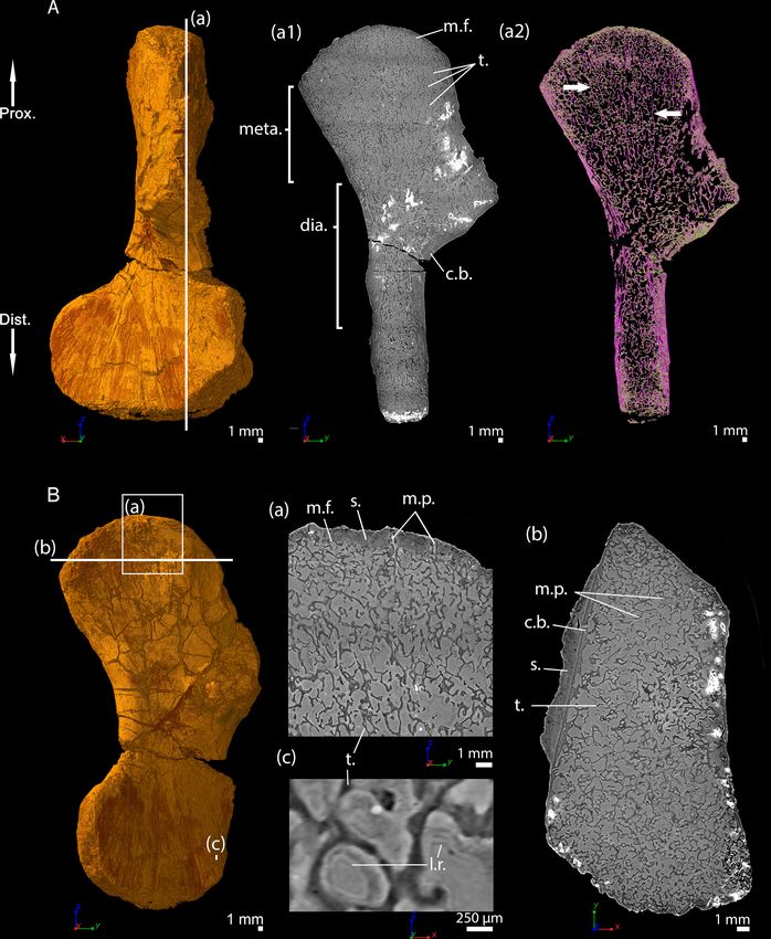
Research article                                                                                              Evolutionary Biology

Table 1. Table summarising the material used.
Skull length measurements and ontogenetic stages determined by Berman et al., 1987b; Sanchez et al., 2008; Sanchez et al.,
2010a and Klembara et al., 2006.
Species                              Collection number                  Skull length (cm)         Ontogenetic stage              Bone
Apateon caducus                      GPIM-N 1297                        1.52                      Juvenile                       Humerus
                                                                                                                                 Radius
                                                                                                                                 Ulna
                                     GPIM-N 1572                        Estimated to 1.60         Adult                          Radius
                                                                                                                                 Ulna
Apateon pedestris                    SMNS 54981                         0.86                      Adult                          Humerus
                                                                                                                                 Radius
                                                                                                                                 Ulna
                                     SMNS 54988                         1.06                      Adult                          Humerus
                                                                                                                                 Radius
                                                                                                                                 Ulna
Seymouria sanjuanensis               MNG 7747                           5.6                       Juvenile                       Humerus
                                     CM 28597                           8.8                       Adult                          Humerus
Discosauriscus austriacus            SNM Z 15568                        6.2                       Subadult                       Humerus
Metoposaurus sp.                     MUZ PGI OS-220/171                 -                         Subadult or adult              Humerus

                                    A. caducus, juvenile specimen GPIM-N 1297, radius and ulna
                                    Both bones exhibit large sequences of cartilage calcification which spread over more than a third of
                                    the total bone length on each side of the long bone (Figure 2C). The mineralisation front (m.f.,
                                    Figure 2C) is located relatively far under the ossification notch (400 mm) (o.n., Figure 2C). Numerous
                                    globuli ossei can be visualised in the metaphysis (g.o., Figure 2Cc1,3). They are unevenly distributed
                                    and their size ranges between 8 and 25 mm. Clusters of chondrocytes can be observed (c.c.,
                                    Figure 2Cc3). The top of the epiphysis probably exhibited a uniform matrix of uncalcified cartilage
                                    before the fossilisation that was not preserved afterwards. The mesh of ossified trabeculae is very
                                    scattered and shows no preferential orientation (t., Figure 2Cc1).

                                    A. caducus, adult specimen GPIM-N 1572, radius and ulna
                                    The epiphysis and metaphysis of the radius and ulna of this individual (Figure 3A) are more hollowed
                                    than those of the specimen GPIM-N 1297, with less cartilaginous matrix between the mineralised tra-
                                    beculae (Figure 3B). Fewer globuli ossei are visible (g.o., Figure 3Bb1). Instead, large empty lacu-
                                    nae can be observed (75 mm) (e.l., Figure 3Bb1). Large bays of erosion open as well between these
                                    lacunae (e.b., Figure 3Bb2). The process of mineralisation therefore seems more advanced but no
                                    obvious trabecular organisation can be observed.

                                    Apateon pedestris, adult specimen SMNS 54981, humerus
                                    In the humerus of SMNS 54981 (Figure 4A), the process of mineralisation seems relatively advanced
                                    as the globuli ossei only remain along a few mineralised trabeculae (g.o., Figure 4B). They are 16 mm
                                    large. In the metaphysis, the cartilage has been removed by erosional process (e.b., Figure 4B). The
                                    uncalcified cartilage in the epiphysis has not been preserved during the fossilisation (at least in this
                                    slide).

                                    A. pedestris, adult specimen SMNS 54981, radius and ulna
                                    The quantity of calcified cartilage is higher in the zeugopod (i.e. radius and ulna) than in the stylopod
                                    (i.e. humerus) (Figure 4Cc1-2). Most of the uncalcified cartilage has been eroded. The calcified carti-
                                    lage is hollowed, thereby forming multiple bays of erosion (e.b., Figure 4Cc2). Nevertheless, the

Estefa et al. eLife 2021;10:e51581. DOI: https://doi.org/10.7554/eLife.51581                                                            5 of 30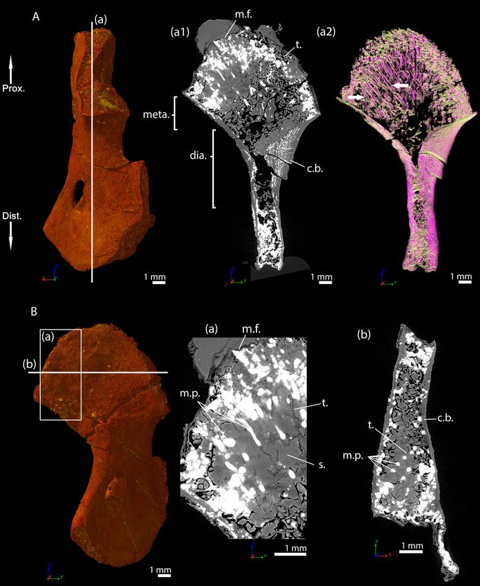
Research article                                                                                                                   Evolutionary Biology

Figure 2. Juvenile specimen of Apateon caducus, GPIM-N 1297. (A) Skeleton. (a) Right limb. (B) Epiphyseal and metaphyseal histology of the proximal
end of the humerus. (C) Epiphyseal and metaphyseal histology of the proximal end of the radius (c2-3) and ulna (c1). Abbreviations: c.b., cortical bone;
c.c., cluster of chondrocytes; c.f., calcification front; c.m., cartilage matrix; dia., diaphysis; e.b., erosion bay; e.l., erosion lacunae; g.o., globuli ossei; H.,
Figure 2 continued on next page

Estefa et al. eLife 2021;10:e51581. DOI: https://doi.org/10.7554/eLife.51581                                                                                  6 of 30
Research article                                                                                                               Evolutionary Biology

Figure 2 continued
humerus; l.r., Liesegang’s rings; meta., metaphysis; m.f., mineralisation front; o.n., ossification notch; Prox., proximal end; R. and U., radius and ulna; t.,
trabeculae.

                                       globuli ossei remain connected to each other by calcified-cartilage trabeculae (c.c.t., Figure 4Cc2)
                                       or mineralised trabeculae (m.t., Figure 4C) present in the metaphysis.

                                       A. pedestris, adult specimen SMNS 54988, humerus
                                       This thin section in the humerus of SMNS 54988 (Figure 5A) shows a very remodelled bone with
                                       large bays of erosion in the cartilaginous matrix (e.b., Figure 5Bb) and only a few remaining globuli
                                       ossei at the surface of the bone trabeculae (g.o., Figure 5Bb). Most of the cartilaginous matrix has
                                       been eroded. There is no preserved cartilage in the epiphysis. The bony trabeculae have no prefer-
                                       ential orientation.

Figure 3. Adult specimen of Apateon caducus, GPIM-N 1572. (A) Skeleton. (a) Right limb. (B) Epiphyseal and metaphyseal histology of the proximal
end of the radius (b1) and ulna (b2). Abbreviations: c.b., cortical bone; c.m., cartilage matrix; dia., diaphysis; e.b., erosion bay; e.l., erosion lacunae; g.o.,
globuli ossei; H., humerus; l.r., Liesegang’s rings; meta., metaphysis; Prox., proximal end; R. and U., radius and ulna.

Estefa et al. eLife 2021;10:e51581. DOI: https://doi.org/10.7554/eLife.51581                                                                              7 of 30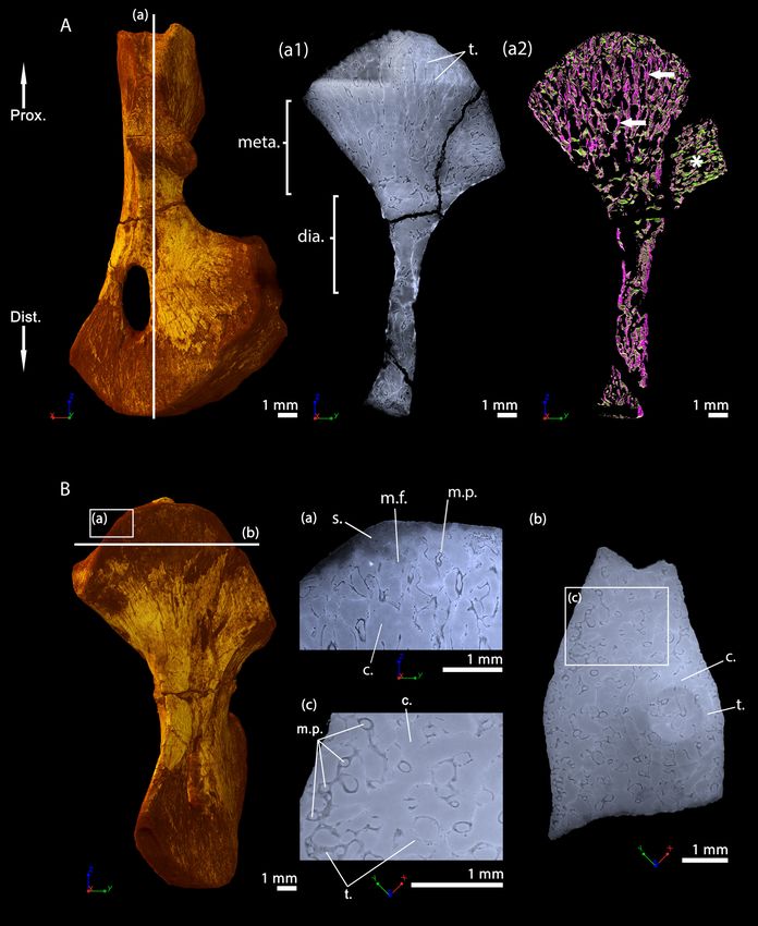
Research article                                                                                                       Evolutionary Biology

Figure 4. Adult specimen of Apateon pedestris, SMNS 54981. (A) Skeleton. (a) Right limb. (B) Epiphyseal and metaphyseal histology of the distal end of
the humerus. (C) Epiphyseal and metaphyseal histology of the proximal end of the radius (c2) and ulna (c1). Abbreviations: c.b., cortical bone; c.c.t.,
Figure 4 continued on next page

Estefa et al. eLife 2021;10:e51581. DOI: https://doi.org/10.7554/eLife.51581                                                                    8 of 30
Research article                                                                                                                Evolutionary Biology

Figure 4 continued
calcified-cartilage trabecula; Dist., distal end; e.b., erosion bay; g.o., globuli ossei; H., humerus; l.r., Liesegang’s rings; meta., metaphysis; m.f.,
mineralisation front; m.t., mineralised trabecula; Prox., proximal end; R. and U., radius and ulna.

                                       A. pedestris, adult specimen SMNS 54988, radius and ulna
                                       As for the zeugopod of the specimen SMNS 54988 (Figure 5Aa), the globuli ossei seem to be
                                       replaced by large empty lacunae (30 mm, e.l., Figure 5C). A certain amount of uncalcified cartilage
                                       has been eroded in the distal epiphyses and metaphyses. Nevertheless, a large amount of cartilage
                                       is still present in the proximal metaphyses of both long bones (c.m., Figure 5C). No or very few tra-
                                       beculae can be observed.

                                       Metoposaurus sp., (sub-)adult specimen MUZ PGI OS-220/171, humerus
                                       Transverse thin sections were made in the metaphysis of the femur (Konietzko-Meier and Sander,
                                       2013) of Metoposaurus diagnosticus krasiejowensis (Sulej, 2002) recently re-diagnosed as

Figure 5. Adult specimen of Apateon pedestris, SMNS 54988. (A) Skeleton. (a) Right limb. (B) Epiphyseal and metaphyseal histology of the distal end of
the humerus. (C) Epiphyseal and metaphyseal histology of the proximal end of the radius and ulna. Abbreviations: c.b., cortical bone; c.m., cartilage
matrix; dia., diaphysis; Dist., distal end; e.b., erosion bay; e.l., erosion lacunae; g.o., globuli ossei; H., humerus; meta., metaphysis; Prox., proximal end;
R. and U., radius and ulna.

Estefa et al. eLife 2021;10:e51581. DOI: https://doi.org/10.7554/eLife.51581                                                                               9 of 30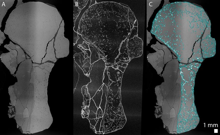
Research article Evolutionary Biology
Metoposaurus krasiejowensis (Brusatte et al., 2015). They revealed a dense trabecular mesh. The
longitudinal virtual thin sections, made with PPC-SRmCT and presented here, were made in the prox-
imal and distal metaphyses of a humerus of Metoposaurus sp. and confirm the presence of a dense
trabecular mesh in the overall humerus (Figure 6Aa1-2). Additionally, a directional coloured light
effect (cf. Materials and method section, Sanchez et al., 2014) shows that this mesh is oriented lon-
gitudinally and exhibits a fan-like shape in the metaphyses (purple trabeculae, Figure 6Aa2). The tra-
becular mesh covers the entire volume of the metaphysis and spreads into the diaphysis
(Figure 6Aa1-2). The mineralisation front (m.f., Figure 6Aa1) contacts the sediment in which the
bone is embedded (Figure 6Ba). The surface of the mineralisation front is irregular. No ossified
epiphysis was found, thereby suggesting that a cartilaginous cap was probably covering the bone.
This cap did not preserve over the fossilisation. In the metaphysis, the trabeculae are homo-
geneously distributed (t., Figure 6Bb). Some remnants of calcified cartilage are visible through Lie-
segang’s rings forming within the cartilage remaining between the metaphyseal trabeculae
(Figure 6Bc). The mean thickness of the trabeculae is 117 mm (Table 2). Tubular structures can be
observed (m.p., Figure 6Ba). They end blindly at the location of the mineralisation front. They are
well defined tubes (248 mm in diameter, Table 2), although anastomosed. They ossified through
endochondral ossification. These tubes are locally slightly eroded (Figures 6Ba and 10A). The size
of these tubes, their intimate connection to each other and their location strongly support their iden-
tification as marrow processes (Haines, 1938).
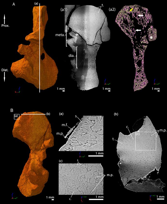
Seymouria sanjuanensis, juvenile specimen MNG 7747, humerus
This specimen was investigated using PPC-SRmCT. The spongiosa occupies the entire bone area
(Figure 7A). The metaphyseal trabeculae are about four times thinner (25 mm on average) than the
diaphyseal trabeculae (94 mm on average, Table 2 and Estefa et al., 2020). A longitudinal section
reveals that the trabecular mesh becomes denser towards the distal and proximal ends of the bone
(Figure 7Aa1-2). As the shape of the bone widens and flattens from midshaft towards the metaphy-
seal surfaces, the longitudinal trabeculae tilt, thereby forming a fan-like configuration (Figure 7Aa2).
In the metaphysis, the trabecular mesh is mostly arranged longitudinally (obviously appearing purple
and progressively shifting to green as the deltopectoral crest tilts to 90 degrees, Figure 7Aa2)
although a few anastomoses (highlighted in green in most of the metaphysis apart from the tilted
region of the deltopectoral crest, Figure 7Aa2) run radially. A few remnants of calcified cartilage
(Francillon-Vieillot et al., 1990) are very rarely visible (Estefa et al., 2020). Marrow processes form
an intricate network, while anastomosing to each other, and connecting to cavities of irregular
shapes and sizes (Figure 7Ba,c and 10B). These tubular structures are around 100 mm in diameter
under the mineralisation front (Table 2). They contact each other when they reach the mineralisation
front (Figure 10B). No ossified epiphysis was found. The humeral epiphysis was probably not pre-
served due to being unmineralised cartilage.
S. sanjuanensis, adult specimen CM 28597, humerus
A longitudinal virtual thin section from the PPC-SRmCT data shows that the trabecular network
remains relatively dense in the metaphyses (Figure 8A) at the adult stage. The trabecular mesh is
longitudinally and radially oriented like a fan although slightly less organised than in the juvenile
specimen (purple trabeculae, Figure 8Aa2). The trabeculae appear to be more remodelled, leaving
large cavities resulting from an intense erosional process (Figure 8Aa2). The cortex is almost inexis-
tent in the metaphysis (Figure 8Bb). The thickness of the trabeculae averages 30 mm (Table 2,
Estefa et al., 2020), which is equivalent to the thickness of the trabeculae in the juvenile metaphysis
(MNG 7747). There is no endosteal bone on the surface of the medullary cavity. Very few remnants
of calcified cartilage were found in the metaphysis of the adult humerus, that is in much lower fre-
quency than in the juvenile specimen (Estefa et al., 2020). The spongiosa contains a few longitudinal
interconnected marrow processes (100 mm in diameter, Figure 8Bb-c and 10C, Table 2). When pres-
ent, these processes exhibit the same distribution as in the juvenile humerus. The epiphyses were
not ossified. A resting surface (yellow arrow, Figure 8Aa2), red arrows, (Figure 8—figure supple-
ment 1A), can be observed 2-to-4 mm under the mineralisation front. It is observed as well in the
distal metaphysis (Figure 8—figure supplement 1A). This resembles Harris lines identified in mam-
mals (Garn et al., 1968; Harris, 1933) and birds (Wegner, 1874). Although these lines are very
Estefa et al. eLife 2021;10:e51581. DOI: https://doi.org/10.7554/eLife.51581 10 of 30Research article Evolutionary Biology Figure 6. Left humerus of a (sub-)adult specimen of Metoposaurus sp., MUZ PGI OS-220/171 imaged using PPC-SRmCT. (A) Frontal view. (a1) Longitudinal virtual thin section (40 mm thick) and (a2) longitudinal virtual thin section of the segmented model of the bone (50 mm thick). The longitudinally-oriented trabeculae are highlighted in purple (white arrows), while the transversally-oriented trabeculae appear in green. (B) Ventral view. (a) Longitudinal virtual thin section of the proximal metaphysis (40 mm thick), (b) transverse virtual thin section made in the metaphysis and (c) Figure 6 continued on next page Estefa et al. eLife 2021;10:e51581. DOI: https://doi.org/10.7554/eLife.51581 11 of 30
Research article                                                                                                               Evolutionary Biology

Figure 6 continued
longitudinal thin section made in the distal metaphysis. Abbreviations: c.b., cortical bone; dia., diaphysis; Dist., distal end; l.r., Liesegang’s rings; meta.,
metaphysis; m.f., mineralisation front; m.p., marrow process; Prox., proximal end; s., sediment; t., trabeculae.

                                       common in mammals (including extant and extinct taxa, Duckler and Van Valkenburgh, 1998), they
                                       have not been comprehensively studied in other groups. We find that they can also be encountered
                                       in groups with no secondary ossification centre such as chelonians (e.g. Centrochelys sulcata, Fig-
                                       ure 8—figure supplement 1B) and crocodilians (e.g. Crocodylus niloticus, P.T. pers. obs.). Harris
                                       lines seem to result from both short- and long-term pressures (e.g. starvation – Park, 1964; disease
                                       and deficiencies – Duckler and Van Valkenburgh, 1998).

                                       Discosauriscus austriacus, subadult specimen SNM Z 15568, humerus
                                       Thin sections were made and described in the distal metaphysis of the femur of the specimen KO224
                                       of D. austriacus (Sanchez et al., 2008). They revealed a dense trabecular mesh and the absence of cal-
                                       cified cartilage. Virtual thin sections from three-dimensional PPC-SRmCT scans in the humerus SNM Z
                                       15568 complete these observations despite the fact that the metaphyseal spongiosa is partly crushed
                                       (Figure 9Aa2). As the femur KO224 (Sanchez et al., 2008), the humerus SNM Z 15568 exhibits a
                                       dense trabecular mesh in both the proximal and distal metaphyses with a fan-like trabecular orienta-
                                       tion (purple trabeculae, Figure 9Aa2). The trabeculae are homogeneously distributed in the metaphy-
                                       sis. They are 54 mm thick on average (Table 2). They are eroded at the base of the metaphysis
                                       (Figure 9Aa1-2). Some tubular marrow processes (average diameter: 111 mm, Table 2) open up
                                       directly towards the diaphysis into the metaphyseal space of the medullary cavity left vacant after ero-
                                       sion (Figures 9B and 10D). The surface of the mineralisation front is irregular (Figure 9Ba). The latter
                                       probably was covered by uncalcified cartilage.

                                       Discussion
                                       The trabecular bone tissues observed in these long bones exhibit characteristics of endochondral
                                       ossification (remnants of calcified cartilage, globuli ossei and/or columnar trabecular mesh) as seen
                                       in stem- (Sanchez et al., 2014; Sanchez et al., 2016) and crown-tetrapods (Estefa et al., 2020;
                                       Francillon-Vieillot et al., 1990; Sanchez et al., 2008; Sanchez et al., 2010a).

                                       Early evolution of tetrapod limb-bone elongation
                                       Although all limb bones studied here have cartilaginous epiphyses, their metaphyseal organisation,
                                       and the underlying long-bone elongation processes, can greatly differ between taxa.
                                          The long-bone elongation in Apateon probably results from the hypertrophying action of scat-
                                       tered cartilaginous cells in the upper part of the metaphysis (Figure 11). The fossils revealed a min-
                                       eralisation front characterised by a large number of globuli ossei (Figures 2–5), which progressively
                                       replaced these hypertrophic chondrocytes, as in urodeles (De Ricqlès, 1964; De Ricqlès, 1965;
                                       Haines, 1938; Quilhac et al., 2014). As the uncalcified cartilage is not preserved in Apateon, it is

Table 2. Microanatomical measurements made on the samples using VGStudio MAX (version 3.2, Volume Graphics Inc, Germany).
The protocol details are provided by Estefa et al., 2020.
                                                            Thickness of the trabeculae (mm)                        Diameter of the marrow processes (mm)
Species                                                     Diaphysis                  Metaphysis                   Metaphysis
Metoposaurus sp.                                            131                        117                          248
(Subadult or Adult, MUZ PGI OS-220/171)
Seymouria sanjuanensis                                      94                         25                           100
(Juvenile, MNG 7747)
S. sanjuanensis                                             79                         30                           100
(Adult, CM 28597)
Discosauriscus austriacus                                   80                         54                           111
(Subadult, SNM Z 15568)

Estefa et al. eLife 2021;10:e51581. DOI: https://doi.org/10.7554/eLife.51581                                                                            12 of 30
Research article Evolutionary Biology Figure 7. Left humerus of a juvenile specimen of Seymouria sanjuanensis, MNG 7747 imaged using PPC-SRmCT. (A) Frontal view. (a1) Longitudinal virtual thin section (40 mm thick), the darker part is an artefact in the original data due to electron reinjection in the synchrotron storage ring (refilling) during the scan and (a2) longitudinal virtual thin section of the segmented model of the bone (250 mm thick). The longitudinally-oriented trabeculae (pointed by horizontal arrows) are highlighted in purple, while the transversally-oriented trabeculae appear in green. Note that, due to the shape of the Figure 7 continued on next page Estefa et al. eLife 2021;10:e51581. DOI: https://doi.org/10.7554/eLife.51581 13 of 30
Research article                                                                                                            Evolutionary Biology

Figure 7 continued
metaphysis, the trabeculae exhibit an overall fan-like configuration which progressively tilts to 90 degrees at the location of the deltopectoral crest
(Asterisk). For that reason, the longitudinal trabeculae appear green and the transverse trabeculae appear purple at this location. (B) Ventral view. (a)
Longitudinal virtual thin section in the proximal metaphysis (40 mm thick), (b) transverse virtual thin section in the metaphysis, the large ring artefact
results from the synchrotron electron refilling visible in a1, (c) detail of (b) showing marrow processes and cavities in transverse section. Abbreviations:
c., cavity; dia., diaphysis; Dist., distal end; meta., metaphysis; m.f., mineralisation front; m.p., marrow process; Prox., proximal end; s., sediment; t.,
trabeculae.

                                      not possible to check whether the growth zone and calcifying zone are distant as in amphibians
                                      (De Ricqlès, 1965; Felisbino and Carvalho, 1999; Felisbino and Carvalho, 2001). However, the
                                      presence of very few bony trabeculae, with no preferential orientation in the metaphyseal region of
                                      Apateon’s long bones (Figures 2–5) strongly support the idea that the ossification did not occur
                                      within a zone of columnar hypertrophic cartilage. Bone elongation was probably followed by miner-
                                      alisation through globuli ossei as previously observed in the temnospondyl dissorophoid Doleserpe-
                                      ton (De Ricqlès, 1963; Figure 11).
                                          On the contrary, the metaphyseal trabecular meshes in the limb bones of Metoposaurus, Seymou-
                                      ria and Discosauriscus all exhibit the same fan-like pattern of longitudinal trabeculae. This implies
                                      that the growth plate comprised longitudinal columns of hypertrophic cells where endochondral
                                      ossification occurred through columnar cartilage-to-bone substitution (Francillon-Vieillot et al.,
                                      1990; Kronenberg, 2003) as seen in turtles and crocodiles (e.g. Haines, 1938; Haines, 1942), lepi-
                                      dosaurs (e.g. Haines, 1969), dinosaurs (De Ricqlès, 1968; Horner et al., 2000; Horner et al.,
                                      2001), birds (e.g. Horner et al., 2001) and mammals (e.g. Jacenko et al., 1993; Figure 11).
                                      Although this fan-like trabecular configuration can be greatly remodelled in extant amniotes, it is
                                      only slightly remodelled here in the adult Seymouria and subadult Discosauriscus (Figures 8 and 9).
                                          Therefore, our study shows that the amniote-like long-bone elongation is more commonly distrib-
                                      uted than previously thought. It is not restrained to the appendicular skeleton of amniotes – includ-
                                      ing Discosauriscus and Seymouria (as demonstrated here), Ophiacodon, Dicynodon and some
                                      kannemeyeriids (De Ricqlès, 1972; Haines, 1938), marine reptiles, Plesiosaurus and Nothosaurus
                                      (Haines, 1938) – neither to that of stem tetrapods (Kamska et al., 2018; Sanchez et al., 2014;
                                      Sanchez et al., 2016; Figure 11). Indeed, our study demonstrates for the first time that certain tem-
                                      nospondyls like Metoposaurus also elongated their appendicular skeleton like amniotes (Figure 11).
                                      We can confidently conclude that (1) endochondral ossification based on the mineralisation of longi-
                                      tudinal columns of hypertrophic cartilage is a primitive process for the elongation of the appendicu-
                                      lar skeleton in tetrapods and that (2) ossification through globuli ossei is restricted to a limited
                                      group of stem- and crown-batrachians (Figure 11).
                                          The mineralisation processes in extant amphibians and amniotes largely differ in many points: (1)
                                      their timing and microstructural relationships (i.e. ossification dependant on a calcified scaffold in
                                      amniotes, Amizuka, 2012, but not in amphibians, Felisbino and Carvalho, 2001), (2) their initiation
                                      (i.e. stacks of hypertrophic cells in amniotes, Lüllmann-Rauch, 2015, versus isolated hypertrophic
                                      cells in amphibians, De Ricqlès, 1965; Quilhac et al., 2014) and (3) their molecular mechanisms (i.e.
                                      collagen type X secreted in amniotes, Gudmann and Karsdal, 2016; versus fibrillar collagens in
                                      amphibians, Quilhac et al., 2014). How could such distinct mineralisation processes play the same
                                      functional role in long-bone elongation in different vertebrates? In addition to a columnar pattern of
                                      trabeculae, several remnants of Liesegang’s rings could be observed in the fossil limb bones of
                                      Metoposaurus (Figure 6Bc and Konietzko-Meier and Sander, 2013) and, to a lesser extent, Sey-
                                      mouria (Estefa et al., 2020) and stem-tetrapods (i.e. Hyneria, Eusthenopteron and
                                      Acanthostega, Kamska et al., 2018; Sanchez et al., 2014; Sanchez et al., 2016). The most parsimo-
                                      nious evolutionary scenario therefore suggests that stem tetrapods were able to produce globular
                                      calcified cartilage although they were elongating their bone through a columnar configuration (Fig-
                                      ure 11). Long-bone elongation exclusively based on the intensive production of globuli ossei would
                                      have been a derived feature emerging within temnospondyls and restricted to extant batrachians
                                      and their close dissorophoid temnospondyl relatives (e.g. Apateon and Doleserpeton). Within
                                      amniotes – including stem amniotes – the globular calcification of the cartilage would have drasti-
                                      cally reduced to be fully abandoned to the benefit of an exclusive columnar elongation (Figure 11).
                                      Two exceptions persist in amniotes: (1) large aquatic animals usually produce a large amount of

Estefa et al. eLife 2021;10:e51581. DOI: https://doi.org/10.7554/eLife.51581                                                                         14 of 30
Research article Evolutionary Biology Figure 8. Right humerus of an adult Seymouria sanjuanensis, CM 28597 imaged using PPC-SRmCT. (A) Frontal view. (a1) Longitudinal virtual thin section (40 mm thick) and (a2) longitudinal section of the segmented model of the bone (450 mm thick). The longitudinally-oriented trabeculae (pointed by horizontal arrows) are highlighted in purple, while the transversally-oriented trabeculae appear in green. Note that, due to the shape of the metaphysis, the trabeculae exhibit an overall fan-like configuration which progressively tilts to 90 degrees at the location of the deltopectoral crest (Asterisk). For Figure 8 continued on next page Estefa et al. eLife 2021;10:e51581. DOI: https://doi.org/10.7554/eLife.51581 15 of 30
Research article                                                                                                              Evolutionary Biology

Figure 8 continued
that reason, the longitudinal trabeculae appear green and the transverse trabeculae appear purple at this location. (B) Dorsal view. (a) Longitudinal
virtual thin section of the proximal metaphysis (40 mm thick), (b) transverse virtual thin section in the metaphysis (40 mm thick), (c) detail of (b) showing
marrow processes and cavities in transverse section. Abbreviations: c., cavity; dia., diaphysis; Dist., distal end; meta., metaphysis; m.f., mineralisation
front; m.p., marrow process; Prox., proximal end; s., sediment; t., trabeculae.
The online version of this article includes the following figure supplement(s) for figure 8:
Figure supplement 1. Humeral microanatomical architecture of the stem amniote Seymouria sanjuanensis (CM 28597) and the tortoise Centrochelys
sulcata.

                                       globular calcified cartilage to balance their buoyancy or in extremely retarded developmental condi-
                                       tions, such as paedomorphosis (e.g. pachy-osteosclerotic amniotes, De Buffrénil et al., 2008;
                                       De Ricqlès and De Buffrénil, 2001; crocodiles, Haines, 1938), (2) diseased amniotes can have
                                       osteosclerotic problems which result in the production and retention of globuli ossei (Gussen, 1967).
                                       In such cases, the production of globuli ossei is not solely located in the epiphysis and does not play
                                       any role in the elongation process of limb bones. Even though these cases reflect derived and/or
                                       rare conditions, they show that amniotes keep the ability to produce globuli ossei although they do
                                       not allocate them to the limb-bone elongation process.

                                       Discussion on the batrachian limb-bone elongation strategy
                                       It was hypothesised that a large number of globuli ossei would be associated with a slow limb-bone
                                       endochondral ossification and development (De Ricqlès, 1972; Haines, 1938). This was based on
                                       the observation of globular calcification in small extant amphibians and neotenic aquatic forms, as
                                       well as the rarity or even absence of globuli ossei in fast growing juvenile mammals and birds
                                       (De Ricqlès, 1972; De Ricqlès, 1979; Haines, 1942; Quilhac et al., 2014). The observations con-
                                       tained herein clearly show that long-bone developmental dynamics does not seem to be the leading
                                       or unique factor for performing one or the other of the elongation and calcification processes.
                                       Indeed, the stem tetrapods Hyneria, Eusthenopteron and Acanthostega (with a humerus remaining
                                       cartilaginous for several years), all exhibit the characteristics of a slow appendicular development
                                       (and slow somatic development as a whole for Eusthenopteron and Acanthostega [Sanchez et al.,
                                       2014; Sanchez et al., 2016]) but only produce very few globuli ossei (Kamska et al., 2018;
                                       Sanchez et al., 2014; Sanchez et al., 2016). On the contrary, they all present an obvious longitudi-
                                       nal metaphyseal spongiosa strongly supporting the development of a hypertrophic columnar carti-
                                       laginous growth plate.
                                           The somatic size and ecology were also hypothesised to play a role in limb-bone elongation strat-
                                       egy (De Ricqlès, 1972). Once again, our data challenge this hypothesis. Both seymouriamorphs,
                                       Seymouria and Discosauriscus, and the temnospondyl Metoposaurus exhibit the same trabecular
                                       pattern despite different somatic sizes (skull length of an adult Seymouria estimated to 9.5 cm,
                                       Berman et al., 2000; skull length of a possibly adult Discosauriscus estimated to 6.2 cm, Klem-
                                       bara, 1995; Klembara, 2009; skull length of an adult Metoposaurus estimated to 40–50 cm,
                                       Sulej, 2007) and distinct ecologies (Seymouria and Discosauriscus being (supposedly) terrestrial,
                                       Berman and Martens, 1993; Klembara, 2009; Klembara and Meszároš, 1992; and Metoposaurus
                                       being aquatic, Schoch and Milner, 2000).
                                           Felisbino and Carvalho, 2001 investigated the limb-bone ossification of the amphibian Rana.
                                       They observed a late calcification and late ossification of the trabeculae in Rana catesbeiana which
                                       did not contribute to the bone elongation (Felisbino and Carvalho, 2001). The authors therefore
                                       suggested that the production of globuli ossei could probably play a greater role in reinforcing the
                                       limb-bone microstructure – for jumping after a certain age – rather than being associated with its
                                       elongation. Because urodeles do not jump despite their late ossification onset, the reasons for them
                                       to produce many globuli ossei could not be justified as such.
                                           The use of exclusive globular calcification and globuli ossei for long-bone endochondral ossifica-
                                       tion and elongation would therefore probably result from the combination of multiple factors shared
                                       by both batrachians and dissorophoids. In order to precisely identify these factors, an extended his-
                                       tological study will have to be carried out within temnospondyls (considering as many environmental
                                       factors as possible, including their ecologies and sizes) to draw strong and broad conclusions on this

Estefa et al. eLife 2021;10:e51581. DOI: https://doi.org/10.7554/eLife.51581                                                                           16 of 30
Research article Evolutionary Biology Figure 9. Right humerus of a subadult Discosauriscus austriacus, SNM Z 15568 imaged using PPC-SRmCT. Due to processing to convert the scan data into a stack of images, the images have been flipped, thereby resulting in a flipped 3D model. (A) Frontal view. (a1) Longitudinal virtual thin section (40 mm thick) and (a2) longitudinal section of the segmented model of the bone (160 mm thick). The longitudinally-oriented trabeculae are highlighted in purple, while the transversally-oriented trabeculae appear in green. (B) Ventral view. (a) Longitudinal virtual thin section of the proximal metaphysis (40 Figure 9 continued on next page Estefa et al. eLife 2021;10:e51581. DOI: https://doi.org/10.7554/eLife.51581 17 of 30
Research article Evolutionary Biology
Figure 9 continued
mm thick) and (b) transverse virtual thin section in the proximal metaphysis (40 mm thick). Abbreviations: c.b., cortical bone; dia., diaphysis; Dist., distal
end; meta., metaphysis; m.f., mineralisation front; m.p., marrow process; Prox., proximal end; s., sediment; t., trabeculae.
evolutionary pattern and the reasons for it to be that restricted in the evolutionary history of tetra-
pods. Nevertheless, the current study shows that amphibians, often considered as models for exhib-
iting primitive tetrapod features, should be regarded as a clade with a significantly derived
evolutionary history, at least with respect to their skeleton.
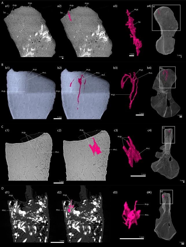
Evolution of limb-bone marrow processes
As the long bones of Apateon were thin sectioned, it was not possible to assess the 3D organisation
of the trabecular mesh nor the calcified cartilage mass to check out the potential presence of mar-
row processes.
The microanatomy of the other taxa (Metoposaurus, Seymouria, Discosauriscus), however, could
be investigated in 3D using PPC-SRmCT. It revealed tubular structures which can be confidently inter-
preted as marrow processes (on the basis of their shape, size and location in the bone), as seen in
extant crocodiles (Haines, 1938). The marrow processes in these groups differ from the closed sys-
tem observed in the stem tetrapod Eusthenopteron (Sanchez et al., 2014). Instead the marrow pro-
cesses in Metoposaurus open up into multilocular spaces in the trabecular mesh of the metaphysis.
In Discosauriscus and Seymouria, the marrow processes lead to a series of small interconnected cavi-
ties (m.c., Figure 10) which connect to other tubular processes and open up into the medullary cav-
ity of the bone shaft. The size and shape of these interconnected cavities are variable. These small
cavities would therefore rather correspond to a primary regionalisation of the marrow environment –
as seen in amniotes like Crocodylus (Haines, 1938). As a result of intense erosion, the tubular mar-
row processes in Discosauriscus more often directly plug into the medullary cavity of the shaft. On
the contrary, the shaft of Metoposaurus is highly crossed by thick trabeculae (Table 2) forming multi-
locular spaces as in Andrias (Sanchez et al., 2014; Figure 10—figure supplement 1).
The tip of the marrow process forming a tube penetrating the hypertrophic cartilage of the
growth plate in crocodiles is located at the level of the mineralisation front, and in mammals, that is
at the base of the calcifying layer of hypertrophic cartilaginous cells (Figure 1A). The tip of the mar-
row process plays a role in initiating endochondral ossification with marrow cells releasing lytic
enzymes that degrade the calcified cartilaginous matrix (Suzuki et al., 1981). In amniotes, the mar-
row process directly connects with the medullary cavity of the shaft (Haines, 1942) where blood ves-
sels supply growth factors to initiate the ossification (Gerber et al., 1999). The bone marrow also
produces haematopoietic cells and stem cells (HSC) which need to remain in regulated microenviron-
ments called niches (Zhang et al., 2003). The latter are located in the metaphysis, in obligatory
proximity with both endothelial and endosteal surfaces (Wilson and Trumpp, 2006). For that reason,
haematopoiesis only occurs in long bones whose shafts are greatly opened (Bazzini et al., 1986;
Tanaka, 1976). In mammals, the medullary cavity of the shaft can be infilled by calcified cartilage or
numerous trabeculae (e.g. pachyostotic condition, De Ricqlès and De Buffrénil, 2001). A study was
conducted on amedullar, pachyostotic long bones of manatees (Trichechus manatus, Bazzini et al.,
1986). Because haematopoiesis cannot be hosted in their long-bone medullary shaft, manatees have
evolved an alternative primary site of haematopoiesis in their vertebral bodies (Bazzini et al., 1986).
In amphibians, bays of erosion progressively form within the cartilage of the medullary cavity during
the development. They can either be isolated forming multilocular spaces separated by bony septa
(sept., Figure 1B1) or forming an open medullary cavity (Figure 1B2). Marrow processes in the
humerus of the aquatic giant salamander Andrias (Figure 10—figure supplement 1) show intercon-
nected tubular structures which open up onto spaces separated by septa in the shaft (Figure 10—
figure supplement 1). Long bones are probably deprived of haematopoietic activity in this taxon as
initial sites of haematopoieisis are located in the liver (Akiyoshi and Inoue, 2012). No haemato-
poietic activity could be observed either in the long bones of the urodele Triturus (Cynops) pyr-
rhogaster exhibiting a multilocular configuration (Tanaka, 1976; Figure 1B1). However, amphibians
with open long-bone medullary cavities (Figure 1B2) produce blood cells (e.g. Rana catesbeiana,
Tanaka, 1976) and exhibit no haematopoietic liver structure (e.g. Akiyoshi and Inoue, 2012). In the
Estefa et al. eLife 2021;10:e51581. DOI: https://doi.org/10.7554/eLife.51581 18 of 30Research article Evolutionary Biology Figure 10. Longitudinal virtual sections and three-dimensional (3D) segmentation from PPC-SRmCT of marrow processes and marrow cavities in the humeral proximal ends of: A, Metoposaurus sp. (MUZ PGI OS-220/171); B, Seymouria sanjuanensis (MNG 7747); C, S. sanjuanensis (CM 28597); D, Discosauriscus austriacus (SNM Z 15568). (a1, b1, c1, d1) Longitudinal virtual thin section (60 mm thick); (a2, b2, c2, d2) marrow processes and cavities segmented; (a3, b3, c3, d3) 3D models of the segmentations. Note that the marrow cavities have not been completely segmented in 3D to allow the Figure 10 continued on next page Estefa et al. eLife 2021;10:e51581. DOI: https://doi.org/10.7554/eLife.51581 19 of 30
Research article                                                                                                              Evolutionary Biology

Figure 10 continued
full visualisation of the marrow processes; (a4, b4, c4, d4) respective locations of a3, b3, c3, d3 in the humeri. Abbreviations: l.e., region of local erosion;
m.c., marrow cavity; m.f., mineralisation front; m.p., marrow process.
The online version of this article includes the following figure supplement(s) for figure 10:
Figure supplement 1. Longitudinal virtual sections and three-dimensional (3D) segmentation from PPC-SRmCT of marrow processes in the humeral
proximal end of (A) Andrias sp.

                                       case of an open medullary cavity, marrow vessels run from a central vein into accessory sinusoids (s.,
                                       Figure 1B2) to form an adequate environment for haematopoietic activity (Tanaka, 1976). The cen-
                                       tralisation of the vascular and marrow systems is therefore crucial for haematopoiesis to occur in
                                       long bones (Tanaka, 1976).
                                          Based on these observations, the full compartmentalisation of the marrow processes in Eusthe-
                                       nopteron (Sanchez et al., 2014), as well as the multilocular arrangement in Metoposaurus (Figure 6),
                                       would probably prevent the formation of a centralised vascular network (as observed in Andrias; Fig-
                                       ure 10—figure supplement 1 or Triturus (Cynops) pyrrhogaster, Tanaka, 1976; Figure 1B1). This
                                       would eventually deprive the marrow cells from HSC niches. We therefore propose that the marrow
                                       processes in Eusthenopteron and Metoposaurus may have only been involved in the induction of
                                       endochondral ossification for the elongation of the fin/limb bone but not in haematopoiesis. The
                                       humerus of the Devonian limbed stem tetrapod, Acanthostega, also exhibits tubular structures under
                                       the mineralisation front of the growth plate (Sanchez et al., 2016). They can be identified as marrow
                                       processes. They open up onto multilocular spaces separated by numerous septa as in Metoposaurus
                                       and Andrias (Figure 10—figure supplement 1). For that reason, it is likely that the vascularisation in
                                       the medullary bone of Acanthostega was not centralised and no marrow haematopoiesis could be
                                       produced in their long bones. The interconnected small cavities opening up into a large medullary
                                       cavity as seen in the terrestrial Permian seymouriamorphs Seymouria and Discosauriscus
                                       (Figure 10B–D) would therefore presumably constitute one of the first forms of microenvironment
                                       for HSC niches. The multiple functions of bone marrow would have been acquired at different times
                                       in the history of tetrapod evolution. Bone-marrow initiation of endochondral ossification already
                                       existed in finned stem tetrapods while trabecular opening/erosion for haematopoiesis could only be
                                       evidenced in the (300-million-year-old) Permian seymouriamorphs so far. The migration of blood-cell
                                       production in long bones would therefore not seem to be an exaptation predating the water-to-land
                                       transition. We intend to investigate the long-bone microanatomy of early tetrapods to identify the
                                       timing of this major evolutionary step and elucidate the question whether haematopoiesis migrated
                                       into bone marrow in the first tetrapods who ventured on land (with body fossil evidence from 360
                                       million years ago) or afterwards when the process of terrestrialisation was a bit more advanced dur-
                                       ing the Carboniferous (350–300 million years ago). This will help clarify the convergent factors – envi-
                                       ronmental conditions (with temperature changes – Weiss and Wislocki, 1956; UV dose –
                                       Kapp et al., 2018) and/or biological factors (e.g. active locomotion – Tanaka, 1976) – accompa-
                                       nying the migration of bone-marrow haematopoietic activity into long bones in both amphibians and
                                       amniotes.

                                       Materials and methods
                                       Materials
                                       We focus on studying the limb-bone growth plate and marrow processes of the temnospondyls A.
                                       pedestris, A. caducus, and Metoposaurus sp., considered as stem amphibians (or at least stem batra-
                                       chians – including anurans and urodeles) by most authors (e.g. Anderson, 2008; Milner, 1988;
                                       Pardo et al., 2017; Ruta and Coates, 2007; Schoch, 2019; Schoch and Milner, 2004;
                                       Sigurdsen and Green, 2011; Trueb and Cloutier, 1991) – although we are aware that some authors
                                       have proposed diverging hypotheses (e.g. Marjanović and Laurin, 2013; Vallin and Laurin, 2004).
                                       We also investigate the bone histology of the seymouriamorphs S. sanjuanensis and D. austriacus,
                                       which we consider stem amniotes following general consensus (e.g. Anderson, 2007;
                                       Klembara et al., 2014; Ruta and Coates, 2007).

Estefa et al. eLife 2021;10:e51581. DOI: https://doi.org/10.7554/eLife.51581                                                                           20 of 30
You can also read