A Circuit Encoding Absolute Cold Temperature in Drosophila
←
→
Page content transcription
If your browser does not render page correctly, please read the page content below
Article

A Circuit Encoding Absolute Cold Temperature in
Drosophila
Graphical Abstract                                               Authors
                                                                 Michael H. Alpert, Dominic D. Frank,
                                                                 Evan Kaspi, ..., Ravi Allada,
                                                                 Alessia Para, Marco Gallio

                                                                 Correspondence
                                                                 marco.gallio@northwestern.edu

                                                                 In Brief
                                                                 Alpert et al. uncover a specialized circuit,
                                                                 from sensory neurons to higher brain
                                                                 centers that processes information about
                                                                 absolute cold temperature in Drosophila.
                                                                 This circuit directly connects
                                                                 thermosensory neurons of the antenna
                                                                 with circadian and sleep centers in the
                                                                 brain, adapting sleep and activity
                                                                 specifically to cold conditions.

Highlights
d   The fly antenna contains 3 types of cold-activated sensory
    neurons

d   Cold cells converge on ‘‘cold’’ domain of the brain
    thermosensory map

d   Second-order TPN-IIs relay absolute cold, below
    D. melanogaster’s favorite 25 C

d   TPN-IIs inhibit DN1a circadian neurons, adapting sleep to
    cold/dark conditions

           Alpert et al., 2020, Current Biology 30, 1–14
           June 22, 2020 ª 2020 Elsevier Inc.
           https://doi.org/10.1016/j.cub.2020.04.038                                              ll
Please cite this article in press as: Alpert et al., A Circuit Encoding Absolute Cold Temperature in Drosophila, Current Biology (2020), https://doi.org/
 10.1016/j.cub.2020.04.038

                                                                                                                                             ll

Article
A Circuit Encoding Absolute
Cold Temperature in Drosophila
Michael H. Alpert,1,2 Dominic D. Frank,1,2 Evan Kaspi,1 Matthieu Flourakis,1 Emanuela E. Zaharieva,1 Ravi Allada,1
Alessia Para,1 and Marco Gallio1,3,*
1Department   of Neurobiology, Northwestern University, Evanston, IL 60208, USA
2These  authors contributed equally
3Lead Contact

*Correspondence: marco.gallio@northwestern.edu
https://doi.org/10.1016/j.cub.2020.04.038

SUMMARY

Animals react to environmental changes over timescales ranging from seconds to days and weeks. An impor-
tant question is how sensory stimuli are parsed into neural signals operating over such diverse temporal
scales. Here, we uncover a specialized circuit, from sensory neurons to higher brain centers, that processes
information about long-lasting, absolute cold temperature in Drosophila. We identify second-order thermo-
sensory projection neurons (TPN-IIs) exhibiting sustained firing that scales with absolute temperature. Strik-
ingly, this activity only appears below the species-specific, preferred temperature for D. melanogaster
(25 C). We trace the inputs and outputs of TPN-IIs and find that they are embedded in a cold ‘‘thermometer’’
circuit that provides powerful and persistent inhibition to brain centers involved in regulating sleep and ac-
tivity. Our results demonstrate that the fly nervous system selectively encodes and relays absolute temper-
ature information and illustrate a sensory mechanism that allows animals to adapt behavior specifically to
cold conditions on the timescale of hours to days.

INTRODUCTION                                                                   absence of specific assays and reagents that would allow one
                                                                               to dis-entangle transient from persistent signals.
Changes in temperature influence animal behavior on both short                    Recent advances in microscopy and the availability of activ-
(seconds to minutes) and long (hours to days and weeks) time-                  ity indicators have made it possible to systematically charac-
scales. Rapid changes in the external temperature are trans-                   terize the responses to temperature simultaneously in large
formed by the nervous system into the percepts of heating and                  numbers of neurons of the mouse trigeminal ganglion (inner-
cooling and used to quickly predict and avoid potentially                      vating the oral cavity) [4] and dorsal horn of the spinal cord
dangerous thermal extremes. Meanwhile, sustained thermal                       (receiving input from neurons innervating the skin) [5]. The re-
conditions below or above the optimal range (perceived as                      sults revealed an unanticipated complexity—with multiple
cold or hot, respectively) trigger specific long-term behavioral               functionally distinct classes of responses characterized by
and autonomic responses, including shivering or sweating;                      different adaptation properties and thresholds of activation.
changes in sleep/wake patterns; and seasonal adaptations,                      However, it is fair to say that, even after more than 200 years
such as hibernation or aestivation. Little is known on how these               of work in this area, we still do not understand how information
responses, happening on vastly different temporal scales, are                  about temperature change and absolute temperature may be
orchestrated in the brain starting from the activity of peripheral             differentially extracted from the activity of sensory neurons
thermosensory neurons.                                                         at the periphery and relayed to the brain to trigger the appro-
   The debate on how absolute temperature and temperature                      priate responses.
change are encoded in the nervous system is an old one and                        Drosophila flies are small poikilothermic animals and are char-
one that is central to our understanding of how sensory stimuli                acterized by robust temperature-evoked behavior and a numer-
are processed on different timescales. Having observed robust                  ically simpler nervous system. On account of these advantages,
adaptation as a result of prolonged exposure to cold tempera-                  work in the fruit fly is providing fundamental insights into the
ture in humans, in the 1800s, Weber argued that only tempera-                  basic principles of temperature sensing and processing in the
ture changes and not absolute temperature stimulate thermo-                    brain.
sensory neurons [1]. In 1950, Hensel and Zotterman rebuked                        In flies, rapid temperature changes are detected by thermo-
this notion, presenting data from cold receptors of the skin that              sensory neurons residing in the last antennal segment, the arista
showed a burst of firing in response to cooling but also persistent            [6]. Each arista contains three thermosensory sensilla, each
activity in response to stable cold [2]. This led to the idea that             housing one hot- and one cold-activated cell. Much like their
temperature change and absolute temperature information                        mammalian counterparts, antennal thermosensory receptor
could be extracted from the complex activity of sensory neurons                neurons (TRNs) respond to the preferred temperature stimulus
[3], but this has been difficult to prove experimentally in the                (cooling for cold-activated TRNs, etc.) with activity that generally

                                                                                   Current Biology 30, 1–14, June 22, 2020 ª 2020 Elsevier Inc. 1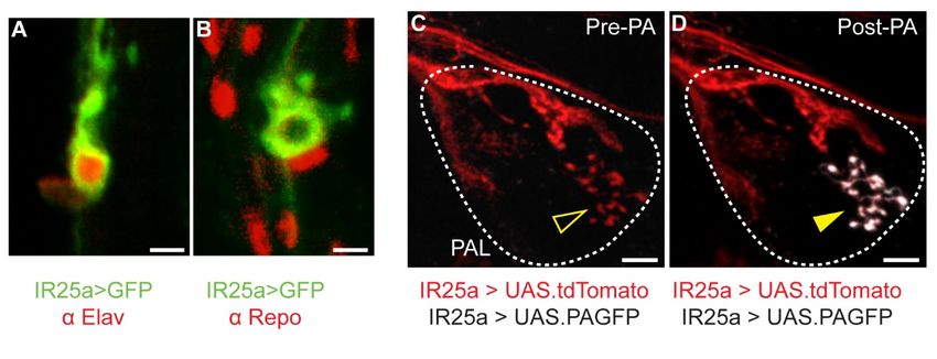
Please cite this article in press as: Alpert et al., A Circuit Encoding Absolute Cold Temperature in Drosophila, Current Biology (2020), https://doi.org/
 10.1016/j.cub.2020.04.038

           ll
                                                                                                                                             Article

scales with stimulus intensity. Non-preferred stimuli instead                  cooling-induced spike (TPN-Is; Figure 1B). In contrast, R60H12
cause a decrease in intracellular calcium [6] and firing rates [7].            TPNs (TPN-IIs) respond to a cold temperature step with an initial
   From their origin at the periphery, the axons of hot- and cold-             calcium spike that accompanies the onset of cooling and that is
activated TRNs of the arista converge onto a brain region called               followed by a rapid decrease to a plateau. The levels of intracel-
the posterior antennal lobe (PAL), where they form adjacent                    lular Ca2+ do not return to baseline for as long as the temperature
‘‘hot’’ and ‘‘cold’’ glomeruli, defining a simple sensory map for              remains in the cold range (Figure 1C), supporting the notion that
the central representation of external temperature [6]. Next in                these cells may not adapt at all under conditions of persistent
line, second-order thermosensory projection neurons (TPNs)                     cold.
collect information from the PAL glomeruli and target higher                       Calcium levels do not allow one to estimate the firing rate of a
brain centers involved in the processing of both innate and                    neuron in stable conditions. To obtain a more direct readout of
learned behavior [8, 9].                                                       TPN-II’s action potential firing under conditions of persistent
   Interestingly, TPN cell types are characterized by different                cold, we next developed a method to perform two-photon
response dynamics to temperature stimuli: ‘‘fast-adapting’’                    guided patch-clamp electrophysiology by targeting TPN-IIs us-
cells respond rapidly but transiently to the onset of a tempera-               ing GFP expression as a guide (again under the control of the se-
ture change, and ‘‘slow-/non-adapting’’ TPNs are characterized                 lective driver R60H12-Gal4) [8].
by persistent activity [8, 9]. Differences in TPN adaptation dy-                   Our patch clamp results confirmed and expanded what we
namics are potentially very significant, as fast-adapting TPNs                 had observed using calcium imaging. The firing rate of non-
may relay temperature change signals, while slow-adapting                      adapting TPN-IIs showed little or no evidence of adaptation;
cells are well positioned to relay information about absolute                  rather, TPN-IIs sustained a remarkably stable level of activity
temperature.                                                                   for the entire duration of the cold step. Only when the temper-
   How are temperature change and absolute temperature sig-                    ature started returning to baseline, TPN-II’s firing rate
nals encoded in the activity of TPNs, relayed to higher brain cir-             decreased, followed by a small inflection in membrane poten-
cuits, and used to inform behavior happening on timescales                     tial (Figures 1D and 1E; note that heating steps caused little
ranging from milliseconds to seconds to days and weeks?                        response in TPN-IIs; Figures 1F and 1G). Protracted patch-
   Here, focusing on cold responses, we set out to identify the                clamp recordings further demonstrated that TPN-IIs display
source and significance of the persistent activity observed in                 little or no adaptation even to extended cold stimuli
slow-adapting TPNs. Our ultimate goal was to understand how                    (25 min; Figures 1H–1J).
the thermosensory system may process information about abso-
lute temperature and how this information may be used to drive                 TPN-II Activity Correlates with Absolute Temperature in
behavior happening on the appropriate timescales.                              the Cold Range
   Our work leads to the identification of a circuit that functions as         Our next goal was to determine whether the persistent activity of
a conduit for information about absolute temperature in the cold               non-adapting TPN-IIs at stable cold conditions indeed scales
range. This circuit displays persistent activity that scales with              proportionally with (and may therefore contain information
temperature, but only at absolute temperatures lower than the                  about) ‘‘absolute’’ temperature rather than correlate with other
fly’s favorite 25 C, and directly targets higher brain centers               stimulus statistics, such as the magnitude of the initial cooling
involved in the control of sleep and activity, adjusting fly behavior          change (Dt).
specifically to cold conditions.                                                   To address this question, we designed temperature steps that
                                                                               either differed in the absolute temperature reached at stable
RESULTS                                                                        conditions (different |t|, same Dt) or in the magnitude of the tem-
                                                                               perature change the preparation experiences on the way to the
A Thermosensory PN Displays Persistent Activity in                             same stable conditions (different Dt, same |t|).
Response to Extended Cold Steps                                                    Our data show that the firing rate of non-adapting TPN-IIs at
To systematically study the adaptation dynamics of TPNs, we                    stable conditions (persistent activity) correlates with absolute
focused on two cold-activated TPN cell types previously                        temperature rather than with stimulus history (Figure 2).
described as fast and slow adapting (TPN-I and II in Figure 1A,                Remarkably, although an initial action potential burst (dynamic
identified by expression of the drivers VT19428 and R60H12,                    activity) was invariably observed in response to cooling (Figures
respectively) [8]. We designed a stimulation protocol that con-                2A–2D), persistent activity (i.e., ‘‘plateau’’ firing rates above
sists of a rapid temperature change (2 C/s) followed by                      baseline firing) only appeared when the absolute temperature
extended stable cold conditions (1–15 min), again followed by                  stabilized at temperatures lower than 25 C (Figures 2B, 2D,
a rapid return to baseline. As before, we initially chose as the               and 2E), i.e., lower than the preferred temperature for
baseline the fly’s preferred temperature of 25 C and stimulus pa-             Drosophila melanogaster. Even when challenged with pro-
rameters (rate of change and temperatures ranges) reasonably                   longed stimuli involving multiple cooling steps, static activity
close to what a fly may encounter in the environment. We then                  only emerged below 25 C (Figure 2F), and only in this range,
used two-photon calcium imaging and recorded TPN responses                     firing rates correlated well with absolute temperature (Figures
to temperature stimulation by targeting expression of the trans-               2G and 2H).
genic calcium indicator GCaMP to each cell type (i.e., under the                   These observations suggest that the non-adapting activity of
control of selective drivers) [8].                                             TPN-IIs may indeed encode information about absolute temper-
   Our results demonstrate that calcium levels in fast-adapting                ature below the preferred range for D. melanogaster, i.e., in what
VT19428 TPNs quickly return to baseline after an initial                       can be described as cold temperature for the fly.

2 Current Biology 30, 1–14, June 22, 2020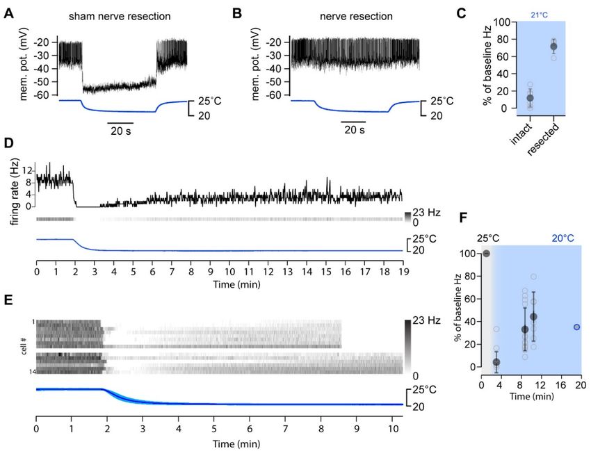
Please cite this article in press as: Alpert et al., A Circuit Encoding Absolute Cold Temperature in Drosophila, Current Biology (2020), https://doi.org/
  10.1016/j.cub.2020.04.038

                                                                                                                                                        ll
Article

  A                        B                                       D                                                F

                          C                                        E                                                G

  H
                                                                                                                                                J

  I

Figure 1. Identification of Thermosensory Projection Neurons Characterized by Non-adapting Responses
(A) Thermosensory projection neurons (TPNs) receive synaptic input from antennal thermoreceptors (TRNs).
(B and C) Two-photon calcium imaging with GCaMP demonstrates significant differences in the adaptation of (B) type-I fast-adapting and (C) type-II slow/non-
adapting TPNs to a cold temperature step (~1 min; DF/F traces are averages of B: 14 cells/3 animals, C: 5 cells/3 animals, ±SD; stimulus trace is an average of the
experiments ± SD).
(D–G) Two-photon guided patch-clamp electrophysiology reveals that TPN-IIs’ firing is indeed non-adapting.
(D) Representative whole-cell current clamp recording from a TPN-II in response to a cold step (~5 C; 1 min). Inset shows x axis expansion during cooling.
(E) Firing rate histogram for TPN-II’s responses to cold (9 animals/9 cells, 3 trials per cell were averaged in each experiment; n = 9 cells ± SEM; temperature [temp.]
trace: average [av.] of 9, ±SEM).
(F and G) TPN-IIs’ are not significantly modulated by heat.
(F) Representative whole-cell current-clamp recording from a TPN-II in response to a hot step (~5 C; 1 min). Inset shows x axis expansion.
(G) Firing rate histogram for TPN-II’s responses to hot (5 cells/4 animals, ±SEM; temp. trace av. of 5, ±SEM).
(H–J) TPN-IIs firing does not return to baseline even for extended cooling steps.
(H) (Top) Representative recording from a TPN-II subjected to extended cooling steps.
(I) Pseudo-colored spike rate histogram of 3 cells in response to a cooling stimulus similar to that in (H) (~20 min recordings; 3 Dt steps as in H; responses aligned
to stimuli).
(J) Quantification (gray lines, mean ± SD for individual cells calculated in regions corresponding numbered bars in I; blue line/shading, mean ± SEM firing rate
across cells; same 3 cells as in I).

The PAL Cold Glomerulus Is Independently Targeted by                                 (harboring the post-synaptic terminals of TPN-IIs; Figures 3A
Distinct Populations of Thermosensory Neurons                                        and 3B) and screened a collection of Gal4 lines for drivers that
Our results raise a number of important questions: where does                        would allow us to identify and selectively manipulate sensory
this absolute cold temperature signal originate (at the sensory                      input to this TPN cell type.
layer or within TPNs)? And what may be the behavioral signifi-                          We discovered that at least 3 cell types independently target
cance of such a persistent cold-evoked activity in the fly brain?                    the cold glomerulus of the PAL: (1) the well-known TRNs of the
We set out to address each question in turn.                                         arista [6] (labeled by a selective driver in Figures 3C and 3D);
   First, we re-evaluated the contribution of the sensory neurons                    (2) a new population of sensory neurons that innervate chamber
of the antenna to TPN-II cold responses. Consistent with previ-                      one of the sacculus, a structure previously shown to be involved
ous observations [8, 9], acute resection of the antennal nerve                       in olfaction and humidity sensing [10, 11] (labeled by a selective
completely abolished TPN-II responses (Figure S1), suggesting                        driver in Figures 3E and 3F); and (3) an unusual cell type that ex-
that all cold-evoked activity originates in thermosensory neurons                    presses the broad sensory co-receptor IR25a and that resides
of the antenna (rather than within TPN-IIs, and see below).                          on the surface of the antennal nerve (note that the terminals of
   We reasoned that any sensory input into TPN-IIs would have to                     this cell type within the PAL are clearly visible after external abla-
come from neurons innervating the cold glomerulus of the PAL                         tion of the antenna, i.e., as a result of degeneration of antennal

                                                                                                                  Current Biology 30, 1–14, June 22, 2020 3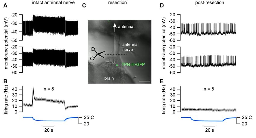
Please cite this article in press as: Alpert et al., A Circuit Encoding Absolute Cold Temperature in Drosophila, Current Biology (2020), https://doi.org/
 10.1016/j.cub.2020.04.038

            ll
                                                                                                                                                 Article

  A                                                                               B

  C                                                                               D

  E                             F                                                                              G                H

Figure 2. TPN-II Neurons Encode Absolute Temperature in the Cold Range
(A–F) Sustained firing of TPN-IIs depends on absolute temperature rather than stimulus history.
(A and B) Representative whole-cell current-clamp recording from a TPN-II in response to a cold step starting from a baseline temperature (A) above or (B) below
25 C (see also Figure S1).
(C and D) Firing rate histograms from TPN-II in response to cooling steps of different sizes (Dt) and settling on distinct absolute temperatures (C) above or (D)
below 25 C (gray line), showing that persistent activity only appears in the cold range (below 25 C; 4 cells/4 animals, ±SD; temp. trace av. of 4, ±SD).
(E) Sustained firing in response to a large stimulus starting at 30 C and settling to ~20 C (3 cells/3 animals, av. ± SD; temp. trace av. of 3, ±SD).
(F) Representative response to a complex stimulus showing persistent firing below 25 C (gray line).
(G) Quantification of firing rates corresponding to numbered regions of interest (ROIs) in (C) and (D) (top: 10 cells/8 animals; bottom: 9 cells/9 animals).
(H) Quantification of TPN-II firing rate changes at stable temperatures below 25 C (readings were taken after ~1 min at each temp.; 9 cells/9 animals).
In (G) and (H), responses from the same cell are connected; colored lines and shading are population averages ± SD (G) or ±SEM (H); gray dots in (H) are averages
of 3 repeats/cell/condition ± SD; note that an f-test demonstrates a significant relationship between firing rates and temperature; p < 0.05.

afferents; Figures 3G and 3H). The location of this cell is similar to            indeed selectively responds to cold temperature stimuli (see
that of the previously described ‘‘anterior cell’’ neurons [12], and              below).
we therefore refer to it as the anterior cold cell (ACc) (Figure 3H,
inset; see Figure S2 for further characterization of this unusual                 Multiple Pre-synaptic Drives Shape the Activity of
sensory neuron).                                                                  TPN-IIs
   The observation that additional sensory cell types (besides the                Our next goal was to determine whether each of the sensory neu-
aristal TRNs) [6] converge onto the cold glomerulus of the PAL                    rons innervating the cold glomerulus indeed provides direct syn-
was unexpected: we have previously described cold-responding                      aptic drive to TPN-II. To test for connectivity, we employed activ-
sensory neurons innervating sensilla located in chamber two of                    ity-dependent, synaptic GFP reconstitution across synaptic
the sacculus, but these neurons are part of an ‘‘hygrosensory                     partners (syb:GRASP) [13].
triad’’ (composed of a dry-, humid-, and cold-activated cell                         Our results suggest that each sensory cell population makes
innervating the same sensillum) and project to a distinct region                  direct functional connections with TPN-II second-order neurons
of the PAL (the ‘‘column’’ or VP1 glomerulus) [10, 11]. The results               (Figures 3P–3R). Using selective drivers, we could directly
of two-color two-photon microscopy instead suggest extensive                      demonstrate synaptic GFP reconstitution between chamber
intermingling between TPN-II dendrites, the terminals of cham-                    one sacculus neurons and TPN-IIs (Figure 3P). We have been
ber one sacculus neurons, and those of arista TRNs within the                     so far unable to identify a driver only active in ACc neurons; to
cold glomerulus of the PAL (Figures 3I–3O). Finally, we were sur-                 test for ACc:TPN-II connectivity, we used R77C10-Gal4—a
prised to find an additional ‘‘internal’’ temperature receptor                    driver active in both the arista cold-activated TRNs and ACc neu-
within the head capsule (ACc; see above), which also selectively                  rons. Robust GFP reconstitution using this line confirmed that
innervates the cold glomerulus. Our results confirm that each of                  one or both of these cell types indeed forms synapses with
these cell types independently innervating the cold glomerulus                    TPN-IIs (Figure 3Q; see [8, 9] for arista TRNs:TPN-II

4 Current Biology 30, 1–14, June 22, 2020
Please cite this article in press as: Alpert et al., A Circuit Encoding Absolute Cold Temperature in Drosophila, Current Biology (2020), https://doi.org/
  10.1016/j.cub.2020.04.038

                                                                                                                                                       ll
Article

 A                                     C                         E                            I
                                                                                                                                   J                        K

                                                                                                                                                            L

                                       D                         F
 B
                                                                                                                                   M                        N

                                                                                                                                                            O

                                        G                        H
                                                                                             P                         Q                      R

 S                                                        T                                                    U

Figure 3. Three Distinct Populations of Peripheral Cold-Sensing Neurons Drive the Activity of TPN-IIs
(A) Confocal micrograph of the fly antenna showing the location of the sacculus (pink box) and arista (blue arrow).
(B) 3D model of the fly brain showing the location of the posterior antennal lobe (PAL) and PAL glomeruli (inset; the glomerulus innervated by cold cells is shown in
blue, additional glomeruli are annotated with standard nomenclature).
(C–H) Selective drivers identify distinct sensory neuron populations targeting the cold glomerulus.
(C and D) A selective driver for arista cold cells labels (C) cell bodies in the arista and (D) their PAL termini.
(E and F) A selective driver for cold cells of the sacculus labels (E) cell bodies innervating chamber I of the sacculus and (F) their termini in the PAL (C and E are
confocal micrographs of the whole antenna; blue, cuticle autofluorescence, green, GFP; scale bars, 50 mm; D and F are single two-photon slices; scale bars,
10 mm).
(G and H) Antenna ablation demonstrates the existence of an unusual ‘‘internal’’ cold receptor also innervating the PAL.
(G) IR25a-Gal4 > UAS-GFP labels many of the antennal sensory neurons innervating PAL glomeruli.
(H) A week following antennal resection, all antennal afferents have degenerated, revealing anterior cold cell (ACc) termini in the PAL. The fluorescent signal can
be traced to one/two cell bodies located on the edge of the antennal nerve (AN) (inset; scale bar, 10 mm; see also Figure S2).
(I–O) Extensive overlap between TPN-II dendrites with both arista/ACc and sacculus termini in the PAL.
(I) A selective split-Gal4 driver reveals TPN-II’s anatomy (two-photon z stack).
(J–O) Two-color two-photon micrograph illustrating spatial overlap between (J–L) TPN-II dendrites (green) and Arista/ACc termini (magenta, single z slice) and
between (M–O) sacculus (green) and arista/ACc termini (magenta).
(P–R) Synaptobrevin GRASP confirms synaptic connectivity between TPN-IIs and (P) sacculus and (Q) arista/ACc; the fact that the syb:GRASP signal in (Q)
persists a week post-antenna ablation (R) shows ACc also connects to TPN-IIs.
(S–U) Two-photon Ca2+ imaging shows sacculus neurons exclusively respond in the cold range. Response profiles of (S) arista, (T) chamber I sacculus, and (U)
ACc neuron termini in response to cooling steps in the hot (bottom, above 25 C, red) or cold (bottom, below 25 C, blue) range are shown.
(S) Arista neurons show both a transient peak in response to cooling and a persistent Ca2+ elevation in both conditions.
(T) Sacculus cold cells only respond when the temperature drops below 25 C (arrows).
(U) ACc neurons show persistent signals both above and below 25 C.
(S)–(U) show averages of 4 animals ± SD. In all PAL panels, scale bars are 10 mm.

                                                                                                                   Current Biology 30, 1–14, June 22, 2020 5
Please cite this article in press as: Alpert et al., A Circuit Encoding Absolute Cold Temperature in Drosophila, Current Biology (2020), https://doi.org/
 10.1016/j.cub.2020.04.038

           ll
                                                                                                                                              Article
                                                                                                          Figure 4. TPN-IIs Target the DN1a Group of
A                             B                                    C
                                                                                                          Dorsal Neurons, Part of the Circadian Clock
                                                                                                          Network
                                                                                                          (A) 3D reconstruction of the TPN-II (blue)-DN1a
                                                                                                          (purple) connection, at the edge of the mushroom
                                                                                                          body (MB) calyx.
                                                                                                          (B and C) Photolabeling with PA-GFP reveals
                                                                                                          DN1as are candidate targets for TPN-IIs. For
                                                                                                          this experiment, (B; pre-photoactivation) TPN-II
                                                                                                          termini are targeted by independent labeling
                                                                                                          with TdTomato, although PA-GFP is expressed
                                                                                                          broadly. Following targeted photoactivation at
                                                                                                          720 nm, (C; white) PA-GFP diffuses to label TPN-II
                                                                                                          targets.
                                                                                                          (D–M) DN1as are identifiable by anatomy and
                                                                                                          because they express a combination of molecular
D                                                    E              N
                                                                                                          clock components. Here, (D) DN1as were labeled
                                                                                                          by GFP expression (green, under the control of a
                                                                                                          selective driver) and immunostained using an anti-
                                                                                                          Clock antibody (CLK, purple; confocal z stack of a
                                                     F     G                                              whole-mount fly brain; note that CLK also labels
                                                                                                          DN1p, DN2, and DN3).
                                                     H                                                    (D–G) DN1 as were labeled by GFP expression
                                                                                                          (green, under the control of a selective driver) and
                                                                                                          immunostained using an anti-Clock antibody
                                                                   O                                      (CLK, purple). (E) is an enlargement from panel (D)
                                                     I     J                                              centered on DN1as; (F) purple channel, (G) green
                                                                                                          channel. (D-G, confocal z stack of a whole-mount
                                                                                                          fly brain; note that CLK also labels DN1p, DN2,
                                                     K                                                    and DN3).
                                                                                                          (H–M) DN1as also express (H–J) period (anti-PER,
                                                                                                          purple) and (K–M) cryptochrome (anti-CRY, pur-
                                                                                                          ple; in all panels, scale bar, 10 mm).
                                                     L     M                                              (N and O) Syb:GRASP demonstrates mono-
                                                                                                          synaptic connectivity between TPN-IIs and
                                                                                                          DN1as.
(N) Enlargement of a 3D reconstruction showing predicted point of synaptic contact.
(O) Synaptic GFP reconstitution is observed between TPN-II and DN1a neurons at the edge of the mushroom body calyx (pseudocolored two-photon stack).

connectivity). Moreover, significant syb:GRASP signal persisted                contribute to shape TPN-II’s responses to temperature but that
even a week following antenna ablation (i.e., following degener-               the absolute cold temperature signal recorded in TPN-IIs is
ation of antennal afferents; Figure 3R). This result suggests that             relayed by chamber one neurons of the sacculus.
ACc neurons are also independently connected to TPN-IIs.                          Interestingly, although calcium transients in the terminals
                                                                               generally correlate well with synaptic release, the activity re-
Chamber I Sacculus Neurons Selectively Respond to                              corded in TPN-IIs does not appear to be a simple summation
Absolute Cold                                                                  of the activity of these pre-synaptic cell types, suggesting addi-
Next, we asked which—if any—of these cold-sensing cell types                   tional processing is likely to occur at this synapse.
may contribute the absolute cold temperature signal recorded in
TPN-IIs. To address this question, we designed a stimulation                   TPN-IIs Have Selective Targets in the Fly Brain
protocol whereby the temperature is stepped down by 5 C                      What is the behavioral significance of this persistent, cold tem-
either from a baseline of 25 C or, alternatively, from a hot base-            perature activity relayed by sensory neurons of the sacculus
line of 30 C. In this setup, we expect that cells responding to ab-           and prominently recorded in specialized second-order neurons
solute cold (rather than cooling) would only respond to the stim-              of the thermosensory system?
ulus starting at the 25 C baseline and dipping into temperatures                 Although many second-order neurons of the thermosensory
that are below the fly’s favorite range (i.e., cold temperatures).             system (TPNs) share common higher brain targets (such as the
   To test for temperature responses, we again used two-photon                 calyx of the mushroom body, lateral horn, and posterior lateral
calcium imaging, measuring calcium transients at the PAL termi-                protocerebrum) [8, 9], non-adapting TPN-IIs exclusively inner-
nals of each cell type by selective targeting of GCaMP. Our re-                vate a microglomerulus at the edge of the calyx [8] (Figure 4A).
sults clearly demonstrate that, although the calcium responses                 We reasoned that this anatomy may underlie selective connec-
of all three cell types appear to have non-adapting components,                tivity in the brain and that revealing TPN-II’s synaptic targets
only the chamber one sacculus neurons exclusively respond to                   may illuminate circuits that utilize specifically cold temperature
cold, but not to cooling (Figures 3S–3U).                                      information to process behavior happening on slow timescales.
   Together with the results of connectivity experiments, this                    Therefore, we used two-photon guided conversion of photo-
observation suggests that all three sensory cell types may                     activatable GFP (PA-GFP) [14] to identify TPN-II’s cellular

6 Current Biology 30, 1–14, June 22, 2020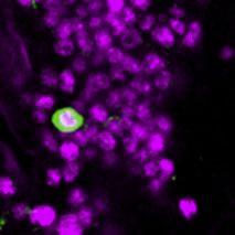
Please cite this article in press as: Alpert et al., A Circuit Encoding Absolute Cold Temperature in Drosophila, Current Biology (2020), https://doi.org/
  10.1016/j.cub.2020.04.038

                                                                                                                                                            ll
Article

 A                                       B

                                         C

 D                                                                             E                                     G                              H

                                                                               F                                      I                              J

 K                                             L                                          M                                             N

Figure 5. TPN-IIs Robustly Inhibit DN1a Activity in Cold Conditions through GABA Release
(A) Schematic representation of the circuit, including cold thermosensory neurons (TRNs) (dark blue), TPN-IIs (light blue), and DN1as (pink).
(B and C) DN1a firing is persistently silenced by cold, correlating with TPN-II activation.
(B and C) Representative whole-cell current-clamp recording (B) from a DN1a neuron and (C) from a TPN-II in response to a cold step (recorded independently).
(D) Average firing rate histograms for DN1as challenged with cooling steps of different amplitude show robust silencing even for small stimuli (blue boxes; Dt = 2,
Dt = 4, Dt = 6; 8 cells/7 animals; av. ± SD). Note that corresponding heating stimuli produce an initial burst in activity but modest persistent modulation (red
box, right; Dt = 2, Dt = 4, Dt = 6; 9 cells/8 animals; av. ± SD).
(E and F) Quantification of firing rates at plateau for (E) cold and (F) hot steps as in (D) (gray lines connect responses from the same cell; colored dots, averages ±
SD).
(G) Whole-mount immunostaining showing that TPN-II (labeled by expression of GFP, white arrow) expresses the inhibitory neurotransmitter GABA (anti-GABA,
pink, scale bar 10 mm).
(H–J) GABA release mediates cold inhibition of DN1as.
(H) Schematic of the experiment.
(I) Average firing rate histograms of DN1a in response to a Dt ~4 C cooling before (black) and after (green) application of the GABA receptor antagonist,
picrotoxin (100 mM; 13 cells/13 animals; av. ± SD), showing that GABA receptor blockade abolishes cold inhibition of DN1as.
(J) Quantification of (I) as trough-to-baseline ratio of firing rate (in first 10 s after cooling), before (black) and after application of picrotoxin (green). Gray circles
connected by lines represent individual neurons; filled circles, population av. ± SD; *p < 0.05 in a paired, one-tailed t test.
                                                                                                                                        (legend continued on next page)

                                                                                                                     Current Biology 30, 1–14, June 22, 2020 7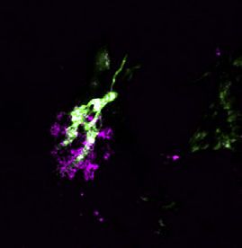
Please cite this article in press as: Alpert et al., A Circuit Encoding Absolute Cold Temperature in Drosophila, Current Biology (2020), https://doi.org/
  10.1016/j.cub.2020.04.038

             ll
                                                                                                                                                           Article

targets. First, we engineered flies where PA-GFP was constitu-                          suggests an additional (albeit transient) heating-evoked drive
tively expressed throughout the brain (under the control of syb-                        to DN1a.
Gal4) and the TPN-II terminals were selectively labeled by a                               The fact that DN1as are powerfully silenced (rather than acti-
red fluorescent protein (Figure 4B). Then, using the red fluores-                       vated) by cold temperature and at the same time are synaptic
cence as a guide, we photo-converted PA-GFP exclusively in a                            targets of cold-activated TPN-IIs suggests that TPN-IIs may be
volume tightly overlapping the TPN-II axon terminals and traced                         inhibitory projection neurons. Indeed, immunohistochemistry
the labeled neurites of next-order neurons as they became illumi-                       demonstrated that TPN-IIs express the inhibitory neurotrans-
nated by diffusion of the photo-converted fluorophore                                   mitter GABA (Figure 5G), and bath application of the GABAA-re-
(Figure 4C).                                                                            ceptor antagonist picrotoxin abolished cold inhibition of DN1as
   Our results reveal that prominent targets of R60H12 cold-acti-                       (Figures 5H–5J).
vated TPNs are two distinctive neurons that have been previ-                               Together, our results suggest that cold-activated TPN-IIs
ously described as components of the circadian network in the                           directly inhibit the activity of the clock neuron cluster DN1a,
Drosophila brain (Figures 4C and 4D). The 1a cluster of ‘‘dorsal                        through a GABA-ergic synapse, in cold conditions. Because
neurons’’ (DN1a) is part of a network of DNs that express key                           clock neurons can exhibit endogenous (clock-regulated)
circadian gene products, such as clock and period (Figures                              rhythms in activity, we next tested the possibility that activity
4D–4J). DN1as are defined by their unique anatomy (including                            rhythms may also shape the firing profile of DN1as, perhaps
projections to the accessory medulla) and by the fact that they                         gating the effect of cold temperature on DN1a activity.
are among a small group of DN1s that express cryptochrome
[15] (Figures 4K–4M). Importantly, DN1as are indeed direct syn-                         DN1as Have Clock-Regulated Rhythms in Activity but
aptic targets of TPN-IIs, as demonstrated by synaptic GRASP                             Are Invariably Inhibited by Cold
(Figures 4N and 4O).                                                                    Previous work has demonstrated that ‘‘posterior’’ DN1s (DN1ps)
                                                                                        possess endogenous mechanisms to modulate firing rates in a
DN1a Neurons Are Directly Inhibited by TPN-IIs in Cold                                  time-of-day-dependent fashion [16]. To test whether DN1as
Conditions                                                                              may display similar properties, we recorded their activity at
What is the effect of cold temperature on DN1a activity? Given                          different times of day and night. Our recordings suggest that,
TPN-II’s firing rates are elevated in cold conditions and scale                         as for DN1ps, DN1as also possess time-of-day-dependent mod-
proportionally with cold temperature, we expected TPN-IIs to                            ulation, with higher firing rates during the first part of the day
drive similar activity in the post-synaptic DN1as. Instead, cold                        (zeitgeber time 0 [ZT0]–4; 12 Hz) as compared to the last
temperature all but shut down activity in this cell type (Figures                       time point of the night (ZT20–24; 6 Hz; Figure 5K). These activ-
5A–5C).                                                                                 ity rhythms are present in both females and males (Figures 5K
    Using temperature steps of different magnitude (and patch-                          and 5L) and are regulated by the molecular clock, as they were
clamp electrophysiology as described above), we observed                                abolished in per01 mutants (Figure 5L). Higher rates of DN1a
that a cooling step as small as 2 C was sufficient to nearly                           firing in the morning may reflect an important role for these cells
silence DN1as (from an initial baseline of nearly 10 Hz) and that                       in the regulation of morning activity and/or in the night-to-day
the firing of this cell type was essentially completely silenced at                     sleep/wake transition. Interestingly, notwithstanding these
temperatures 4 C or 6 C below the 25 C baseline (Figures 5D                          rhythms of activity, cold steps of 4 C or 6 C (from a baseline of
and 5E). As expected, and consistent with the properties of                             25 C) were effective in significantly reducing DN1a activity at
TPN-IIs, DN1a’s inhibition by cold temperature was very persis-                         all time points tested (Figures 5M and 5N). Hence, in the absence
tent (showing limited recovery in stable cold conditions) and de-                       of other external stimuli, cold temperature should be effective in
pended on the presence of the antennae (Figure S3).                                     silencing DN1a activity at all times of day and night.
    In contrast to strong responses to cold, DN1as demonstrated
little persistent modulation by heat (i.e., when the temperature                        Cold Temperature Has Both an Acute and Persistent
was stepped above the 25 C range; Figures 5D and 5F). This                             Effect on Fly Activity and Sleep
is again consistent with the limited response recorded from                             What is the functional significance of this powerful cold inhibition
TPN-IIs in the hot range. Interestingly, we did observe a rapid,                        of DN1a? The DN1ps have been abundantly implicated in the
heating-induced burst in DN1a firing that could not be directly                         regulation of sleep, including the onset and extent of the after-
correlated with the responses of the pre-synaptic TPN-IIs (Fig-                         noon ‘‘siesta’’ flies enjoy in hot days [17–19], as well as the regu-
ure 5D; compare with Figures 1F and 1G). This observation                               lation of sleep patterns in cold days [20]. At least one previous

(K) Baseline firing rates of DN1as at 25 C measured during different times of day (white) and night (gray). Firing rates from different cells (black circles) were
grouped in 4-h bins (filled circles; mean ± SD). Morning rates (pink) were significantly higher than evening ones (purple; n = 39 cells/32 animals; *p < 0.05; unpaired
one-tailed t test).
(L) Circadian rhythms of DN1a firing are absent in per01 mutants. Nighttime and daytime firing rates of DN1as at 25 C recorded from wild-type (WT) (*p < 0.05;
unpaired one-tailed t test) and period mutant flies are shown (per01; NS, not significant difference; unpaired one-tailed t test; black circles are individual cells; filled
circles indicate av. ± SD; n = 35 cells/21 animals; recordings are from +/Y and per01/Y male flies).
(M and N) DN1as are inhibited by cooling at all ZTs.
(M) Mean firing rate of DN1as in response to a cooling step (blue box, from 25 C; individual gray traces represent averages; ZTs as in L; ZT20–24 and ZT0–4 are
colorized as in K; envelope: ±SEM, n = 40 cells/32 animals).
(N) Quantification of change in firing frequency in response to different cooling steps (from 25 C) during the night (ZT20–24, purple) and day (ZT0–4, pink; gray
circles connected by lines indicated individual cells; filled circles indicate mean ± SD; *p < 0.05; paired one-tailed t test; 14 cells/10 animals; see also Figure S3).

8 Current Biology 30, 1–14, June 22, 2020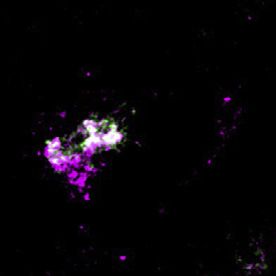
Please cite this article in press as: Alpert et al., A Circuit Encoding Absolute Cold Temperature in Drosophila, Current Biology (2020), https://doi.org/
  10.1016/j.cub.2020.04.038

                                                                                                                                                           ll
Article

  A                                            B                                       C                                        D

  E                                     F                                             G                                         H

  I                                            J                                      K                                         L

Figure 6. Genetic Silencing of Either DN1a or TPN-II Perturbs Normal Daytime Sleep Restructuring by Cold
In wild-type flies, cold temperature has both acute and persistent effects on daytime activity and sleep. In (A), (F), (G), and (I)–(K), activity and sleep were quantified
in 30-min bins in 2 consecutive days per condition (B and C: 1 day/condition). Schematics on top illustrate the experimental design. Data plots represent sleep
(above) and activity bar graphs (below) and are averages ± SEM across days and across individual flies; filled circles in sleep plots and black dots above activity
bars indicate time points that are significantly different between cold (18 C, blue) and 25 C (gray) conditions (p < 0.05; paired two-sided t test); dark shades
indicate lights off (night); ZT, zeitgeber time.
(A) In wild-type animals, cold conditions increase morning sleep and suppress morning activity (ZT0–3); in contrast, cold reduces both sleep and activity in the
evening (ZT6–12). Moreover, the onset of evening sleep is advanced (green arrowhead in A). As a result, the net effect of cold is an advancement of daytime sleep.
(B) Morning cold (ZT0–3) rapidly suppresses activity and increases sleep; following the return to 25 C, sleep and activity quickly return to normal levels.

                                                                                                                                        (legend continued on next page)
                                                                                                                     Current Biology 30, 1–14, June 22, 2020 9
Please cite this article in press as: Alpert et al., A Circuit Encoding Absolute Cold Temperature in Drosophila, Current Biology (2020), https://doi.org/
10.1016/j.cub.2020.04.038
ll
Article
publication suggests that DN1as may also be involved in the blocker of synaptic transmission (tetanus toxin light chain) [27]
regulation of daytime sleep [21]. and monitored the effect of this manipulation on sleep and
Here, our goal was to test the possibility that DN1as may be activity.
directly involved in the regulation of activity and sleep by temper- Remarkably, genetic silencing of DN1a’s output at 25 C
ature and that their powerful inhibition by cold may be of signif- partially mimicked cold conditions: under LD cycles, the onset
icance for the regulation of activity/sleep patterns in persistent of siesta sleep was advanced even at 25 C, resembling the
cold conditions. Unlike previous studies, our experiments could response to cold conditions observed in controls (Figures 6G
be guided by knowledge of the specific thermal range that mod- and 6H; see Figures 6F and 6I for controls). This suggests
ulates DN1a’s activity via connections with the antennal TRNs/ that reducing the output of DN1as in the morning may be a
TPN-II circuit (Figure 6). key mechanism for daytime siesta sleep advancement by
At the normal rearing temperature of 25 C (and under 12 h cold temperature. Interestingly, blocking DN1a’s output also
light:12 h dark, or LD, cycles), fly behavior is characterized by prevented the plastic remodeling of sleep by cold temperature
peaks of activity corresponding to the late night-early morning in the evening (ZT6–12). This time, constitutive block of DN1a
transition (‘‘morning peak’’) and to the end of the day (‘‘evening output produced a stable (i.e., temperature-independent)
peak’’; see Figure 6A, gray bar graph). Fly sleep is generally sleep profile more similar to the control’s 25 C conditions,
defined as inactivity that persists for 5 min or longer [22, 23], as DN1a > tetanus toxin light chain (TNT) flies did not reduce
and as such, most of fly sleep occurs at night, yet flies (male flies the amount of evening sleep in response to cold (Figures 6G
in particular) [24] also display a prominent mid-day siesta (Fig- and 6H).
ure 6A, black plot)—perhaps to avoid potentially hot/dry condi- As cold temperature suppresses DN1a firing in both morning
tions in the mid-day [25]. and evening (Figure 5), this observation is incompatible with sim-
Cold temperature (18 C) has both an acute and persistent ef- ple models, in which silencing DN1a invariably results in more
fect on fly activity and sleep: cold conditions rapidly suppress sleep, irrespective of time of day. Instead, our results suggest
morning activity (and increase morning sleep; Figure 6A, blue that the appropriate timing and extent of DN1a activity may be
bar graphs and lines). Activity in the evening is also reduced; in crucial for the dynamic regulation of daytime sleep patterns
addition, the onset of evening activity is advanced (arrow in bar and for their plastic adaptation to changes in the external
graph, Figure 6A). The overall effect of cold on daytime sleep is temperature.
that the siesta is advanced to earlier time points—potentially Next, we tested the potential impact of TPN-II silencing. Our
an adaptation to the fact that cooler conditions normally accom- previous results suggest that TPN-IIs provide powerful inhibitory
pany the seasonal shortening of days (Figure 6A, blue plot; see drive to DN1as in cold conditions. Consistent with this, silencing
also [26]). TPN-II’s output had no effect on sleep at 25 C. After a shift to
Interestingly, this acute shift in siesta sleep can be ascribed to 18 C, siesta sleep of TPN-II > TNT flies advanced normally. Inter-
independent and reversible effects of cold temperature on sleep estingly, constitutive block of TPN-II’s output again prevented
in the morning and evening: in LD conditions, a defined 3 h cold sleep restructuring in the evening, producing a stable sleep pro-
step in the morning increases sleep (but, following return to 25 C, file similar to that of DN1a > TNT flies (Figures 6K and 6L;
fly behavior returns to normal; Figure 6B), although a similar compare to Figures 6G and 6H).
defined cold step in the evening decreases sleep (Figure 6C; The fact that reducing the output of inhibitory neurons (TPN-
see Figure 6D for quantifications). IIs) had the same (rather than the opposite) effect on evening
sleep to that obtained by silencing their targets (DN1as) is again
Silencing DN1a Output Mimics Cold Conditions consistent with the notion that the dynamics of DN1a activity
Are DN1as involved in the restructuring of daytime sleep in (rather than their net output at a given time point) may be impor-
response to cold temperature? To test the potential involvement tant to determine the appropriate pattern of daytime sleep, so
of DN1as (and TPN-IIs) in this process, we first developed ge- that locking the system in one state may produce similar
netic reagents to selectively target each cell type for genetic effects.
silencing. Starting from broader drivers, we created intersec- Notably, the overall daytime sleep profile of TPN-II > TNT flies
tional split-Gal4 lines narrowly active in either DN1as (Figure 6E) at 18 C was remarkably similar to that of DN1a > TNT flies at
or non-adapting TPN-IIs (see Figure 3I and STAR Methods for 25 C (both in the morning and evening; compare Figure 6K,
details). Next, we used these drivers to express a transgenic blue plot with Figure 6G, black plot). This daytime sleep profile
(C and D) Evening cold (C; ZT6–12) decreases sleep so that, together, morning and evening effects recapitulate all day cold conditions (n = 31 animals in A, 26 in B
and C; see D for quantifications).
(E) A split-Gal4 driver allows selective targeting of DN1as (shown driving GFP; two-photon z stack; scale bar, 20 mm).
(F–L) Silencing DN1as or TPN-IIs using selective split-Gal4s perturbs sleep restructuring by cold temperature.
(F, I, and J) Control genotypes (n = 30 in F; n = 61 in I; n = 62 in J).
(G) Silencing DN1as by expression of tetanus toxin light chain (TNT) partially mimics cold conditions, producing flies that sleep more in the morning even at 25 C
and that fail to restructure their afternoon sleep in response to cold (n = 19 animals).
(K and L) Silencing (K) TPN-II output with TNT also produces flies that fail to restructure afternoon sleep in response to cold (n = 52 animals; see H and L for
quantifications; in all boxplots, box edges: 25th and 75th percentiles; thick lines: median; whiskers: data range; gray dots: individual data points/flies; *p < 0.05 in
paired two-sided t test comparing 25 C versus 18 C within genotype or two-way ANOVAs with a Bonferroni correction for multiple comparisons across ge-
notypes/temperatures).
10 Current Biology 30, 1–14, June 22, 2020Please cite this article in press as: Alpert et al., A Circuit Encoding Absolute Cold Temperature in Drosophila, Current Biology (2020), https://doi.org/
10.1016/j.cub.2020.04.038
ll
Article
A B F
G
H
C D E
I
J K M N O P
L
Q R S T
Figure 7. Sleep and DN1a Activity Are Modulated by the Opposing Pushes of Light and Cold Temperature
(A and B) Cold and dark synergize to increase sleep across the day.
(A) (Top) Behavioral protocol used to evaluate sleep on flies entrained in 12 h light-dark (LD) cycles (white box: day [lights on]; black box: night [lights off]; gray box:
subjective day [lights off]). (Bottom) Sleep plot for two independent groups of control (wild-type) flies during a single LD day at 25 C and in the following dark day at
either 18 C (blue line; n= 19 animals, ±SEM) or 25 C is shown (gray line; N = 19 animals, ±SEM; filled circles indicate time points that are significantly different
between conditions; p < 0.05; unpaired two-sided t test).
(B) Quantification of total sleep in the indicated intervals (box edges: 25th and 75th percentiles; thick lines: median; whiskers: data range; gray dots: individual data
points/flies; *p < 0.05 in unpaired two-sided t test).
(C–E) In the dark, suppressing DN1a output by TNT expression mimics cold conditions, increasing sleep across the day.
(C) (Top) Behavioral protocol. (Bottom) Sleep in DN1a > TNT flies (orange trace; n = 25 animals), UAS-TNT/+ (gray; n = 32 animals) and DN1a-Gal4/+ flies is shown
(black; n = 31 animals; all traces are av. ± SEM; circles, significantly different from both controls in two-way ANOVA; p < 0.05).
(D and E) Quantification of total sleep in the indicated intervals for genotypes in (C) (box edges: 25th and 75th percentiles; thick lines: median; whiskers: data range;
gray dots: individual data points/flies; *p < 0.05; two-way ANOVA with a Bonferroni correction for multiple comparisons across genotypes).
(F–I) Optogenetic activation of TPN-II produces an acute increase in sleep.
(F) Protocol used (3 consecutive days represented top to bottom); red shading indicates optogenetic activation.
(G) Sleep pattern of TPN-II > Chrimson flies fed all-trans retinal (red trace; 25 animals) or control food (black; 27 animals—note that retinal is essential for Chrimson
function; traces: av. ± SEM; circles, significantly different from controls in two-sided t tests; p < 0.05).
(H and I) Quantification of total sleep in the indicated intervals (H) ZT0–3 on day 2 and (I) ZT6–12 on day 4 (box edges: 25th and 75th percentiles; thick lines: median;
whiskers: data range; gray dots: individual data points [flies]; *p < 0.05; unpaired, two-sided t test).
(legend continued on next page)
Current Biology 30, 1–14, June 22, 2020 11Please cite this article in press as: Alpert et al., A Circuit Encoding Absolute Cold Temperature in Drosophila, Current Biology (2020), https://doi.org/
10.1016/j.cub.2020.04.038
ll
Article
may perhaps represent a default state of the system that results targets of light-responsive ventral lateral neurons (LNvs) (a core
from manipulations that disconnect it from external temperature component of the clock circuit) [28]. Importantly, the ‘‘small’’
drive. LNvs or sLNvs have been described as master regulators of
Despite this potentially complex interaction, our results sug- morning activity [29] and have been suggested to form reciprocal
gest that the circuit composed of inhibitory TPN-IIs and DN1a connections with DN1as [21].
plays a key role in mediating the restructuring sleep and activity Our results suggest that indeed DN1as respond to light as well
patterns in cold conditions. as to sLNv signaling. First, we observed light responses in DN1a
(Figures 7J–7L; see [29]). Next, we showed that ‘‘artificial’’ acti-
Dark and Cold Synergize to Suppress Morning vation of sLNvs using P2X2 results in an increase of DN1a firing
Wakefulness (Figures 7M and 7N). Finally, we recorded an increase in firing
In the environment, the seasonal arrival of cold temperatures is rates in DN1as upon focal application of the neuropeptide
often accompanied with darker conditions and shorter days. pigment dispersing factor (PDF) (normally expressed by sLNvs
Next, we tested the potential impact of light on DN1a function [30] and for which DN1as express the cognate receptor PDF re-
and fly behavior in the cold. Under light/dark cycles in constant ceptor [PDFR] [31]; Figures 7Q and 7R).
cold (LD at 18 C), control flies robustly wake up at the first Strikingly, light exposure, artificial sLNv activation, and PDF
appearance of light (‘‘lights on’’) but quickly return to sleep within application were each able to partially overcome cold inhibition
60–90 min (Figure 6); our results also demonstrate that, at 25 C, of DN1as (Figures 7K, 7L, 7O, 7P, 7S, and 7T), suggesting that
blocking DN1a output significantly increases morning sleep, light and/or LNv signaling (through PDF release) could drive
partially mimicking cold conditions (Figures 6G and 6H). Interest- DN1as even in cold conditions and explaining the dominant ef-
ingly, when switched to constant dark/cold conditions (DD at fect of light in setting the beginning of daytime activity.
18 C), control animals nearly fail to wake up in the morning alto- Together, our results demonstrate functional connectivity be-
gether (Figure 7A, blue line; see Figure 7B for quantification). tween sLNvs and DN1as and suggest that DN1a activity is
Blocking the output of DN1as mimics this effect of cold, so shaped by signals from the circadian clock and modulated by
that—in the absence of a light signal—most DN1a > TNT flies the opposing pushes of light and cold temperature, dynamically
fail to wake up in the morning even at 25 C (Figures 7C–7E). Op- shifting the pattern of daytime sleep to better adapt to changing
togenetic activation of TPN-IIs was sufficient to reproduce this environmental conditions.
effect, generating animals that largely ignore the lights on signal
in the morning (Figures 7F–7H; note that, in dark conditions, cold DISCUSSION
temperature also produces a significant increase in evening
sleep, an effect reproduced by DN1a silencing and optogenetic In this work, we uncover a complete circuit, from sensory neu-
activation of TPN-IIs; Figures 7F, 7G, and 7I). rons to circadian and sleep centers, that processes information
Hence, light and cold have powerfully antagonistic effects on about absolute cold temperature to exert influence on fly
morning wakefulness. Based on these results, we tested the behavior in the timescale of minutes to hours to days.
interaction of light and temperature in regulating DN1a firing The circuit we describe is composed of sensory neurons of the
rates. antenna (including newly identified thermosensory neurons only
active in the cold) and of specialized second-order thermosen-
DN1as Directly Integrate Light and Cold Temperature sory projection neurons of the PAL and provides persistent inhi-
Signals bition to the DN1a cluster of circadian neurons to adapt sleep/
Ample evidence suggests that DN1a activity may be directly or activity patterns specifically to cold conditions.
indirectly modulated by light. For example, DN1as express the Our data show that ‘‘absolute temperature’’ and ‘‘temperature
blue light receptor cryptochrome [15] and, in the larva, are known change’’ signals can be extracted by second-order neurons from
(J) Circuit schematic including TPN-IIs (light blue), DN1as (pink), and sLNvs (orange).
(K and L) DN1as are excited by light.
(K) Light produces robust increases in firing rate at 25 C (black) and at 20 C (blue; gray circles connected by lines represent individual cells; filled circles are av. ±
SD; *p < 0.05; paired one-tailed t test).
(L) Representative whole-cell recordings from a single DN1a neuron before and during light stimulation (yellow box) at 25 C (black) or 20 C (blue).
(M–P) Artificial activation of sLNvs drives fire rate increases in DN1a and can overcome cold inhibition.
(M) Experiment schematic. sLNvs express the exogenous ATP receptor P2X2 and can be activated by pressure ejection of ATP (20 mM, green), while patch clamp
records activity in DN1a (pink).
(N) ATP (20 mM, green) can drive an increase in DN1a firing at 25 C in animals in which sLNvs express P2X2 (green trace, 4 cells/animals), but not in control
animals (driver without the receptor, gray/black trace; 6 cells/4 animals).
(O) ATP can also overcome cold inhibition of DN1as (4 cells/4 animals av. ± SEM; the green trace at the bottom of N and O is Alexa Fluor 594 fluorescence, a dye
included as a marker in the ATP solution; arbitrary fluorescence units ± SEM).
(P) Quantification of (N) and (O) (gray circles connected by lines represent individual cells; filled circles are av. ± SD; *p < 0.05; paired one-tailed t test).
(Q–T) The neuropeptide PDF can increase DN1a firing to overcome inhibition in the cold.
(Q) Experiment schematic. PDF is pressure ejected (50 mM, yellow), while patch clamp records activity in DN1a (pink).
(R–T) PDF can drive an increase in DN1a firing both at (R) 25 C or (S) 20 C (9 cells/6 animals av. ± SD; the yellow line at the bottom of R and S represent the
approximate time of the PDF puff).
(T) Quantification of (R) and (S) (gray circles connected by lines represent individual cells; filled circles are av. ± SD; *p < 0.05; paired one-tailed t test; all ex-
periments were done at ZT 0–8).
12 Current Biology 30, 1–14, June 22, 2020Please cite this article in press as: Alpert et al., A Circuit Encoding Absolute Cold Temperature in Drosophila, Current Biology (2020), https://doi.org/
10.1016/j.cub.2020.04.038
ll
Article
the activity of peripheral thermoreceptors and demonstrate that B Lead Contact
persistent signaling in sensory circuits mediates long-lasting B Materials Availability
changes in behavior, beyond the rapid responses that are gener- B Data and Code Availability
ally well understood. Moreover, our results illustrate how the fly d EXPERIMENTAL MODEL AND SUBJECT DETAILS
nervous system selectively encodes and relays absolute cold B Fly Strains
temperature information to adapt behavior specifically to cold d METHOD DETAILS
conditions. B Characterization of Gal4, LexA and split-Gal4 drivers
What may be the significance of this sensory mechanism for B Electrophysiology
the animal’s natural behavior? Thermal conditions are well known B Immunohistochemistry
to exert long-lasting changes in physiology and behavior, but due B Fluorescence Microscopy and Image Analysis
to the pervasive nature of temperature itself, such changes do not B Antennae Anatomy
necessarily require input from a sensory circuit. For example, on B Ablation of Antennae
the timescale of days and weeks, cold temperature promotes the B Calcium Imaging
alternative splicing of clock genes [26, 32], directly affecting the B Circadian Behavioral Experiments
dynamics of the molecular clock. The sensory mechanism we d QUANTIFICATION AND STATISTICAL ANALYSIS
discover here allows the animal to respond both rapidly and B Detection of Action Potentials
persistently to cold conditions. Such a mechanism may be impor- B Temperature
tant to bridge the gap between behavioral responses on the time- B Time of Day Cellular Effects
scale of minutes to hours and biochemical changes that may take B Cellular Effects of Light
days to fully set in (and may be difficult to reverse). B Pressure Ejected Chemicals
In a small poikilotherm, cold (the range of temperature below B Calcium Imaging
the optimal species-specific value determined by the biochem- B Circadian Behavior
istry of the animal) profoundly impacts motility and the ability
to process stimuli. Cold temperature can quickly render a fly un- SUPPLEMENTAL INFORMATION
able to move rapidly or fly away [33], and it is well known that
Supplemental Information can be found online at https://doi.org/10.1016/j.
larger insects, such as bumble bees, have evolved adaptations
cub.2020.04.038.
to ensure that their internal temperature is sufficient to support
flight once they leave the hive [34]. We speculate that, for
ACKNOWLEDGMENTS
example, it may be adaptive for a fly to ‘‘sleep in’’ on a cold,
dark morning until the conditions are met for it to warm up suffi- We thank Meghana Holla, Eilene Ni and David Kocoj for assistance, Evdokia
ciently as to rapidly avoid predation. If cold conditions indeed Menelaou for help with spike analysis, Graham Robinson for graphical abstract
persist, the new sleep/wake pattern may become further rein- artwork, and Lindsey Macpherson, Tiffany Schmidt, Bridget Lear, Clark Rose-
forced by stable biochemical/molecular changes and become nsweig, and members of the Gallio lab for comments on the manuscript. Work
in the Aallada Lab is supported by NIH grant R01NS106955 and the US
part of a new seasonal pattern of activity.
Department of the Army grant W911NF-1-6-1-0584P00001. Work in the Gallio
Following up on the TPN-II targets, our work also identifies lab is supported by NIH grant R01NS086859, the Pew Scholars Program in the
DN1a neurons as a key node for the integration of sensory infor- Biomedical Sciences, and a McKnight Technological Innovations in Neurosci-
mation with internally regulated drives for rest and activity. We ence Award (to M.G.). M.H.A. was supported by training grant NIH
show that DN1as are powerfully and persistently inhibited by T32HL007909, and D.D.F. was supported by NIH F31NS093873.
cold temperature but also that they have clock-regulated
rhythms of activity, respond to light, and receive excitatory drive AUTHOR CONTRIBUTIONS
from sLNvs (which are part of the endogenous pacemaker and
M.G., M.H.A., and D.D.F. designed the study, analyzed the data, and wrote the
are in turn also activated by light) [35]. Together, our results paper with critical input from all authors; M.H.A. performed all recordings with
demonstrate how information about external conditions (light initial assistance from M.F. and Ca2+ imaging for F1. D.D.F. performed circuit
and temperature) is directly relayed to a circadian/sleep center mapping, Ca2+ imaging (F3), and anatomy. E.K. performed and analyzed sleep
in the brain and integrated with internal drives to adapt sleep and activity recordings with assistance from M.H.A. E.E.Z. performed immu-
and wake cycles to changing external conditions. nohistochemistry. A.P. assisted with recombinant and transgenic reagents.
R.A. provided critical expertise.
Our results open a window on the temporal structure of sen-
sory signaling in the fly thermosensory system and reveal
DECLARATION OF INTERESTS
how—even within sensory modality—distinct neural circuits
can operate on different temporal scales to drive appropriate The authors declare no competing interests.
behavioral responses.
Received: February 27, 2020
Revised: March 31, 2020
STAR+METHODS
Accepted: April 16, 2020
Published: May 21, 2020
Detailed methods are provided in the online version of this paper
and include the following: REFERENCES
d KEY RESOURCES TABLE 1. Weber, E.H., (1842). Handworterbuch der Physiologie: 1 (Friedrich Vieweg
d RESOURCE AVAILABILITY und Sohn).
Current Biology 30, 1–14, June 22, 2020 13You can also read